Article

Histological and Histochemical Analysis of Austrocedrus chilensis Trees Healthy and Infected with Phytophthora austrocedri

by
Oscar Troncoso
1 and
Alina G. Greslebin
2,*
1
Facultad de Ingeniería, Universidad Nacional de la Patagonia San Juan Bosco, Chubut 9200, Argentina
2
Consejo Nacional de Investigaciones Científicas y Técnicas (CONICET), Facultad de Ciencias Naturales y Ciencias de la Salud, Universidad Nacional de la Patagonia, Chubut 9200, Argentina
*
Author to whom correspondence should be addressed.
Forests 2025, 16(7), 1073; https://doi.org/10.3390/f16071073 (registering DOI)
Submission received: 23 May 2025 / Revised: 19 June 2025 / Accepted: 24 June 2025 / Published: 27 June 2025
(This article belongs to the Section Forest Health)

Abstract

The endemic Patagonian conifer, Austrocedrus chilensis, is threatened by the pathogen Phytophthora austrocedri. This study presents the first histological and histochemical analysis of A. chilensis affected by this pathogen. We examined the stem tissues of naturally infected adult trees (over 30 years old) and artificially inoculated saplings (8–12 years old) to identify the pathogen’s colonization strategies and the tree’s histological responses. Using light and scanning electronic microscopy along with several histochemical techniques (Lugol, toluidine blue, vanillin-HCl, Phloroglucinol, Calcofluor white, and aniline blue), we found that P. austrocedri can grow in all active tissues, leading to cambium and parenchyma necrosis. The pathogen spreads through sieve cells and tracheids, moving to the adjacent cells via sieve plates and bordered pits and colonizing nearby parenchyma cells. We observed loss of starch in necrotic tissues. In contrast, starch accumulation and an increase in the number of polyphenolic cells occur in the healthy areas adjacent to the margins of the lesion, indicating a tree’s induced defense mechanisms. The tree’s responses include cambium reprogramming, which leads to the formation of traumatic resin ducts, alterations in cell shape and size, and the deposition of phenolic compounds. We analyze the tree responses and discuss their potential relationship with a methyl jasmonate-induced defense and a hypersensitive-like response.

1. Introduction

Phytophthora austrocedri Gresl. & E.M. Hansen (Peronosporales) is a forest pathogen that affects trees in the Cupressaceae family. It was first identified in Patagonia, Argentina, where it causes a disease known as “Mal del Ciprés” or Austrocedrus root disease (ARD) in the native Austrocedrus chilensis (D. Don) Pic. Ser. & Bizarri [1,2]. Other endemic Cupressaceae species have also been shown to be susceptible to this pathogen [3]. It has also been reported in England, Scotland, Germany, and Iran, where it is consistently associated with plant species from the Cupressaceae family [4,5,6]. The pathogen belongs to clade 8 of the Phytophthora phylogenetic tree, which includes other significant forest pathogens, such as P. ramorum Werres, De Cock & Man in ’t Veld and P. lateralis Tucker & Milbrath 1942 [7].
External symptoms of A. chilensis trees include chlorosis, foliage necrosis, and resin exudates emerging from the basal area of the tree bole. The pathogen first invades the roots and then extends from the dead roots to the stem. Internally, the main symptom is necrotic lesions in the inner bark, extending from killed roots, which affect both the phloem and xylem, as shown by the positive detection in both tissues [2]. The external resin exudates are frequently associated with the advancing margins of active lesions, suggesting a possible link to the tree’s response. Olate et al. [8] reported that the resin from infected trees exhibited significant differences in its diterpene profiles compared to resin from healthy trees, as well as a notable inhibition of mycelial growth on agar plates.
Vélez et al. [9] found that A. chilensis saplings inoculated with P. austrocedri experienced a significant decrease in photosynthesis. This decline was followed by a notable reduction in stomatal conductance and stem-specific hydraulic conductivity, as well as the development of significant stem lesions. Histological examination of the affected xylem revealed necrosis in the parenchyma rays, along with the presence of hyphae and resinous materials in the tracheids and rays beneath the lesions. These findings may be related to the observed decrease in hydraulic conductivity.
The study of infected tissues provides valuable information on the attack strategies of the pathogen and the defense mechanisms of the plant. Although histopathology studies of Phytophthora spp. have been conducted in different tree species, they remain relatively scarce, with most focusing primarily on the root system [10,11,12,13,14], but also on trunk tissues [15,16,17]. These studies have revealed that certain Phytophthora species can infect only the phloem and cambium of the stem, while others are capable of colonizing the xylem as well [14,18]. Xylem colonization can impair hydraulic conductivity, whereas phloem necrosis can interfere with the translocation of nutrients to the roots, leading to starch accumulation in the leaves, stomatal closure, and a decline in photosynthesis and growth rates. Therefore, the histology of affected trees can contribute to explaining the symptoms of the disease and inferring the tree’s defense strategy.
Trees exhibit both constitutive and induced defense systems. The constitutive system includes resin accumulation cells and ducts in the phloem or xylem, storage cells for toxic substances (e.g., phenolic compounds) in the phloem, and mechanical resistance structures in the bark, such as suberized and lignified cells, stone cells, and calcium oxalate crystals [19].
The induced defense system involves de novo synthesis or activation of a wide range of defense chemicals, including terpenoids, phenolic compounds, pathogenesis-related (PR) proteins, and enzymes. Induced responses include the hypersensitive response (HR), characterized by cell death at the site of infection [20], which helps prevent the advance of biotrophic pathogens. The role of Phytophthora elicitors in the HR has been studied in some pathosystems [21], and their potential involvement in the physiological response of A. chilensis has been hypothesized [9]. Another response is the induced systemic resistance (ISR), which is associated with metabolic pathways sensitive to jasmonic acid and ethylene [22]. Methyl jasmonate induces the production of traumatic resin ducts and increases resin toxicity due to changes in its composition of phenolic compounds and terpenoids [19,23,24,25,26]. It also increases the number of polyphenolic parenchyma (PP) cells in the areas adjacent to the affected tissues and activates the expression of PR proteins [19,23,24,25,26].
Although the effect of P. austrocedri infection on the physiology of A. chilensis has been reported, and some causes related to the indirect action of pathogen elicitors and the direct action of the pathogen have been hypothesized, detailed histopathological and histochemical studies of affected tissues are needed to elucidate the mechanisms involved, a relevant research gap which has not been previously addressed. This study presents the microscopic, ultramicroscopic (SEM), and histochemical analysis of A. chilensis tissues affected by P. austrocedri, both from adult trees and saplings. Based on these data, a hypothetical model is proposed to describe the stem colonization process, the effect of the pathogen on stem tissues, and the tree’s defensive responses.

2. Materials and Methods

2.1. Saplings Inoculation

Austrocedrus chilensis saplings were obtained from the nursery of the Institute of Biotechnology Esquel (INBIES, National University of Patagonia, Esquel, Chubut, Argentine). The saplings were between 8 and 12 years old, with a base diameter greater than 1 cm but less than 2 cm. Inoculation was conducted following traditional stem-inoculation procedures [2,16]. Briefly, bark cores with a diameter of 5 mm were aseptically removed using a borer. Discs of tomato-juice agar (TA), taken from the edges of 15-day-old cultures of P. austrocedri (Isolate AG271, GenBank accession number JX121856.1), were placed into the holes and covered with the removed bark (Figure S1A, Supplementary Materials). A piece of sterilized, moist muslin cloth was then placed over each inoculation, covered with aluminum foil, and sealed with plastic film. Control saplings received uninfected TA discs. The saplings were kept under natural environmental conditions (average temperature, 6 °C; average humidity, 74%; average photoperiod, 12 h) and watered as needed. After 60 days, small incisions were made in the outer bark to identify the necrotic area and assess its extent. Once the limits of the lesion were identified, 1 cm-wide cross-sections of the stem were cut from three distinct lesion zones: (1) the advancing zone, (2) the middle zone, and (3) the oldest zone (closest to the inoculation point). Additionally, a cross-section of the unaffected tissue 1 cm beyond the margin of the lesion was taken. This procedure was repeated both above and below the inoculation points, resulting in eight samples (duplicates from the four zones) for each sapling. One sample from each duplicate was designated for light microscopy and histochemical analysis, while the other was prepared for scanning electron microscopy (SEM) analysis. Re-isolation attempts were made from the edges of the lesions, and ELISA immunoassays were performed on the necrotic tissues associated with each inoculation. In total, six P. austrocedri-inoculated and three mock-inoculated plants were analyzed.

2.2. Naturally Infected Adult Trees

Naturally infected trees were selected from an affected forest in the “Valle 16 de Octubre” area (43°10′ S, 71°42′ W), where the disease is widely distributed [27]. We inspected the inner bark of symptomatic trees at the base of the trunk to identify typical necrotic lesions indicative of P. austrocedri infection, following the criteria described in [2]. From this, we selected and sampled six trees that showed one or more active lesions. Wedge-shaped samples were taken from each necrotic lesion area on each infected tree at the advancing, middle, and oldest zones of the lesion (Figure S1B,C, Supplementary Materials). Additionally, we took a wedge from the apparently healthy area 5 cm above the margin of the necrotic lesion. To prevent confusion later due to color changes resulting from tissue oxidation, the necrotic and healthy areas of the wood samples were immediately delineated in the field upon extraction (Figure S1C,D, Supplementary Materials). For comparison, healthy control samples were collected from trees in an unaffected cypress forest located at 43°32′38.24″ S 71°26′49.62″ W, near the town of Corcovado. One wedge-shaped sample was taken from each of the three sampled healthy trees.
To confirm the identity of the pathogen causing the lesion, isolation in selective media PAR [1] from the advancing area of the lesion was attempted in the field. In the laboratory, ELISA immunoassays (DAS ELISA reagent set for Phytophthora, ADGIA Inc., Elkhart, IN, USA) were conducted on samples taken from the advancing zone of each lesion. In the laboratory, two blocks measuring 1 cm wide and 2 cm long (which include both xylem and phloem) were extracted from each wood wedge sample. One block was designated for light microscopy and histochemical analysis, while the other was prepared for scanning electron microscopy (SEM) analysis. For samples that showed infection, the blocks were taken from the central area of the necrotic lesion. For healthy samples, as well as for samples above the lesion’s margin, the blocks were cut from the center of the wedge.

2.3. Sample Processing

Sample processing followed standard protocol for wood processing for microscopical analysis [28]. Microscopic sections were prepared using a Leica Hn 40 microtome equipped with disposable Patho Cutter II blades (35°, 80 mm). Observations were conducted using a Leica light microscope with epifluorescence capabilities. Microphotographs were taken with a Canon EOS Rebel T3i digital camera.
Saplings: Quadrangular blocks measuring 1 cm × 1 cm were cut from stem samples and sliced into cross and radial sections, each 10 µm thick. Adult trees: Blocks were sliced into cross, radial, and tangential sections, each 10 µm thick. The slices were then processed using various staining and histochemical techniques. Then, slices were observed under a light microscope, and the best 10 slices from each sample section from each sampled sapling and tree were mounted for analysis.

2.4. Light-Microscopy Examination

For the study conducted under the light microscope, we utilized three different plant histology techniques:
  • Safranin-Fast Green Staining: This method was applied following [29], with some modifications. The process begins by decolorizing the samples in sodium hypochlorite (0.4 g·L−1) for 3 min, followed by three rinses with distilled water. The samples are then dehydrated in 60% ethanol for 5 min, after which they are stained in a saturated solution of safranin in 80% ethanol for 5 min. The dehydration continues in 96% ethanol for another 5 min, and the samples are subsequently stained with a saturated solution of Fast Green in 96% ethanol for 10 s. Immediately after this, the samples are transferred to absolute ethanol (100%) for 10 s, completing the dehydration with xylene. Finally, the sections are mounted with clear acrylic and allowed to dry for 24 h at room temperature.
  • Cotton Blue Staining: The slices are first decolorized using a 30% hydrogen peroxide solution for 1.5 h. They are then rinsed with distilled water 3 to 4 times before being immersed in a solution of Cotton Blue (0.05%) in lactophenol for 24 h. After staining, the slices are rinsed again with distilled water 3 to 4 times and subsequently mounted in lacto glycerol.
  • Water Mounting: Mounted in distilled water without the use of any stain.

2.5. Histochemical Analyses

The following histochemical analyses were performed on cross-sections of healthy and infected trees:
Concentrated Lugol’s reagent was used to differentiate structural carbohydrates, which appeared yellow, from non-structural carbohydrates, which stained blue-black. Transverse sections were placed in a drop of Lugol, covered with cover slips, and immediately observed under a light microscope [30].
A toluidine blue solution (0.1% in distilled water) was used to reveal lignin (blue) and polyphenols (turquoise green), as well as carbohydrates (lilac) and oils (colorless) [30]. Transverse sections were immersed in a drop of dye for one minute, then rinsed with distilled water to eliminate excess stain.
Condensed tannins were identified using the vanillin-HCl technique. The slides were immersed in a saturated vanillin solution in 95% ethanol for 5 min and then mounted with a drop of 9 N HCl. Tannins were visualized as an orange-reddish coloration [30].
Lignin presence was analyzed following the method described by Mauch-Mani and Slusarenko [31]. Transverse sections were immersed in a 1% phloroglucinol solution in 70% ethanol and stored overnight in the dark. The next day, the slides were treated with a drop of hydrochloric acid (HCl) for 5 min. After this, the sections were mounted in water and observed under a light microscope within 20 min. The presence of lignin was indicated by a reddish-purple stain.
Microscopic examination under fluorescence, to detect callose deposits, was performed with two different stains: Calcofluor white and aniline blue. For Calcofluor white staining, sections were treated with a 0.01% solution of Calcofluor White (Fluorescent Brightener 28, Sigma-Aldrich), then mounted in distilled water and observed. For aniline blue staining, sections were submerged in 2.5% Aniline Blue solution in 2% acetic acid (Sigma-Aldrich, St. Louis, MO, USA) for 10 min, then mounted in distilled water. These sections were observed using an epifluorescence system with an excitation filter 340–380 nm, dichroic mirror 400 nm, barrier filter LP 425 nm cube.

2.6. Scanning Electron Microscopy (SEM)

Microscopic cross and radial sections (10 µm thick) from wood blocks of saplings and adult trees were prepared as previously described. These sections were placed between glass slides for drying, using only the weight of the upper glass for pressure. Additionally, samples were prepared using the splitting method, which involves manually cleaving the wood along the longitudinal radial section. In this method, the blocks were dried in an oven at 70 °C for 7 days. After drying, the samples were split, and the exposed surface was cleaned under a binocular microscope with tweezers and compressed air. The cross-sections and split samples were then mounted on stubs using double-sided conductive tape for subsequent gold metallization. This metallization was performed in a Denton Vacuum Mod. Desk IV device under the following conditions: 34 mA, 39 mTorr, 90% power, and for 60 s. The analyses were conducted using a LEOL microscope, model JSM 6610 LV, in high vacuum mode, equipped with a secondary electron detector. The equipment settings varied based on the sample type, with a voltage range of 5 to 25 kV and a working distance of 10 to 19 mm. The processing and acquisition of micrographs took place at the Scanning Electron Microscopy and Microanalysis Laboratory of the Argentine Institute of Snow, Glaciology, and Environmental Sciences (IANIGLA), CCT CONICET-Mendoza, Mendoza, Argentine.

3. Results

3.1. Histological Description of Healthy Tissues

Microscopic examination of the healthy tissues of affected trees, both saplings and adult trees, revealed that cell aspect and organization were consistent with previous descriptions [32,33]. Briefly, the phloem was composed of sieve cells, axial and radial parenchyma, and fibers arranged in regular, tangential rows (Figure 1A,E). In cross-section, sieve cells were rectangular with rounded vertices (Figure 1A,E, black arrowheads) and rounded parenchyma cells (Figure 1A,E, white arrowheads). Two types of fibers were observed: (a) “mature fibers,” the more frequent type, which were rectangular in cross-section, thick-walled, lignified, and had a narrow lumen (Figure 1A, grey arrow); and (b) “immature fibers,” observed only in active phloem near the vascular cambium (Figure S2A, Supplementary Materials), which were also rectangular in cross-section but had thin to slightly thick walls, a wide lumen, and were only slightly lignified, as indicated by a weak reaction to phloroglucinol. The arrangement in each row was as follows: sieve cells, parenchyma, sieve cells, fibers. This regular arrangement gradually became looser towards the inactive phloem, where the parenchyma cells were bigger and swollen, and the fiber row was disorganized and discontinued. Healthy tissues contained scattered resin ducts mainly in the inactive phloem (Figure 1C). In cross-section, the vascular cambium consisted of a thin layer of three to six tangential rows of thin-walled, rectangular cells (Figure 1A, black arrow). In addition, the xylem was composed of tracheids (Figure 1A,E, grey arrowhead) and parenchyma rays.

3.2. Histological Analysis of Affected Stem Tissues of Saplings and Adult Trees

3.2.1. Pathogen Recovery from Affected Tissues

Phytophthora austrocedri was re-isolated from all advancing zones of necrotic lesions in saplings. In adult trees, it was re-isolated from 78% of active necrotic lesions (seven out of nine), which represented 60% of the total number of lesions sampled (twelve). All samples tested positive in the ELISA tests. Detailed information can be found in Tables S1 and S2 of the Supplementary Materials.

3.2.2. Saplings

Microscopic examination of the tissues affected by P. austrocedri revealed that the first tissue affected is the vascular cambium. This was evident from the observation that the cambium in areas adjacent to the lesion, which appeared macroscopically “healthy” (normal color, no apparent necrosis), was already necrotic, while no symptoms were observed in the phloem adjacent to this necrotic cambium (Figure 1D, black arrow). This necrosis of the vascular cambium, evidenced by its discoloration and the reduction in thickness due to cell collapse, was observed throughout the necrotic lesion, extending even beyond its borders.
In the phloem region of the necrotic lesion (Figure 1B), the axial and radial parenchyma cells were completely necrotic and collapsed, forming a distinct dark line (Figure 1B, white arrowhead). The sieve cells appeared empty and more rounded in cross-section due to the expansion caused by the collapse of the adjacent parenchyma cells (Figure 1B, black arrowheads). In some cases, alterations were observed in the area adjacent to the lesion margin, including cell deformation, enlargement, and disorganization (Figure 2A,B).

3.2.3. Adult Trees

The symptoms in the tissues of naturally infected adult trees were consistent with those observed in artificially inoculated saplings. In the advancing zone of the lesion, and a few millimeters beyond its margin, necrosis of the vascular cambium was observed, which then extended toward the phloem, causing collapse of parenchyma cells (Figure 1F, white arrow).
Traumatic resin duct: Another notable alteration, evident in both saplings and adult trees as well as in all specimens studied, was the formation of numerous traumatic resin ducts (TRD) in the most active zone of the phloem, typically near the vascular cambium (Figure 1G,H and Figure 2C). These ducts often formed a network or merged into larger ducts or resin pockets (Figure S2B, Supplementary Materials). All samples analyzed, from both saplings and adult trees, showed abundant TRD formation in the active phloem, while such ducts were absent in the most active zones of the healthy phloem. In healthy tissues, scattered resin ducts were observed, but they were not abundant and were usually located toward the less active zone of the phloem (Figure 1C). It was observed that excess resin was transferred radially through the parenchyma rays, spilling into adjacent tissues, both in the phloem and xylem, causing clogging of the conduction elements (Figure 2D).
In the xylem associated with the advancing zone of the necrotic lesion caused by P. austrocedri, deformation of the shape of tracheids and parenchyma rays was observed in cross-section. Parenchyma rays appeared widened and deformed, with thickened cell walls and resinous contents spilling into the tracheids, blocking their lumen (Figure 2B, yellow arrow, and Figure S2C, Supplementary Materials). Another alteration observed in the xylem, particularly in adult trees, was the formation of rod- or plate-shaped trabeculae, either solitary or, more frequently, in single, double, or triple arrangements [34].

3.3. Presence of the Pathogen in the Tissues

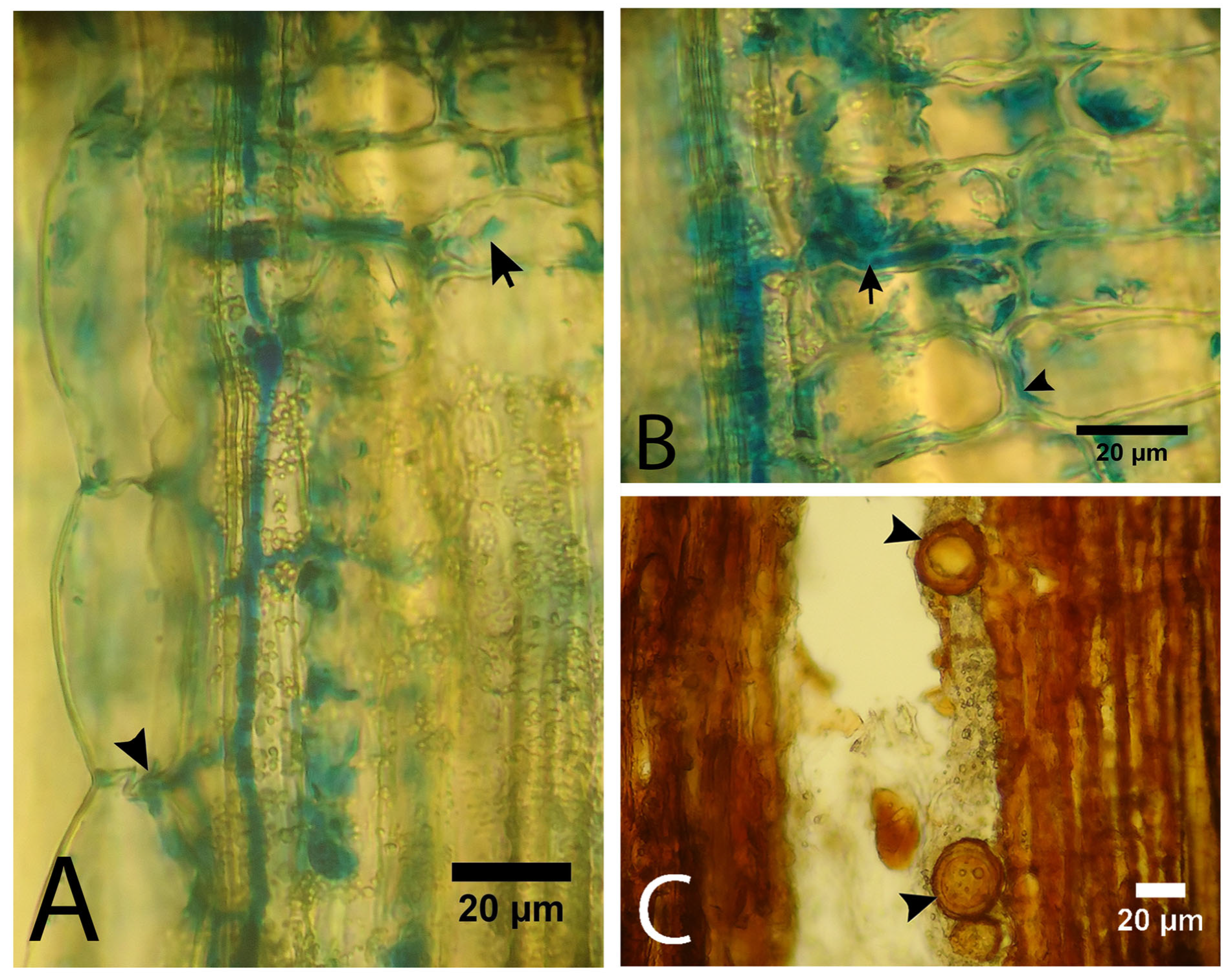
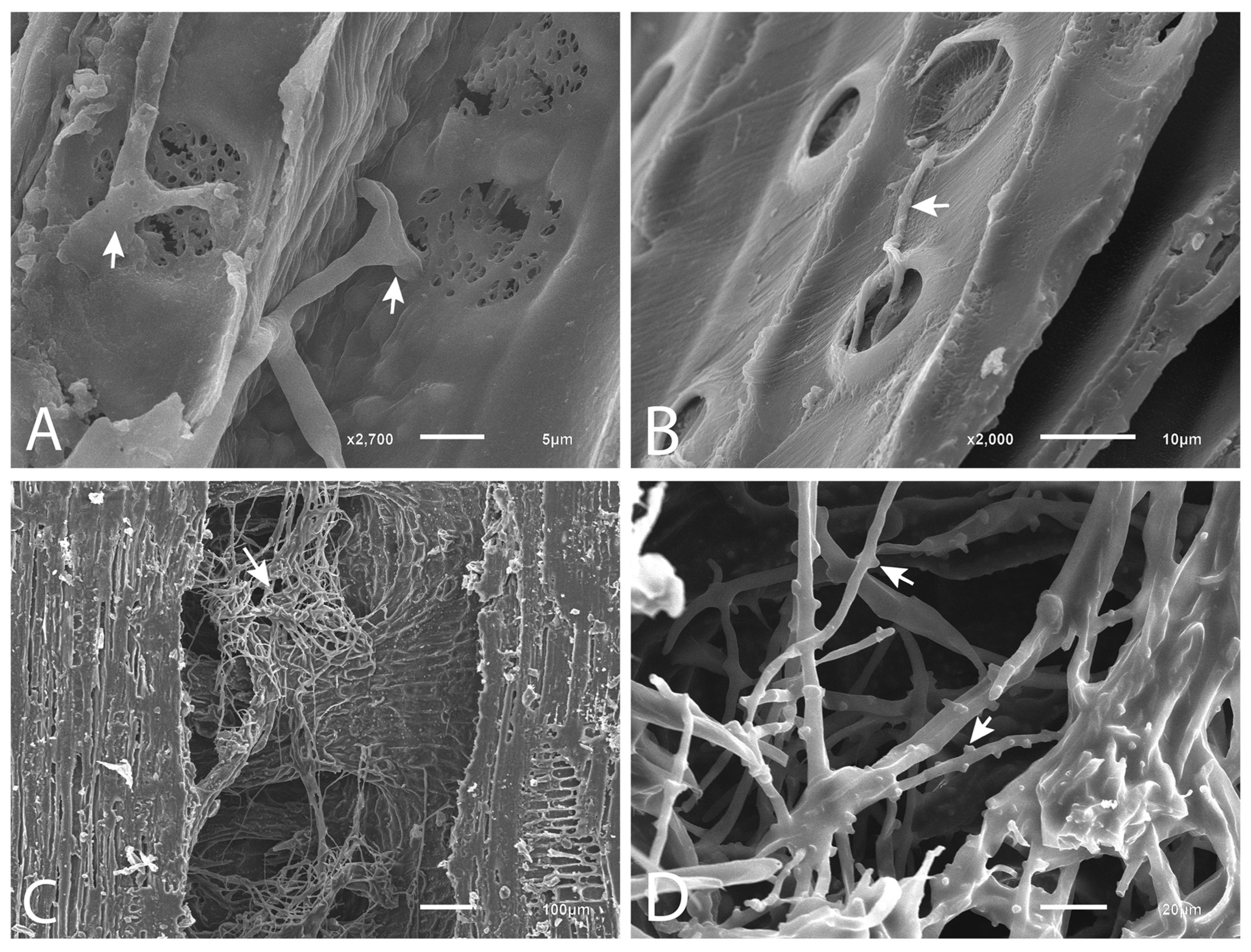
The pathogen was detected in both the phloem and xylem of saplings and adult trees (Figure 3 and Figure 4). In the phloem, it was observed that the hyphae developed along the sieve cells, with branches extending toward the axial (Figure 3A) and radial (Figure 3B) parenchyma, where they developed both inter- and intracellularly. Figure 4A and Figure S2D, Supplementary Materials, show the movement of hyphae between sieve cells through the sieve plates. In adult trees, oospores were observed in the phloem, especially in the resin ducts (Figure 3C), where large, dense masses of hyphae were also present (Figure 4C).
In the xylem, hyphae were observed entering the tracheids from the parenchyma rays and growing along the tracheids through the bordered pits (Figure 4B). The characteristic lateral swellings of P. austrocedri hyphae [1] were observed on the hyphae (Figure 4D), confirming their identity. Occasionally, isolated globose structures with less thickened walls than the oospores were observed (Figure S2E, Supplementary Materials). They could correspond to chlamydospores, although these have not been reported in in vitro cultures of the species.

3.4. Histochemical Analysis

3.4.1. Evaluation of the Presence of Starch

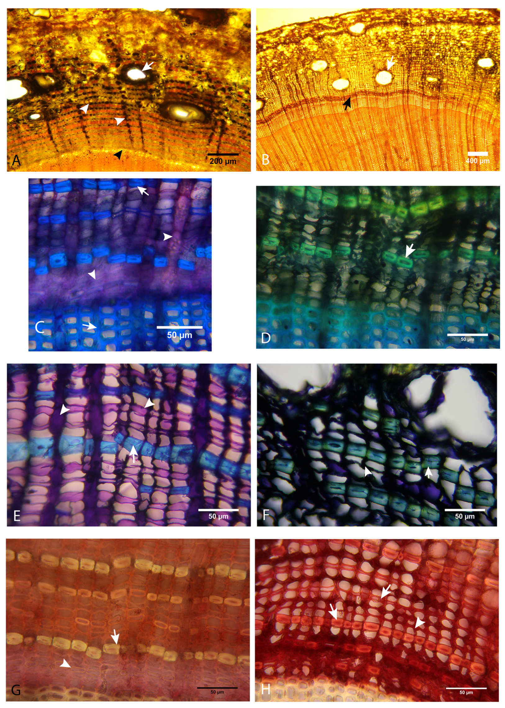
In Lugol, mock-inoculated healthy plants showed the presence of starch mainly in the radial parenchyma, in the axial parenchyma cells of the active phloem zone (Figure 5A, white arrowheads), and in the subsidiary cells of the normal resin ducts (Figure 5A, white arrow). In contrast, the vascular cambium displayed a uniform yellow color, indicating the absence of starch (Figure 5A, black arrowhead).
In the area of the necrotic lesion of the inoculated plants, the cambium appeared darker due to necrosis (Figure 5B, black arrow). The phloem showed a loss of starch in the axial and radial parenchyma, and in the subsidiary cells of the normal resin ducts (Figure 5B, white arrow), while an accumulation of starch was observed in the transition zone between the margin of the necrotic lesion and the healthy tissues. The presence of starch in the tissues decreased toward the older zones of the lesion, being very low in the middle zone and almost absent in the oldest zone. In the traumatic resin ducts, unlike the normal ones, no starch was detected in the initial epithelial cells or in the adjacent ones, indicating a difference in the structure of the epithelial and subsidiary cells between normal and traumatic resin ducts.

3.4.2. Evaluation of the Presence of Phenolic Compounds

In mock-inoculated saplings and healthy adult trees, toluidine blue staining revealed the presence of carbohydrates (indicated by lilac coloration) in vascular cambium cells, in axial and radial parenchyma cells of the phloem (Figure 5C,E, white arrowheads), and in epithelial and subsidiary cells of the resin ducts. Fibers and tracheids appeared light blue, indicating lignin presence (Figure 5C,E, white arrows), while sieve cells were either hyaline or pink.
In the advancing zone of the lesion, a clear contrast was observed between the healthy and affected zones. The affected phloem did not exhibit the lilac color indicating the presence of carbohydrates. Instead, the black color of the necrotic parenchyma cells and the white color of the empty sieve cells were observed (Figure 5D,F and Figure S2F, Supplementary Materials). Phloem fibers showed a color change from light blue to greenish/turquoise, indicating the presence of phenolic compounds. This was observed in both saplings (Figure 5D, white arrow) and adult trees (Figure 5F, white arrow) throughout the entire lesion.

3.4.3. Evaluation of the Presence of Tannins

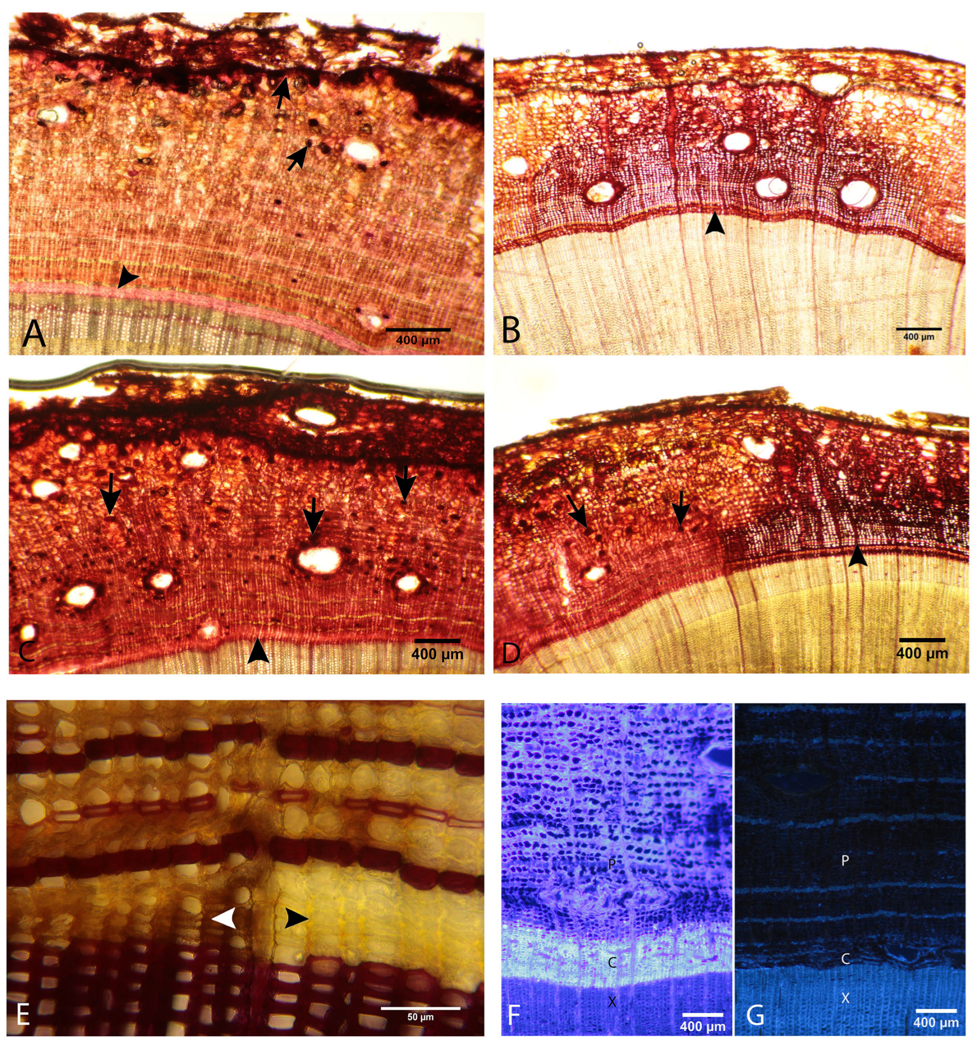
Healthy tissue, stained with vanillin, displayed a light pink coloration in the vascular cambium (Figure 5G, white arrowhead, and Figure S2G, Supplementary Materials) and also in the axial and radial parenchyma and sieve cells. The fibers were hyaline and refringent (Figure 5G, white arrow, and Figure S2G, Supplementary Materials). Healthy tissues showed a higher concentration of tannins only in the periderm, in the subsidiary cells of the resin ducts, and in some isolated cells of the axial parenchyma corresponding to polyphenolic parenchyma cells (PP cells) (Figure 6A, black arrows).
Inoculated saplings displayed a noticeable change in coloration in the affected tissues, which became dark reddish, indicating an increase in tannin deposition (Figure 5H and Figure 6B). The walls of the sieve cells and fibers showed a reddish color (Figure 5, white arrowhead and white arrows) throughout the necrotic lesion, indicating a higher concentration of tannins compared to healthy tissue. In the asymptomatic zone, above the advancing lesion, a significant increase in PP cells was observed, indicating a tree response that would explain the deposition of tannins in the cell walls of the affected area (Figure 6C, black arrows). Furthermore, a more reddish coloration of the tissues was observed, suggesting a higher tannin concentration than in tissues of healthy plants. The same was observed in the areas laterally adjacent to the lesion (Figure 6D, black arrows).
The same characteristics were observed in the tissues of naturally infected adult trees: increased tannins in the walls of the sieve cells and fibers, as well as collapsed parenchyma cells (Figure S2G,H, Supplementary Materials).

3.4.4. Evaluation of the Presence of Lignin

A positive reaction to phloroglucinol was observed in the fibers and tracheids of both healthy and affected tissues (Figure 6E and Figure S2A, Supplementary Materials). No remarkable differences were observed between healthy and affected tissues.

3.4.5. Histochemical Analysis with Epifluorescence

Analysis of healthy and infected tissues using Calcofluor Fluorescent Bright 28 (Figure 6F,G and Figure S2I,J, Supplementary Materials) under UV light staining showed a generalized loss of fluorescence in the affected tissues compared to the healthy ones. In healthy tissues, fluorescence was observed primarily in cambium, sieve cells, parenchyma cells of the resin ducts, and in xylem tracheids (Figure 6F). The tissues of the necrotic lesion showed a total loss of fluorescence except for fibers and tracheids (Figure 6G). The reduction in fluorescence increased toward older areas of the lesion.
No differences were detected in aniline blue staining between healthy and affected tissues that could be attributed to an increase in callose deposits (Figure S2K,L, Supplementary Materials).

4. Discussion

This study confirmed that P. austrocedri can successfully colonize nearly all stem tissues of A. chilensis, growing within them and completing its life cycle. The pathogen was found in both the phloem and the xylem. Scanning electron microscopy images showed the characteristic lateral swelling in the hyphae, a trait previously reported for this species [1], which further confirms its identity. Additionally, numerous oogonia and oospores were observed, primarily in the phloem, which is reported for the first time in A. chilensis. This information is crucial from an epidemiological perspective. These structures can remain viable within tree tissues for long periods, which represents a significant challenge for eradication and increases the risk of pathogen spread through the movement of infected wood.
The pathogen’s development in the stem tissues aligns with findings reported for other Phytophthora species, which typically infect the roots first before spreading to the stem. Specifically, the pathogen initially advances through the inner bark (cambium and phloem) and then extends radially toward the xylem [16,17,35,36]. In A. chilensis, the pathogen initially affects the cambium, leading to necrosis even in areas that appear healthy above the lesion’s margin. This was observed in both sapling and adult trees, aligning with the findings of Oh and Hansen [37]. These authors found that P. lateralis colonized the cambial cells of both susceptible and resistant Port-Orford-Cedar seedlings. In the resistant seedlings, colonization was restricted to the cambium and the outermost layers of sieve and parenchyma cells. In contrast, in the susceptible seedlings, colonization extended to the sieve cells and parenchyma of the active phloem. From these observations, we can infer that the first tissue affected is the cambium, suggesting that it is the plant’s most susceptible tissue. Following the cambium necrosis, the phloem, particularly the axial parenchyma, becomes the next tissue to be impacted. Hyphae grow along the sieve cells and colonize the axial parenchyma cells both inter- and intracellularly. A study by Vieytes Blanco et al. [14] on different Phytophthora species affecting Black Alder found that, depending on the specific Phytophthora species, alterations were more pronounced in the axial parenchyma compared to the radial parenchyma, or vice versa. In the case of P. austrocedri, similar damages are observed in both types of parenchyma. Eventually, colonization of the xylem occurs through the radial parenchyma, with hyphae reaching the tracheids and developing along them through the bordered pits.
The tree responds to P. austrocedri’s attack likely by reprogramming the stem cells of the vascular cambium, affecting both the phloem and xylem. This results in different structural and chemical changes in the affected tissues. One consistent response observed in both saplings and adult trees was the formation of traumatic resin ducts and resin pockets. The formation of traumatic resin ducts is induced by methyl jasmonate (MJ) through ethylene production, which triggers the reprogramming of the cambial zone for duct formation [23,38]. Hudgins et al. [38] demonstrated that the response to MJ varies across different conifer families: in Pinaceae and Taxodiaceae, MJ induces the formation of traumatic resin ducts in the xylem, while in Auraucariaceae and Cupressaceae, it induces duct formation in the phloem, as observed in A. chilensis.
The formation of resin pockets associated with the necrotic lesions of P. austrocedri has been reported previously [2], and the profuse external exudation from them is a diagnostic symptom of the disease. In addition, it has been shown that resin from traumatic ducts has a diterpene composition that differs from that of healthy trees and that some of its fractions have fungistatic activity [8]. It would be interesting to investigate whether there are differences in the phenolic compound profile between traumatic and normal resin, given the PP cell activity in response to the pathogen attack reported in this work. Oh and Hansen [37] reported bark resin exudates near necrotic areas in resistant Port-Orford-Cedar infected by P. lateralis and found that resin crystals accumulated more abundantly in the tissues of inoculated resistant individuals than in healthy or inoculated susceptible ones, suggesting that the deposition of resin is a defensive response of the tree.
Another response, observed at the margins of the lesion in some cases, was an alteration in cell morphology characterized by an increase in size, a change in shape, and a loss of normal cell arrangement. Similar observations have been reported in other pathosystems [17,37,39], where cells in the cambium and phloem appear distorted or collapsed and disorganized, even in the absence of visible hyphae [37]. The alteration is frequently accompanied by the deposition of defense substances in the cell walls [17,37,39]. This reaction serves as a structural barrier to prevent further pathogen spread and limit the infected area [40]. The deposition of callose [17,37], lignin [14,39], and other phenolic compounds [12,41,42] is a recorded response reaction to Phytophthora spp. attack in various plant species. In all infected A. chilensis specimens, the accumulation of phenolic compounds, including tannins, was observed. This aligns with the increased presence of PP cells noted in the tissues adjacent to the lesion. The deposition of phenols is also likely a defense response induced by MJ, as reported by Hudgins et al. [38] for several conifer species, including a species of Cupressaceae (Cupressus macrocarpa).
This mechanism could also explain the starch accumulation observed in the areas adjacent to the advancing lesion zone, as PP cells contain significant starch reserves, which may be related to their ability to produce a rapid and sustained biochemical response [40]. Nagle et al. [42] found that resistant coast live oaks contain higher amounts of some constitutive soluble phenolics (e.g., ellagic acid, tyrosol derivative, and an unidentified one) compared to susceptible oaks. This finding agrees with Ockels et al. [41], who reported a marked increase in the phloem content of some constitutive soluble phenols, especially ellagic and gallic acids.
Lignin deposition was not detected, except occasionally in the “immature” fibers, similar to what was reported by [38]. However, since this deposition was observed infrequently, it cannot be considered a characteristic response in A. chilensis. Likewise, no significant deposition of callose was observed, suggesting that this compound does not play a significant role in tree defense, as also found by [12] in the P. cinnamomi–Castanea spp. pathosystem.
Similarly to our observations, Giesbrecht et al. [17] reported diminished fluorescence in calcofluor of Notholithocarpus densiflorus tissues affected by P. ramorum. This decrease in fluorescence could indicate some degree of alteration of structural carbohydrates in damaged or dead cells. Still, more studies are needed to determine whether it is related to the breakdown of carbohydrates or to the deposition of other substances that can mask fluorescence.
For some broadleaf species, the formation of tyloses in the xylem has been described as a response to Phytophthora spp. attack [43] or as damage induced by the pathogen [14,16], producing embolisms in the vessels that reduce hydraulic conductivity [16]. Tylosis is not possible in A. chilensis, whose xylem is composed exclusively of tracheids. However, a drastic reduction in hydraulic conductivity due to P. austrocedri attack on saplings has been reported [9]. The evidence found in this work allows us to assume that this is mainly due to the clogging of the tracheids by the abundant resin flow from the traumatic ducts, the blockage caused by pathogen structures (hyphae, oospores), and the formation of trabeculae [34], a phenomenon that has not been reported in other Phytophthora pathosystems with forest species.
Histological and histochemical analyses of the tissues from both young and adult A. chilensis individuals affected by P. austrocedri contribute to understanding how the pathogen colonizes stem tissues, how it spreads along these tissues, as well as the damage it causes. These analyses also provide insights into the tree’s response to infection.
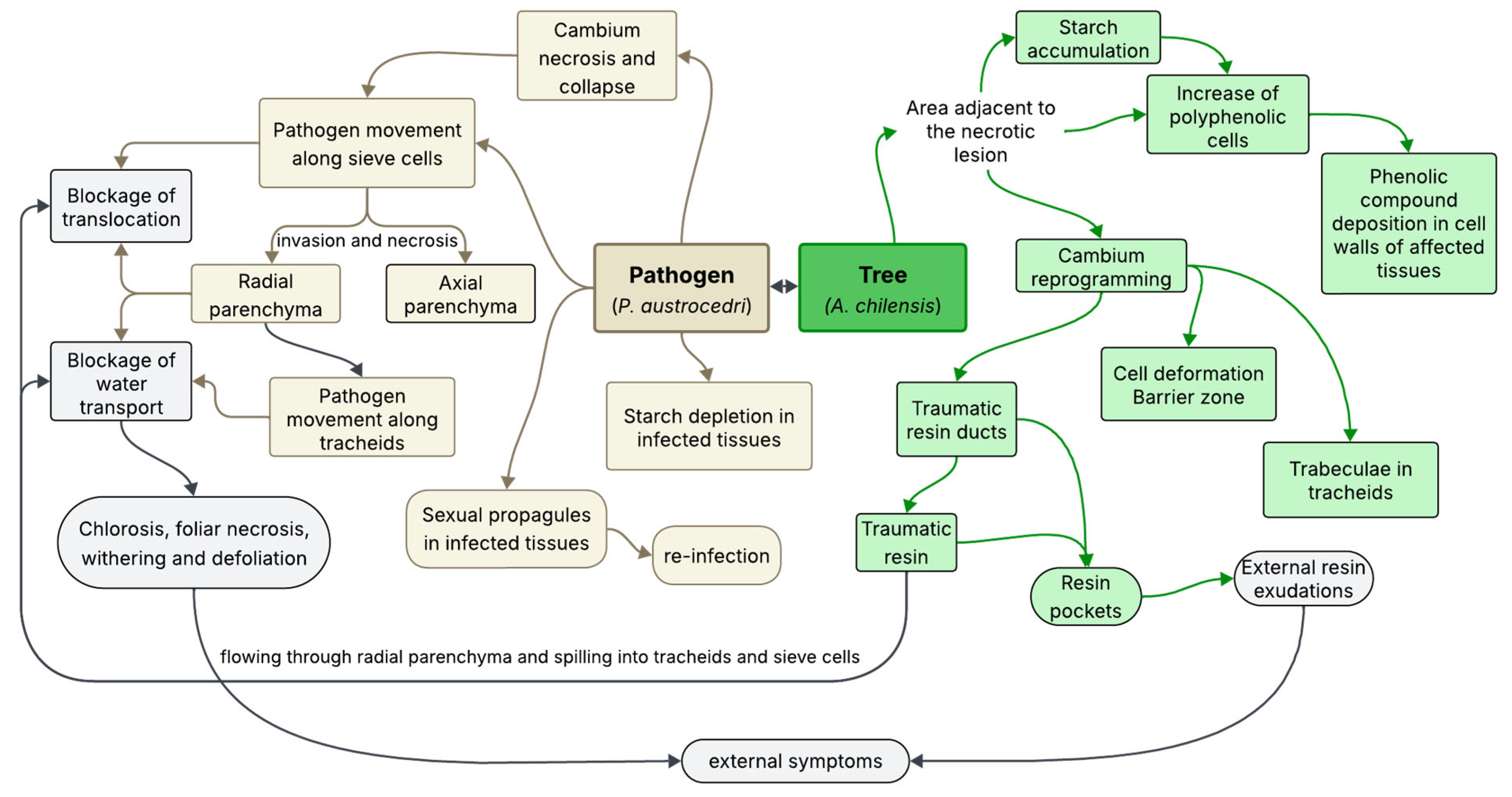
Figure 7 presents a schematic summary of these processes, inferred from the results of the present study. Understanding this process helps explain the external symptoms of the disease. The blockage of sieve cells and tracheids affects nutrient translocation and water transport, respectively, leading to the primary symptoms: chlorosis, wilting, and defoliation. These symptoms result from a combination of necrosis in the radial parenchyma and blockages caused by the pathogen’s structures, as well as traumatic resin that spills into the vascular tissues. Finally, when necrosis affects the inner bark around all or a significant portion of the tree’s perimeter, tree death may occur due to water imbalance, disruption of translocation leading to root death, or a combination of both.

5. Conclusions

In this pathogen–host interaction, it has been confirmed that stem damage starts in the vascular cambium, where necrosis is evident before the symptoms become visible in the tree’s bark. The response of A. chilensis to P. austrocedri attack shows common elements with responses observed in other forest species [21]. The rapid necrosis of the cambium, noted even beyond the advancing zone of the lesion, and the browning of the affected tissues, evidencing the accumulation of phenolic compounds, are compatible with a hypersensitive response (HR) [12]. This agrees with previous results on the physiological response of A. chilensis to pathogen inoculation, which also supports the hypothesis of an HR or HR-like response mechanism [9]. On the other hand, the formation of traumatic resin ducts and resin, along with the increase in PP cells, phenols, and tannins, is induced by methyl jasmonate (MJ), which is widely reported in other conifer species as a response to various pathogens and pests [19,24]. The relationship between MJ and HR remains unclear. Some studies have reported that MJ can induce HR [44,45], while others suggest it may inhibit it [46,47]. In the case of A. chilensis, both MJ-induced responses and HR-type responses were observed simultaneously, which suggests that there may not be an inhibitory effect. However, further research is needed to confirm whether MJ induces HR.
The mechanisms observed in A. chilensis do not seem to be effective enough to limit the progression of the disease. Early necrosis of the cambium fails to circumscribe the pathogen, which is undoubtedly in its necrotrophic phase. Similarly, traumatic resin does not appear to play a significant role in limiting the pathogen, as P. austrocedri has been isolated from resin-impregnated tissues (Greslebin pers. comm.). Gavira [48] and Fernández et al. [12], in their comparative histopathological studies of Phytophthora spp. infection in susceptible and resistant individuals, found that both groups exhibit the same response mechanism, but resistant individuals respond more quickly and effectively. While resistance to P. austrocedri has not been conclusively proven in A. chilensis, potentially resistant individuals have been recorded and are currently under study (Vélez, pers. comm.). If resistant individuals are identified, it would be valuable to conduct a study similar to the present one to assess similarities and differences in their response to the pathogen.
The results of this study establish anatomical and epidemiological bases for developing strategies focused on early detection, monitoring, and integrated management of P. austrocedri in Argentine forests. The presence of abundant oospores in tree tissues confirms that they serve as a source of inoculum, capable of acting as a reinfection agent or infecting areas free of the pathogen if transported there. This is particularly important for A. chilensis, a highly valued wood species, since Argentine legislation permits the use of dead trees. As a result, transporting these trees out of the affected area for sawmilling, as well as treating the waste, should include management practices to prevent the spread of the disease.

Supplementary Materials

The following supporting information can be downloaded at: https://www.mdpi.com/article/10.3390/f16071073/s1, Figures S1–S2: Another complementary photograph of affected tissues not included in the main manuscript. Figure S1: (A) Inoculated sapling. (B,C) Sampled adult trees. (D) Fresh wedge-shaped sample in the field, showing delineated necrotic and healthy areas. (E) The same sample as in (D), in the laboratory; blocks for light microscopy and SEM analysis are indicated by black arrows. Figure S2: (A) Cross-section of the phloem of a healthy sapling in phloroglucinol showing the two types of fibers: “mature”, thick-walled fibers, and immature fibers with slightly thickened walls (black arrow). (B) Resin pocket formed by the merging of traumatic resin ducts in a natural infected adult tree. (C) Deformed parenchyma ray in the xylem associated with the necrotic lesion of an inoculated sapling. (D) SEM microphotograph (splitting of necrotic phloem) of a naturally infected adult tree: Phythophthora austrocedri hyphae in sieve cells crossing through sieve plates. (E) Chlamydospore-like structure in the xylem of a naturally infected adult tree. (F) Cross-sections, stained with toluidine blue, of an infected sapling, showing the transition between unaltered and necrotic tissues. Unaffected tissues (left) exhibit the presence of carbohydrates (lilac) in the axial and radial parenchyma. Necrotic tissues (right) show no carbohydrates and deposition of phenolic compounds in fibers, evidenced by the green tints (indicated by the white arrowheads). (G) Cross-section of a healthy adult tree in vanillin stain exhibits cambium cells in pink (indicated by a white arrowhead) and fibers in a brilliant pale yellow (shown by black arrowheads). (H) A cross-section of a naturally infected adult tree in vanillin stain reveals tannin deposition in fibers (shown by white arrowheads), indicated by the red color. (I,J) These figures display the details of healthy (I) and naturally infected (J) adult tree tissues observed using calcofluor staining. Healthy tissues exhibit significantly higher fluorescence compared to the affected tissues, particularly in the cambium (indicated by the black arrowheads). Both photographs were taken with the same exposure time of 2 seconds. (K,L) These figures present the details of healthy (K) and naturally infected (L) adult tree tissues observed using aniline blue staining. No significant differences were detected between the two types of tissues; however, fluorescence was slightly higher in the healthy tissues. Both photographs were taken with the same exposure time of 5 s. Table S1: Results of re-isolation and ELISA immunoassay of necrotic lesions from inoculated saplings. Table S2: Results of isolation and ELISA immunoassay of necrotic lesions from naturally infected adult trees.

Author Contributions

Conceptualization, O.T. and A.G.G.; methodology, O.T. and A.G.G.; validation, O.T. and A.G.G.; formal analysis, O.T. and A.G.G.; investigation, O.T.; resources, O.T. and A.G.G.; writing—original draft preparation, A.G.G.; writing—review and editing, O.T. and A.G.G.; visualization, O.T. and A.G.G.; supervision, A.G.G.; project administration, A.G.G.; funding acquisition, O.T. and A.G.G. All authors have read and agreed to the published version of the manuscript.

Funding

This research was funded by the National Agency for the Promotion of Science, Technology, and Innovation (ANPCyT, FONCyT), grant number PICT 2012-0733. National University of Patagonia, grant for postgraduate studies.

Data Availability Statement

The original contributions presented in this study are included in the article/Supplementary Materials. Further inquiries on additional photographs can be directed to the corresponding author.

Acknowledgments

We sincerely thank Ken Troncoso Imai for his assistance with the preparation of the figures.

Conflicts of Interest

The authors declare no conflicts of interest. The funders had no role in the design of the study; in the collection, analyses, or interpretation of data; in the writing of the manuscript; or in the decision to publish the results.

References

  1. Greslebin, A.G.; Hansen, E.M.; Sutton, W. Phytophthora austrocedrae sp. nov., a new species associated with Austrocedrus chilensis mortality in Patagonia. Mycol. Res. 2007, 111, 308–316. [Google Scholar] [CrossRef] [PubMed]
  2. Greslebin, A.G.; Hansen, E.M. Pathogenicity of Phytophthora austrocedrae on Austrocedrus chilensis and its relation with mal del ciprés in Patagonia. Plant Pathol. 2010, 59, 604–612. [Google Scholar] [CrossRef]
  3. Taccari, L.E.; Greslebin, A.G.; Salomón, M.E.S.; Vélez, M.L.; Santini, A. Two conifer species native to Patagonia threatened by Phytophthora austrocedri. For. Pathol. 2019, 49, e12496. [Google Scholar] [CrossRef]
  4. Green, S.; Elliot, M.; Armstrong, A.; Hendry, S.J. Phytophthora austrocedrae emerges as a serious threat to juniper (Juniperus communis) in Britain. Plant Pathol. 2015, 64, 456–466. [Google Scholar] [CrossRef]
  5. Werres, S.; Elliot, M.; Greslebin, A. Phytophthora austrocedrae Gresl. & E.M. Hansen. JKI Datasheets Plant Diseases and Diagnosis. 2014. Available online: https://forestphytophthoras.org/sites/default/files/educational_materials/3008-12007-1-PB.pdf (accessed on 20 June 2022).
  6. Mahdikhani, M.; Matinfar, M.; Aghaalikhani, A. First report of Phytophthora austrocedri causing phloem lesions and bronzing on Cupressus sempervirens in northern Iran. New Dis. Rep. 2017, 36, 10. [Google Scholar] [CrossRef]
  7. Cooke, D.E.L.; Drenth, A.; Duncan, J.M.; Wagels, G.; Brasier, C.M. A Molecular Phylogeny of Phytophthora and Related Oomycetes. Fungal Genet. Biol. 2000, 30, 17–32. [Google Scholar] [CrossRef]
  8. Olate, V.R.; Vélez, M.L.; Greslebin, A.; Schmeda-Hirschmann, G. Phytophthora austrocedri Elicitates Changes in Diterpene Profile of Austrocedrus chilensis. Molecules 2015, 20, 15084–15097. [Google Scholar] [CrossRef]
  9. Vélez, M.L.; Silva, P.V.; Troncoso, O.A.; Greslebin, A.G. Alteration of physiological parameters of Austrocedrus chilensis by the pathogen Phytophthora austrocedrae. Plant Pathol. 2012, 61, 877–888. [Google Scholar] [CrossRef]
  10. Gómez, F.J.R.; Navarro-Cerrillo, R.M.; Sánchez-Cuesta, R.; Pérez-De-Luque, A. Histopathology of infection and colonization of Quercus ilex fine roots by Phytophthora cinnamomi. Plant Pathol. 2015, 64, 605–616. [Google Scholar] [CrossRef]
  11. Redondo, M.Á.; Pérez-Sierra, A.; Abad-Campos, P.; Torres, L.; Solla, A.; Reig-Armiñana, J.; García-Breijo, F. Histology of Quercus ilex roots during infection by Phytophthora cinnamomi. Trees 2015, 29, 1943–1957. [Google Scholar] [CrossRef]
  12. Fernandes, P.; Machado, H.; Silva, M.D.C.; Costa, R.L. A Histopathological Study Reveals New Insights Into Responses of Chestnut (Castanea spp.) to Root Infection by Phytophthora cinnamomi. Phytopathology 2021, 111, 345–355. [Google Scholar] [CrossRef]
  13. Nave, C.; Schwan, J.; Werres, S.; Riebesehl, J. Alnus glutinosa Threatened by Alder Phytophthora: A Histological Study of Roots. Pathogens 2021, 10, 977. [Google Scholar] [CrossRef]
  14. Vieites-Blanco, C.; Colangelo, M.; Camarero, J.J.; Caballol, M.; García Breijo, F.J.; Štraus, D.; Oliva, J. Pathogenicity of Phytophthora and Halophytophthora species on black alder and the host histological response. Mycol. Prog. 2023, 22, 71. [Google Scholar] [CrossRef]
  15. Brown, A.V.; Brasier, C.M. Colonization of tree xylem by Phytophthora ramorum, P. kernoviae and other Phytophthora species. Plant Pathol. 2007, 56, 227–241. [Google Scholar] [CrossRef]
  16. Parke, J.L.; Oh, E.; Voelker, S.; Hansen, E.M.; Buckles, G.; Lachenbruch, B. Phytophthora ramorum Colonizes Tanoak Xylem and Is Associated with Reduced Stem Water Transport. Phytopathology 2007, 97, 1558–1567. [Google Scholar] [CrossRef] [PubMed]
  17. Giesbrecht, M.B.; Hansen, E.M.; Kitin, P. Histology of Phytophthora ramorum in Notholithocarpus densiflorus bark tissues. N. Z. J. For. Sci. 2011, 41, S89–S100. [Google Scholar]
  18. Jung, T.; Pérez-Sierra, A.; Durán, A.; Jung, M.H.; Balci, Y.; Scanu, B. Canker and decline diseases caused by soil- and airborne Phytophthora species in forests and woodlands. Persoonia-Mol. Phylogeny Evol. Fungi 2018, 40, 182–220. [Google Scholar] [CrossRef] [PubMed]
  19. Franceschi, V.R.; Krokene, P.; Christiansen, E.; Krekling, T. Anatomical and chemical defenses of conifer bark against bark beetles and other pests. New Phytol. 2005, 167, 353–376. [Google Scholar] [CrossRef]
  20. Goodman, R.N.; Novacky, A.J. The Hypersensitive Reaction in Plants to Pathogens: A Resistance Phenomenon; APS Press: St. Paul, MN, USA, 1994; 253p. [Google Scholar]
  21. Oßwald, W.; Fleischmann, F.; Rigling, D.; Coelho, A.C.; Cravador, A.; Diez, J.; Dalio, R.J.; Jung, M.H.; Pfanz, H.; Robin, C.; et al. Strategies of attack and defence in woody plant–Phytophthora interactions. For. Pathol. 2014, 44, 169–190. [Google Scholar] [CrossRef]
  22. Pieterse, C.M.; van Loon, L.C. Salicylic acid-independent plant defence pathways. Trends Plant Sci. 1999, 4, 52–58. [Google Scholar] [CrossRef]
  23. Hudgins, J.W.; Franceschi, V.R. Methyl Jasmonate-Induced Ethylene Production Is Responsible for Conifer Phloem Defense Responses and Reprogramming of Stem Cambial Zone for Traumatic Resin Duct Formation. Plant Physiol. 2004, 135, 2134–2149. [Google Scholar] [CrossRef] [PubMed]
  24. Krokene, P.; Nagy, N.E.; Solheim, H. Methyl jasmonate and oxalic acid treatment of Norway spruce: Anatomically based defense responses and increased resistance against fungal infection. Tree Physiol. 2008, 28, 29–35. [Google Scholar] [CrossRef]
  25. Yu, X.; Zhang, W.; Zhang, Y.; Zhang, X.; Lang, D.; Zhang, X. The roles of methyl jasmonate to stress in plants. Funct. Plant Biol. 2018, 46, 197–212. [Google Scholar] [CrossRef] [PubMed]
  26. Nagy, N.E.; Franceschi, V.R.; Solheim, H.; Krekling, T.; Christiansen, E. Wound-induced traumatic resin duct development in stems of Norway spruce (Pinaceae): Anatomy and cytochemical traits. Am. J. Bot. 2000, 87, 302–313. [Google Scholar] [CrossRef] [PubMed]
  27. La Manna, L.; Carabelli, F.; Gómez, M.; Matteucci, S.D. Disposición espacial de parches de Austrocedrus chilensis con síntomas de defoliación y mortalidad en el Valle 16 de Octubre. Bosque 2008, 29, 23–32. [Google Scholar] [CrossRef]
  28. Tardif, J.C.; Conciatori, F. Microscopic examination of wood: Sample preparation and techniques for light microscopy. In Plant Microtechniques and Protocols; Yeung, E., Stasolla, C., Sumner, M., Huang, B., Eds.; Springer: Cham, Switzerland, 2015; pp. 373–415. [Google Scholar] [CrossRef]
  29. D’Ambrosio de Argüeso, A. Manual de Técnicas en Histología Vegetal, 1st ed.; Ed Hemisferio Sur S.A: Buenos Aires, Argentina, 1986; 83p. [Google Scholar]
  30. Nava, L.R.; Isaías, A.Q.; Olvera, C.D.L.P.P. Comparación histoquímica de albura y duramen de tres especies de Quercus. Madera y Bosques 1999, 5, 27–41. [Google Scholar] [CrossRef]
  31. Mauch-Mani, B.; Slusarenko, A.J. Production of Salicylic Acid Precursors Is a Major Function of Phenylalanine Ammonia-Lyase in the Resistance of Arabidopsis to Peronospora parasitica. Plant Cell 1996, 8, 203–212. [Google Scholar] [CrossRef]
  32. De Magistris, A.A.; Castro, M.A. Bark anatomy of southern South American Cupressaceae. IAWA J. 2001, 22, 367–383. [Google Scholar] [CrossRef]
  33. Castro, M.A. Cortezas. Especies Leñosas de los Bosques Andinos Patagónicos Argentina; LOLA: Buenos Aires, Argentina, 2009; 280p. [Google Scholar]
  34. Troncoso, O.; Greslebin, A. Trabeculae in Patagonian mountain cypress (Austrocedrus chilensis) associated with Phytophthora austrocedri infection. IAWA J. 2018, 39, 209–220. [Google Scholar] [CrossRef]
  35. Tippett, J.T.; Shea, S.R.; Hill, T.C.; Shearer, B.L. Development of Lesions Caused by Phytophthora cinnamomi in the Secondary Phloem of Eucalyptus marginata. Aust. J. Bot. 1983, 31, 197–210. [Google Scholar] [CrossRef]
  36. Davison, E.M.; Stukely, M.J.; Crane, C.E.; Tay, F.C.S. Invasion of phloem and xylem of woody stems and roots of Eucalyptus marginata and Pinus radiata by Phythophtora cinnamomi. Phytopathology 1994, 84, 335–340. [Google Scholar] [CrossRef]
  37. Oh, E.; Hansen, E.M. Histopathology of Infection and Colonization of Susceptible and Resistant Port-Orford-Cedar by Phytophthora lateralis. Phytopathology 2007, 97, 684–693. [Google Scholar] [CrossRef]
  38. Hudgins, J.W.; Christiansen, E.; Franceschi, V.R. Induction of anatomically based defense responses in stems of diverse conifers by methyl jasmonate: A phylogenetic perspective. Tree Physiol. 2004, 24, 251–264. [Google Scholar] [CrossRef] [PubMed]
  39. Pogoda, F.; Werres, S. Histological studies of Phytophthora ramorum in Rhododendron twigs. Can. J. Bot. 2004, 82, 1481–1489. [Google Scholar] [CrossRef]
  40. Franceschi, V.R.; Krekling, T.; Berryman, A.A.; Christiansen, E. Specialized phloem parenchyma cells in Norway spruce (Pinaceae) bark are an important site of defense reactions. Am. J. Bot. 1998, 85, 601–615. [Google Scholar] [CrossRef] [PubMed]
  41. Ockels, F.S.; Eyles, A.; McPherson, B.A.; Wood, D.L.; Bonello, P. Phenolic Chemistry of Coast Live Oak Response to Phytophthora ramorum Infection. J. Chem. Ecol. 2007, 33, 1721–1732. [Google Scholar] [CrossRef] [PubMed]
  42. Nagle, A.M.; Mcpherson, B.A.; Wood, D.L.; Garbelotto, M.; Bonello, P. Relationship between field resistance to Phytophthora ramorum and constitutive phenolic chemistry of coast live oak. For. Pathol. 2011, 41, 464–469. [Google Scholar] [CrossRef]
  43. Philips, D.; Grant, B.R.; Weste, G. Histological changes in the roots of an avocado cultivar, Duke 7, infected with Phytophthora cinnamomi. Phytopathology 1986, 77, 691–698. [Google Scholar] [CrossRef]
  44. Repka, V.; Fischerova, I.; Silharova, K. Methyl jasmonate induces a hypersensitive-like response of grapevine in the absence of avirulent pathogens. Vitis-Geilweilerhof 2001, 40, 5–10. [Google Scholar]
  45. Repka, V.; Fischerová, I.; Šilhárová, K. Methyl Jasmonate is a Potent Elicitor of Multiple Defense Responses in Grapevine Leaves and Cell-Suspension Cultures. Biol. Plant. 2004, 48, 273–283. [Google Scholar] [CrossRef]
  46. Rao, M.V.; Lee, H.-I.; Creelman, R.A.; Mullet, J.E.; Davis, K.R. Jasmonic Acid Signaling Modulates Ozone-Induced Hypersensitive Cell Death. Plant Cell 2000, 12, 1633–1646. [Google Scholar] [CrossRef] [PubMed]
  47. Andi, S.; Taguchi, F.; Toyoda, K.; Shiraishi, T.; Ichinose, Y. Effect of Methyl Jasmonate on Harpin-Induced Hypersensitive Cell Death, Generation of Hydrogen Peroxide and Expression of PAL mRNA in Tobacco Suspension Cultured BY-2 Cells. Plant Cell Physiol. 2001, 42, 446–449. [Google Scholar] [CrossRef] [PubMed]
  48. Gaviria, A.E.; Patiño, L.F.; Morales, J.G. Histological changes induced during the biotrophic phase of infection of three potato varieties by Phytophthora infestans (Mont.) de Bary. J. Plant Prot. Res. 2019, 59, 465–478. [Google Scholar] [CrossRef]
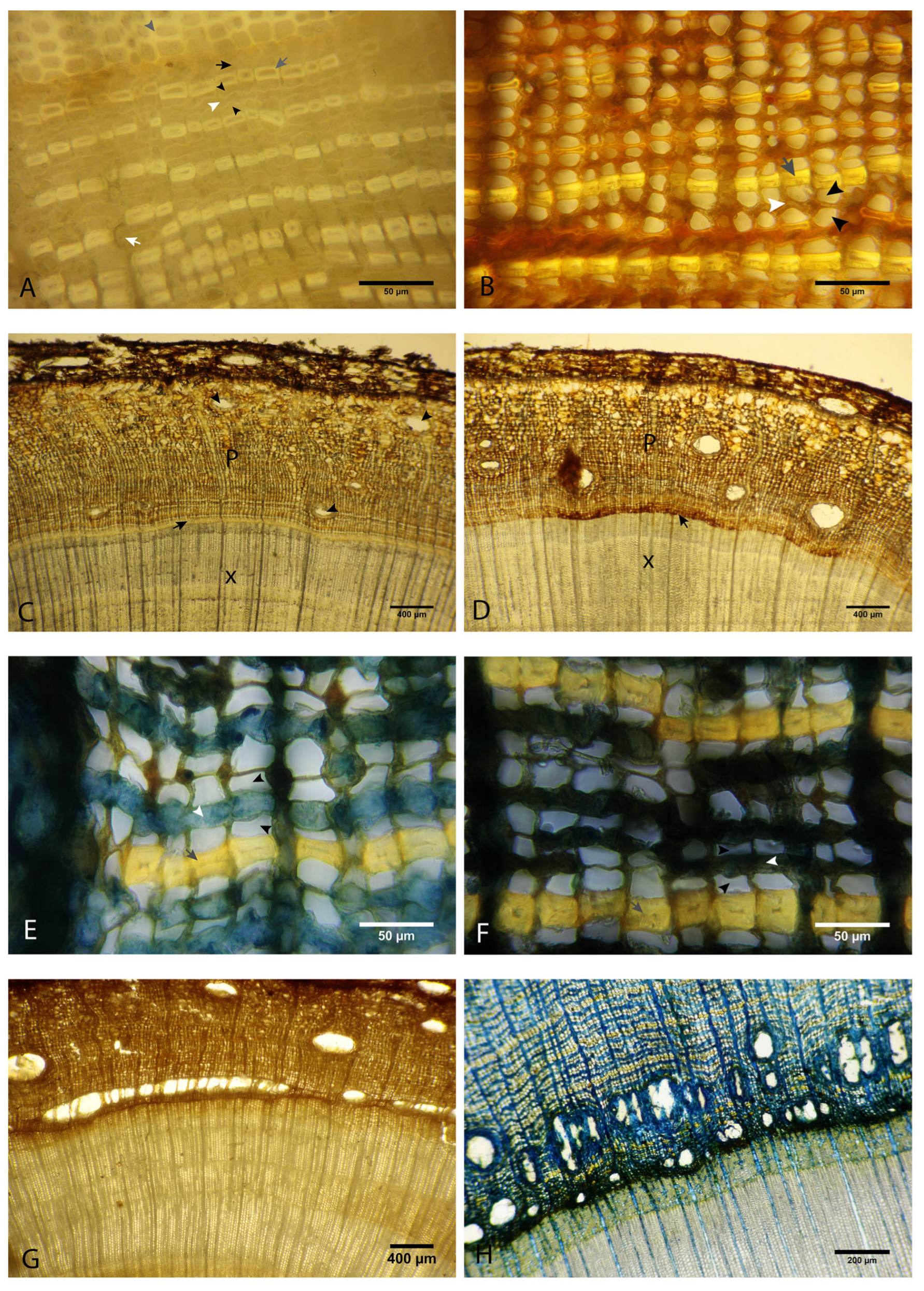
Figure 1. A. chilensis tissues of healthy (A,C,E) and infected (B,D,F,G,H) saplings and adult trees. (A) Stem cross-section of a healthy sapling: xylem, composed of tracheids (indicated by grey arrowhead), cambium (shown by a black arrow), phloem showing the arrangement in rows of fibers (indicated by a grey arrow), sieve cells (indicated by black arrowheads), and axial parenchyma (indicated by a white arrowhead). The white arrow indicates a parenchyma ray. (B) Phloem cross-section of an infected sapling: necrotic and collapsed axial parenchyma (shown by a white arrowhead); sieve cells (indicated by a black arrowhead), empty and expanded due to the collapse of the adjacent parenchyma cells; fibers (shown by a grey arrow). (C,D) Cross-section of a healthy sapling (C) and an infected (D) sapling in water: X indicates xylem and P indicates phloem; the black arrow shows necrotic cambium a few millimeters beyond the lesion margin. (E,F) Cross-section of phloem of a healthy (E) and infected (F) adult tree in cotton blue: sieve cells (shown by black arrowheads), axial parenchyma (indicated by white arrowheads), and fibers (shown by grey arrows). (G) Infected sapling and (H) infected adult tree showing traumatic resin ducts.
Forests 16 01073 g001
Figure 2. SEM microphotograph of cross-sections of affected tissues. (A,B) Cross-sections of infected sapling encompassing the transition between unaltered and necrotic tissues: unaltered cambium (panel A, indicated by a white arrow), deformed cambial and parenchyma cells in the margin of the lesion (panels A and B, indicated by white arrowheads), collapsed cambium in the area of the lesion (panel B, indicated by a white arrowhead), and deformed parenchyma ray (panel B, shown by a yellow arrowhead). (C,D) Cross-section of naturally infected adult tree: (C) Traumatic resin ducts. (D) Traumatic resin duct (indicated by a white arrow) and spilled resin in the adjacent tissues (shown by a white arrowhead).
Forests 16 01073 g002
Figure 3. Phytophthora austrocedri in radial sections of A. chilensis phloem in cotton blue stain: (A) Inoculated sapling: Hyphae developing along the sieve cells, with branches extending toward the axial (indicated by a black arrowhead) and radial (indicated by a black arrow). (B) Hyphae growing inter- (indicated by a black arrow) and intracellularly (indicated by a black arrowhead) in the radial parenchyma. (C) Naturally infected adult tree: oospores of P. austrocedri in a resin pocket (black arrowheads).
Forests 16 01073 g003
Figure 4. SEM microphotograph (splitting) of adult tree tissues showing the presence of the pathogen. (A) Hyphae of P. austrocedri in phloem (indicated by white arrows) crossing through sieve cells. (B) Hyphae of P. austrocedri in xylem (shown by a white arrow) crossing through a bordered pit. (C) Mass of hyphae in a resin duct (indicated by a white arrow). (D) Detail of the hyphae showing the characteristic lateral swellings of P. austrocedri hyphae (shown by white arrows).
Forests 16 01073 g004
Figure 5. Histochemical analysis of healthy and infected tissues. (A,B) Saplings’ stem cross-sections stained with Lugol: (A) Healthy tissues show the presence of starch in the radial and axial parenchyma cells (indicated by white arrowheads) of the active phloem, as well as in the subsidiary cells of normal resin ducts (indicated by a white arrow). The healthy cambium appears bright yellow (shown by a black arrowhead). (B) In infected tissues, there is a generalized loss of starch in parenchyma cells and phloem subsidiary cells of the resin ducts (indicated by a white arrow). The necrotic cambium appears brown (shown by a black arrow). (C,F) Histochemical analysis using toluidine blue stain: (C) The healthy sapling displays cambium and parenchyma cells in a pink to purple color (indicated by a white arrowhead), while fibers and tracheids are bright blue (shown by white arrows). (D) The infected sapling reveals green fibers (indicated by white arrows), indicating the deposition of phenolic compounds. Additionally, the phloem appears black and exhibits collapsed parenchyma cells. (E) The healthy adult tree shows axial and radial parenchyma in purple (indicated by a white arrowhead) and fibers in bright blue (shown by a white arrow). (F) The infected adult tree, consistent with the infected saplings, exhibits green tints in the fibers (indicated by a white arrow) and black, collapsed parenchyma cells (shown by a white arrowhead). (G,H) Histochemical analysis of saplings’ stem cross-sections stained with vanillin: (G) Healthy tissues exhibit cambium cells in pink (indicated by a white arrowhead) and fibers in a brilliant pale yellow (shown by a white arrow). (H) Infected tissues display red tints in the fibers (indicated by white arrows), as well as in the cambium and phloem cells (shown by a white arrowhead), indicating phenolic compound deposition.
Forests 16 01073 g005
Figure 6. (A,D) Cross-section of saplings in vanillin: (A) Healthy sapling displays the cambium in pale pink (indicated by a black arrowhead) and reveals tannin deposits within the bark, along with scattered PP cells (indicated by black arrows). (B) Infected sapling shows an increase in tannin deposition within the affected cambium and phloem, demonstrated by a dark red color (shown by the black arrowhead). (C) A cross-section of an infected sapling taken 1 cm beyond the margin of the lesion exhibits a noticeable increase in PP cells (indicated by black arrows) and elevated tannin content in the cambium (shown by the black arrowhead), as evidenced by the red coloration. (D) A cross-section of an infected sapling reveals a necrotic lesion (indicated by the black arrowhead) and the surrounding tissues. Near the edge of the lesion, there is an increase in PP cells (indicated by black arrows), along with red tints in the cambium and phloem, indicating higher tannin content in the cells. (E) A transverse section of an infected sapling stained with phloroglucinol shows the boundary between the necrotic lesion (indicated by a white arrowhead) and the unaffected tissue (indicated by a black arrowhead). The necrotic tissues appear brown and collapsed, contrasting with the unaffected tissue. There is no significant difference in lignin content between the affected and unaffected areas. (F,G) Cross-section of a healthy (F) and naturally infected (G) adult tree in calcofluor stain. The fluorescence intensity is significantly higher in healthy tissues compared to infected tissues, especially in the cambium and the youngest areas of phloem. X indicates xylem, C indicates cambium, and P indicates phloem.
Forests 16 01073 g006
Figure 7. Schematic summary of the infection process and tree response.
Forests 16 01073 g007
Disclaimer/Publisher’s Note: The statements, opinions and data contained in all publications are solely those of the individual author(s) and contributor(s) and not of MDPI and/or the editor(s). MDPI and/or the editor(s) disclaim responsibility for any injury to people or property resulting from any ideas, methods, instructions or products referred to in the content.