Article

Preliminary Report of Three Entomopathogenic Fungi as Potential Biocontrol Agents Against the Oak Wilt Vector, Platypus koryoensis

1
Department of Forest Healing, Catholic Kwandong University, Gangneung 25601, Republic of Korea
2
Department of Forest Environmental Resources, Gyeongsang National University, Jinju 52828, Republic of Korea
3
Institute of Agriculture and Life Science, Gyeongsang National University, Jinju 52828, Republic of Korea
4
Tree Pathology and Mycology Laboratory, College of Forest and Environmental Sciences, Kangwon National University, Chuncheon 24341, Republic of Korea
5
Department of Environment and Forest Resources, Chungnam National University, Daejeon 34134, Republic of Korea
*
Authors to whom correspondence should be addressed.
Forests 2025, 16(6), 1009; https://doi.org/10.3390/f16061009
Submission received: 13 May 2025 / Revised: 9 June 2025 / Accepted: 14 June 2025 / Published: 16 June 2025
(This article belongs to the Special Issue Pathogenic Fungi in Forest)

Abstract

:
Entomopathogenic fungi are a group of fungi that infect and kill insects to obtain nutrients, thereby contributing to the natural regulation of insect populations. In recent years, they have been increasingly utilized as biological control agents, particularly in response to the rising prevalence of pesticide-resistant pests in agricultural systems. Representative examples include Beauveria bassiana and Metarhizium anisopliae, which are regarded as natural enemies of pests in agroecosystems. Since the first report of Korean oak wilt disease in 2004, the disease has continuously spread across the country and causes severe damage to deciduous oak species, especially Quercus mongolica. Although many efforts have been made to effectively control the disease, including chemical treatments, the control efficacy was shown to be low, and given the environmental side effects arising from the use of insecticides, there has been a demand for alternative control strategies. Integrated Pest Management in forests promotes ecological sustainability by reducing chemical pesticide use, conserving biodiversity, and enhancing long-term forest health. In this study, to mitigate issues with disease management strategies, assessments were made on three entomopathogenic fungi, B. bassiana, M. anisopliae, and Purpureocillium lilacinum, as potential biological control agents against oak wilt disease and its insect vector, Platypus koryoensis. In this regard, we investigated the insecticidal efficacy and LT50 of each entomopathogenic fungus, and the results showed that all three entomopathogenic fungal strains exhibited fast insecticidal effects against the insect vector, P. koryoensis, with M. anisopliae showing the fastest action, recording a lethal time to 50% mortality (LT50) of 58.7 h. The spores of M. anisopliae were found to be sensitive to high temperatures, while demonstrating a relatively high germination rate under UV exposure and strong initial germination ability at low temperatures.

1. Introduction

Oak trees are one of the broad-leaved deciduous species of most significant economic and ecological value within the forest ecosystems that are widely distributed across South Korea, where these species collectively cover over 25% of South Korea’s forested area. In South Korea, five oak species predominantly found across the country include Quercus acutissima, Q. aliena, Q. mongolica, Q. serrata, and Q. variabilis [1].
Oak wilt mortality was first detected in Q. mongolica in Gyeonggi Province, South Korea, in 2004 and has subsequently spread throughout the country [2,3], inflicting considerable damage on forest ecosystems. Although the number of affected trees has gradually decreased due to the continuous monitoring, prevention, and control strategies for the disease, the number of affected trees surged in 2006, reaching a peak of 331,000 damaged trees in 2011. Since 2017, the number of damaged trees has stabilized at around 150,000 to 170,000 annually (http://www.forest.go.kr (accessed on 1 March 2022)).
The pathogen, Dryadomyces quercus-mongolicae, is linked to this oak wilt mortality, facilitated by mass infestations of the wood-boring ambrosia beetle vector, Platypus koryoensis. This wood-boring ambrosia beetle transmits the fungal inoculum into oak trees that are stressed or weakened by various factors during its boring activity, facilitating systemic infection and wilting [4,5,6,7]. The wood-boring ambrosia beetle, P. koryoensis, was reported to be distributed domestically as early as the 1930s. Given the fact that the distribution of P. koryoensis has been documented in Russia and Korea, it is suggested that the wood-boring ambrosia beetle is a native species in these regions [8].
It is widely accepted that entomopathogenic fungi play a role in regulating the density of host insects within natural ecosystems. These fungi exhibit high host specificity, targeting only the intended pest insects and thereby posing no known toxicity to the environment, livestock, or humans [9]. Due to the development of resistance to chemical insecticides in pests and the challenges posed by hard-to-control pests, many studies have focused on biological or environmentally friendly control methods. The representative entomopathogenic fungi being studied include Beauveria bassiana, B. brongniartii, Hirsutella thompsonii, Isaria fumosorosea, Lecanicillium muscarium, Metarhizium anisopliae, and Nomuraea, which are commercially available [9,10]. In addition, Purpureocillium lilacinum has been increasingly recognized beyond its traditional nematophagous role—namely, for its entomopathogenic properties. In this regard, it was shown that P. lilacinum significantly reduced egg hatching and juvenile survival of M. incognita in vitro and decreased root galling in eggplants, while also enhancing plant growth and photosynthetic pigment levels [11].
In cases where entomopathogenic fungi have been utilized for environmentally friendly pest control in agriculture, an isolate of I. fumosorosea FG340 is a representative example of their application in controlling thrips that affect cucumbers [12]. In addition, the efficacy of entomopathogenic fungi as an environmentally friendly control strategy against cherry scale insects, a pest of cherry trees, has been evaluated using B. bassiana ARP14 [13].
In this study, the Tenebrio molitor baiting system was employed to isolate entomopathogenic fungi from the soil in South Korea, and those that were successfully recovered were subjected to identification based on morphology and DNA sequence comparisons. In addition, three strains, B. bassiana GHA, B. bassiana KACC 43988, and M. anisopliae KACC 40969, were obtained from KACC and utilized in this study. Different fungal strains were assessed to select those suitable for use as control agents against P. koryoensis, the vector of oak wilt disease, and to provide foundational data. This involved evaluating their stability under various environmental conditions, as well as analyzing their insecticidal efficacy and median lethal time (LT50) against the insect vector.

2. Materials and Methods

2.1. Sampling of the Insect Vector

The insect vector, Platypus koryoensis, was targeted in the Suri-Mt. area located in Gunpo City [37.401388, 126.894722] and the forested area of Jung-dong in Hwaseong City [37.211944, 127.148611], where significant infestation was observed in Quercus mongolica trees showing numerous entry holes by the insect vector and weakened vitality due to previous-year damage. Collection was conducted using mass trapping systems and multi-funnel traps (Lindgren multiple-funnel traps) [14], and only vigorous individuals, confirmed within three days post-capture, were used for subsequent experiments.

2.2. Isolates of Entomopathogenic Fungi

The entomopathogenic fungus used in the experiment was isolated from soil collected in a Quercus dentata stand within the Mulhyanggi Arboretum, Osan City, Gyeonggi Province [37.165000, 127.056388]. Isolation was performed using the Tenebrio molitor baiting system, in which larvae of the mealworm beetle (Tenebrio molitor) were employed to selectively attract and recover fungal pathogens from the soil.
The collected soil was sieved through a 2 mm mesh, and approximately 100 mL of the sieved soil was placed in a container, into which five T. molitor larvae were introduced. The containers were incubated at approximately 25 °C for 14 days, during which the soil was mixed once daily, and the larvae were monitored for signs of fungal infection. Infected larvae were surface-sterilized by immersion in 3% sodium hypochlorite solution for 3 min, followed by two rinses in sterile distilled water, each for 3 min. The larvae were then placed on 90 mm Petri dishes lined with filter paper moistened with sterile distilled water and incubated at 25 °C for more than 7 days to induce sporulation. Fungal isolates were purified on potato dextrose agar (PDA, Difco, Detroit, MI, USA) and subcultured on Sabouraud dextrose agar (SDA, Difco, Detroit, MI, USA) for use in the experiment, which served as a conducive medium for the growth of pathogenic fungi due to its lower pH and high dextrose content.
The fungal isolates were further obtained from the Korea Agricultural Culture Collection (KACC), with one isolate of Beauveria bassiana and one isolate of Metarhizium anisopliae. Additionally, one isolate of B. bassiana GHA was obtained from the Department of Biological Resources at Andong National University and used in this study.
Although four isolates were initially considered—two isolates of B. bassiana (GHA and KACC 43988), one M. anisopliae (KACC 40969), and one Purpureocillium lilacinum (GG18031) recovered from soil—only three were eventually used in the insect bioassays. B. bassiana KACC 43988 was excluded due to poor sporulation and low viability during pre-testing. This selection ensured consistency and reliability in subsequent bioefficacy evaluations.

2.3. Identification of Entomopathogenic Fungi

2.3.1. Morphology

SDA medium was dispensed at 15 mL per 90 mm Petri dish, followed by streak inoculation to observe fungal colony growth. Morphological identification was conducted using the slide culture technique, and observations were made under a phase-contrast microscope [15].
Morphological characteristics of the fungal colonies were assessed by inoculating 100 µL of a conidial suspension (5 × 10⁷ conidia/mL) onto solid media, followed by incubation in the dark at 25 °C for 15 days. For slide culture, fungal mycelia grown on the medium were cut into 5 mm discs (Ø 5 mm) along with the medium and placed onto slide glasses, covered with cover slips, and incubated in the dark at 25 °C for 4 days. After incubation, hyphae and conidia were observed using a phase-contrast microscope at 400× magnification.

2.3.2. Sequence Comparisons

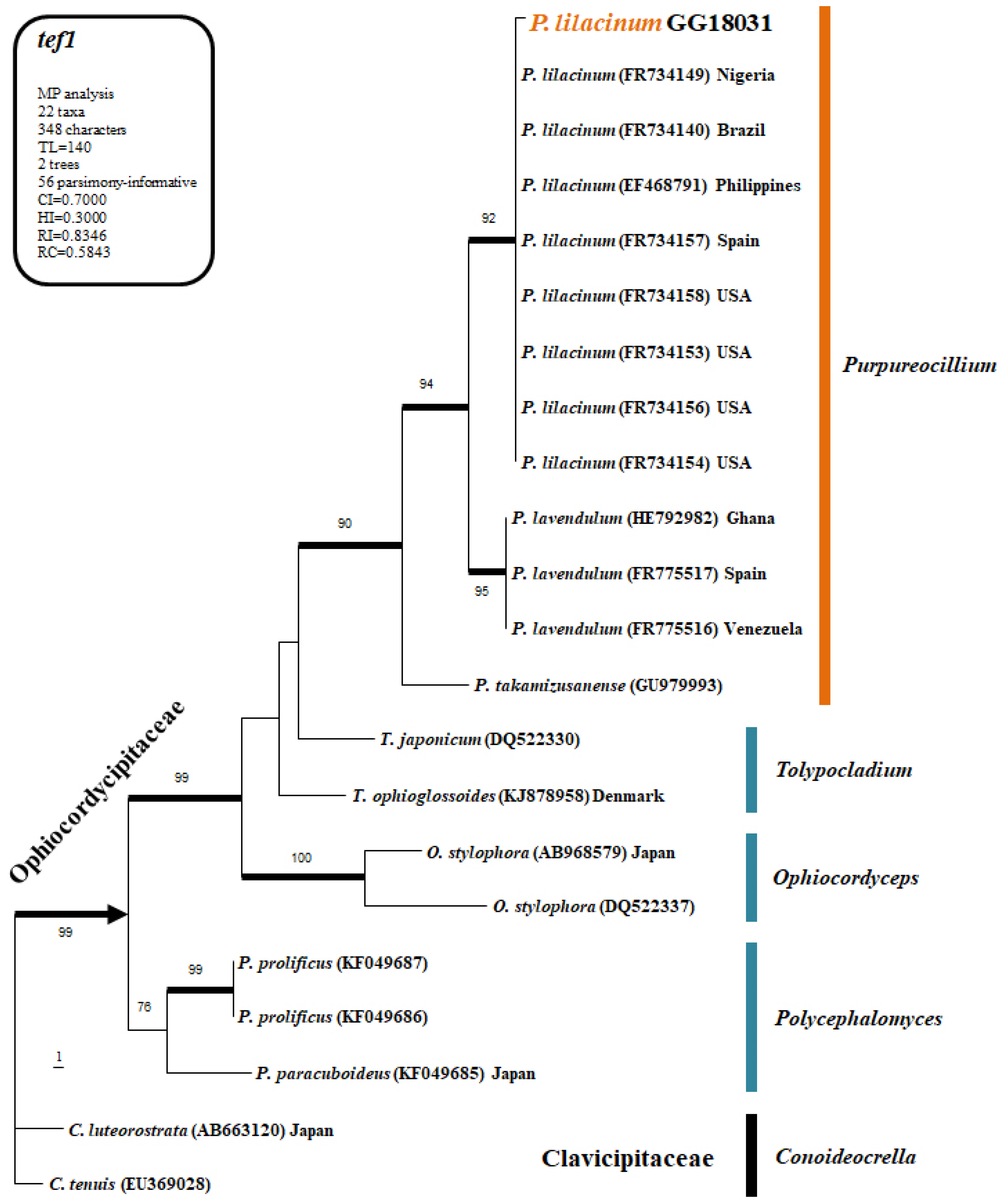
The fungal isolate obtained from soil was subjected to genomic DNA extraction, and sequence analysis was performed using the ITS1, ITS4, and tef1 primers. The PCR products were sent to Bionic Inc. (Seoul, Republic of Korea) for sequencing. The obtained sequences were aligned using ClustalX v.1.81 and edited and curated with PHYDIT v. 3.2. Phylogenetic analysis was conducted using PAUP v. 4.0 b10 for Maximum Parsimony (MP) bootstrap analysis and MrBayes v. 3.1.2 for Bayesian Inference (BI). For the heuristic search to generate the most parsimonious tree, optimization was performed with 1000 random addition sequence replications and tree bisection-reconnection (TBR) branch swapping. Only trees with bootstrap values of 70% or higher were used in the final phylogenetic tree.

2.4. Evaluation of Control Efficacy

The four entomopathogenic fungal isolates were cultured on SDA and PDA media at 25 °C for 10–14 days to induce sporulation. The conidia that developed on the surface were collected inside a clean bench using 0.02% Tween 20. The number of conidia was counted using a hemacytometer, and a conidial suspension (1.0 × 10⁸ conidia/mL) was prepared for subsequent use.
A total of 100 µL of conidial suspension (1 × 10⁸ conidia/mL) was dispensed into each sterilized 25 mL glass vial, amounting to approximately 1 × 10⁷ conidia per vial. The suspension was evenly distributed and air-dried for 24 h at room temperature to allow the liquid to evaporate, leaving only the conidia inside. Five P. koryoensis beetles were then introduced into each vial and sealed with a loosely fitting cap to prevent escape. For the control group, an identical volume of 0.02% Tween-20 solution (without spores) was applied and similarly dried to ensure consistent experimental conditions. The vials were incubated at 25 °C, 40% humidity, and in the dark, with 5 beetles per vial and 3 replicates per treatment.
The P. koryoensis beetles introduced into the vials air-dried at room temperature were examined under a compound microscope at 8 h intervals to determine their survival or death. Individuals showing no movement were considered to have died from infection. The LT50 (lethal time for 50% mortality) for each entomopathogenic fungus was measured. In subsequent observations, the presence of fungal infection in the beetles was confirmed using a microscope.

2.5. Evaluation of the Heat, UV Stability, and Cold Tolerance for Germination of Entomopathogenic Fungal Spore

2.5.1. Evaluation of Heat Stability

Fungal conidia were induced to form by incubating on SDA medium at 25 °C in the dark. After conidia were collected, a conidial suspension (100 µL, 1 × 10⁸ conidia/mL with 0.02% Tween-20) was prepared and placed in sterilized 1.5 mL microtubes. The suspension was then subjected to treatment in a water bath at 45 °C for 1, 2, 3, and 4 h. The control group was treated with the same conidial suspension at 25 °C.
The conidial suspension treated at high temperatures was inoculated with 10 µL into a 96-well plate, with each well containing 200 µL of PDB medium. The plate was incubated at 25 °C in the dark for 48 h. Fungal mycelial growth was then assessed at a wavelength of 650 nm using the VersaMaxPLUS ROM v1.17. The heat stability was evaluated with 5 replicate experiments, and the stability index was expressed as a percentage by measuring the absorbance of the treatment groups relative to the control group.

2.5.2. Evaluation of UV Stability

Fungal conidia were induced to form by incubating on SDA medium at 25 °C in the dark. After conidia were collected, a conidial suspension (100 µL, 1 × 10⁸ conidia/mL with 0.02% Tween-20) was prepared and placed in a 96-well plate. The suspension was then exposed to UV treatment for 30 min, 1 h, and 2 h using a UV lamp. After UV treatment, 10 µL of the conidial suspension was inoculated into a 96-well plate containing 200 µL of PDB medium
The conidial suspension was incubated at 25 °C in the dark for 48 h. Fungal mycelial growth was then assessed at a wave-length of 650 nm using SoftMax Pro [Molecular Devices, VersaMaxPLUS ROM v1.17]. The UV stability was evaluated with 5 replicate experiments, and the stability index was expressed by measuring the absorbance of the treatment groups relative to the control group.

2.5.3. Evaluation of Cold Germination Ability

Fungal conidia were induced to form by incubating on SDA medium at 25 °C in the dark. After conidia were collected, a conidial suspension (1 × 10⁷ conidia/mL with 0.02% Tween-20) was prepared. A 50 µL aliquot of the suspension was then inoculated onto PDA medium using the streak plate method.
The treated media were incubated in the dark for 15 days at 4 °C and 15 °C, respectively. Conidial germination was observed at 1, 2, 3, 6, 10, and 15 days using a phase-contrast microscope (LEICA DM5500B, Wetzlar, Germany) at magnifications of 200×, 400×, or 600×. A total of 100 conidia were randomly observed three times for each time point. The control group was treated at 25 °C with the same conidial suspension. The experiment was performed with 5 replicates.
For the cold germination assay, a lower concentration of 1 × 10⁷ conidia/mL was intentionally used to facilitate clearer microscopic observations by avoiding spore clumping or overgrowth. This concentration was sufficient to accurately monitor individual germination events over time. The higher concentration (1 × 10⁸ conidia/mL) used in other assays was optimized for uniform coating and stronger bioassay efficacy.

3. Results

3.1. Identification of Entomopathogenic Fungi

3.1.1. Morphology

The fungal strain grown on SDA medium exhibited white mycelial growth and, over time, displayed the formation of purplish conidia. Microscopic observation revealed the presence of distinct conidiophores on the aerial mycelium, with the conidiophores branching and producing long, chain-like conidia at the tips. Based on the morphological features including spindle-shaped conidia (2–4 µm) with a thick center and tapered, pointed ends, the identity of the isolate obtained in this study was preliminarily confirmed as Purpureocillium sp. based on the previous report [16].

3.1.2. Phylogenetic Analysis

The phylogenetic results for the ITS (28 taxa) and tef1 (22 taxa) datasets produced based on the MP analysis are shown in Figure 1 and Figure 2, respectively. In the ITS region, GG18031 (GenBank Accession no. PV762045 for ITS) was confirmed to belong to the genus Purpureocillium with high bootstrap support (MPBS = 87), clustering with Purpureocillium takamizusanensis and P. lavendulum (Figure 1). Furthermore, it formed a group with other P. lilacinum sequences, including the reference sequence (FR734101), with a bootstrap value of 53. In the tef1 analysis, GG18031 (GenBank Accession no. PV764424 for tef1) clustered with P. lilacinum sequences, showing even higher bootstrap support (MPBS = 92) than in the ITS analysis (Figure 2). Therefore, based on the results of this study, the soil-isolated strain GG18031 was identified as P. lilacinum.

3.2. Evaluation for the Control Efficacy

The pesticidal activity of the selected three strains was tested against the control treatment, showing significantly higher insecticidal effects (Figure 3). Specifically, the strains M. anisopliae KACC40969 and P. lilacinum GG18031 exhibited survival rates of 33.33% and 46.67%, respectively, after 3 days, demonstrating faster insecticidal effects compared to B. bassiana GHA, which had a survival rate of 86.67%.
Although mortality in the control group was observed with an LT50 of 137.76 h, mortality in all treatment groups occurred significantly earlier, with M. anisopliae KACC40969 inducing the most rapid death (LT50 = 58.7 h) (Table 1). Fungal infection was confirmed microscopically only in the treated groups. The control mortality is attributed to factors such as handling stress and confinement. Despite the delayed decline in control beetles, the significant difference in onset and rate of mortality validates the pesticidal effects of the fungal treatments.

3.3. Evaluation of the Heat, UV Stability, and Cold Tolerance for Germination of Entomopathogenic Fungal Spore

3.3.1. Evaluation of Heat Stability

The spores of insect pathogenic fungal strains were treated at 45 °C for 1 h, 2 h, 3 h, and 4 h, and their stability at high temperatures was assessed using a 96-well plate and VersaMaxPLUS ROM v1.17. In the control treatment (25 °C), the growth rate of M. anisopliae KACC40969 was higher than that of B. bassiana GHA, P. lilacinum GG18031, and B. bassiana KACC43988. The spore growth rates of each strain were found to be 56% for M. anisopliae KACC40969, 20% for P. lilacinum GG18031, and 3% for B. bassiana GHA.
However, P. lilacinum GG18031 treated at 45 °C showed the highest growth rate compared to the other strains, and its growth rate also remained high with increasing treatment time. Furthermore, except for M. anisopliae KACC40969, the growth rates of spores treated at high temperature (45 °C) were higher than those of spores treated at the control temperature (25 °C). These results indicate that the spore growth rate of M. anisopliae KACC40969 is most sensitive to high temperatures, leading to a significant reduction in growth (Figure 4).

3.3.2. Evaluation of UV Stability

UV treatment was applied to the spores of the strains, and their UV stability was assessed using a 96-well plate and VersaMaxPLUS ROM v1.17. The growth rates under no UV treatment were found to be 69% for M. anisopliae KACC40969, 24% for P. lilacinum GG18031, and 5% for B. bassiana GHA, indicating that M. anisopliae KACC40969 had the highest growth rate compared to the other insect pathogenic fungi.
The growth rates of the strains after UV treatment for 30 min, 1 h, and 2 h also showed that M. anisopliae KACC40969 had higher growth rates compared to the other strains. The growth rates over time were analyzed as 61%, 70%, and 66%, respectively (Figure 5).

3.3.3. Low-Temperature Germination Capacity

The spore germination capacity of the strains was evaluated at 4 °C, 15 °C, and 25 °C. The results showed that none of the four strains germinated at 4 °C, indicating a high sensitivity to low temperatures. At 15 °C, the B. bassiana GHA strain reached 100% germination by day 6, while at 25 °C, all three strains achieved 100% germination by day 3 (Table 2).

4. Discussion

In this study, the potential of three entomopathogenic fungal strains, Metarhizium anisopliae KACC40969, Beauveria bassiana GHA, and Purpureocillium lilacinum GG18031, that can be used as a bio-control agent was evaluated against the insect vector, Platypus koryoensis, responsible for transmitting the fungus associated with oak wilt disease in South Korea [8,17].
Analysis of insecticidal efficacy and LT50 revealed that all three fungal strains significantly reduced the survival rate of the insect vectors, with treated groups surviving for 6 days post-infection compared to 9 days in the untreated control. Among the strains tested, M. anisopliae KACC40969 showed the most rapid effect, with the shortest LT50 of 58.7 h. These results suggest that eco-friendly control methods can reduce reliance on chemical pesticides and serve as effective biological control strategies. In conclusion, this study demonstrates that entomopathogenic fungi represent a promising alternative for managing P. koryoensis. Furthermore, these findings may provide a valuable foundation for the development of sustainable forest management strategies in the future.
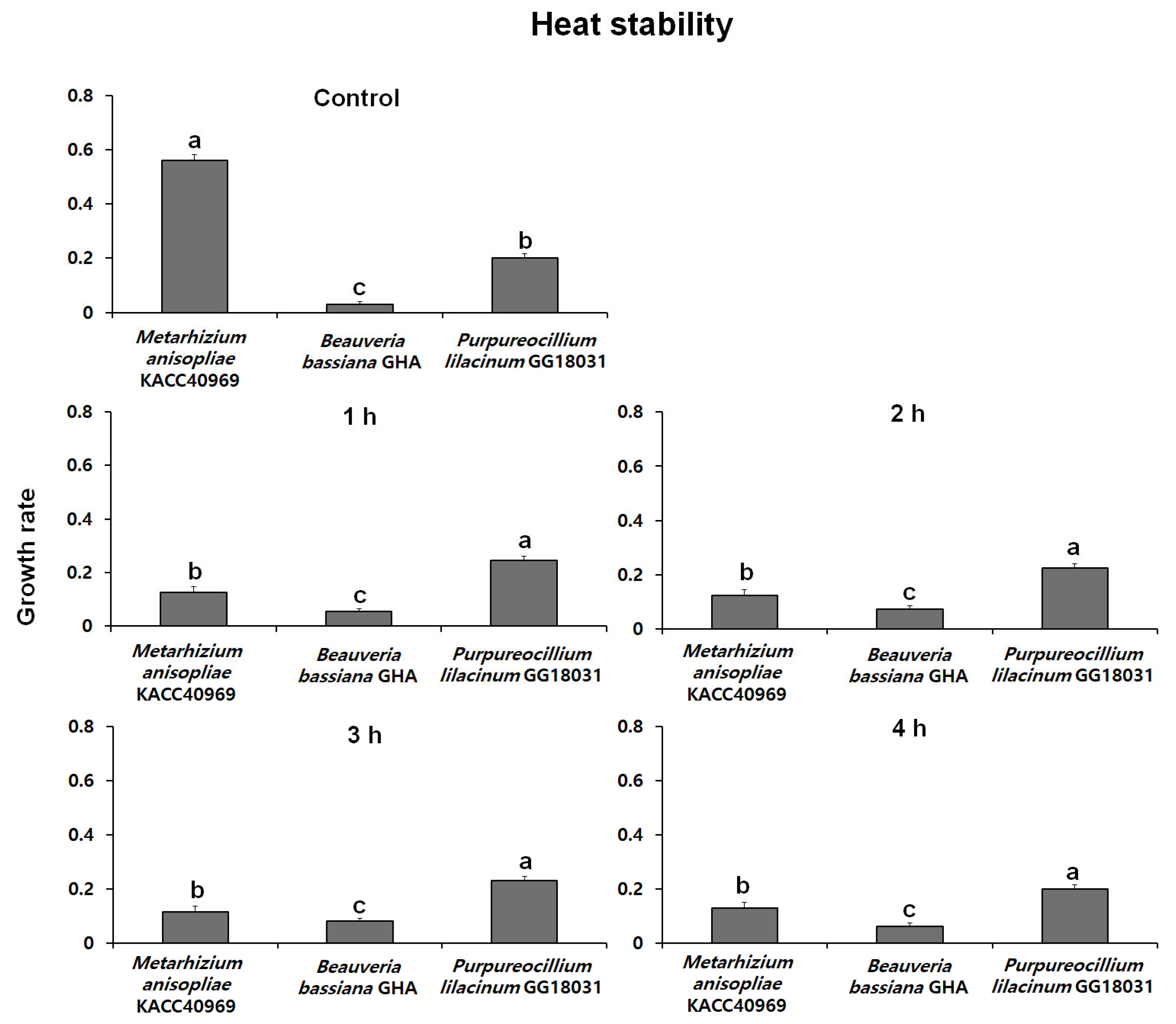
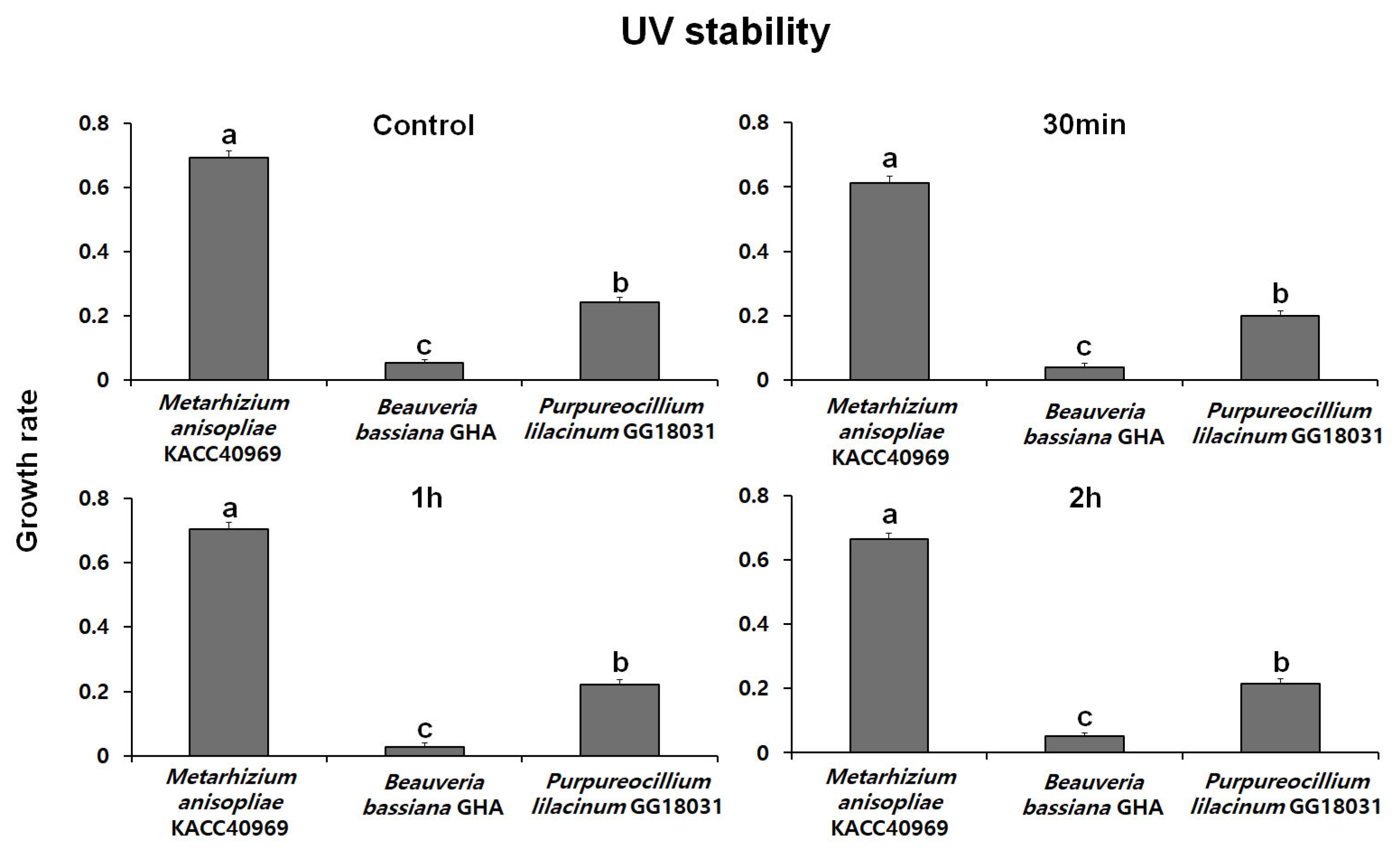
Thermal stability at 45 °C was evaluated over time using spores produced from each fungal strain. The growth rates of B. bassiana GHA and P. lilacinum GG18031 remained unchanged compared to the control regardless of exposure duration. In contrast, M. anisopliae KACC40969 exhibited a markedly reduced growth rate following high-temperature treatment, indicating a high sensitivity to heat stress. In addition, analysis of spore germination under ultraviolet (UV) exposure showed no significant differences compared to the control in all strains. However, M. anisopliae KACC40969 exhibited a relatively higher germination rate compared to the other entomopathogenic fungal strains. Spore germination was completely inhibited at 4 °C for all tested strains, indicating a temperature threshold between 4 °C and 15 °C. At 15 °C, all strains reached 100% germination, although at a slower rate than at 25 °C. These findings highlight that while low temperatures delay the germination process, they do not compromise the viability of the fungal spores. This suggests that the selected strains are capable of functioning effectively across a moderate temperature range relevant to temperate forest environments.
While previous studies on the efficacy of entomopathogenic fungi have largely focused on crops and ornamental plants [9,10,12], this study provides meaningful insights by focusing on a forest pathosystem. Specifically, we assessed the experimental efficacy and practical applicability of a biological control strategy targeting the insect vector [8] responsible for transmitting pathogenic fungi that cause severe damage to oak species—one of the dominant broadleaf tree groups in Korea. These findings contribute to expanding the scope of biological control research into forest ecosystems and highlight the potential of environmentally friendly management strategies for major forest pests and diseases, which might include the use of biological control agents, aiming to minimize non-target toxicity, preserve soil microbial integrity, and reduce disruptions to food webs and broader ecosystem processes [18,19].
While this study provides important insights under controlled conditions, the observed mortality in the control group points to the need for further refinement in bioassay methodology, such as improving ventilation or reducing stress-related factors in confined test environments. This limitation has also been consistently mentioned in previous research [20,21]. Future field trials should also consider these environmental stresses, alongside temperature, humidity, and UV exposure, which can influence fungal efficacy. Moreover, broader soil sampling and formulation improvements should be considered for subsequent research to enhance isolate diversity and environmental resilience [22].

Author Contributions

Conceptualization, D.-H.L. and K.S.; methodology, J.H.L., N.K.K., D.-H.L. and K.S.; software, J.H.L. and K.S.; validation, J.H.L., N.K.K., D.-H.L. and K.S.; formal analysis, J.H.L.; investigation, J.H.L.; resources, J.K.L. and K.S.; data curation, J.H.L. and D.-H.L.; writing—original draft preparation, J.H.L.; writing—review and editing, D.-H.L. and K.S.; supervision, J.K.L., D.-H.L. and K.S. All authors have read and agreed to the published version of the manuscript.

Funding

This research was supported by Chungnam National University and the National Research Foundation of Korea (NRF) (No. RS-2023-00250589).

Data Availability Statement

The data presented in this study are available upon request from the corresponding authors. The data are not publicly available due to institutional policy.

Conflicts of Interest

The authors declare no conflicts of interest.

References

  1. Korea Forest Research Institute. Economic Tree Species O2 Oak; KFRI: Seoul, Republic of Korea, 2012; p. 250, (In Korean Without English Abstract). [Google Scholar]
  2. Cha, S.; Lee, W.-K.; Kim, M.; Lee, S.G.; Jo, H.-W.; Choi, W.I. Characterizing the spatial distribution of oak wilt disease using remote sensing data. J. Korean For. Soc. 2017, 106, 310–319. [Google Scholar]
  3. Korea Forest Research Institute. Annual Report of Monitoring for Forest Insect Pests and Diseases in Korea; SeongMunSa: Seoul, Republic of Korea, 2014. [Google Scholar]
  4. Choi, W.I. Influence of global warming on forest coleopteran communities with special reference to ambrosia and bark beetles. J. Asia Pac. Entomol. 2011, 14, 227–231. [Google Scholar] [CrossRef]
  5. Biedermann, P.H.W.; Vega, F.E. Ecology and evolution of insect–fungus mutualisms. Annu. Rev. Entomol. 2020, 65, 431–455. [Google Scholar] [CrossRef] [PubMed]
  6. Hulcr, J.; Stelinski, L.L. The ambrosia symbiosis: From evolutionary ecology to practical management. Ann. Rev. Entomol. 2017, 62, 285–303. [Google Scholar] [CrossRef] [PubMed]
  7. Lee, J.-S.; Haack, R.A.; Choi, W.I. Attack pattern of Platypus koryoensis (Coleoptera: Platypodidae) in relation to crown dieback of Mongolian oak in Korea. Environ. Entomol. 2011, 40, 1363–1369. [Google Scholar] [CrossRef] [PubMed]
  8. Choi, W.I.; Lee, D.H.; Jung, J.B.; Park, Y.S. Oak decline syndrome in Korean forests: History, biology, and prospects for Korean oak wilt. Forests 2022, 13, 964. [Google Scholar] [CrossRef]
  9. Lacey, L.A.; Frutos, R.; Kaya, H.K.; Vail, P. Insect pathogens as biological control agents: Do they have a future? Biol. Control 2001, 21, 230–248. [Google Scholar] [CrossRef]
  10. Shah, P.A.; Pell, J.K. Entomopathogenic fungi as biological control agents. Appl. Microbiol. Biotechnol. 2003, 61, 413–423. [Google Scholar] [CrossRef] [PubMed]
  11. Khan, M.; Tanaka, K. Purpureocillium lilacinum for plant growth promotion and biocontrol against root-knot nematodes infecting eggplant. PLoS ONE 2023, 18, e0283550. [Google Scholar] [CrossRef] [PubMed]
  12. Han, J.H.; Jeong, H.J.; Lee, M.R.; Choi, S.N.; Kim, D.Y.; Ahn, S.H.; Park, J.W. Insecticidal Effect of Entomopathogenic fungus, Isaria fumosorosea FG340 to Thrips palmi. Korean J. Pestic. Sci. 2020, 24, 374–380. [Google Scholar] [CrossRef]
  13. Kim, S.; Kehinde, B.O.; Park, J.; Raju, M.R.; Hassan, M.R.; Lim, U.T. Insecticidal activity of an entomopathogenic fungi against Pseudaulacaspis prunicola (Hemiptera: Diaspididae). In Proceedings of the 2023 Korean Society of Applied Entomology Fall Meeting and Extraordinary General Assembly, Pyeongchang, Republic of Korea, 25–27 October 2023. [Google Scholar]
  14. Lindgren, B.S. A multiple funnel trap for scolytid beetles (Coleoptera). Can. Entomol. 1983, 115, 299–302. [Google Scholar] [CrossRef]
  15. Sigler, L.; Sutton, D.A.; Gibas, C.F.C.; Summerbell, R.C.; Noel, R.K.; Iwen, P.C. Phialosimplex, a new anamorphic genus associated with infections in dogs and having phylogenetic affinity to the Trichocomaceae. Med. Mycol. 2010, 48, 335–345. [Google Scholar] [CrossRef] [PubMed]
  16. Luangsa-Ard, J.; Houbraken, J.; van Doorn, T.; Hong, S.B.; Borman, A.M.; Hywel-Jones, N.L.; Samson, R.A. Purpureocillium, a new genus for the medically important Paecilomyces lilacinus. FEMS Microbiol. Lett. 2011, 321, 141–149. [Google Scholar] [CrossRef] [PubMed]
  17. Lee, D.H.; Jung, J.M.; Seo, S.T. Population genetic structure of Raffaelea quercus-mongolicae indicates a recent fungal introduction event to Jeju Island from inland areas of South Korea. Plant Pathol. 2021, 70, 1871–1882. [Google Scholar] [CrossRef]
  18. Sánchez-Bayo, F. Indirect effect of pesticides on insects and other arthropods. Toxics 2021, 9, 177. [Google Scholar] [CrossRef] [PubMed]
  19. Wan, N.F.; Fu, L.; Dainese, M.; Kiær, L.P.; Hu, Y.Q.; Xin, F.; Goulson, D.; Woodcock, B.A.; Vanbergen, A.J.; Spurgeon, D.J.; et al. Pesticides have negative effects on non-target organisms. Nat. Commun. 2025, 16, 1360. [Google Scholar] [CrossRef] [PubMed]
  20. Mishra, S.; Kumar, P.; Malik, A. Effect of temperature and humidity on pathogenicity of native Beauveria bassiana isolate against Musca domestica L. J. Parasit. Dis. 2015, 39, 697–704. [Google Scholar] [CrossRef] [PubMed]
  21. Couceiro, J.D.C.; Fatoretto, M.B.; Demétrio, C.G.B.; Meyling, N.V.; Delalibera, Í., Jr. UV-B radiation tolerance and temperature-dependent activity within the entomopathogenic fungal genus Metarhizium in Brazil. Front. Fungal Biol. 2021, 2, 645737. [Google Scholar] [CrossRef] [PubMed]
  22. Nguyen, M.H.; Park, I.K.; Lee, J.K.; Lee, D.H.; Shin, K. Antifungal activity of culture filtrate from endophytic fungus Nectria balsamea E282 and its fractions against Dryadomyces quercus-mongolicae. Forests 2024, 15, 332. [Google Scholar] [CrossRef]
Figure 1. One of the eight most parsimonious trees inferred from a heuristic analysis of ITS sequences. Broad black branches indicated MPBS > 80%. Only MPBS values > 50% are shown above or below branches. Conoideocrella luteorostrata was used as the outgroup.
Forests 16 01009 g001
Figure 2. One of the two most parsimonious trees inferred from a heuristic analysis of tef1 sequences. Broad black branches indicated MPBS > 90%. Only MPBS values > 70% are shown above or below branches. Conoideocrella luteorostrata was used as the outgroup.
Forests 16 01009 g002
Figure 3. Survivorship of adult P. koryoensis after exposure to each entomopathogenic fungus.
Forests 16 01009 g003
Figure 4. Effect of higher temperature on fungal germination of each entomopathogenic fungus at different exposure times. Different letters indicate a significant difference (p < 0.05) among treatments by Tukey’s HSD test.
Forests 16 01009 g004
Figure 5. Effect of UV radiation on fungal germination of each entomopathogenic fungus at different exposure times. Different letters indicate a significant difference (p < 0.05) among treatments by Tukey’s HSD test.
Forests 16 01009 g005
Table 1. Statistical comparison of each entomopathogenic fungus against adults of P. koryoensis.
LT50 aC.I. bSlop ± SEχ2 (df c)
M. anisopliae KACC4096958.7 a47.32–69.554.32 ± 0.696.54 (5)
P. lilacinum GG1803172.79 ab61.55–83.046.14 ± 1.171.11 (4)
B. bassiana GHA90.17 bc80.90–98.6310.98 ± 2.260.79 (4)
Control137.76 d124.41–151.437.26 ± 1.213.01 (7)
a Lethal time (hours) to 50% mortality; b confidence interval (95%); c degree of freedom. LT50 followed by the same letter is not significantly different based on the 95% confidence interval.
Table 2. Effect of lower temperature on fungal germination of different entomopathogenic fungus at different exposure times. Different letters indicate a significant difference (p < 0.05) among treatments by Tukey’s HSD test.
Germination Rate (%)
TemperatureFungus NameDay 1Day 2Day 3Day 6Day 10Day 15
4 °CM. anisopliae KACC409690.000.000.000.000.00
B. bassiana GHA0.000.000.000.000.00
P. lilacinum GG180310.000.000.000.000.00
15 °CM. anisopliae KACC409690.80 a19.47 a34.00 b64.07 b93.27 b100
B. bassiana GHA0.60 a19.87 a49.87 a100 a100 a
P. lilacinum GG180310.00 b0.07 b3.40 c30.33 c100 a
25 °CM. anisopliae KACC4096923.47 a98.07 a100 a
B. bassiana GHA13.73 b92.40 b100 a
P. lilacinum GG180311.93 c39.93 c100 a
Disclaimer/Publisher’s Note: The statements, opinions and data contained in all publications are solely those of the individual author(s) and contributor(s) and not of MDPI and/or the editor(s). MDPI and/or the editor(s) disclaim responsibility for any injury to people or property resulting from any ideas, methods, instructions or products referred to in the content.

