Article

Phlebia formosana Strain SMF410-5-1 and Auricularia cornea Strain ME1-1 Display Potential in Wood Degradation and Forest Waste Reutilization

1
Co-Innovation Center for Sustainable Forestry in Southern China, Nanjing Forestry University, Nanjing 210037, China
2
Fujian Yangkou State Forest Farm, Nanping 353299, China
3
The Connecticut Agricultural Experiment Station Valley Laboratory, Windsor, CT 06095, USA
*
Author to whom correspondence should be addressed.
Forests 2025, 16(5), 795; https://doi.org/10.3390/f16050795
Submission received: 18 March 2025 / Revised: 2 May 2025 / Accepted: 3 May 2025 / Published: 9 May 2025
(This article belongs to the Section Wood Science and Forest Products)

Abstract

:
Wood waste, primarily composed of lignin, cellulose, and hemicellulose, which is typically disposed of through burning and crushing, poses environmental challenges. However, conventional wood waste disposal methods present critical limitations, such as environmental pollution and resource waste. To develop sustainable processing strategies to dispose wood waste, we identified two fungal isolates, SMF410-5-1 and ME1-1, from decayed wood trunks, demonstrating high lignocellulose-degrading enzyme activities, including laccase (Lac, 125.7 U/mL), manganese peroxidase (MnP, 89.3 U/mL), and lignin peroxidase (LiP, 67.9 U/mL). Isolates of ME1-1 and SMF410-5-1 both exhibited superior poplar lignin degradation, while SMF410-5-1 excelled in coniferous wood weight losses, which reached 19.7% for pine after 180 days post inoculation. Moreover, biochemical analyses revealed that isolates of ME1-1 and SMF410-5-1 accelerated the degradation by producing various lignocellulose-degrading enzymes to hydrolyze wood waste. In addition, through multi-locus phylogenetic analysis using sequences of the internal transcribed spacer (ITS), large subunit ribosomal RNA (LSU), and RNA polymerase II second largest subunit (RPB2), SMF410-5-1 and ME1-1 were identified as Phlebia formosana and Auricularia cornea, respectively. This study provides novel insights into fungal-driven biodegradation, offering eco-friendly solutions for forest waste recycling and supporting circular bioeconomy strategies.

1. Introduction

Wood waste is generated through a variety of activities, such as logging, timber production, and wood processing [1,2]. The acceleration of urbanization has led to the increasing consumption of forest resources, resulting in a substantial growth in wood waste [1]. Various traditional methods have been employed for the disposal of wood waste, such as landfilling, incineration, and coarse recycling by crushing. However, these disposal methods are usually associated with significant drawbacks. For instance, the burning of wood waste releases a large number of harmful greenhouse and toxic gases and causes air pollution [3]. The process of crushing and coarse recycling of wood waste results in notable costs for transportation and management [4]. Disposing of synthetic panels in landfills presents a risk of toxic substance leakage into groundwater, which can further contaminate groundwater [5]. Disposal strategies currently not only demonstrate incomplete recycling efficiency for wood waste but also impose substantial ecological burdens through greenhouse gas emissions and particulate matter release. Therefore, it is imperative for more effective biodegradation strategies for wood waste to be developed.
Wood waste is mainly composed of lignin (15%–35%), cellulose (30%–60%), and hemicellulose (20%–40%), along with protein and minerals [6,7]. Together, lignin, cellulose, and hemicellulose collectively account for over 90% of its dry weight [6,7]. Lignin has an intricate three-dimensional structure and consists of a high molecular weight aromatic polymer with biologically stable ether or ester linkages [8]. Lignin is primarily responsible for the adhesive properties of plants, which bind the cellulose fibers together to constitute their structural framework [9]. This characteristic renders it insoluble and resistant to acid hydrolysis. Although short-term thermal modification alters the chemical and structural properties of wood waste, the degree and order of degradation of wood waste are a matter of many parameters, mainly depending on the wood rotting fungi [10]. Additionally, the dense structure of lignin impedes the access of degrading enzymes to cellulose and hemicellulose, thereby diminishing the efficiency of wood waste enzymatic digestion [9,11]. This poses a well-recognized challenge for microbial degradation [11,12]. Consequently, the high lignin content and low protein levels in forestry waste limit its potential applications [13,14]. Therefore, the degradation of lignin is a prerequisite for the efficient utilization of wood waste.
Numerous microorganisms, including fungi and bacteria, have the ability to degrade lignocellulose [11,15,16]. In particular, wood-rotting fungi are able to break down lignocellulose into water and carbon dioxide through lignocellulose-degrading enzymes [11,15,16]. White-rotting fungi produce lignin-degrading enzymes, such as laccase (Lac), manganese peroxidase (MnP), lignin peroxidase (LiP), versatile peroxidase, and dye-decolorizing peroxidase [17], initiating lignin degradation through one-electron oxidation by enzymatic catalysis, forming free radicals with high redox potential and opening aromatic rings. Notably, laccase (Lac), lignin peroxidase (LiP), and manganese peroxidase (MnP) are important for the lignin degradation process [18]. Wood-rotting fungi play a vital role in the material cycle within ecosystems. The use of white-rotting fungi for wood waste resolution is highly desirable due to its practicality, safety, and environmental sustainability [19]. White-rotting fungi have been employed to degrade lignocellulose in biopulping, biological fuel, the feed industry, and composting [20,21,22,23]. For example, white-rotting fungi mediate biopulping through the secretion of ligninolytic enzymes, which selectively remove lignin to enhance pulp brightness and paper strength properties [20]. The application of wood-rotting fungi for wood waste degradation presents a promising strategy for the circular utilization of wood resources.
It expands the microbial resource available for lignocellulose degradation for the development of environmentally friendly and sustainable strategies for wood waste recycling and valorization. Our study aimed to identify wood-rotting fungal isolates capable of efficiently degrading wood waste. Through substrate-based preliminary screening, two isolates, SMF410-5-1 and ME1-1, were selected for their high lignin-degrading enzyme activities. Subsequent analyses revealed that these strains produced abundant lignocellulolytic enzymes and were highly effective in decomposing wood waste from various tree species. Considering those functions in decayed wood, these strains will be applied to the degradation of wood waste and more fields. Moreover, these findings provide valuable insights into the biodegradation potential of wood-rotting fungi and offer a promising basis for the sustainable bioconversion and utilization of lignocellulosic waste.

2. Materials and Methods

2.1. Isolation of Fungal Isolates and Culture Conditions

In June 2021, decayed tree trunks with visible fungal colonization were collected from the Nanjing Forestry University campus (32°4′41″ N, 118°48′34″ E), mainly comprising hardwood species (e.g., Populus spp.) and softwood species (e.g., Pinus spp.). These decayed trunks were cut into small pieces (2~3 × 2~3 mm) and surface sterilized in 75% alcohol for 30 s. Then, the wood pieces were immersed in 1% sodium hypochlorite (NaClO) for 90 s, followed by three rinses with sterile water [24]. Subsequently, the wood pieces were put on 2% potato dextrose agar (PDA) plates and cultured in the dark at 25 °C for 5 days. The hyphal tips were subsequently transferred to fresh PDA plates for the purification of fungal isolates [24].
Isolates of ME1-1 and SMF410-5-1 were used to detect the ability of degraded wood waste, and these strains have been deposited in the China Center for Type Culture Collection (CCTCC) with the access numbers of CCTCC AF 2024044 and M 20231538, respectively. The reference strain Pleurotus ostreatus (PO; CGMCC5.784) was purchased from China General Microbiological Culture Collection Center (CGMCC) and served as the positive control in this study.
All fungal isolations were maintained on potato dextrose agar (PDA) plates at 25 °C in this study. For total DNA extraction, strains were incubated in PD broth media with shaking for 2 days and harvested.

2.2. Screening of Potential Wood-Rotting Fungi

Laccase-producing fungal strains were screened using guaiacol as a substrate through plate assays [25]. In our study, candidate isolates were inoculated on PDA plates supplemented with 0.5 g/L guaiacol (Sangon, Shanghai, China; A600490). Fungal colonies demonstrating guaiacol-derived brown halo formation were prioritized as laccase-producing candidate isolates.
In order to screen the fungal isolates for secreted peroxidase activity, aniline blue was used as a chromogenic indicator [26]. Each fungal isolate was inoculated on the PDA plates supplemented with 0.1 g/L aniline blue (Sangon, Shanghai, China; A500083). The formation of a discolored zone around the colony presents peroxidase secretion.
To screen cellulose-producing isolates, fungal isolates were inoculated on carboxymethyl cellulose sodium (CMC-Na) plates for 6 days [27]. The plates were then stained with 1 mg/mL Congo red (Sangon, Shanghai, China; A600324) for 10 min and subsequently decolorized with 1 M NaCl. The formation of a discolored circle was used to assess the cellulase-producing ability of the tested isolates.
We assessed the ability of the fungus to produce diverse hydrolytic enzymes by calculating the ratio of the discoloration (D) to the fungal colony size (d). The experiment was conducted three times, with each treatment having three replicates.

2.3. Enzyme Activity Assays of Laccase, Manganese Peroxidase, and Lignin Peroxidase

Mycelial plugs (~5 mm in diameter) were harvested with a sterile 5 mm cork borer when mycelial colonization had reached two-thirds of the agar plate, inoculated in 100 mL of PDB, and shaken at 160 rpm in an orbital shaking incubator at 28 °C for 4 days. One milliliter of fermentation filtrate of each isolate was collected and centrifuged at 6000 rpm for 10 min, and the prepared supernatant was used to detect different enzyme activities of laccase (Lac), manganese peroxidase (Mnp), and lignin peroxidase (Lip) using a UV–visible spectrophotometer. In brief, the Lac activity was measured using its catalysis of the substrate ABTS (2,2’-Azinobis (3-ethylbenzothiazoline-6-sulfonic acid) Ammonium Salt) at a wavelength of 420 nm [28,29]. The Mnp activity was analyzed by the oxidation of guaiacol in the presence of Mn2+ at a wavelength of 465 nm [29]. The LiP activity was determined based on the oxidation of the dye Azure B at a wavelength of 651 nm [30]. All experiments were conducted three times, and the treatment had three replicates.

2.4. Assays of Wood Weight Loss

Six mycelial plugs of tested strains (~5 mm in diameter) were inoculated onto autoclaved wood chips (5 g in weight) of poplar (Populus delotides×P. euramericana. cv. ‘Nanlin895’), pine (Pinus massoniana), and Chinese fir (Cunninghamia lanceolata), respectively, without additional nutrients and kept at 25 °C in a dark environment within the incubator. The weight loss of the wood chips was measured at 3 and 6 months after post-inoculation using the following formula:
ω (%) = (W1 − W2)/W1 × 100%
ω(%) represents the weight loss rate; W1 (g) and W2 (g) indicate the absolute dry weights of the wood sample pre-inoculation and post-inoculation, respectively. The experiment was conducted twice, with each treatment having three replicates.

2.5. Degradation of Wood Lignin, Cellulose, and Hemicellulose

The contents of lignin, cellulose, and hemicellulose in wood samples were determined using assay kits (#BC4200, #BC4280, #BC4445, Solarbio, Beijing, China) following the protocol provided. In brief, approximately 5 g of sawdust of poplar, Chinese fir, and pine was sampled; 0.5 mL of distilled water and 0.75 mL of concentrated sulfuric acid were added to the sample, then mixed and ice-bathed for 30 min. The supernatant was collected by centrifuging at high speed and diluted 20 times for testing. The lignin, cellulose, and hemicellulose contents were measured by the manufacturer’s instructions [31]. The experiment was conducted twice, with each treatment having three replicates.
The degradation efficiency of each component in wood was calculated at 90 and 180 days post-infection (dpi), respectively, using the following formula:
DR = (m0 × M1 − m1 × M2) / m0 × M1 × 100%
DR (%) represents the degradation rate; m0 (g) represents the dry weight of the wood specimen before inoculation; m1 (g) represents the dry weight of the wood sample after inoculation with wood-rotting fungi; M1 (%) indicates the content of each component before inoculation; M2 (%) represents the content of the component after inoculation with wood-rotting fungi.

2.6. Phylogenetic Analysis

The genomic DNA of fungal isolates was extracted using the method described [32]. The sequences of ITS, LSU, and RPB2 of the tested isolates were amplified with the primers listed in Table S1. The fragments were purified and sequenced by Shanghai Sangon Biotechnology Company. For phylogenetic analysis, reference sequences of ITS, LSU, and RPB2 from other strains were downloaded from the NCBI database accessed on 1 May 2023 (https://www.ncbi.nlm.nih.gov/). The phylogenetic analysis of isolate SMF410-5-1 employed ITS/LSU sequences, whereas isolate ME1-1 was characterized using ITS/LSU/RPB2 sequences. The sequences of ITS, LSU, and RPB2 were aligned and edited using Molecular Evolutionary Genetic Analysis (MEGA 7.0) with Clustal W [33]. The multi-locus phylogenetic tree was constructed using Maximum Likelihood (ML) and Bayesian inference (Bl) analyses with IQ-TREE v.1.6.8 and MrBayes v.3.2.6, respectively. The tree was drawn with branch lengths measured in substitutions per site. The interior-branch test was performed using 1000 bootstrap replications to evaluate the relative stability of the branches.

2.7. Statistical Analysis

Statistical analyses were performed using Prism 8.0 (GraphPad) software. Statistical significance was calculated using one-way ANOVA followed by Tukey’s honest significance test. All error bars represent the standard deviation (SD) of the mean.

3. Results

3.1. Isolation and Screening of Wood-Rotting Fungal Isolates

Eight fungal isolates were obtained from the decayed trunks, with visible fungal colonization of Chinese fir and other tree species. With the exception of isolate PA-2, ME1-1, LZ, SMF410-5-1, and JH-1 displayed significant laccase activity when exposed to guaiacol (Figure S1A; Table S2). Among them, isolates ME1-1 and SMF410-5-1 produced the highest levels of laccase. (Figure 1A). When these fungal isolates were inoculated on the PDA supplemented with aniline blue, the isolates of ME1-1 and SMF410-5-1 also demonstrated relatively higher peroxidase secretion (Figure S1B and Figure 1B). Then, we found that the isolate of SMF410-5-1 displayed the best performance of producing cellulase on PDA plates supplemented with CMC-Na (Figure S1C and Figure 1C). Based on these findings, isolates of ME1-1 and SMF410-5-1 exhibiting stronger laccase, peroxidase, and cellulase production ability were selected as candidate isolates for further analysis.

3.2. Quantitation of Laccase, Manganese Peroxidase, and Lignin Peroxidase of ME1-1 and SMF410-5-1

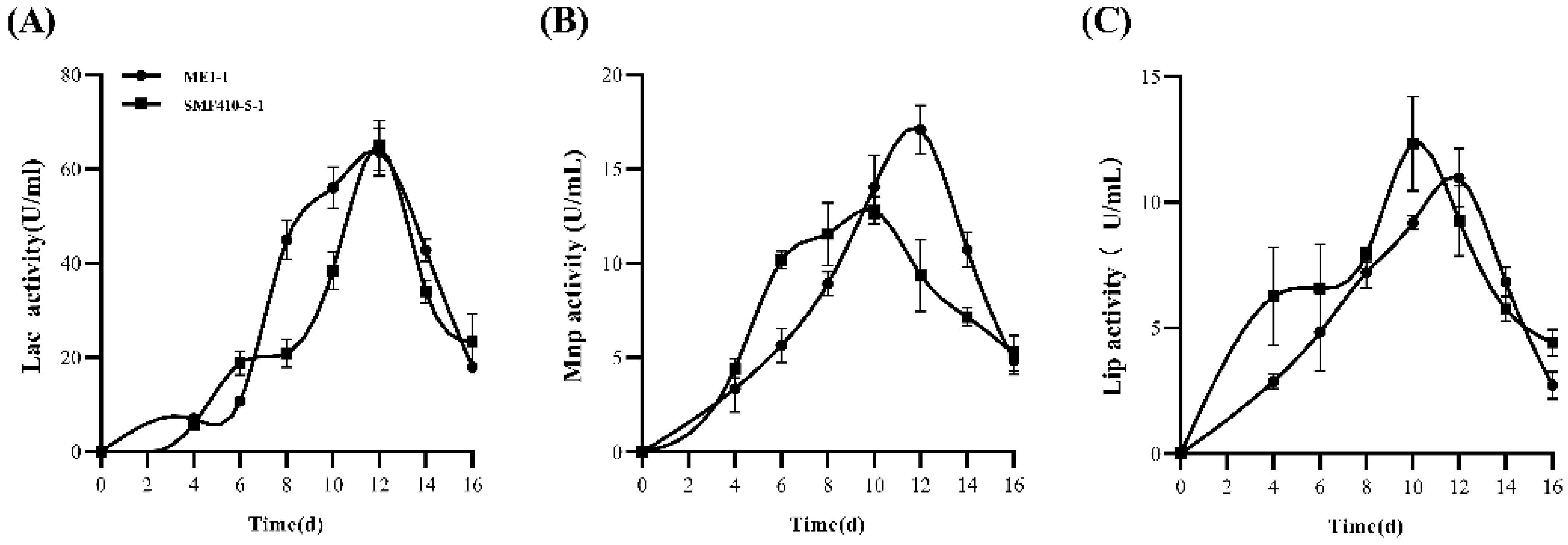
To investigate the activity of laccase, manganese peroxidase, and lignin peroxidase, the liquid culture filtrates of the SMF410-5-1 and ME1-1 isolates were analyzed quantitatively. The results showed that laccase activity in SMF410-5-1 gradually increased from 0 to 16 days post-inoculation (dpi), peaking at 64.9 U/mL at 12 dpi. Similarly, isolate ME1-1 exhibited a comparable trend, reaching 63.6 U/mL at 12 dpi (Figure 2A). Manganese peroxidase (Mnp) activity of SMF410-5-1, reaching 12.8 U/mL, occurred at 10 dpi. The ME1-1 isolate exhibited the peak Mnp activity of 17.1 U/mL on day 12 (Figure 2B).
Lignin peroxidase (Lip) activity was also detected in the culture filtrates of both SMF410-5-1 and ME1-1. Lip activity of SMF410-5-1 peaked at 12.33 U/mL on day 10. The Lip activity of ME1-1 peaked on day 12 (Figure 2C). The activities of laccase, manganese peroxidase, and lignin peroxidase were not detected in the control (blank liquid PD medium). These results indicate that both SMF410-5-1 and ME1-1 isolates can effectively secrete laccase, manganese peroxidase, and lignin peroxidase, facilitating wood waste degradation.

3.3. Wood Weight Loss Caused by ME1-1 and SMF410-5-1

Wood weight loss caused by the fungal isolates ME1-1 and SMF410-5-1 was evaluated at 90 and 180 dpi, respectively. ME1-1 caused significant weight loss in poplar wood chips, with a loss of 44.8 ± 3.27% at 90 dpi, which was significantly higher than that caused by the isolate SMF410-5-1 (7.33 ± 0.58%) and reference isolate PO (19.6 ± 2.60%). The reference isolate PO is characterized by its unique set of ligninolytic enzymes necessary for the degradation of wood [34]. A similar weight loss trend of poplar wood chips was observed at 180 dpi (Figure 3A).
However, when these fungal isolates were inoculated on the wood chips of Chinese fir, SMF410-5-1 resulted in the highest wood weight loss at 90 dpi. After 180 dpi, both ME1-1 and SMF410-5-1 resulted in wood weight loss rates of approximately 13.9 ± 1.10% and 14.7 ± 0.81%, which were significantly higher than those of the isolate PO and the blank control (Figure 3B). When the isolates tested were inoculated on the pine wood chips, they exhibited varying wood degradation efficiencies. At 90 dpi, the isolate SMF410-5-1 caused the highest wood weight loss. After 180 days of inoculation, SMF410-5-1 displayed the highest wood degradation efficiency, with a loss rate of 19.7 ± 1.50% of pine wood weight (Figure 3C). These data indicated that the tested fungal isolates have varying abilities to degrade different wood substrates. Isolate ME1-1 showed a stronger degradation effect on poplar wood, while SMF410-5-1 had a better performance in degrading the woods of Chinese fir and pine.

3.4. Degradation of Wood Lignin, Cellulose, and Hemicellulose by M1E-1 and SMF410-5-1

ME1-1 and SMF410-5-1 caused significant wood weight loss in poplar, Chinese fir, and pine. To determine whether this weight loss was attributed to the degradation of lignin, cellulose, and hemicellulose, we further assessed the breakdown of these components by the fungal isolates. Lignin degradation assays on poplar revealed that ME1-1 had the strongest lignin degradation rate at 90 dpi, reaching approximately 30%, which further increased to 40.7 ± 2.52% at 180 dpi (Figure 4A). Cellulose degradation analysis indicated that SMF410-5-1 had a stronger ability to degrade poplar cellulose than PO and ME1-1 (Figure 4B). Upon inoculation of the tested isolates on poplar wood, the poplar hemicellulose was significantly degraded. Among these fungal isolates, the reference isolate PO showed the strongest ability to degrade poplar hemicellulose, followed by ME1-1 and SMF410-5-1 (Figure 4C).
To assess lignin, cellulose, and hemicellulose degradation in Chinese fir, the isolates PO, ME1-1, and SMF410-5-1 were inoculated individually on Chinese fir wood and analyzed at 90 and 180 dpi. Among these fungal isolates, SMF410-5-1 displayed the strongest lignin degradation capability on Chinese fir, with the degradation rate reaching 21.8 ± 2.80% at 180 dpi, significantly higher than those of PO and ME1-1 (Figure 4D). At 90 dpi, the isolate SMF410-5-1 showed the strongest cellulose degradation ability. SMF410-5-1 also demonstrated superior performance in degrading Chinese fir cellulose, with a 33.4 ± 1.72% degradation rate after 180 dpi (Figure 4E). Similarly, at both 90 and 180 dpi, SMF410-5-1 also showed the strongest ability to degrade Chinese fir hemicellulose among all tested isolates (Figure 4F).
In pine wood, lignin degradation assays demonstrated that SMF410-5-1 exhibited the highest degradation rate among the tested fungal isolates, achieving a degradation rate of 37.7 ± 0.96% (Figure 4G). Among these isolates, SMF410-5-1 had the strongest capability of degrading pine cellulose (Figure 4H). At 90 dpi, SMF410-5-1 exhibited the highest hemicellulose degradation rate, reaching 15.74 ± 1.54%. At 180 dpi, SMF410-5-1 displayed a similar hemicellulose degradation ability to PO, and it was significantly stronger than that of ME1-1 (Figure 4I). In conclusion, these results indicate that both ME1-1 and SMF410-5-1 degrade the major components of lignin, cellulose, and hemicellulose of various types of wood. Furthermore, ME1-1 and SMF410-5-1 have different capabilities of degrading lignin, cellulose, and hemicellulose across different tree species.

3.5. Molecular Identification of ME1-1 and SMF410-5-1

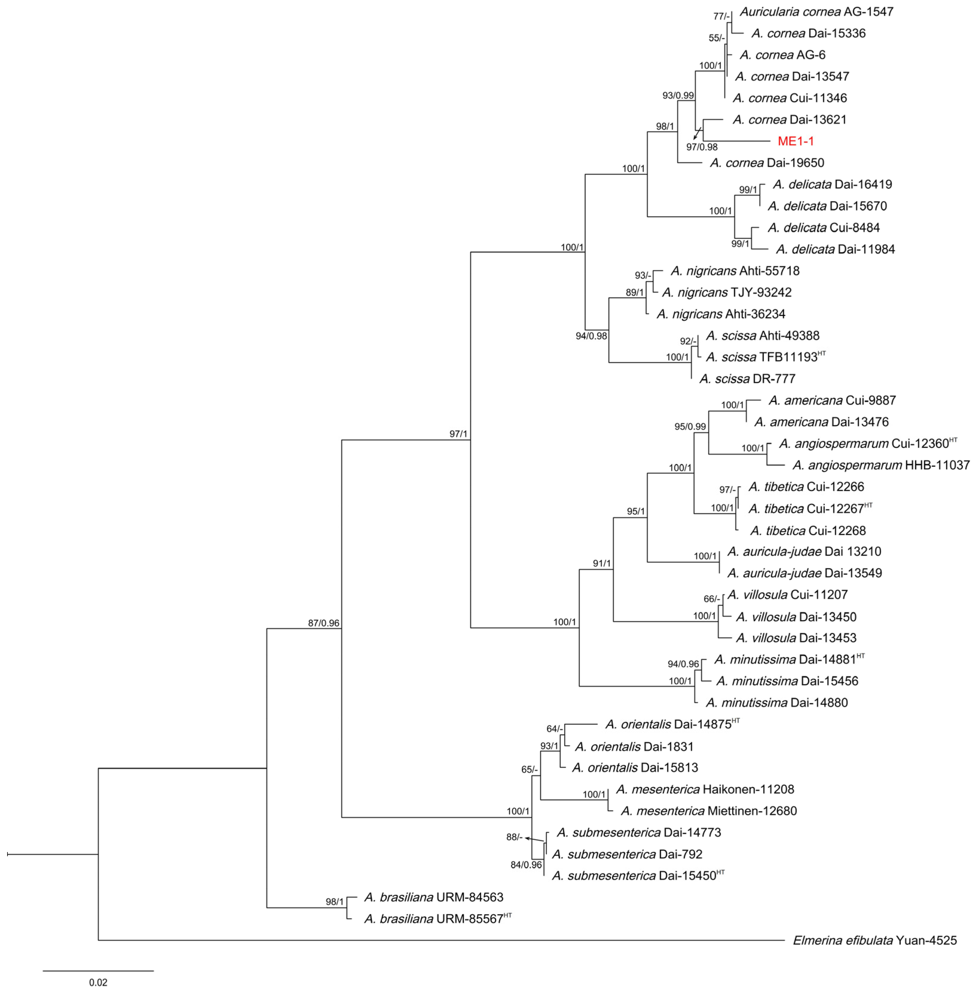
Gene fragments of ITS, LSU, and RPB2 from the ME1-1 isolate were successfully cloned, sequenced, and deposited in GenBank under the accession numbers OR608007, OR608009, and OR611928. The similarities of ITS, LSU, and RPB2 between the ME1-1 isolate and the Auricularia cornea strain were 96.56%, 99.71%, and 96.49%, respectively (Table S3). To further identify the ME1-1 isolate, phylogenetic analysis using the concatenated sequences of ITS, LSU, and RPB2 showed that ME1-1 and authentic strains of A. cornea formed a monophyletic clade, supported by a significantly higher bootstrap value (Figure 5). The phylogenetic tree indicate that ME1-1 belongs to A. cornea.
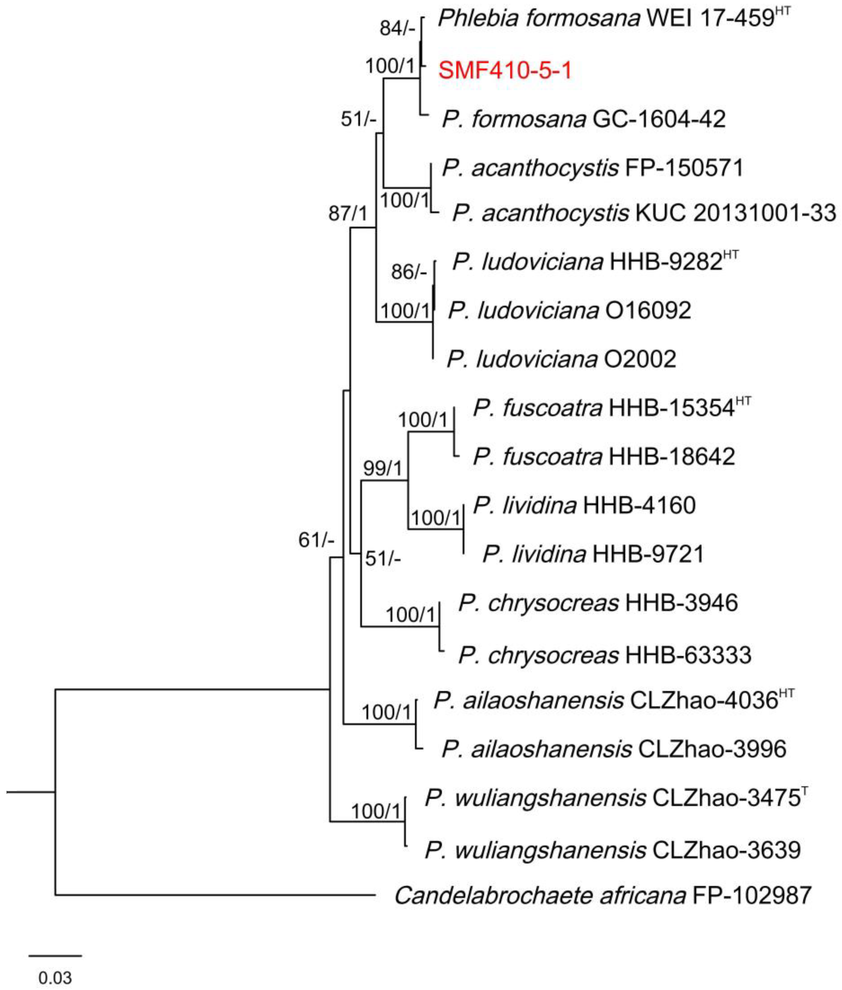
To identify the SMF410-5-1 isolate, the sequences of ITS and LSU were also successfully obtained by PCR. Then, these sequences were deposited in GenBank, under the accession numbers OR608008 and OR608010. Similarities of ITS and LSU in the SMF410-5-1 were 99.50% and 99.55% compared to the Phlebia formosana strain (Table S3). The phylogenetic tree derived from concatenated sequences of the ITS and LSU suggests that SMF410-5-1 belongs to P. formosana (Figure 6). Isolates of ME1-1 and SMF410-5-1 have been deposited in the China Center for Type Culture Collection (CCTCC) with the access numbers CCTCC AF 2024044 and M 20231538, respectively.

4. Discussion

Wood waste is an abundant and renewable resource on Earth, and its reutilization offers significant ecological and economic benefits [35]. Lignin, cellulose, and hemicellulose are the main components of plant cell walls and constitute the majority of plant biomass [36]. It is a complex process to build polymers of lignin, cellulose, and hemicellulose in the cell wall. The complexity necessitates pre-treatment techniques, such as enzymes, to degrade these components [36,37]. Thus, we found that the wood-rotting fungal isolates ME1-1 and SMF410-5-1 exhibited a strong ability to degrade lignin, cellulose, and hemicellulose, highlighting their potential for the cyclic utilization of forest wood waste.
Compared to traditional methods, wood-rotting fungi have been considered as the primary microorganisms involved in the degradation of wood waste [19]. Wood-rotting fungi can produce a diverse pool of enzymes capable of decomposing various crop and tree residues, including beech [38], Populus tomentosa [39], and wheat straw [40]. These hydrolases, especially laccase, manganese peroxidase, and lignin peroxidase, play crucial roles in the breakdown of lignocellulosic biomass, including lignin, cellulose, and hemicellulose [41]. The utilization of wood-rotting fungi for wood waste management offers distinct advantages to reduce energy consumption [42]. Wood-rotting fungi exhibit unparalleled enzymatic versatility, employing synergistic lignocellulolytic enzymes to holistically deconstruct complex plant biomass [42,43]. In this study, we isolated wood-rotting fungal isolates from decayed wood trunks. Among them, isolates ME1-1 and SMF410-5-1 showed a stronger ability to produce laccase, peroxidase, and cellulase. In a liquid medium, these isolates also demonstrated high enzymatic activity of laccase, manganese peroxidase, and lignin peroxidase to degrade the major components of the plant cell wall. Compared to Canoderma lucidum, a common white-rotting fungus produced laccase abundantly, reaching 20 U/mL in the PDB medium on the sixth day. However, its laccase significantly increased to 68.75 U/mL when corncob was added as a substrate [44]. In our study, isolates of ME1-1 and SMF410-5-1 reached peak laccase activities of 63.63 U/mL and 64.95 U/mL in the PDB liquid medium after 12 days. Therefore, we suggest that isolates of ME1-1 and SMF410-5-1 possess robust laccase activity, which could potentially be further optimized through substrate supplementation, pH adjustment, or other strategies. Isolates of SMF410-5-1 and ME1-1 exhibited manganese peroxidase (Mnp) activities of 12.8 U/mL and 15.5 U/mL, respectively, which were significantly higher than other white-rotting fungi, such as Lenzites betulina and Trametes versicolor [45]. Therefore, we suggest that isolates of SMF410-5-1 and ME1-1 possess significant potential in degrading wood waste for microbial biofertilizers. Furthermore, when treated with the wood-rotting fungal isolates ME1-1 and SMF410-5-1, the biomasses of poplar, Chinese fir, and pine were significantly decreased compared to the positive control P. ostreatus. Additionally, the principal components in the plant cell wall of wood waste exhibited a significant decrease. The findings indicate that the isolates ME1-1 and SMF410-5-1 may contribute to the wood waste degradation process by producing and secreting hydrolases to break down lignin, cellulose, and hemicellulose. Additionally, wood-rotting fungal degradation aligns with circular economy principles by converting waste into reusable outputs [46]. Critically, the advantages of degraded wood waste by wood-rotting fungi prompted us to further investigate the biotechnological potential of isolates ME1-1 and SMF410-5-1 for industrial applications.
To further explore and apply these fungal isolates, we identified ME1-1 and SMF410-5-1 as Auricularia cornea (ITS 96.56%, LSU 99.71%, RPB2 96.49%) and Phlebia formosana (ITS 99.50%, LSU 99.55%), respectively, with phylogenetic analysis. The wood-rotting fungal isolate ME1-1 belongs to the genus Auricularia (Basidiomycota), which has both nutritional and medicinal applications [47]. In this study, we found that re-inoculation of strain SMF410-5-1, originally isolated from naturally decaying pine wood, into pine wood waste resulted in a higher colonization capacity compared to isolates from other sources. Thus, the isolate SMF410-5-1 exhibited a stronger ability to degrade wood waste derived from pine spp. These findings indicated that isolate SMF410-5-1 was well adapted to abundant secondary metabolites produced by coniferous trees. Therefore, it is suggested that the isolate SMF410-5-1 may have significant advantages in the biodegradation and cycling of pine wood product waste and dead pine trees. For instance, in Asia, large numbers of pine trees have died due to infestations by the pine wilt nematode Bursaphelenchus xylophilus [48,49]. However, since B. xylophilus is a critical quarantine invasive species and threatens forest ecosystems worldwide [50], local harmless treatment of dead pine trees is particularly important for disease prevention and control. The wood-rotting fungi identified in this study demonstrated efficient degradation of pine wood, providing more options for the sustainable reutilization of wood waste.

5. Conclusions

Collectively, our data suggest that the wood-rotting fungal isolates ME1-1 and SMF410-5-1 possess strong capabilities to degrade lignin, cellulose, and hemicellulose, indicating their potential for the sustainable utilization of forest wood waste. Although these isolates can produce abundant hydrolases to decompose wood waste under laboratory conditions, current evidence regarding their industrial effectiveness remains limited. In future studies, we aim to explore the biosynthetic pathways of various degradative enzymes, which may facilitate their efficient application in industrial settings.

Supplementary Materials

The following supporting information can be downloaded at: https://www.mdpi.com/article/10.3390/f16050795/s1, Figure S1: Screening of wood-rotting fungal isolates producing laccase, peroxidase, and cellulose; Table S1: Primers used for PCR amplification and DNA sequences; Table S2: Fungal isolates used for activity of laccase, peroxidase, and cellulase in the study; Table S3: The similarity analysis of sequences ITS, LSU, and RPB2 for isolates ME1-1 and SMF410-5-1; Table S4: Fungal strains and sequences of ITS, LSU and RPB2 used for phylogenetic analysis in the study.

Author Contributions

J.Y. and L.H. designed the research. H.-L.Q., J.H., J.-L.R. and Y.R. performed experiments. H.-L.Q., Y.R., J.Y. and J.-H.H. analyzed data. Y.R., J.-H.H. and D.-W.L. wrote the manuscript. J.Y., D.-W.L. and L.H. edited the manuscript. All authors have read and agreed to the published version of the manuscript.

Funding

This study was funded by the National Key R & D Program of China (Grant number 2023YFD1401304), Qing Lan Project, and Priority Academic Program Development of Jiangsu Higher Education Institutions (PAPD).

Data Availability Statement

The data that support the findings of the study are provided in the Supplemental File.

Conflicts of Interest

The authors declare no competing interests.

References

  1. Mancini, M.; Rinnan, A. Classification of waste wood categories according to the best reuse using FT-NIR spectroscopy and chemometrics. Anal. Chim. Acta. 2023, 1275, 341564. [Google Scholar] [CrossRef] [PubMed]
  2. Pazzaglia, A.; Gelosia, M.; Giannoni, T.; Fabbrizi, G.; Nicolini, A.; Castellani, B. Wood waste valorization: Ethanol based organosolv as a promising recycling process. Waste Manag. 2023, 170, 75–81. [Google Scholar] [CrossRef]
  3. Aliaño-González, M.J.; Gabaston, J.; Ortiz-Somovilla, V.; Cantos-Villar, E. Wood Waste from Fruit Trees: Biomolecules and Their Applications in Agri-Food Industry. Biomolecules 2022, 12, 238. [Google Scholar] [CrossRef]
  4. de Klerk, S.; Ghaffariyan, M.R.; Miles, M. Leveraging the entrepreneurial method as a tool for the circular economy: The case of wood waste. Sustainability 2022, 14, 1559. [Google Scholar] [CrossRef]
  5. Huang, Z.; Gao, N.; Liu, M.; Lin, Y.; Chang, G.; Deng, L.; Huang, H. Emissions of nitrogenous pollutants in chemical looping gasification of high nitrogen wood waste using a K-modified copper slag oxygen carrier. J. Therm. Anal. Calorim. 2022, 147, 9725–9735. [Google Scholar] [CrossRef]
  6. Tuomela, M.; Vikman, M.; Hatakka, A.; Itävaara, M. Biodegradation of lignin in a compost environment: A review. Bioresour. Technol. 2000, 72, 169–183. [Google Scholar] [CrossRef]
  7. Chaverri, R.H.; Figueroa, J.B. Biomass: Biorefinery as a model to boost the bioeconomy in Costa Rica, a review. Agron. Mesoam. 2021, 32, 1047–1070. [Google Scholar]
  8. Ong, S.S.; Wickneswari, R. Expression profile of small RNAs in Acacia mangium secondary xylem tissue with contrasting lignin content—Potential regulatory sequences in monolignol biosynthetic pathway. BMC Genom. 2011, 12, S13. [Google Scholar] [CrossRef] [PubMed]
  9. Balat, M. Production of bioethanol from lignocellulosic materials via the biochemical pathway: A review. Energy Convers. Manag. 2011, 52, 858–875. [Google Scholar] [CrossRef]
  10. Kamperidou, V. Chemical and structural characterization of poplar and black pine wood exposed to short thermal modification. Drv. Ind. 2021, 72, 155–167. [Google Scholar] [CrossRef]
  11. Gurovic, M.S.V.; Viceconte, F.R.; Bidegain, M.A.; Dietrich, J. Regulation of lignocellulose degradation in microorganisms. J. Appl. Microbiol. 2023, 134, lxac002. [Google Scholar] [CrossRef]
  12. Banu Jamaldheen, S.; Kurade, M.B.; Basak, B.; Yoo, C.G.; Oh, K.K.; Jeon, B.H.; Kim, T.H. A review on physico-chemical delignification as a pretreatment of lignocellulosic biomass for enhanced bioconversion. Bioresour. Technol. 2022, 346, 126591. [Google Scholar] [CrossRef]
  13. Datsomor, O.; Gou-Qi, Z.; Miao, L. Effect of ligninolytic axenic and coculture white-rot fungi on rice straw chemical composition and in vitro fermentation characteristics. Sci. Rep. 2022, 12, 1129. [Google Scholar] [CrossRef] [PubMed]
  14. Van Soest, P.J. Rice straw, the role of silica and treatments to improve quality. Anim. Feed Sci. Tech. 2006, 130, 137–171. [Google Scholar] [CrossRef]
  15. Den, W.; Sharma, V.K.; Lee, M.; Nadadur, G.; Varma, R.S. Lignocellulosic biomass transformations via greener oxidative pretreatment processes: Access to energy and value-added chemicals. Front. Chem. 2018, 6, 141. [Google Scholar] [CrossRef]
  16. Lama-Muñoz, A.; Del Mar Contreras, M.; Espínola, F.; Moya, M.; Romero, I.; Castro, E. Characterization of the lignocellulosic and sugars composition of different olive leaves cultivars. Food Chem. 2020, 329, 127153. [Google Scholar] [CrossRef] [PubMed]
  17. Yuan, J.M.; Li, H.; Xiao, L.P.; Wang, T.P.; Ren, W.F.; Lu, Q.; Sun, R.C. Valorization of lignin into phenolic compounds via fast pyrolysis: Impact of lignin structure. Fuel 2022, 319, 123758. [Google Scholar] [CrossRef]
  18. Shi, K.; Liu, Y.; Chen, P.; Li, Y. Contribution of Lignin Peroxidase, Manganese Peroxidase, and Laccase in Lignite Degradation by Mixed White-Rot Fungi. Waste Biomass Valor. 2021, 12, 3753–3763. [Google Scholar] [CrossRef]
  19. Janusz, G.; Pawlik, A.; Sulej, J.; Swiderska-Burek, U.; Jarosz-Wilkolazka, A.; Paszczynski, A. Lignin degradation: Microorganisms, enzymes involved, genomes analysis and evolution. FEMS Microbiol. Rev. 2017, 41, 941–962. [Google Scholar] [CrossRef]
  20. Dunn, K.; Unruh Snyder, L.; McCarter, J.; Frey, G.; Idassi, J.; Schnake, D.; Cubbage, F. Bioeconomic assessment of an alley cropping field trial in North Carolina, US: Tree density, timber production, and forage relationships. Sustainability 2021, 13, 11465. [Google Scholar] [CrossRef]
  21. Kumar, A.; Gautam, A.; Dutt, D. Bio-pulping: An energy saving and environment-friendly approach. Phys. Sci. Rev. 2020, 5, 20190043. [Google Scholar] [CrossRef]
  22. Peng, L.; Ma, R.; Jiang, S.; Luo, W.; Li, Y.; Wang, G.; Xu, Z.; Wang, Y.; Qi, C.; Li, Y.; et al. Co-composting of kitchen waste with agriculture and forestry residues and characteristics of compost with different particle size: An industrial scale case study. Waste Manag. 2022, 149, 313–322. [Google Scholar] [CrossRef]
  23. Verma, S.; Midha, V.K.; Choudhary, A.K. Optimization of parameters for alkali pretreatment on coir fiber for biomass production using TOPSIS. J. Nat. Fibers 2022, 19, 3038–3050. [Google Scholar] [CrossRef]
  24. Lee, B.H.; Kwon, W.J.; Kim, J.Y.; Park, J.S.; Eom, A.H. Differences among endophytic fungal communities isolated from the roots of Cephalanthera longibracteata collected from different sites in Korea. Mycobiology 2017, 45, 312–317. [Google Scholar] [CrossRef] [PubMed]
  25. Thrimothi, D.; Sujatha, E.; Swetha, K.G.; Krishna, G. Isolation, Screening, Identification, and Assessment of Laccase-Producing Fungi Isolated From Different Environmental Samples. Biosci. Biotechnol. Res. Asia 2023, 20, 1303–1315. [Google Scholar] [CrossRef]
  26. Ali, N.S.; Huang, F.; Qin, W.; Yang, T.C. A high throughput screening process and quick isolation of novel lignin-degrading microbes from large number of natural biomasses. Biotechnol. Rep. 2023, 39, e00809. [Google Scholar] [CrossRef]
  27. Helal, G.A.; Khalil, R.R.; Galal, Y.G.; Soliman, S.M.; Abd Elkader, R.S. Studies on cellulases of some cellulose-degrading soil fungi. Arch. Microbiol. 2022, 204, 65. [Google Scholar] [CrossRef]
  28. Sharma, S.; Murty, D.S. Enhancement of laccase production by optimizing the cultural conditions for Pleurotus sajor-caju in solid-state fermentation. J. Pure Appl. Microbiol. 2021, 15, 958–967. [Google Scholar] [CrossRef]
  29. Nurika, I.; Aristya, Y.I.; Azizah, N.; Sunyoto, N.M.; Suhartini, S.; Bugg, T.D.; Barker, G.C. Enhancement of the ligninolytic activity of Lysinibacillus sphaericus by the addition of MnSO4 and its impact on subsequent methane production from Oil Palm Empty Fruit Bunches (OPEFB). Bioresour. Technol. Rep. 2023, 22, 101394. [Google Scholar] [CrossRef]
  30. Arora, D.S.; Gill, P.K. Comparison of two assay procedures for lignin peroxidase. Enzyme Microb. Technol. 2001, 28, 602–605. [Google Scholar] [CrossRef]
  31. Liang, R.; Zhao, J.; Li, B.; Cai, P.; Loh, X.J.; Xu, C.; Chen, P.; Kai, D.; Zheng, L. Implantable and degradable antioxidant poly(ε-caprolactone)-lignin nanofiber membrane for effective osteoarthritis treatment. Biomaterials 2020, 230, 119601. [Google Scholar] [CrossRef] [PubMed]
  32. Bian, J.Y.; Fang, Y.L.; Song, Q.; Sun, M.L.; Yang, J.Y.; Ju, Y.W.; Li, D.W.; Huang, L. The fungal endophyte Epicoccum dendrobii as a potential biocontrol agent against Colletotrichum gloeosporioides. Phytopathology 2021, 111, 293–303. [Google Scholar] [CrossRef] [PubMed]
  33. Cui, W.L.; Lu, X.Q.; Bian, J.Y.; Qi, X.L.; Li, D.W.; Huang, L. Curvularia spicifera and Curvularia muehlenbeckiae causing leaf blight on Cunninghamia lanceolata. Plant Pathol. 2020, 69, 1139–1147. [Google Scholar] [CrossRef]
  34. Baldrian, P.; Gabriel, J. Lignocellulose degradation by Pleurotus ostreatus in the presence of cadmium. FEMS Microbiol. Lett. 2003, 220, 235–240. [Google Scholar] [CrossRef]
  35. Velvizhi, G.; Goswami, C.; Shetti, N.P.; Ahmad, E.; Pant, K.K.; Aminabhavi, T.M. Valorisation of lignocellulosic biomass to value-added products: Paving the pathway towards low-carbon footprint. Fuel 2021, 313, 122678. [Google Scholar] [CrossRef]
  36. Kang, X.; Kirui, A.; Dickwella Widanage, M.C.; Mentink-Vigier, F.; Cosgrove, D.J.; Wang, T. Lignin-polysaccharide interactions in plant secondary cell walls revealed by solid-state NMR. Nat. Commun. 2019, 10, 347. [Google Scholar] [CrossRef]
  37. Houfani, A.A.; Anders, N.; Spiess, A.C.; Baldrian, P.; Benallaoua, S. Insights from enzymatic degradation of cellulose and hemicellulose to fermentable sugars—A review. Biomass Bioenergy 2020, 134, 105481. [Google Scholar] [CrossRef]
  38. Fukasawa, Y.; Osono, T.; Takeda, H. Decomposition of Japanese beech wood by diverse fungi isolated from a cool temperate deciduous forest. Mycoscience 2005, 46, 97–101. [Google Scholar] [CrossRef]
  39. Rustøen, F.; Høiland, K.; Heegaard, E.; Boddy, L.; Gange, A.C.; Kauserud, H.; Andrew, C. Substrate affinities of wood decay fungi are foremost structured by wood properties not climate. Fungal Ecol. 2023, 63, 101231. [Google Scholar] [CrossRef]
  40. Veloz Villavicencio, E.; Mali, T.; Mattila, H.K.; Lundell, T. Enzyme Activity Profiles Produced on Wood and Straw by Four Fungi of Different Decay Strategies. Microorganisms 2020, 8, 73. [Google Scholar] [CrossRef]
  41. Park, G.W.; Gong, G.T.; Joo, J.C.; Song, J.; Lee, J.; Lee, J.; Kim, H.T.; Ryu, M.H.; Sirohi, R.; Zhuang, X.; et al. Recent progress and challenges in biological degradation and biotechnological valorization of lignin as an emerging source of bioenergy: A state-of-the-art review. Renew. Sustain. Energy Rev. 2022, 157, 112025. [Google Scholar] [CrossRef]
  42. Chai, Y.Z.; Bai, M.; Chen, A.W.; Peng, L.; Shao, J.H.; Luo, S.; Deng, Y.C.; Yan, B.H.; Peng, C. Valorization of waste biomass through fungal technology: Advances, challenges, and prospects. Ind. Crops Prod. 2022, 188, 115608. [Google Scholar] [CrossRef]
  43. Chen, L.; Zhang, X.; Zhang, M.; Zhu, Y.; Zhuo, R. Removal of heavy-metal pollutants by white rot fungi: Mechanisms, achievements, and perspectives. J. Clean. Prod. 2022, 354, 131681. [Google Scholar] [CrossRef]
  44. Yuliana, T.; Putri, N.Z.; Komara, D.Z.; Mardawati, E.; Lanti, I.; Rahimah, S. Study of Ganoderma lucidum in Laccase Production using Corncob and Paddies Straw Substrates on Submerged Fermentation System. Pak. J. Biol. Sci. 2020, 23, 1060–1065. [Google Scholar] [CrossRef]
  45. Cui, T.; Yuan, B.; Guo, H.; Tian, H.; Wang, W.; Ma, Y.Q.; Li, C.Z.; Fei, Q. Enhanced lignin biodegradation by consortium of white rot fungi: Microbial synergistic effects and product mapping. Biotechnol. Biofuels 2021, 14, 162. [Google Scholar] [CrossRef]
  46. Liu, S.; Chen, Y.Y.; Sun, Y.F.; He, X.L.; Song, C.G.; Si, J.; Liu, D.M.; Gates, G.; Cui, B.K. Systematic classification and phylogenetic relationships of the brown-rot fungi within the Polyporales. Fungal Divers. 2023, 118, 1–94. [Google Scholar] [CrossRef]
  47. Phithakrotchanakoon, C.; Mayteeworakoon, S.; Siriarchawatana, P.; Kitikhun, S.; Harnpicharnchai, P.; Wansom, S.; Eurwilaichitr, L.; Ingsriswang, S. Beneficial bacterial-Auricularia cornea interactions fostering growth enhancement identified from microbiota present in spent mushroom substrate. Front. Microbiol. 2022, 13, 1006446. [Google Scholar] [CrossRef]
  48. Futai, K. Pine wood nematode, Bursaphelenchus xylophilus. Annu. Rev. Phytopathol. 2013, 51, 61–83. [Google Scholar] [CrossRef]
  49. Xiao, Y.; Guo, Q.; Xie, N.; Yuan, G.; Liao, M.; Gui, Q.; Ding, G. Predicting the global potential distribution of Bursaphelenchus xylophilus using an ecological niche model: Expansion trend and the main driving factors. BMC Ecol. Evol. 2024, 24, 48. [Google Scholar] [CrossRef]
  50. Ding, X.; Guo, Y.; Ye, J.; Wu, X.; Lin, S.; Chen, F.; Zhu, L.; Huang, L.; Song, X.; Zhang, Y.; et al. Population differentiation and epidemic tracking of Bursaphelenchus xylophilus in China based on chromosome-level assembly and whole-genome sequencing data. Pest Manag. Sci. 2022, 78, 1213–1226. [Google Scholar] [CrossRef]
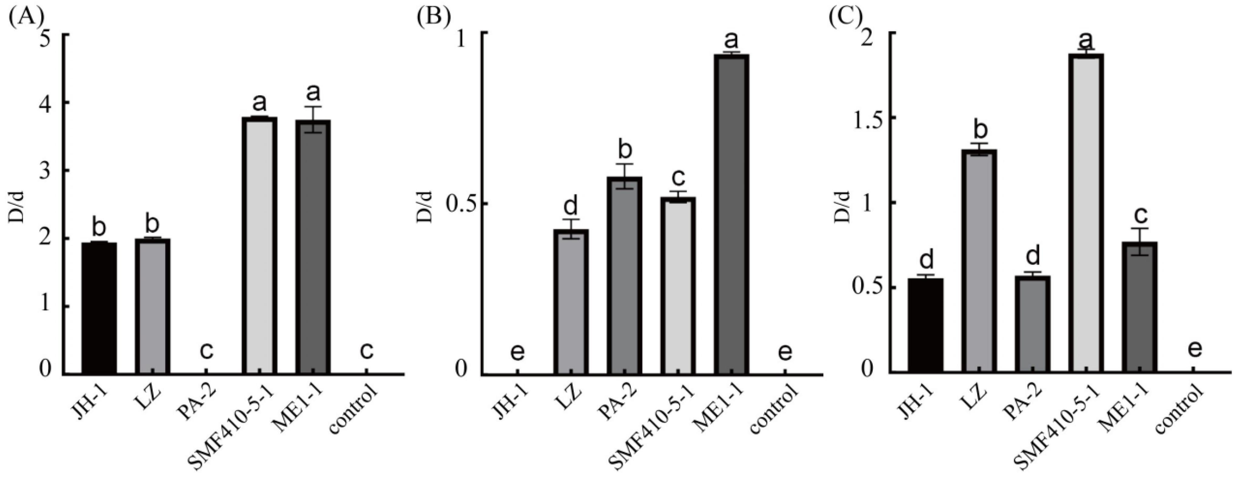
Figure 1. Screening of wood-rotting fungi producing laccase, peroxidase, and cellulase. Potential wood-rotting fungal isolates were inoculated onto potato dextrose agar (PDA) plates supplemented with guaiacol (A), aniline blue (B), and CMC-Na (C) for evaluation of laccase, peroxidase, and cellulase production, respectively. “D” indicates the diameter of discolored circles, while “d” represents the diameter of the colony. Isolate ZSL5-7 inoculated on PDA medium served as the control, with no detectable LiP, MnP, or Lac activities.. Error bars represent means ± standard deviation (SD; n = 3). Different letters denote significant differences among different treatments at p < 0.05 according to Tukey’s test.
Forests 16 00795 g001
Figure 2. Evaluation of lignocellulose-degrading enzyme activity of SMF410-5-1 and ME1-1. The activities of laccase (A), manganese peroxidase (B), and lignin peroxidase (C) were measured in the cultured supernatants of SMF410-5-1 and ME1-1. The square and circle represent isolates SMF410-5-1 and ME1-1. The means ± SD (n = 3) of representative data from three independent experiments are presented.
Forests 16 00795 g002
Figure 3. Weight losses of sawdust by inoculation with ME1-1 and SMF410-5-1. Wood-rotting fungal isolates ME1-1 and SMF410-5-1 were inoculated onto 5 g sawdust of poplar (A), Chinese fir (B), and pine (C). Mass losses were measured at 90 and 180 days post-inoculation (dpi). Pleurotus ostreatus (PO) served as the positive control. The sawdust without inoculated fungi was used as the negative control. Error bars represent the SD (n = 3). Different letters (a–d/A–D) indicate statistically significant differences (Tukey’s test, p < 0.05).
Forests 16 00795 g003
Figure 4. Measurement of principal components in lignocellulosic biomass after inoculation with wood-rotting fungi. Lignin, cellulose, and hemicellulose degradation rates of poplar (AC), Chinese fir (DF), and pine (GI) were measured, respectively. Wood-rotting fungal isolates ME1-1 and SMF410-5-1 were inoculated onto 5 g sawdust of poplar, Chinese fir, and pine, respectively. After 90 and 180 dpi, the lignin, cellulose, and hemicellulose of sawdust were detected. Pleurotus ostreatus (PO) served as the positive control, while uninoculated sawdust was used as the negative control. Error bars represent the SD (n = 3) of three replicates. Different letters (a–d/A–D) indicate statistically significant differences (Tukey’s test, p < 0.05).
Forests 16 00795 g004
Figure 5. Phylogenetic tree of isolate ME1-1 with related taxa derived from concatenated sequences of the ITS, LSU, and RPB2 sequences using bayesian inference (BI)·and maximum likelihood (ML) methods. Bootstrap support values from ML ≥ 50% and BI posterior values ≥ 0.9 are shown at nodes (ML/BI). Elmerina efibulata Yuan 4525 was used as the outgroup. The red text designates the isolate ME1-1. Bar = 0.01 substitutions per nucleotide position.
Forests 16 00795 g005
Figure 6. Phylogenetic tree of the isolate SMF410-5-1 with related taxa derived from ITS and LSU sequences using bayesian inference (BI)·and maximum likelihood (ML) methods. Bootstrap support values from ML ≥ 50% and BI posterior values ≥ 0.9 are shown at nodes (ML/BI). Candelabrochaete africana FP-102987-Sp was used as the outgroup. The red text designates the isolate SMF410-5-1. Bar = 0.04 substitutions per nucleotide position.
Forests 16 00795 g006
Disclaimer/Publisher’s Note: The statements, opinions and data contained in all publications are solely those of the individual author(s) and contributor(s) and not of MDPI and/or the editor(s). MDPI and/or the editor(s) disclaim responsibility for any injury to people or property resulting from any ideas, methods, instructions or products referred to in the content.