Article

Comparative Analysis of Endophytic Bacterial Microbiomes in Healthy and Phytoplasma-Infected European Blueberry Plants

by
Martynas Dėlkus
,
Juliana Lukša-Žebelovič
,
Marija Žižytė-Eidetienė
,
Algirdas Ivanauskas
,
Deividas Valiūnas
* and
Elena Servienė
State Scientific Research Institute Nature Research Centre, Akademijos St. 2, LT-08412 Vilnius, Lithuania
*
Author to whom correspondence should be addressed.
Forests 2025, 16(5), 758; https://doi.org/10.3390/f16050758 (registering DOI)
Submission received: 26 March 2025 / Revised: 24 April 2025 / Accepted: 28 April 2025 / Published: 29 April 2025
(This article belongs to the Special Issue Recent Scientific Developments in Forest Pathology)

Abstract

:
Phytoplasma infections pose a significant threat to the ecological equilibrium and economic worth of Vaccinium myrtillus L., the plant’s overall well-being and capacity for fruit production. This study utilized next-generation sequencing techniques targeting the V3–V4 region of 16S rRNA genes to examine the endophytic bacterial communities present in both healthy and infected samples with ‘Candidatus Phytoplasma pruni’ and ‘Candidatus Phytoplasma trifolii’ related strains. Our findings revealed a total of 1.286 million raw paired-end reads across sequenced samples, which, after quality filtering, resulted in 58,492 high-quality reads without chloroplasts and 1670 amplicon sequence variants (ASVs). Infected plants exhibited statistically higher ASV richness (325 ± 71.5) than healthy plants (231 ± 21.9). This divergence suggests that, although more unique taxa were present in infected plants, their distribution was uneven or phylogenetically clustered, resulting in no significant differences in other diversity indices. However, other alpha diversity metrics did not show significant differences between the groups. Beta diversity analyses also indicated no significant differences in community composition between healthy and infected samples. The taxonomic analysis revealed that both groups were dominated by the Pseudomonadota phylum (~47.6%). However, infected plants displayed a higher prevalence of the Acidobacteriota and Myxococcota phyla, whereas healthy plants exhibited a higher prevalence of the Actinomycetota phylum. The data presented in this study suggest that ‘Candidatus Phytoplasma’ infection may result in mild changes to the bacterial community structure within V. myrtillus. These data provide insights into phytoplasma disease-related changes in the microbial diversity of the plant host.

1. Introduction

Vaccinium is a widespread genus of shrubs. These species are widely used in homeopathic medicine and as food supplements [1]. The European blueberry (Vaccinium myrtillus L.) is a Holarctic species native to continental Northern Europe, the British Isles, Western Canada, and the Western United States [2,3]. Traditionally, its leaves and berries have been used in folk medicine for their potential anti-inflammatory, antioxidant, and metabolic benefits [4]. Although European blueberry dietary supplements are commercially available, the empirical evidence supporting their effectiveness in promoting health or mitigating disease conditions is limited [5]. Beyond human health, these bioactive properties suggest that V. myrtillus may host a microbiome with potential ecological functions, including defence against pathogens and adaptation to environmental stress. Understanding the endophytic bacterial communities of this plant, especially under biotic stress such as phytoplasma infection, can reveal microbiome-mediated mechanisms that influence both plant health and its ecological role within forest ecosystems.
Plant pathogens represent a substantial threat to worldwide food production, contributing to an estimated up to 28% decrease in annual crop yields [6]. Extensive research has examined the relationships between plants and pathogens. The primary focus of these studies has often been on the binary interactions between plants and pathogens under different environmental conditions [7]. There is growing interest in understanding the structure and function of the plant microbiome to leverage its potential for plant growth promotion and disease resistance [8,9]. Alterations in the plant microbiome mediated by microbes can trigger the plant’s immune defences. Plant growth conditions and environmental factors shape the structure and diversity of the plant microbiome, with plant immunity being the main component influencing the structure of microbial community [10,11]. Recent research suggests that the plant microbiome can influence the plant’s cellular immune response [11].
Phytoplasmas are a group of obligate intracellular bacteria that colonize the phloem of plants, causing widespread diseases in various crops and wild plant species [12]. Unlike many bacterial pathogens, phytoplasmas lack a cell wall and rely on insect vectors for transmission between host plants [13]. Based on partial 16S ribosomal RNA gene sequence RFLP analyses and phylogenetic analyses, phytoplasmas are taxonomically classified into groups or subgroups. As unculturable bacteria, they are provisionally categorized under the Candidatus status [14,15,16]. These pathogens infect a broad spectrum of plants. The most dominant symptoms are yellowing, witches’ broom, and stunting [17]. Phytoplasma-associated diseases have been documented in Vaccinium species across Europe and North America, caused by strains related to ‘Candidatus Phytoplasma pruni’ [18,19,20], ‘Candidatus Phytoplasma trifolii’ [21], and ‘Candidatus (Ca.) Phytoplasma (P.) asteris’ [22]. Infections of Vaccinium sp. plants with ‘Ca. P. pruni’, and ‘Ca. P. trifolii’-related strains have been reported in Lithuania too [23,24] To our knowledge, no confirmed insect vectors in blueberries have been identified for ‘Ca. P. pruni’ (in particular 16SrIII-F) and ‘Ca. P. trifolii’-related strains in Lithuania. Currently, phytoplasma infections in Lithuania are not managed, which enables infected European blueberries to function as reservoirs, potentially disseminating the disease to other understory vegetation and disrupting forest ecosystems. Given blueberries’ ecological and economic significance, investigating these infections is vital for comprehending their impact, impeding further propagation, and formulating future management approaches. To promote environmental sustainability, there is a need to develop innovative natural biocontrol strategies to replace chemical pesticides for managing blueberry cultivation. The endophytic bacterial community within the plant plays a critical role in defending the host and facilitating pathogen infection [25]. Our studies compared the endophytic microbiomes of healthy and infected blueberry leaves to investigate how pathogens influence microbial community composition. Earlier research by Mažeikienė et al. [26] has explored the cultivable in vitro endophytic bacteria inhabiting healthy V. myrtillus blueberry leaves but a comprehensive assessment of the microbiome under phytoplasma influence is lacking. Cultivation-based studies have traditionally focused on a limited subset of fast-growing, easily culturable bacteria, which may not fully represent the microbial diversity present within plant tissues. These methods often underestimate community complexity and tend to overlook slow-growing or unculturable taxa, including those that may be ecologically significant. In contrast, next-generation sequencing (NGS) technologies such as 16S rRNA amplicon sequencing provide a culture-independent means of profiling entire microbial communities at higher taxonomic resolution. This approach allows the detection of rare or novel taxa and reveals broader community shifts, including subtle changes that may occur in response to phytoplasma infection. Our study builds on prior cultivation-based work by offering a more comprehensive overview of the endophytic bacterial diversity in healthy and infected blueberry leaves. In our study, we performed an analysis of healthy and infected blueberry plants inhabiting microbial communities based on reads derived from high-throughput sequencing. This study is the first to describe the composition of bacterial endophytic microbiome in non-symptomatic and symptomatic blueberry plants infected by ‘Ca. P. trifolii’ and ‘Ca. P. pruni’-related strains.

2. Materials and Methods

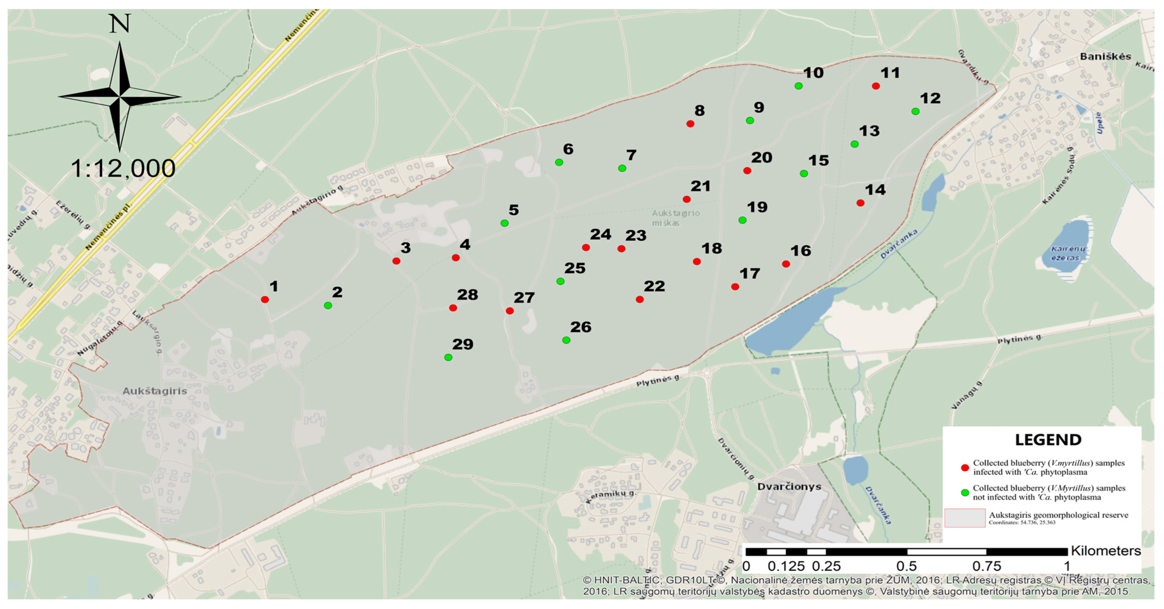
2.1. Sample Collection

The European blueberry (Vaccinium myrtillus) samples were collected from Vilnius County, Lithuania, from the Aukštagiris geomorphological reserve (54°44′1″ N; 25°21′58″ E) on 15–16 August 2023. Randomly, 29 plants showing phytoplasma-like symptoms and 29 asymptomatic plants (growing at least 1 m away from infected individuals) were chosen from various parts of the geomorphological reserve (Figure 1). The shoots of plants were cut down and transported to the laboratory in separate zip-lock bags under freezing conditions. All collected samples were divided into smaller portions. Leaves were surface sterilized by washing in 0.25% NaOCl for 1 min, followed by 70% ethanol for 40 s, and later washed in sterile water thrice. Each plant sample yielded more than 190 mg of fresh leaf tissue. The material was processed under sterile conditions using autoclaved tools and clean benches. The samples were then halved: one-half was kept at −80 °C until DNA extraction and phytoplasma identification, and the other half—approximately 75 mg—was transferred to a 2 mL Eppendorf tube containing DNA/RNA Shield (Zymo Research®, Irvine, CA, USA) and stored at −20 °C until bacterial microbiome sequencing.

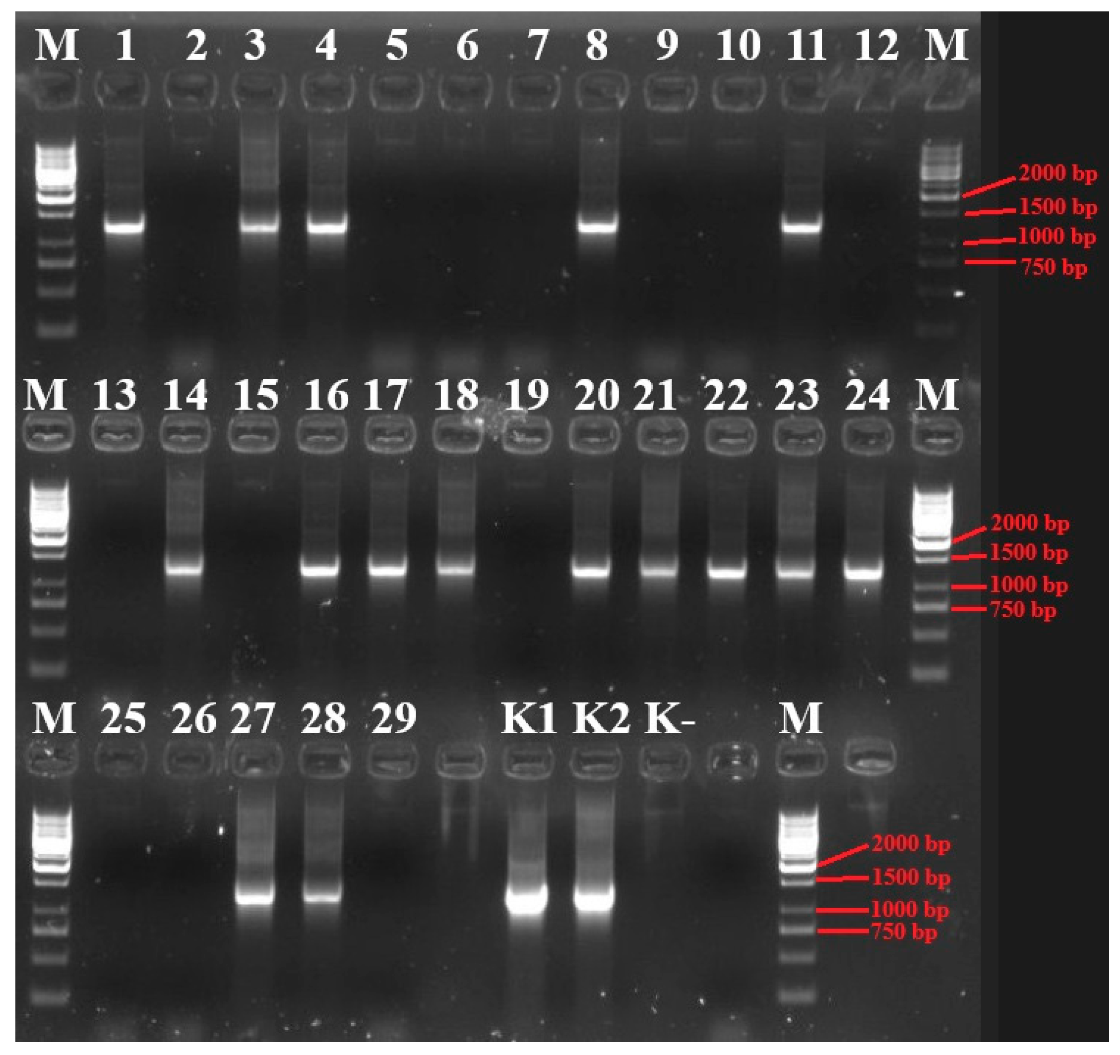
2.2. DNA Extraction, Phytoplasma Detection and Identification

Total genomic DNA was extracted from blueberry leaves using a Genomic DNA Purification Kit (Thermo Fisher Scientific Baltics, Vilnius, Lithuania) according to the manufacturer’s instructions. The concentration and purity of the extracted DNA were assessed using a NanoDrop™ spectrophotometer (Thermo Fisher Scientific) and Qubit® 4 Fluorometer (Invitrogen, Waltham, MA, USA). DNA samples used for sequencing exhibited A260/280 ratios between 1.8 and 2.0, and A260/230 ratios above 1.8, indicating sufficient purity and concentration for downstream PCR amplification and sequencing. Polymerase chain reaction amplification of the phytoplasma 16S rRNA gene fragment was carried out using universal phytoplasma primer pairs P1/P7 [27,28] and R16F2n/R16R2 [29]. The reaction conditions were conducted for 35 cycles under the following parameters: denaturation at 95 °C for 1 min, annealing at 55 °C for 2 min, and extension at 72 °C for 3 min as described previously [30].
The 1.2 kb size PCR product of 16S rDNA fragment was cloned in Esherichia coli using the InsTAclone PCR Cloning Kit (Thermo Fisher Scientific Baltics). Recombinant plasmids were purified using GeneJET Plasmid Miniprep Kit (Thermo Fisher Scientific Baltics) and cloned inserts were sequenced using Sanger sequencing (State Scientific Research Institute Nature Research Centre, Laboratory of Molecular Ecology, Vilnius, Lithuania). Accession numbers PV339489 and PV339918 were assigned to VAC-L2 designated phytoplasma strain 16S rDNA sequence and to VacRWB2 designated phytoplasma strain 16S rDNA sequence, respectively. Nucleotide sequences were analysed by using the NCBI website BLAST option [31] and iPhyClassifier [32].

2.3. Bacterial DNA Amplification, Library Preparation and Sequencing

Sample processing and sequencing were conducted by ZymoBIOMICS® Targeted Sequencing Service (Zymo Research, Irvine, CA, USA). DNA extraction was performed using the ZymoBIOMICS®-96 MagBead DNA Kit (Zymo Research, Irvine, CA, USA). For bacteria identification, the V3–V4 region of the 16S rRNA gene was amplified using 341F/785R primer pair (5′-CCTACGGGNGGCWGCAG-3′/5′-GACTACHVGGGTATCTAATCC-3′) and targeted library was created using the Quick-16S™ NGS Library Preparation Kit (Zymo Research, Irvine, CA, USA). The final PCR products were quantified with qPCR fluorescence readings and pooled together based on equal molarity. Library clean-up was performed using the Select-a-Size DNA Clean & Concentrator™ (Zymo Research, Irvine, CA, USA), then quantified with TapeStation® (Agilent Technologies, Santa Clara, CA, USA) and Qubit® (Thermo Fisher Scientific, Waltham, WA, USA). The ZymoBIOMICS® Microbial Community DNA Standard (Zymo Research, Irvine, CA, USA) was used as a positive control and negative controls were included to monitor potential contamination. Sequencing was conducted on an Illumina® Nextseq™ platform with paired-end sequencing (2 × 300 bp), using a P1 reagent kit (600 cycles). A 30% PhiX spike-in was included to enhance sequence diversity. All raw sequencing data generated in this study are available at the Sequence Read Archive (SRA) of the National Center for Biotechnology Information (NCBI), under accession PRJNA1238772. For this analysis, samples were pooled into six composite libraries representing three infected groups (IP1, IP2, IP3) and their corresponding healthy controls (HP1, HP2, HP3), based on phytoplasma infection type and geographic proximity. Pooling was implemented to equalize sequencing depth, control inter-sample variability, and reduce technical and cost-related constraints while still enabling comparative analysis between infected and healthy plants. Individual samples are under the following accession numbers: IP_1—SRR32786535, IP_2—SRR32786534, IP_3—SRR32786533, HP_1—SRR32786538, HP_2—SRR32786537, HP_3—SRR32786536.

2.4. Bioinformatic Analysis

Bioinformatic analyses were conducted using QIIME2 (v2024.10) [33]. Primer sequences from raw data were trimmed from the 5′ end using cutadapt plugin in Qiime2 [34]. Denoising, quality filtering, chimera removal, and Amplicon Sequence Variant (ASV) inference were conducted using the DADA2 plugin in QIIME2 [35]. Sequences were truncated to 270 bp (forward reads) and 300 bp (reverse reads) to remove low-quality bases, and a maximum expected error threshold of 2.6 was applied to further filter low-quality reads. ASVs were taxonomically assigned using the qiime feature-classifier classify-sklearn plugin with pre-trained Naïve Bayes classifier based on Greengenes2 v2024.09 database [36]. Sequences identified as “chloroplast” were removed prior to downstream analysis to avoid confounding plant-derived taxa. A midpoint-rooted phylogenetic tree was constructed using the qiime phylogeny align-to-tree-mafft-fasttree pipeline, including alignment with MAFFT and tree generation with FastTree using default values [37]. Alpha and beta diversity metrics were calculated using the qiime diversity core-metrics-phylogenetic plugin pipeline at a rarefaction depth determined by the lowest read sample (5460). Pairwise comparisons of alpha diversity metrics between groups were performed using Kruskal–Wallis test within Qiime2 using default parameters. Beta diversity distances were compared using permutational multivariate analysis of variance (PERMANOVA) using qiime diversity beta-group-significance command [38]. Patterns of beta diversity were assessed using Weighted and Unweighted UniFrac distances, visualized via principal coordinate analysis (PCoA) generated in QIIME2’s Emperor tool [39]. To further explore microbial community differences, differential abundance analysis was conducted using Analysis of Composition of Microbiomes with Bias Correction (ANCOM-BC), implemented in QIIME2. ANCOM-BC was used to account for compositionality and uneven sequencing depth, estimating taxa log-fold changes while controlling the false discovery rate (FDR) [40]. Taxa were considered significantly differentially abundant based on an adjusted p-value threshold of p ≤ 0.05 and an absolute LFC cutoff of ≥1.1, to capture both enriched (LFC > 1.1) and depleted (LFC < –1.1) taxa with meaningful biological relevance.

3. Results and Discussion

3.1. Molecular Identification of Phytoplasma in Blueberry Plants

Vaccinium myrtillus samples were collected from the Aukštagiris geomorphological reserve. Twenty-nine symptomatic and twenty-nine asymptomatic blueberry plants growing in proximity were gathered. All symptomatic plants exhibited three or more characteristic phytoplasma-related symptoms, including stunting, reduced leaf size, yellowing (16SrVI-A phytoplasma) and/or reddening (16SrVI-A phytoplasma), shortened internodal distances, and stem deformities forming witches’-broom structures (Figure 2).
Phytoplasma 16S rRNA gene analysis was performed on both symptomatic and asymptomatic blueberry samples. A 1.2 kb size phytoplasma-specific 16S rDNA fragment was amplified using universal primer pairs P1/P7 and R16F2n/R16R2n in nested PCRs (Figure 3). BLAST 1.30+ and iPhyClassifier confirmed phytoplasma infection in 16 (from 29) symptomatic plants, while no phytoplasma DNA was detected in any asymptomatic samples.
Blueberry plants showing symptoms of ‘Candidatus Phytoplasma’ infection—stunting, leaf size reduction, discolouration (yellowing and reddening), shortened internodes, stem deformities and witches’ broom—were confirmed to be infected using PCR product 16S rRNR gene sequence analyses. These analyses showed the pathogen as belonging to the 16SrIII-F and 16SrVI-A phytoplasma subgroups, that are related to ‘Ca. P. pruni’ and ‘Ca. P. trifolii’, respectively. Data revealed that all ‘Ca. P. pruni’ and ‘Ca. P. trifolii’ strains (their 16S rDNA) within their respective subgroups were genetically identical. Representative sequences from each phytoplasma group were deposited to the GenBank database under accession numbers PV339489 (designated as VAC-L2) and PV339918 (designated as VacRWB2). The infected plant samples were divided into three groups: the first group consisted of five plant samples that contained ‘Ca. P. pruni’-related (16SrIII-F phytoplasma subgroup) strain; the second group consisted of five plant samples that contained ‘Ca. P. trifolii’-related (16SrVI-A phytoplasma subgroup) strains; and the third group consisted of six plant samples that contained mixed infections of both phytoplasma subgroups (16SrIII-F, 16SrVI-A). Similar associations between ‘Candidatus Phytoplasma’ infections and symptomatic blueberry plants have been noted in other regions of Lithuania. The infection data of Vaccinium myrtillus samples gathered from the Aukštagiris geomorphological reserve match the prior research conducted in Europe and Lithuania [18,24].

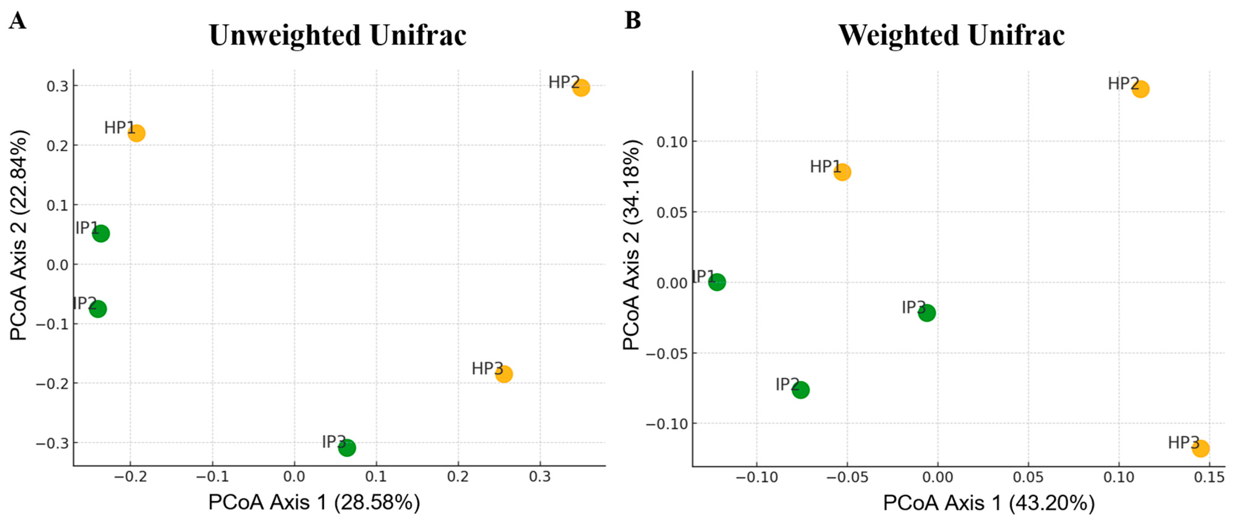
3.2. Diversity and Richness of Bacterial Microbiota of Healthy and Infected with ‘Candidatus Phytoplasma’ Vaccinium Myrtillus Plants

The bacterial community composition of healthy and phytoplasma-infected plants was analysed using NGS of the V3–V4 region of 16S rRNA genes. The V3–V4 hypervariable region of the 16S rRNA gene is one of the most commonly targeted regions in microbiome studies due to its balance of taxonomic resolution, sequencing efficiency, and bioinformatic tractability [41]. V. myrtillus samples were divided and pooled into two groups and three subgroups. Two groups consist of infected plants (IP) and healthy plants (HP). Each group was further divided into three subgroups based on infection status and their respective counterparts growing in proximity. IP1 represents pooled infected plants related to ‘Candidatus Phytoplasma pruni’, while HP1 indicates healthy plants growing near IP1. IP2—pooled infected plants related to ‘Candidatus Phytoplasma trifolii’, HP2 as their healthy counterparts. IP3—pooled infected plants with ‘Ca. P. pruni’ and ‘Ca. P. trifolii’ mix. HP3—near the IP3 growing healthy plants. A total of 1.286 million raw paired-end reads were generated across 6 samples (Table S2). The number of reads per sample varied from 107,082 to 367,802, with an average of 214,415. After pre-processing and quality filtering, 58,492 high-quality reads were obtained, averaging 9749 sequences per sample. The DADA2 denoising approach generated a total of 1670 amplicon sequence variants (ASVs). The number of ASVs detected in individual samples ranged from 212 to 407. The infected samples (IP) exhibited a higher number of ASVs compared to healthy samples (HP) (977 and 963, respectively). The IP samples displayed significantly higher average ASV count (325 ± 71.5), than HP samples (231 ± 21.9). However, it is important to note that the use of pooled samples may influence diversity results by masking individual-level variation within groups. While this strategy improves consistency across libraries and reduces noise from inter-individual variability, it also reduces the resolution of intra-group comparisons and may affect statistical power. Future studies should incorporate individually sequenced replicates to confirm and extend these findings. Based on ASV diversity, statistically significant differences were detected between HP and IP samples (p = 0.0495, Kruskal–Wallis test). (Figure 4).
Sequencing depth was comprehensive enough to estimate microbial diversity in all individual samples. Other alpha diversity metrics, such as the Shannon diversity index and the phylogenetic diversity index (Faith’s PD), did not reveal statistically significant differences in bacterial diversity among the HP and IP samples (Figure 4). Beta diversity was assessed using weighted and unweighted UniFrac distances, with statistical comparisons performed using PERMANOVA. Results indicated no statistically significant differences among sample groups (p > 0.05). Principal coordinate analysis (PCoA) based on unweighted and weighted UniFrac distances of bacterial microbiota demonstrated a slight separation between healthy and phytoplasma-infected blueberry plants (Figure 5A,B). Although the PCoA plots suggest some visual separation between healthy and infected groups, the PERMANOVA results did not indicate statistically significant differences. This apparent discrepancy may arise due to relatively high intra-group variability or limited sample size, which reduces statistical power. Such visual distinctions, while informative, should be interpreted cautiously in the absence of supporting statistical significance.
The observed increase in ASV richness in infected V. myrtillus plants suggests that phytoplasma infection may contribute to a more diverse bacterial community. This finding contrasts with previous cultivation-based studies, where pathogens typically correlate with a reduction in microbial diversity due to immune activation and microbiome dysbiosis [42,43]. One potential explanation is that phytoplasma infections may induce physiological changes in the host plant, such as altered nutrient availability, phloem disruption, or immunosuppression, which could facilitate colonization by a broader range of bacterial taxa. Alternatively, changes in plant exudates due to infection might create new ecological niches that support increased microbial diversity.
Despite the increase in ASV richness, beta diversity analyses indicate that overall bacterial community composition remains similar between healthy and infected plants. This suggests that while infection may lead to the incorporation of additional microbial taxa, it does not substantially alter the structure of the core microbiome. This pattern is inconsistent with prior research on pathogen–plant interactions [44], which often report significant microbiome restructuring in response to infection. These findings contribute to understanding the impact of phytoplasma infections on the plant’s endophytic microbiome. However, further research is needed to explore the specific mechanisms by which phytoplasma infections influence microbial communities. Such investigations could uncover subtle changes in microbiome composition and function that may not be captured by conventional diversity metrics. The increased ASV richness observed in infected plants may be attributed to phytoplasma-induced physiological changes such as altered nutrient allocation, phloem blockage, or immune modulation. These factors can create novel microenvironments or reduce host immune filtering, enabling colonization by a broader range of bacterial taxa. For instance, impaired phloem transport may cause localized nutrient accumulation, promoting microbial growth, while pathogen-induced immune suppression may reduce microbial exclusion at the tissue level [45].

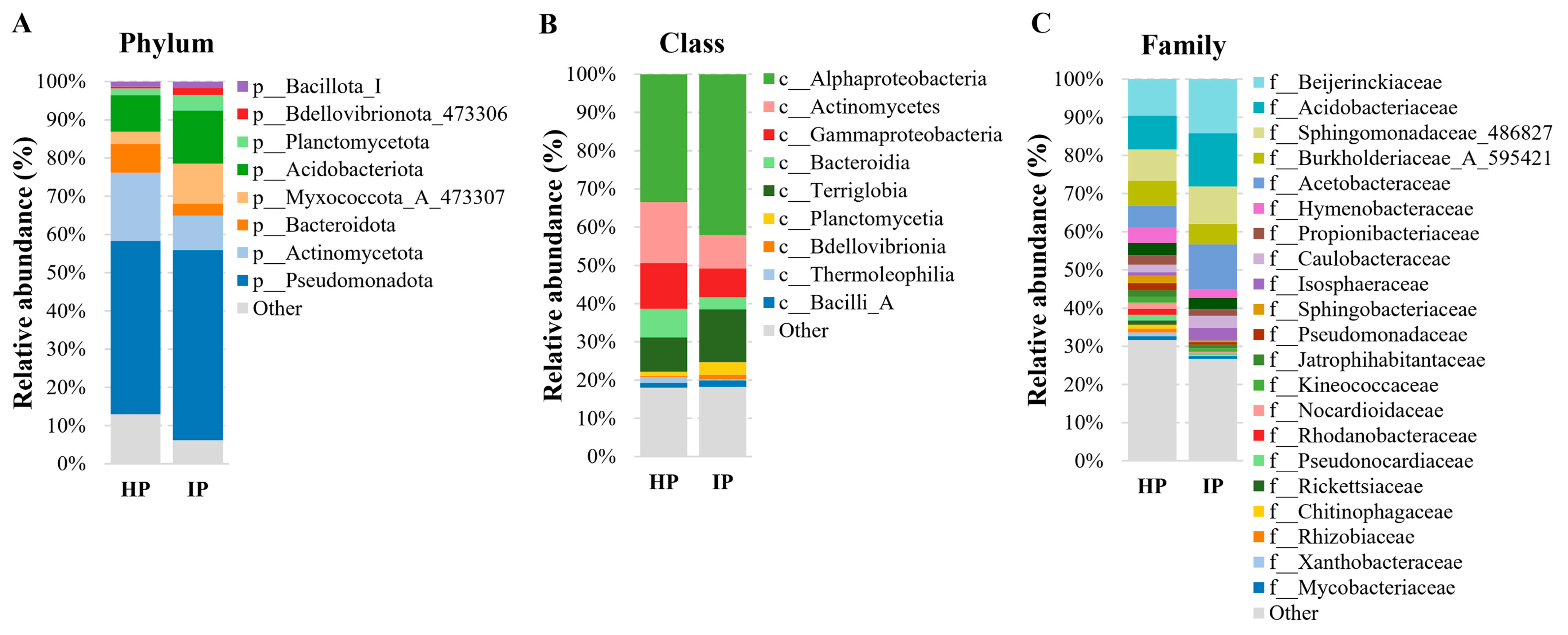
3.3. Bacterial Community Profiling of Healthy and Infected with Phytoplasma V. myrtillus Plants

Both infected and healthy blueberry plants carried bacterial DNA sequences assigned to the four predominant phyla: Pseudomonadota, Actinomycetota, Acidobacteriota, and Myxococcota, which collectively comprised over 83% of the bacterial population. The most dominant phylum in both infected and healthy blueberry plants was Pseudomonadota, comprising 45.4% in HP and 49.8% in IP (Figure 6, Table S1). The relative abundances of Actinomycetota (17.8% in IP vs. 9.0% in HP), Acidobacteriota (13.82% in IP vs. 9.5% in HP), and Myxococcota (10.56% in IP vs. 3.3% in HP) were marginally higher in infected samples, suggesting potential infection-associated shifts in microbial composition (Figure 6, Table S1). The Alphaproteobacteria class comprised the predominant bacterial class in both infected and healthy blueberry plants (33.5% HP and 42.2% IP), succeeded by Actinomycetes (16.0% HP and 8.6% IP), Gammaproteobacteria (11.9% HP and 7.6% IP), and Terriglobia (9.0% HP and 13.8% IP). The prevalence of these members varied little between HP and IP samples (Figure 6, Table S1). The unidentified class of the Myxococcota phylum (designated as XYA12-FULL-58-9) exhibited significantly greater dominance in IP samples (10.4%) compared to HP samples (2.6%) (Figure 6, Table S1). The Beijerinckiaceae family was the predominant bacterial family in both diseased and healthy blueberry plants (9.6% HP and 14.3% IP), succeeded by Acidobacteriaceae (8.9% HP and 13.8% IP), Sphingomonadaceae (8.2% HP and 9.8% IP), and Acetobacteraceae (5.8% HP and 11.9% IP). The prevalence of these members varied little between HP and IP samples (Figure 6, Table S1). There were a lot more of the unidentified family XYA12-FULL-58-9 in IP samples (10.2%) than in HP samples (2.6%). This family is in the Myxococcota phylum. Overall, plants that had different phytoplasma infections (IP.1—‘Ca. P. pruni’; IP.2—‘Ca. P. trifolii’; IP.3—‘Ca. P. trifolii’ and ‘Ca. P. pruni’ mix) had high similarity between them. On the other hand, healthy plant bacterial communities had more diversity between different samples (Figure 6, Table S1).
The bacterial communities of healthy and infected V. myrtillus were very different, but there was a core group of phyla that was always present in both groups of plants. Pseudomonadota was the largest phylum in the V. myrtillus endophytic microbiome. It heads most bacteria in both healthy (45.4%) and infected (49.8%) plants. In this phylum, the Beijerinckiaceae family, specifically the Methylobacterium genus (Methylobacterium cerastii), was slightly more predominant in healthy plants. Those bacteria are recognized as plant-associated bacteria [46], typically located in the plant endosphere [47]. This genus contributes to plant growth and health by engaging in plant–microbe interactions [48]. The genera Beijerinckia genus (Beijerinckia mobilis) and Lichenibacterium genus (Lichenibacterium minor) were a bit more prevalent in infected plants. The Beijerinckia genus is known as a plant growth promoter and nitrogen fixator [49]. The Lichenibacterium genus promotes nutrient cycling by decomposing organic materials [50]. This bacterial genus is known for its pathogen inhibition, and for boosting the microbial diversity of plants [51]. These microorganisms are significant contributors to the plant’s microbiome, promoting its growth and resistance to biotic and abiotic stress.
Acidobacteriota was one of the predominant phyla in both groups, with 9.51% in healthy plants and 13.8% in infected plants. In this phylum, the Acidobacteriaceae family, specifically the Granulicella genus, which includes species such as Granulicella acidiphila, Granulicella sibirica, Granulicella aggregans, Granulicella tundricola, and other unidentified species, was a bit more prevalent in infected plants. Granulicella species can break down complex polymers which are prevalent in organic matter [52]. This bacteria genus produces exopolysaccharides (EPS), which serve as a carbon source for other microorganisms, promoting microbial interactions and nutrient cycling in soil ecosystems. This process is particularly important in nutrient-deficient environments [53]. Granulicella spp. plays a significant role in enhancing plant defence mechanisms by altering the rhizosphere microbiome, particularly in the presence of biochar. This leads to increased resistance to pathogens, through mechanisms that may involve microbial diversity and potential activation of plant immune responses [54].
The Actinomycetota phylum bacteria were found in both healthy (17.8%) and infected (9.1%) plants. One of these phylum members, the Microbacteriaceae family, comprising mostly unidentified species, was common in both groups. The Microbacteriaceae family, which includes many unidentified species, plays a significant role in the breakdown of pollutants, contributing to the remediation of damaged habitats [55].
Finally, the Myxococcota phylum was more prevalent in infected (10.6%) than in healthy (3.3%) plants, suggesting that these bacteria may be multiplying in response to infection. Members of the Myxococcota phylum are well known for their social behaviour and predatory capabilities, including the secretion of hydrolytic enzymes and antimicrobial compounds used to lyse other microbial cells. The increased prevalence of these bacteria in infected V. myrtillus plants may indicate ecological restructuring within the microbiome. Phytoplasma infections are known to alter host physiology, which may create favourable niches for predatory or opportunistic bacteria. The presence of Myxococcota could reflect increased bacterial turnover or shifts in community competition, potentially driven by immune suppression or altered nutrient exudation [56]. These bacteria modulate bacterial populations, inhibit competitors or pathogens, and may flourish in various settings. The species potentially enhances plant health by modulating microbial populations and facilitating the breakdown of organic materials [57]. Its predatory characteristics may also inhibit the proliferation of dangerous microorganisms, fostering a healthier microbial population [58].

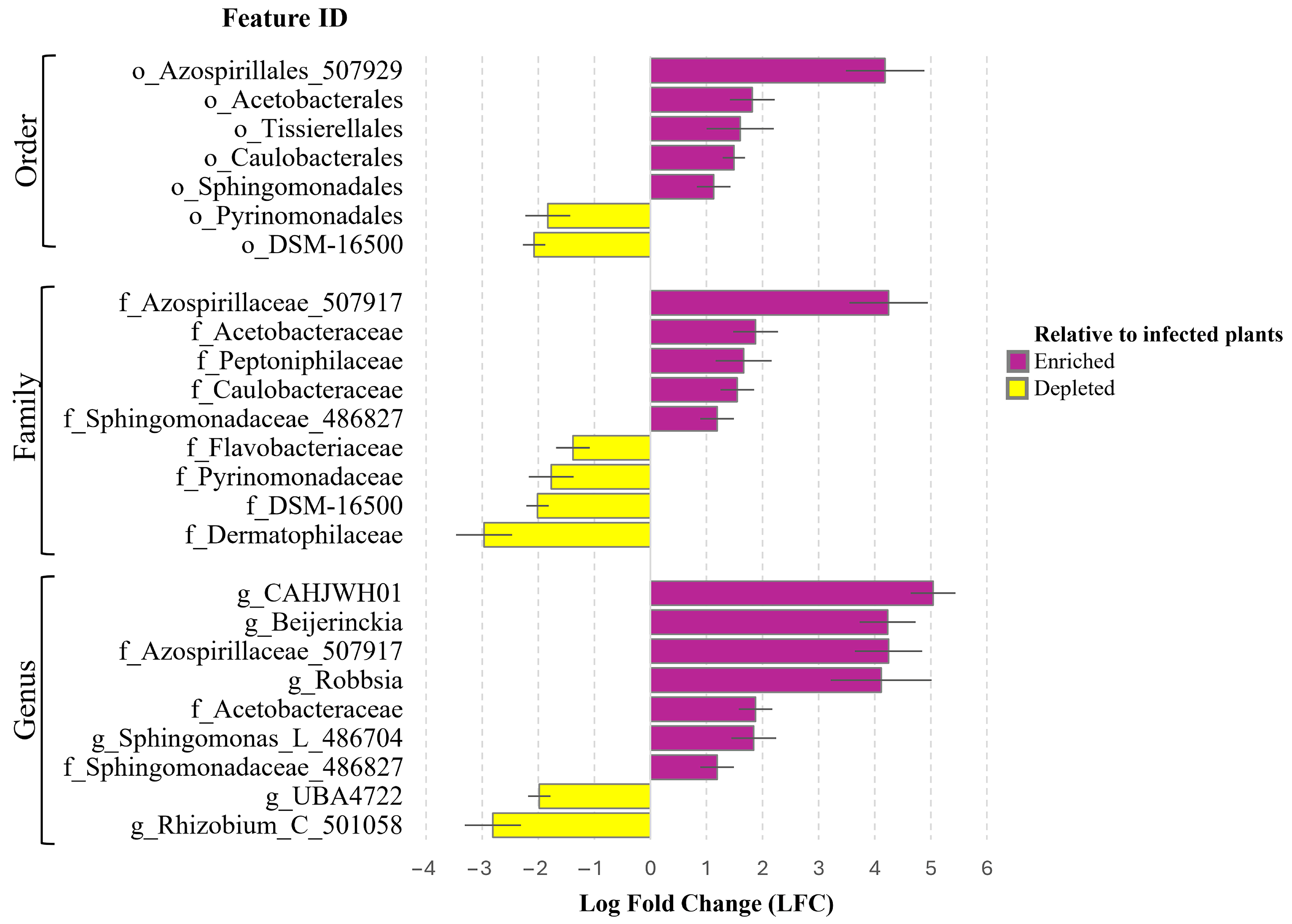
3.4. ANCOM-BC Analysis for Identifying Significant Taxonomic Differences and Potential Biomarkers

To better understand the bacterial communities in blueberry plants, we used ANCOM-BC analysis to identify taxa with significant differences between infected and healthy blueberry plants at the family, order, and genus taxonomic levels (Figure 7). The order Azospirillales (507929) (4.19 LFC) and family Azospirillaceae (507917) (4.25 LFC) demonstrated strong associations with infected plants. Moderate associations were observed in Acetobacterales (1.82 LFC) order and Acetobacteraceae family (1.88 LFC), Tissierellales order (1.60 LFC), and Peptoniphilaceae family (1.67 LFC), Caulobacterales order (1.49 LFC) and Caulobacteraceae family (1.55 LFC), and Sphingomonadales order (1.12 LFC) and Sphingomonadaceae (486827) family (1.19 LFC), exhibited negligible differences in their presence between healthy and infected plant samples. On the other hand, certain bacteria taxa demonstrated decreased abundance in infected plants. Pyrinomonadales order (−1.83 LFC), Pyrinomonadaceae (−1.77 LFC), and Flavobacteriaceae families (−1.38 LFC) showed a weak to moderate association with healthy plants. An unclassified order within the class Gammaproteobacteria (designated as DSM-16500) (−2.07 LFC) order and (−2.01 LFC) family, and Dermatophilaceae family (−2.97 LFC), exhibited moderate to strong association with healthy plants.
The study at the genus taxonomical level showed changes in the microbiome composition that were linked to infection status. An unclassified genus within the Caulobacteraceae (designated as CAHJWH01) (5.04 LFC) family exhibited the most substantial association with infected plants, followed by Beijerinckia (4.23 LFC), an unclassified genus within the family Azospirillaceae (507917) (4.25 LFC) and Robbsia (4.11 LFC). Moderate associations were observed for an unclassified genus within Acetobacteraceae (1.88 LFC) and Sphingomonas (L_486704) (1.18 LFC). A minimal association was found for an unclassified genus within the Sphingomonadaceae family (designated as 486827) (1.19 LFC). On the other hand, several bacterial genera demonstrated increased association with healthy plants. These included an unclassified genus (UBA4722) of the Gammaproteobacteria class (−1.98 LFC) and Rhizobium genus (C_501058) (−2.81 LFC), showing a stronger association with healthy plants.
The results revealed subtle yet significant shifts in the bacterial communities between infected (IP) and healthy (HP) blueberry plants. A strong association was observed between infected plants and bacterial taxa belonging to the Azospirillales order, Azospirillaceae family, and unknown bacteria genus of Azospirillaceae family. The enrichment of Azospirillaceae in infected plants is particularly noteworthy, as this family is known for its strong plant growth-promoting traits, including nitrogen fixation, production of phytohormones such as indole-3-acetic acid (IAA), and enhancement of root architecture. These traits can help plants tolerate biotic stress, including pathogen pressure. It is possible that their increased abundance reflects a compensatory response by the microbiome to phytoplasma-induced stress or damage, potentially contributing to plant resilience. Similarly, Beijerinckia, a genus within the Beijerinckiaceae family, is associated with nitrogen fixation and carbon cycling in nutrient-poor soils. Its increased presence in infected plants may be related to changes in nutrient availability caused by phloem disruption or altered plant metabolism. These functional traits suggest that certain microbial taxa may act as opportunistic colonizers or beneficial responders under phytoplasma-induced stress and could serve as potential candidates for biocontrol or plant health monitoring [59]. Members of the Azospirillaceae family are well known for nitrogen fixation and plant growth-promoting properties, contributing to plant resilience under biotic and abiotic stress conditions [60]. These bacteria may mitigate the effects of pathogens on plants, by improving nutrient cycling or reducing plant stress [61]. Members of the Azospirillales order, particularly within the genus Azospirillum, are known for their ability to produce phytohormones such as indole-3-acetic acid (IAA), gibberellins, and cytokinins, which enhance root development and nutrient uptake [62]. They also fix atmospheric nitrogen and release siderophores that can improve iron availability to the plant. Importantly, Azospirillum spp. have been shown to trigger induced systemic resistance (ISR) in plants, priming the immune system for faster or stronger responses to pathogen attack [63]. Infected plants also exhibited strong to moderate associations with bacterial taxa from the Sphingomonadaceae, Acetobacteraceae, and Caulobacteraceae families. These microbial families exhibit distinct metabolic and ecological behaviour, yet they share similar effects on the plant microbiome. Their capacity to promote plant growth [64], enhance nutrient cycling [65], and outcompete pathogens [66] are fundamental to their contribution to plant health.
Healthy V. myrtillus plants exhibited a strong association with members of Gammaproteobacteria bacteria. The members of this class are known for their ability to degrade organic compounds [67], participate in nitrogen or sulphur cycling [68], and influence plant growth [69]. Their prevalence in healthy plants suggests a beneficial role in maintaining plant health and microbiome stability [70]. Strong to moderate associations were also observed in several orders, families, and genera, including the Actinomycetales order, Dermatophilaceae family, Pyrinomonadales order, and Pyrinomonadaceae family. Both families are associated with the degradation of complex organic compounds [69]; however, Dermatophilaceae is often associated with pathogenicity [71], while the Pyrinomonadaceae family members may play beneficial roles in the plant microbiome [72]. The Rhizobium genus is strongly associated with healthy plants. These genera can enhance nitrogen fixation, and nutrient uptake, and can improve soil health and fertility, suggesting an important role in sustaining plant nutrition and microbiome stability.

4. Conclusions

This study provides new insights into the impact of ‘Candidatus Phytoplasma’ on the endophytic bacterial microbiome of Vaccinium myrtillus L. plants. The meta-analysis revealed that infection is associated with an increase in ASV richness, suggesting a greater number of bacterial taxa in infected plants. However, other diversity metrics, including Shannon and Faith’s phylogenetic diversity indices, did not show a statistically significant link between infection and health status. This finding indicates that while the number of distinct bacterial taxa increases, the overall diversity and phylogenetic range remain relatively stable. Analysis of microbiome communities indicated that the most dominant phylum was Pseudomonadota in both healthy and infected plants. Acidobacteriota and Myxococcota were more prevalent in infected plants than in healthy plants. Taxonomic analysis of lower levels revealed specific bacteria were more dominant in infected plants, while healthy plants had more diverse microbiomes. Notably, an unidentified genus within the Myxococcota phylum was significantly more prevalent in infected plants. The ANCOM-BC analysis revealed that phytoplasma infection is associated with the Azospirillales order and Azospirillaceae family members. These bacteria may thrive under the altered physiological conditions induced by infection. An unclassified genus in the family Caulobacteraceae had the strongest link with infected plants. It was followed by Beijerinckia, an unclassified genus in the family Azospirillaceae, and Robbsia. These bacteria suggest a potential ecological role in the infected plant’s microbiome, either as adaptive colonizers or as indirect contributors to disease progression. On the other hand, healthy plants are associated with the Gammaproteobacteria and Dermatophilaceae members. At the genus level, UBA4722 (Gammaproteobacteria) and Rhizobium showed a strong association with healthy plants. The observed microbiome shifts highlight the influence of the ’Candidatus phytoplasma’ on the blueberry microbiome. This effect favours certain bacterial taxa that may sustain the infectious environment while diminishing those that promote plant health. While microbial profiling has potential as a diagnostic tool for detecting phytoplasma infections, additional validation is required to determine its practical applicability in disease monitoring and management. Future research should incorporate metagenomic and transcriptomic approaches to explore the metabolic functions of enriched bacterial taxa and their potential interactions with phytoplasma infections. The cross-sectional design captures a single time point, which limits our ability to assess dynamic changes in microbiome composition over the course of infection. Additionally, the use of pooled samples, while reducing biological noise, may mask individual variation in microbial communities. Future research should consider longitudinal sampling to better understand microbiome shifts during different stages of infection and symptom progression. Furthermore, longitudinal studies tracking microbiome changes over the course of infection could help establish causal relationships between microbial shifts and disease progression.

Supplementary Materials

The following supporting information can be downloaded at: https://www.mdpi.com/article/10.3390/f16050758/s1, Table S1: Endophytic bacterial (V3-V4) microbial community taxonomy abundance count of European blueberry (Vaccinium myrtillus L.) from phylum to species level. Figure S1: Nested PCR products derived using primers R16F2n/R16R2n from European blueberry (Vaccinium myrtillus L.) plant samples. M—GeneRuler 100 bp Plus DNA Ladder (Thermo Fisher Scientific, Vilnius, Lithuania), fragment sizes: 3000, 2000, 1500, 1200, 1000, 900, 800, 700, 600, 500, 400, 300, 200, 100; (raw data of Figure 3). Table S2: Sequencing summary and alpha diversity metrics of microbial community in healthy (HP) and infected (IP) plant samples. Figure S2: Distribution of endophytic bacteria on phytoplasma-infected plant with ‘Candidatus Phytoplasma pruni’—related strain (IP.1), infected plant with ‘Candidatus Phytoplasma trifolii’—related strain (IP2), infected plant with ‘Candidatus Phytoplasma trifolii’—related strain and ‘Candidatus Phytoplasma pruni’—related strain (IP.3)—healthy plant (HP.1, HP.2, HP.3) blueberry plants at phylum (A), class (B) and family (C) levels.

Author Contributions

Conceptualization, E.S. and D.V.; methodology, E.S. and D.V.; formal analysis, M.D. and J.L.-Ž.; investigation, M.D., M.Ž.-E. and A.I.; resources, M.D. and E.S.; data curation, J.L.-Ž., E.S. and M.D.; writing—original draft preparation, M.D.; writing—review and editing, M.D., M.Ž.-E., A.I., J.L.-Ž., D.V. and E.S.; visualization, M.D. and J.L.-Ž.; supervision, E.S. and D.V. All authors have read and agreed to the published version of the manuscript.

Funding

This research received no external funding.

Data Availability Statement

Data will be made available upon request.

Conflicts of Interest

The authors declare no conflicts of interest.

References

  1. Edger, P.P.; Iorizzo, M.; Bassil, N.V.; Benevenuto, J.; Ferrão, L.F.V.; Giongo, L.; Hummer, K.; Lawas, L.M.F.; Leisner, C.P.; Li, C.; et al. There and back again; historical perspective and future directions for Vaccinium breeding and research studies. Hortic. Res. 2022, 9, uhac083. [Google Scholar] [CrossRef] [PubMed]
  2. Cignarella, A.; Nastasi, M.; Cavalli, E.; Puglisi, L. Novel lipid-lowering properties of vaccinium myrtillus L. leaves, a traditional antidiabetic treatment, in several models of rat dyslipidaemia: A comparison with ciprofibrate. Thromb. Res. 1996, 84, 311–322. [Google Scholar] [CrossRef]
  3. Kopystecka, A.; Kozioł, I.; Radomska, D.; Bielawski, K.; Bielawska, A.; Wujec, M. Vaccinium uliginosum and Vaccinium myrtillus—Two Species—One Used as a Functional Food. Nutrients 2023, 15, 4119. [Google Scholar] [CrossRef] [PubMed]
  4. Shamilov, A.A.; Olennikov, D.N.; Pozdnyakov, D.I.; Bubenchikova, V.N.; Garsiya, E.R.; Larskii, M.V. Caucasian blueberry: Comparative study of phenolic compounds and neuroprotective and antioxidant potential of Vaccinium myrtillus and Vaccinium arctostaphylos Leaves. Life 2022, 12, 2079. [Google Scholar] [CrossRef] [PubMed]
  5. Azari, H.; Morovati, A.; Gargari, B.P.; Sarbakhsh, P. Beneficial effects of blueberry supplementation on the components of metabolic syndrome: A systematic review and meta-analysis. Food Funct. 2022, 13, 4875–4900. [Google Scholar] [CrossRef]
  6. Ristaino, J.B.; Anderson, P.K.; Bebber, D.P.; Brauman, K.A.; Cunniffe, N.J.; Fedoroff, N.V.; Finegold, C.; Garrett, K.A.; Gilligan, C.A.; Jones, C.M.; et al. The persistent threat of emerging plant disease pandemics to global food security. Proc. Natl. Acad. Sci. USA 2021, 118, e2022239118. [Google Scholar] [CrossRef]
  7. Bertaccini, A. Plants and phytoplasmas: When bacteria modify plants. Plants 2022, 11, 1425. [Google Scholar] [CrossRef]
  8. Trivedi, P.; Leach, J.E.; Tringe, S.G.; Sa, T.; Singh, B.K. Plant–microbiome interactions: From community assembly to plant health. Nat. Rev. Microbiol. 2020, 18, 607–621. [Google Scholar] [CrossRef]
  9. Noman, M.; Ahmed, T.; Ijaz, U.; Shahid, M.; Azizullah, N.; Li, D.; Manzoor, I.; Song, F. Plant–microbiome crosstalk: Dawning from composition and assembly of microbial community to improvement of disease resilience in plants. Int. J. Mol. Sci. 2021, 22, 6852. [Google Scholar] [CrossRef]
  10. Fitzpatrick, C.R.; Copeland, J.; Wang, P.W.; Guttman, D.S.; Kotanen, P.M.; Johnson, M.T.J. Assembly and ecological function of the root microbiome across angiosperm plant species. Proc. Natl. Acad. Sci. USA 2018, 115, E1157–E1165. [Google Scholar] [CrossRef]
  11. Kim, H.; Lee, K.K.; Jeon, J.; Harris, W.A.; Lee, Y. Domestication of Oryza species eco-evolutionarily shapes bacterial and fungal communities in rice seed. Microbiome 2020, 8, 20. [Google Scholar] [CrossRef]
  12. Wei, W.; Zhao, Y. Phytoplasma taxonomy: Nomenclature, classification, and identification. Biology 2022, 11, 1119. [Google Scholar] [CrossRef] [PubMed]
  13. Trivellone, V.; Wei, W.; Filippin, L.; Dietrich, C.H. Screening potential insect vectors in a museum biorepository reveals undiscovered diversity of plant pathogens in natural areas. Ecol. Evol. 2021, 11, 6493–6503. [Google Scholar] [CrossRef] [PubMed]
  14. Pérez-López, E.; Luna-Rodríguez, M.; Olivier, C.; Dumonceaux, T. The underestimated diversity of phytoplasmas in Latin America. Int. J. Syst. Evol. Microbiol. 2016, 66, 492–513. [Google Scholar] [CrossRef] [PubMed]
  15. Schneider, B.; Seemueller, E.; Smart, C.D.; Kirkpatrick, B.C. Phylogenetic classification of plant pathogenic mycoplasma-like organisms or phytoplasmas. Mol. Diagn. Proced. Mycoplasmol. 1995, 1, 369–380. [Google Scholar] [CrossRef]
  16. Zhao, Y.; Wei, W.; Lee, I.; Shao, J.; Suo, X.; Davis, R. Construction of an interactive online phytoplasma classification tool, iPhyClassifier, and its application in analysis of the peach X-disease phytoplasma group (16SrIII). Int. J. Syst. Evol. Microbiol. 2009, 5, 5607–5612. [Google Scholar] [CrossRef]
  17. Wei, W.; Trivellone, V.; Dietrich, C.H.; Zhao, Y.; Bottner-Parker, K.D.; Ivanauskas, A. Identification of phytoplasmas representing multiple new genetic lineages from phloem-feeding leafhoppers highlights the diversity of phytoplasmas and their potential vectors. Pathogens 2021, 10, 352. [Google Scholar] [CrossRef]
  18. Wright, A.A.; Shires, M.; Molnar, C.; Bishop, G.; Johnson, A.; Frias, C.; Harper, S.J. Titer and Distribution of ‘Candidatus Phytoplasma Pruni’ in Prunus Avium. Phytopathology 2022, 112, 1406–1412. [Google Scholar] [CrossRef]
  19. Ivanauskas, A.; Valiūnas, D.; Jomantienė, R.; Picciau, L.; Davis, R.E. Possible insect vectors of ‘Candidatus Phytoplasma asteris’ and ‘Ca. Phytoplasma pruni’-related strains in Lithuania. Zemdirb.-Agric. 2014, 101, 313–320. [Google Scholar] [CrossRef]
  20. Lee, I.; Polashock, J.; Bottner-Parker, K.D.; Bagadia, P.G.; Rodriguez-Saona, C.; Zhao, Y.; Davis, R.E. New subgroup 16SrIII-Y phytoplasmas associated with false-blossom diseased cranberry (Vaccinium macrocarpon) plants and with known and potential insect vectors in New Jersey. Eur. J. Plant Pathol. 2014, 139, 399–406. [Google Scholar] [CrossRef]
  21. Dėlkus, M.; Žižytė-Eidetienė, M.; Ivanauskas, A.; Valiunas, D. First report of lingonberry stunted yellows disease of Vaccinium vitis-idaea associated with ‘Candidatus Phytoplasma trifolii’-related phytoplasma strain in Lithuania. Plant Dis. 2024, 108, 1391. [Google Scholar] [CrossRef] [PubMed]
  22. Pradit, N.; Mescher, M.C.; Wang, Y.; Vorsa, N.; Rodriguez-Saona, C. Phytoplasma infection of cranberries benefits non-vector phytophagous insects. Front. Ecol. Evol. 2019, 7, 181. [Google Scholar] [CrossRef]
  23. Valiunas, D.; Alminaite, A.; Jomantiene, R.; Davis, R.E.; Maas, J.L. Possible cause of European blueberry disease is related to North American milkweed yellows phytoplasma. J. Plant Pathol. 2004, 86, 135–140. [Google Scholar]
  24. Dėlkus, M.; Žižytė-Eidetienė, M.; Ivanauskas, A.; Valiunas, D. First report of ‘Candidatus Phytoplasma trifolii’-related strain associated with Vaccinium reddish witches’-broom disease of European blueberry in Lithuania. Plant Dis. 2025, 109, 709. [Google Scholar] [CrossRef]
  25. Yurgel, S.N.; Ajeethan, N.; Smertenko, A. Response of plant-associated microbiome to plant root colonization by exogenous bacterial endophyte in perennial crops. Front. Microbiol. 2022, 13, 863946. [Google Scholar] [CrossRef]
  26. Mažeikienė, I.; Frercks, B.; Burokienė, D.; Mačionienė, I.; Šalaševičienė, A. Endophytic community composition and genetic-enzymatic features of cultivable bacteria in Vaccinium myrtillus L. in forests of the Baltic-Nordic region. Forests 2021, 12, 1647. [Google Scholar] [CrossRef]
  27. Deng, S.; Hiruki, C. Amplification of 16S rRNA genes from culturable and nonculturable Mollicutes. J. Microbiol. Methods 1991, 14, 53–61. [Google Scholar] [CrossRef]
  28. Schneider, B.; Goldstein, H.W.; Smith, D.B. The ASA Framework: An Update. Pers. Psychol. 1995, 48, 747–773. [Google Scholar] [CrossRef]
  29. Gundersen, D.E.; Lee, I.M.; Schaff, D.A.; Harrison, N.A.; Chang, C.J.; Davis, R.E.; Kingsbury, D.T. Genomic diversity and differentiation among phytoplasma strains in 16S rRNA groups I (aster yellows and related phytoplasmas) and III (X-disease and related phytoplasmas). Int. J. Syst. Bacteriol. 1996, 46, 64–75. [Google Scholar] [CrossRef]
  30. Lee, I.; Gundersen-Rindal, D.E.; Davis, R.E.; Bartoszyk, I.M. Revised Classification scheme of phytoplasmas based on RFLP analyses of 16S rRNA and ribosomal protein gene Sequences. Int. J. Syst. Bacteriol. 1998, 48, 1153–1169. [Google Scholar] [CrossRef]
  31. National Center for Biotechnology Information. Available online: https://www.ncbi.nlm.nih.gov/ (accessed on 10 December 2024).
  32. Zhao, Y.; Wei, W.; Lee, I.M.; Shao, J.; Suo, X.; Davis, R.E. The iPhyClassifier, an Interactive Online Tool for Phytoplasma Classification and Taxonomic Assignment. In Phytoplasma; Dickinson, M., Hodgetts, J., Eds.; Methods in Molecular Biology; Humana Press: Totowa, NJ, USA, 2013; Volume 938, pp. 329–338. [Google Scholar] [CrossRef]
  33. Bolyen, E.; Rideout, J.R.; Dillon, M.R.; Bokulich, N.A.; Abnet, C.C.; Al-Ghalith, G.A.; Alexander, H.; Alm, E.J.; Arumugam, M.; Asnicar, F.; et al. Reproducible, interactive, scalable, and extensible microbiome data science using QIIME 2. Nat. Biotechnol. 2019, 37, 852–857. [Google Scholar] [CrossRef] [PubMed]
  34. Martin, M. Cutadapt removes adapter sequences from high-throughput sequencing reads. EMBnet J. 2011, 17, 10. [Google Scholar] [CrossRef]
  35. Callahan, B.J.; McMurdie, P.J.; Rosen, M.J.; Han, A.W.; Johnson, A.J.A.; Holmes, S.P. DADA2: High-resolution sample inference from Illumina amplicon data. Nat. Methods 2016, 13, 581–583. [Google Scholar] [CrossRef] [PubMed]
  36. McDonald, D.; Jiang, Y.; Balaban, M.; Cantrell, K.; Zhu, Q.; Gonzalez, A.; Morton, J.T.; Nicolaou, G.; Parks, D.H.; Karst, S.M.; et al. Greengenes2 unifies microbial data in a single reference tree. Nat. Biotechnol. 2024, 42, 715–718. [Google Scholar] [CrossRef] [PubMed]
  37. Katoh, K.; Standley, D.M. MAFFT Multiple Sequence Alignment Software Version 7: Improvements in performance and usability. Mol. Biol. Evol. 2013, 30, 772–780. [Google Scholar] [CrossRef]
  38. Anderson, M.J. A new method for non-parametric multivariate analysis of variance. Aust. Ecol. 2001, 26, 32–46. [Google Scholar] [CrossRef]
  39. Vázquez-Baeza, Y.; Pirrung, M.; Gonzalez, A.; Knight, R. EMPeror: A tool for visualizing high-throughput microbial community data. GigaScience 2013, 2, 16. [Google Scholar] [CrossRef]
  40. Lin, H.; Peddada, S.D. Analysis of compositions of microbiomes with bias correction. Nat. Commun. 2020, 11, 3514. [Google Scholar] [CrossRef]
  41. Hrovat, K.; Dutilh, B.E.; Medema, M.H.; Melkonian, C. Taxonomic resolution of different 16S rRNA variable regions varies strongly across plant-associated bacteria. ISME Commun. 2024, 4, ycae034. [Google Scholar] [CrossRef]
  42. Michalko, J.; Medo, J.; Ferus, P.; Konôpková, J.; Košútová, D.; Hoťka, P.; Barta, M. Changes of endophytic bacterial community in mature leaves of Prunus laurocerasus L. during the seasonal transition from winter dormancy to vegetative growth. Plants 2022, 11, 417. [Google Scholar] [CrossRef]
  43. Huo, D.; Malacrinò, A.; Lindsey, L.E.; Benitez, M. Subtle responses of soil bacterial communities to corn-soybean-wheat rotation. Phytobiomes J. 2023, 7, 392–400. [Google Scholar] [CrossRef]
  44. Vlasselaer, L.; Crauwels, S.; Lievens, B.; De Coninck, B. Unveiling the microbiome of hydroponically cultivated lettuce: Impact of Phytophthora cryptogea infection on plant-associated microorganisms. FEMS Microbiol. Ecol. 2024, 100, fiae010. [Google Scholar] [CrossRef] [PubMed]
  45. Kwak, M.; Jeong, H.; Madhaiyan, M.; Lee, Y.; Sa, T.; Oh, T.K.; Kim, J.F. Genome Information of Methylobacterium oryzae, a Plant-Probiotic Methylotroph in the Phyllosphere. PLoS ONE 2014, 9, e106704. [Google Scholar] [CrossRef]
  46. Knief, C.; Ramette, A.; Frances, L.; Alonso-Blanco, C.; Vorholt, J.A. Site and plant species are important determinants of the Methylobacterium community composition in the plant phyllosphere. ISME J. 2010, 4, 719–728. [Google Scholar] [CrossRef]
  47. Grossi, C.E.M.; Fantino, E.; Serral, F.; Zawoznik, M.S.; Porto, D.F.D.; Ulloa, R.M. Methylobacterium sp. 2A is a plant growth-promoting rhizobacteria that has the potential to improve potato crop yield under adverse conditions. Front. Plant Sci. 2020, 11, 71. [Google Scholar] [CrossRef] [PubMed]
  48. De Araújo Diniz, P.F.; De Oliveira, L.E.M.; Lopes, N.A.; Florentino, L.A.; De Carvalho, T.S.; De Souza Moreira, F.M. Bactérias diazotróficas em solos sob seringueira. Rev. Bras. De Ciência Do Solo 2012, 36, 1426–1433. [Google Scholar] [CrossRef]
  49. Delgado-Baquerizo, M.; Gallardo, A.; Covelo, F.; Prado-Comesaña, A.; Ochoa, V.; Maestre, F.T. Differences in thallus chemistry are related to species-specific effects of biocrust-forming lichens on soil nutrients and microbial communities. Funct. Ecol. 2014, 29, 1087–1098. [Google Scholar] [CrossRef]
  50. Cernava, T.; Müller, H.; Aschenbrenner, I.A.; Grube, M.; Berg, G. Analyzing the antagonistic potential of the lichen microbiome against pathogens by bridging metagenomic with culture studies. Front. Microbiol. 2015, 6, 620. [Google Scholar] [CrossRef]
  51. Costa, O.Y.; Pijl, A.; Kuramae, E.E. Dynamics of active potential bacterial and fungal interactions in the assimilation of acidobacterial EPS in soil. Soil Biol. Biochem. 2020, 148, 107916. [Google Scholar] [CrossRef]
  52. De Tender, C.; Vandecasteele, B.; Verstraeten, B.; Ommeslag, S.; Kyndt, T.; Debode, J. Biochar-enhanced resistance to botrytis cinerea in strawberry fruits (But Not Leaves) is associated with changes in the rhizosphere microbiome. Front. Plant Sci. 2021, 12, 700479. [Google Scholar] [CrossRef]
  53. Shahid, M.J.; Al-Surhanee, A.A.; Kouadri, F.; Ali, S.; Nawaz, N.; Afzal, M.; Rizwan, M.; Ali, B.; Soliman, M.H. Role of microorganisms in the remediation of wastewater in floating treatment wetlands: A review. Sustainability 2020, 12, 5559. [Google Scholar] [CrossRef]
  54. Muñoz-Dorado, J.; Marcos-Torres, F.J.; García-Bravo, E.; Moraleda-Muñoz, A.; Pérez, J. Myxobacteria: Moving, killing, feeding, and surviving together. Front. Microbiol. 2016, 7, 781. [Google Scholar] [CrossRef]
  55. Murphy, C.L.; Yang, R.; Decker, T.; Cavalliere, C.; Andreev, V.; Bircher, N.; Cornell, J.; Dohmen, R.; Pratt, C.J.; Grinnell, A.; et al. Genomes of novel Myxococcota reveal severely curtailed machineries for predation and cellular differentiation. Appl. Environ. Microbiol. 2021, 87, e01706–e01721. [Google Scholar] [CrossRef] [PubMed]
  56. Pérez, J.; Moraleda-Muñoz, A.; Marcos-Torres, F.J.; Muñoz-Dorado, J. Bacterial predation: 75 years and counting! Environ. Microbiol. 2016, 18, 766–779. [Google Scholar] [CrossRef] [PubMed]
  57. Cassán, F.; Coniglio, A.; López, G.; Molina, R.; Nievas, S.; De Carlan, C.L.N.; Donadio, F.; Torres, D.; Rosas, S.; Pedrosa, F.O.; et al. Everything you must know about Azospirillum and its impact on agriculture and beyond. Biol. Fertil. Soils 2020, 56, 461–479. [Google Scholar] [CrossRef]
  58. Cassan, F.D.; Coniglio, A.; Amavizca, E.; Maroniche, G.; Cascales, E.; Bashan, Y.; de-Bashan, L.E. The Azospirillum brasilense type VI secretion system promotes cell aggregation, biocontrol protection against phytopathogens and attachment to the microalgae Chlorella sorokiniana. Environ. Microbiol. 2021, 23, 6257–6274. [Google Scholar] [CrossRef] [PubMed]
  59. Compant, S.; Samad, A.; Faist, H.; Sessitsch, A. A Review on the Plant Microbiome: Ecology, Functions, and Emerging Trends in Microbial Application. J. Adv. Res. 2019, 19, 29–37. [Google Scholar] [CrossRef]
  60. Asaf, S.; Numan, M.; Khan, A.L.; Al-Harrasi, A. Sphingomonas: From diversity and genomics to functional role in environmental remediation and plant growth. Crit. Rev. Biotechnol. 2020, 40, 138–152. [Google Scholar] [CrossRef]
  61. Wilhelm, R.C. Following the terrestrial tracks of Caulobacter—Redefining the ecology of a reputed aquatic oligotroph. ISME J. 2018, 12, 3025–3037. [Google Scholar] [CrossRef]
  62. Spaepen, S.; Vanderleyden, J.; Remans, R. Indole-3-Acetic Acid in Microbial and Microorganism-Plant Signaling. FEMS Microbiol. Rev. 2007, 31, 425–448. [Google Scholar] [CrossRef]
  63. Sun, W.; Shahrajabian, M.H.; Wang, N. A Study of the Different Strains of the Genus Azospirillum Spp. on Increasing Productivity and Stress Resilience in Plants. Plants 2025, 14, 267. [Google Scholar] [CrossRef] [PubMed]
  64. Wang, F.; Wei, Y.; Yan, T.; Wang, C.; Chao, Y.; Jia, M.; An, L.; Sheng, H. Sphingomonas sp. Hbc-6 alters physiological metabolism and recruits beneficial rhizosphere bacteria to improve plant growth and drought tolerance. Front. Plant Sci. 2022, 13, 1002772. [Google Scholar] [CrossRef] [PubMed]
  65. Msomi, N.N.; Padayachee, T.; Nzuza, N.; Syed, P.R.; Kryś, J.D.; Chen, W.; Gront, D.; Nelson, D.R.; Syed, K. In Silico analysis of P450s and their role in secondary metabolism in the bacterial class gammaproteobacteria. Molecules 2021, 26, 1538. [Google Scholar] [CrossRef] [PubMed]
  66. Zhou, Z.; Liu, Y.; Pan, J.; Cron, B.R.; Toner, B.M.; Anantharaman, K.; Breier, J.A.; Dick, G.J.; Li, M. Gammaproteobacteria mediating utilization of methyl-, sulfur- and petroleum organic compounds in deep ocean hydrothermal plumes. ISME J. 2020, 14, 3136–3148. [Google Scholar] [CrossRef]
  67. Benhizia, Y.; Benhizia, H.; Benguedouar, A.; Muresu, R.; Giacomini, A.; Squartini, A. Gammaproteobacteria can modulate legumes of the genus hedysarum. Syst. Appl. Microbiol. 2004, 27, 462–468. [Google Scholar] [CrossRef]
  68. Baldwin, S.A.; Khoshnoodi, M.; Rezadehbashi, M.; Taupp, M.; Hallam, S.; Mattes, A.; Sanei, H. The microbial community of a passive biochemical reactor treating arsenic, zinc, and Sulfate-Rich seepage. Front. Bioeng. Biotechnol. 2015, 3, 00027. [Google Scholar] [CrossRef]
  69. Lacombe-Harvey, M.; Brzezinski, R.; Beaulieu, C. Chitinolytic functions in actinobacteria: Ecology, enzymes, and evolution. Appl. Microbiol. Biotechnol. 2018, 102, 7219–7230. [Google Scholar] [CrossRef]
  70. Brescini, L.; Fioriti, S.; Morroni, G.; Barchiesi, F. Antifungal combinations in dermatophytes. J. Fungi 2021, 7, 727. [Google Scholar] [CrossRef]
  71. Pérez-Jaramillo, J.E.; Carrión, V.J.; De Hollander, M.; Raaijmakers, J.M. The wild side of plant microbiomes. Microbiome 2018, 6, 143. [Google Scholar] [CrossRef]
  72. Matse, D.T.; Huang, C.; Huang, Y.; Yen, M. Effects of coinoculation of Rhizobium with plant growth promoting rhizobacteria on the nitrogen fixation and nutrient uptake of Trifolium repens in low phosphorus soil. J. Plant Nutr. 2019, 43, 739–752. [Google Scholar] [CrossRef]
Figure 1. Map with location of collected European blueberry (Vaccinium myrtillus) plant samples from Aukštagiris geomorphological reserve (54,736; 25,363). Locations where infected V. myrtillus plants were found are marked with red dots, and locations with not infected V. myrtillus plants are marked with green dots.
Forests 16 00758 g001
Figure 2. European blueberry (Vaccinium myrtillus L.) (A)—infected plant with ‘Candidatus Phytoplasma pruni’-related strain (IP.1); (B)—infected plant with ‘Candidatus Phytoplasma trifolii’-related strain (IP2); (C)—infected plant with ‘Candidatus Phytoplasma trifolii’-related strain and ‘Candidatus Phytoplasma pruni’-related strain (IP.3); (D)—healthy plant (HP.1); (E)—healthy plant (HP.2); (F)—healthy plant (HP.3).
Forests 16 00758 g002
Figure 3. Nested PCR products derived using primer pairs P1/P7 and R16F2n/R16R2n from European blueberry (V. myrtillus) plant samples. M—GeneRuler 1 kb DNA Ladder (Thermo Fisher Scientific, Vilnius, Lithuania), fragment sizes: 10,000, 8000, 6000, 5000, 4000, 3500, 3000, 2500, 2000, 1500, 1000, 750, 500, 250 bp; K1 and K2—positive control; K—negative control; 1–29—collected symptomatic V. myrtillus plant samples.
Forests 16 00758 g003
Figure 4. Alpha diversity analysis of healthy (HP) and phytoplasma-infected (IP) V. myrtillus plants microbiota. ASV—amplicon sequence variants. The asterisk indicates a statistically significant difference (p ˂ 0.05).
Forests 16 00758 g004
Figure 5. Comparison of bacterial microbiota on healthy (HP) and phytoplasma-infected (IP) V. myrtillus plants by principal coordinate analysis. Plots were counted using weighted (B) and unweighted (A) UniFrac.
Forests 16 00758 g005
Figure 6. Relative abundance of bacterial microorganisms in healthy (HP) and phytoplasma-infected (IP) blueberry plants at the phylum (A), class (B), and family (C) levels.
Forests 16 00758 g006
Figure 7. Differential abundance analysis (ANCOM-BC) identifying bacterial taxa significantly associated with infection status in blueberry plants. Bars represent log2 fold changes (LFC) in relative abundance of taxa in infected versus healthy plants. Taxa enriched in infected plants are shown in purple, whereas depleted taxa (i.e., enriched in healthy plants) are shown in yellow.
Forests 16 00758 g007
Disclaimer/Publisher’s Note: The statements, opinions and data contained in all publications are solely those of the individual author(s) and contributor(s) and not of MDPI and/or the editor(s). MDPI and/or the editor(s) disclaim responsibility for any injury to people or property resulting from any ideas, methods, instructions or products referred to in the content.

