Article

Endophytic Bacteria in Forest Protection: Pseudomonas silvicola Controls Pine Needle Blight in Masson Pine

1
Co-Innovation Center for Sustainable Forestry in Southern China, Nanjing Forestry University, Nanjing 210037, China
2
Suichuan County Institute of Tea Science, Ji’an 343900, China
*
Author to whom correspondence should be addressed.
Forests 2025, 16(4), 650; https://doi.org/10.3390/f16040650
Submission received: 27 February 2025 / Revised: 31 March 2025 / Accepted: 4 April 2025 / Published: 8 April 2025
(This article belongs to the Section Forest Health)

Abstract

:
Pine needle blight of Pinus massoniana caused by pathogens of the Pestalotiopsis genus is a destructive disease worldwide, especially in young forests. Chemical fungicides accelerate the formation of resistant strains among plant pathogenic fungi, which makes microbial biocontrol particularly important. In this study, we identified Neopestalotiopsis camelliae-oleiferae as a new pathogen of pine needle blight in P. massoniana via pathogen isolation, inoculation, pathogenicity assays, morphology observations, and multilocus phylogenetic analyses of the ITS, TEF1, and TUB2 regions. PSM-6, an endophytic bacterium, was subsequently isolated from pine needles and was shown to have excellent antagonistic activity against N. camelliae-oleiferae in vitro. Based on the morphology, physiology, and molecular analysis, we identified this strain as P. silvicola. The extracellular secondary metabolites of PSM-6 were further proven to cause the shrinkage and collapse of pathogen hyphae. The decreased disease index and mortality indicated that pretreatment with PSM-6 may effectively protect pine seedlings from pathogen infection. In addition, PSM-6 exhibited broad-spectrum antifungal activity in several phytopathogenic fungi, including Fusarium graminearum, Botrytis cinerea, and Verticillium dahliae. These findings establish PSM-6 as a promising biocontrol agent, offering an environmentally friendly alternative to chemical fungicides for managing pine needle blight and other fungal diseases.

1. Introduction

Masson pine (Pinus massoniana Lamb.) has significant economic value [1]. As pioneer species in artificial secondary forests, pine trees contribute to soil and water conservation [2]; their bioactive compounds are gaining attention for their potential applications in medicines [3,4]. P. massoniana is also valuable for ornamental landscaping and construction, such as for wood-based panels, polymer composite materials, and industrial raw materials. A variety of pathogens can infect masson pine; for example, pinewood nematodes can infect pine trees and cause pine wilt disease [5], Lecanosticta acicola can lead to brown spot needle blight [6], and blight disease is a significant foliar disease affecting young pine forests worldwide, especially in China [7]. This disease is caused by the pathogenic fungus Pestalotiopsis funereal, primarily infecting young needles near the tree crown. Initial symptoms appear as dark brown to yellowish-brown segmented lesions, which gradually spread along the entire needle, leading to extensive damage [8]. Pine needle blight can lead to premature needle drop and, in severe cases, cessation of growth. Pestalotiopsis sensu is a large genus of asexual endophytes. Modern taxonomic systems divide it into three genera: Pestalotiopsis sensu stricto, Neopestalotiopsis, and Pseudopestalotiopsis [9]. It includes an increasing number of species recognized as emerging plant pathogens. Among them, Pestalotiopsis funerea has been reported to infect multiple host plants; for example, Pestalotiopsis spp. caused blueberry leaf spots and stem cankers in 2022 in China [10]; Pestalotiopsis microspora caused leaf blight of banana in 2021 in Bangladesh [11]; Neopestalotiopsis rosae and N. siciliana made avocado symptoms of stem and wood lesion [12], which seriously affected the Italian avocado industry. These findings highlight the expanding host range and geographic spread of Pestalotiopsis-associated diseases. Owing to its substantial economic and ecological losses, the prevention and treatment of this disease have become urgent priorities.
Current pathogen control measures rely primarily on chemical pesticides [13]. These methods have advantages and disadvantages. In recent years, the drawbacks of chemical pesticide use have been very obvious, including the emergence of pathogen resistance and the accumulation of chemical pollutants in the environment. As a result, environmentally friendly and effective biological control has emerged as a promising alternative [14]. Biological control, derived from natural systems, is nonpolluting and offers sustainable pest management. This method is cost-effective and provides long-term benefits [15], and it ensures the safety of humans [16] and animals [17] while reducing the risk of pests developing resistance [18]. The efficiency of a biological control strategy can significantly reduce the dependence on chemical pesticides. Biological control can offer viable and eco-friendly alternatives to traditional pest management methods [19,20].
The genus Pseudomonas is widely distributed in natural environments [21] and survives in various habitats, such as water and soil, and within plants and animals [22]. Owing to their roles as plant growth-promoting and biocontrol agents, Pseudomonas strains have been extensively studied for their ability to protect the environment [23]. Research has demonstrated that Pseudomonas spp. can suppress pathogens by secreting antimicrobial compounds and promoting the expression of plant resistance-related enzymes [24]. The common antimicrobial substances secreted by Pseudomonas spp. are 2,4-diacetylphloroglucinol, phenazines [25], pyrrolnitrin [26], pyocyanin, and pseudomonic acid. P. putida HB3S-20 effectively colonizes cotton plants and induces systemic resistance, whereas P. alcaliphila strain Ej2 inhibits the growth of Magnaporthe oryzae and enhances rice stress resistance. P. protegens Pf-5 controls maize brown rot by producing siderophores and antifungal compounds such as lipopeptides and polyketides [27]. The P. protegens CHA0 produces a unique cyclic lipopeptide called orfamide H to suppress rice blast disease [28]. These examples illustrate the promising potential of Pseudomonas strains in sustainable plant disease management.
The pathogenic fungi infecting pine needle blight are increasingly being reported due to the evolution of pathogens; however, research on the biological control of pine needle blight in P. massoniana is limited. In this study, we identified N. camelliae-oleiferae as a newly reported pathogen of P. massoniana needle blight and confirmed its pathogenicity using Koch’s postulates. Additionally, we isolated a potent antagonistic bacterium, PSM-6, from pine needle microbiota, which exhibited strong inhibitory effects against the pathogen. Remarkably, PSM-6 showed the ability to successfully recolonize pine tissues, suggesting its potential as an effective biocontrol agent. Our findings not only provide new insights into the pathogens driving pine needle blight but also offer a sustainable strategy for disease management through microbial antagonism.

2. Materials and Methods

2.1. Pathogen Isolation

The fungal pathogen responsible for pine needle blight in P. massoniana was isolated from infected needle samples collected in 2022 at the Nanjing Forestry University nursery (118°49′0.887″ E, 32°4′44.681″ N). A large-scale outbreak of pine needle blight was observed in the nursery. Twenty diseased needle samples were randomly collected, and small segments (0.3 × 0.3 cm) were excised from the symptomatic-healthy tissue margin. The segments were surface sterilized with 75% ethanol for 30 s, followed by the addition of 1% NaClO for 1 min [29]. The samples were then rinsed three times in sterile distilled water for 1 min each [30]. After drying on sterile filter paper, the segments were inoculated onto potato dextrose agar media (PDA, peeled potato (200 g), glucose (20 g), and agar (15–20 g)) in 1000 mL ddH2O. Five segments were placed per plate. The plates were incubated at 25 °C for three days to promote fungal growth. The mycelia from the edge of the fungal colonies were then transferred to fresh PDA plates for purification [31].

2.2. Fungal Pathogenicity Test

Koch’s postulates were verified [32], and pathogenicity tests were conducted on five representative isolates (W1, W2, W3, W4, and W5) with the highest isolation rates from the needles. Eighteen healthy 2-year-old P. massoniana seedlings were selected and planted in pots (16 cm in diameter, 17 cm in height) with a mixture of sandy soil and organic fertilizer (pH 5.0–6.5). The needle surfaces were gently wounded with a sterile needle to facilitate infection. Each seedling was uniformly sprayed with 5 mL of a spore suspension containing 1 × 10⁶ conidia/mL for each fungal isolate. Each isolate was tested in triplicate, while the control group seedlings were treated with sterile ddH2O.
After inoculation, the seedlings were covered with transparent plastic bags for 24 h to maintain humidity. A minimum space of 50 cm was maintained between seedlings to prevent cross-contamination. All treated plants were grown in a controlled greenhouse at Nanjing Forestry University (relative humidity > 70%, temperature 25 ± 2 °C, 12 h light/dark cycle). The seedlings were monitored for four weeks, during which disease development was recorded by observing the development of characteristic symptoms to confirm the pathogenicity of the fungal isolates.

2.3. Morphological and Molecular Identification of the Pathogen

The fungal isolates confirmed to be pathogenic in previous studies were further analyzed via morphological and molecular identification. Pathogens were cultured on PDA media for 3 days at 25 °C, and mycelia were scraped from the culture surface for DNA extraction via the CTAB method [33]. Morphological characteristics were observed to provide preliminary identification. A Canon EOS M50 Mark II camera was used to photograph the PDA plates after 7 days of incubation to identify colony features, including the color, texture, and growth rate. After 15 days of incubation, the conidial masses on the PDA medium were examined via a SteREO Discovery V20 stereomicroscope (Zeiss, Oberkochen, Germany). Conidia size, morphology, conidiophores, and pigmentation were analyzed under an Axio Imager M2 microscope (Carl Zeiss, Oberkochen, Germany). For statistical reliability, conidial measurements were taken from 30 replicates via Zen 3.0 software.
For molecular identification, PCR amplification was performed via three primer pairs: ITS (ITS1/ITS4) [34], TuB2 (Bt-a/Bt-2b) [35], and TEF1 (EF1-728F/EF2) [36]. The detailed primer and PCR conditions are provided in Table S1. The PCR products were electrophoresed on a 2% agarose gel at 160 V for 20 min and then purified. The DNA was sequenced by Sangon Biotech Co., Ltd. (Shanghai, China). The obtained sequences were analyzed via BLAST in GenBank, and reference sequences were downloaded from NCBI for phylogenetic analysis (Table S2). Sequence alignment and editing were conducted with BioEdit version 7.0.9.0 [37]. The sequences of ITS, TuB2, and TEF1 were analyzed with PhyloSuite v1.2.1 [38]. Phylogenetic analysis was performed via both maximum likelihood (ML) and Bayesian inference (BI) methods with IQtree v1.6.8 [39]. The phylogenetic tree was visualized via FigTree v1.4.4 [40]. Pseudopestalotiopsis ampullacea served as the outgroup for phylogenetic analysis [41,42].

2.4. Screening of Endophytic Bacteria from Pine Trees

Healthy needle samples collected from P. massoniana trees, as described previously, were surface sterilized following the same protocol. The sterilized needles were ground into a paste in a sterile mortar. After allowing the homogenate to settle briefly, the supernatant of the homogenate was aspirated with a pipette gun and collected in a sterile centrifuge tube as a stock solution. Serial 10-fold dilutions of the supernatant were prepared, resulting in concentrations of 10−2, 10−4, 10−5, 10−6, and 10−7. For each dilution, 100 μL of the bacterial suspension was evenly spread onto Luria agar (peptone (10 g), yeast extract (5 g), NaCl (5 g), and agar (15–20 g), 1000 mL ddH2O, pH adjusted to 7.0 with a l M NaOH solution) and nutrient agar (beef powder (3 g), peptone (10 g), NaCl (5 g), and agar (15–20 g) per 1000 mL ddH2O, pH adjusted to 7.0 with a l M NaOH solution) plates [43]. Each dilution was tested in triplicate. The plates were incubated at 28 °C for 3 days. Bacterial colonies with distinct morphological characteristics (e.g., shape, color, size) were selected and purified through subculturing. These purified bacterial isolates were cultured for further analysis and stored at −80 °C in 20% glycerol for long-term preservation.

2.5. Screening of Effective Endophytic Bacteria via a Plate Antagonism Assay

The purified bacterial isolates were inoculated into LB liquid media and incubated in a shaking incubator (200 rpm) at 28 °C for 24 h. A plate antagonism assay was then performed using the fungal pathogen identified in previous studies as the target organism. The fungal pathogen was cultured on PDA plates for 5 days and prepared as 5 mm-diameter mycelial plugs via a sterile punch. These plugs were placed at the center of 70 mm PDA plates, and the bacterial suspensions (optical density at 600 nm [OD600 = 2.0]) were streaked along the plate, 2 cm from the edge of the medium, via a sterile inoculation loop. Plates streaked with LB medium without bacterial cultures served as a negative control. Each treatment was performed in triplicate [44]. The plates were incubated in the dark at 25 °C until fungal growth on the control plates nearly covered the entire PDA plate surface [45]. Antifungal activity was evaluated by observing the inhibition of fungal growth around the bacterial streak. Isolates that exhibited significant inhibition zones were subjected to repeated antagonism assays to confirm their activity. The bacterial isolate demonstrating the most substantial inhibitory effect was selected for further studies. The bacteriostasis rate was calculated via the following formula:
B a c t e r i o s t a s i s   r a t e % = C o n t r o l   g r o u p   m y c e l i a l   g r o w t h   r a d i u s T r e a t m e n t   g r o u p   m y c e l i a l   g r o w t h   r a d i u s C o n t r o l   g r o u p   m y c e l i a l   g r o w t h   r a d i u s C a k e   r e d i u s × 100 %

2.6. Morphological and Physicochemical Properties of Effective Endophytic Bacteria

The effective endophytic bacteria we identified were cultured on LB and PDA agar plates via the streaking method. The plates were incubated at 28 °C for 3 days. After incubation, we recorded the colony morphology, including the shape, color, size, surface texture, odor, transparency, viscosity, and any changes in the color of the medium. Gram-staining was performed following standard steps: initial dyeing, mordant dyeing, decolorization, and counterstaining [46], Staphylococcus aureus served as a Gram-positive control to validate the staining procedure. The antagonistic bacteria isolated with the best inhibition potential were further characterized on the basis of standard microbiological protocols outlined in the Identification System Manual of Common Bacteria and Bergey’s Manual of Determinative Bacteriology [47,48]. A series of biochemical tests were conducted to evaluate extracellular enzymatic activity and plant growth-promoting (PGP) properties. These tests include the methyl red test, the cellulolytic test, the Voges-Proskauer (V-P) test, the phosphate and potassium solubilization test, the catalase test, the gelatin liquefaction test, the nitrogen fixation assay, indole-3-acetic acid (IAA) production, siderophore production, and amylase production.

2.7. Determination of the Taxonomic Status of Selected Endophytic Bacteria

The endophytic bacteria that exhibit antagonistic activity against the pathogen determine the taxonomic status. DNA was extracted via the freeze–thaw method [49]. A 1 mL aliquot of the bacterial culture was incubated for 24 h and then centrifuged at 12,000 rpm for 1 min at 4 °C. The supernatant was discarded, and the bacterial pellet was resuspended in 100 μL of ddH2O. The suspension was subjected to freeze–thaw treatment at 100 °C for 5 min, followed by incubation at −20 °C for 10 min; this cycle was repeated twice. After the final freeze–thaw cycle, the suspension was centrifuged again at 12,000 rpm for 10 min, and the DNA supernatant was collected. The extracted bacterial DNA was amplified via PCR with primers targeting the 16S rDNA [50] and gyrB [51] genes (the primer sequences and reaction conditions are detailed in Table S1). The PCR products were electrophoresed on a 2% agarose gel at 160 V for 20 min. After electrophoresis, the PCR products were purified and sequenced by Shanghai Sangon Biotech Co. Ltd. (Nanjing, China). The obtained sequences were submitted to GenBank for BLAST searches to identify closely related bacterial species. Reference sequences were downloaded in FASTA format for phylogenetic analysis. The 16S rRNA and gyrB gene sequences of PSM-6 were searched via NCBI, and the most similar sequences (high percentage identity) were used to construct a phylogenetic tree. The tree was generated via the neighbor-joining method via MEGA 11 software (1000 bootstrap replicates) [52].

2.8. Effects of Bacterial Volatile Organic Compounds (VOCs)

To explore the bacteriostatic mechanism of biocontrol bacteria against pathogenic fungi, we performed a series of coculture experiments. The approaches were designed to preliminarily investigate the potential mechanisms involved in their antagonistic activity against pathogenic fungi. The initial step involved evaluating whether bacterial VOCs inhibit the growth of pathogenic fungi. To prevent contact between bacterial fermentation broth and fungal mycelium, dual-culture plates were prepared by dividing Petri dishes into two equal sections containing LB medium (for bacteria) and PDA medium (for fungi) [53]. A 5 mm diameter fungal culture plug was placed at the center of the PDA section. Bacterial suspensions (10 µL, 30 µL, 60 µL, and 100 µL) cultured for 24 h [OD600 = 2.0] were inoculated onto the LB medium section. Plates containing LB medium without bacteria served as a negative control. After 48 h of incubation, pathogenic fungal growth was observed to determine the inhibitory effects of bacterial VOCs.

2.9. Effect of Sterile Bacterial Fermentation Supernatant

The inhibitory effects of bacterial fermentation products were investigated by culturing a single bacterial colony in liquid LB medium at 28 °C and 200 rpm for 24 h. The fermentation broth was centrifuged at 8000 rpm for 2 min at 4 °C to collect the supernatant. The collected supernatant was filtered through a 0.2-µm bacterial filter to ensure sterility. The supernatant was added to PDA medium at 1:5, 1:10, and 1:20 ratios and then inoculated with the pathogenic fungi. After 48 h of incubation at 25 °C, fungal growth was observed and compared with that of the control group treated with LB medium.

2.10. Effect of Bacterial Crude Extracts

The antifungal activity of bacterial metabolites was assessed by growing cultures on PDA plates for 5 and 7 days. In brief, the bacterial biomass was extracted with ethyl acetate, and after a 24 h incubation period, the organic phase was collected. This phase was subsequently concentrated under reduced pressure using rotary evaporation. To maximize metabolite recovery, this extraction procedure was repeated in five consecutive cycles. Crude extracts diluted with an equal volume of methanol, and the crude extracts (5 µL, 10 µL, 20 µL, and 40 µL dilutions) were added to PDA plates. The plates were then inoculated with the pathogenic fungi and incubated at 25 °C. Fungal growth was monitored after 48 h to determine the inhibitory effects of the crude bacterial extracts.

2.11. Pot Experiment for the Disease Control Test

The inhibitory efficacy of the selected bacteria against pine needle blight, a pathogenic fungal disease in P. massoniana, was examined. A pot experiment was conducted with two-year-old potted pine seedlings (cultivation conditions are the same as 2.3). Four treatment groups were established to assess the effectiveness of disease management. Fungal spore suspensions and bacterial suspensions were prepared as follows: the pathogenic fungus was cultured on PDA plates for more than 20 days until black conidial masses formed. The spores were subsequently collected in sterile water to create a spore suspension with a concentration of 1 × 10⁶ conidia/mL. The bacterial culture was grown in LB liquid medium at 28 °C for 24 h, followed by centrifugation at 8000 rpm for 2 min. The bacterial pellet was resuspended in sterile ddH2O, and the OD600 was adjusted via a LAMBDA 365 spectrometer (OD600 = 0.8).
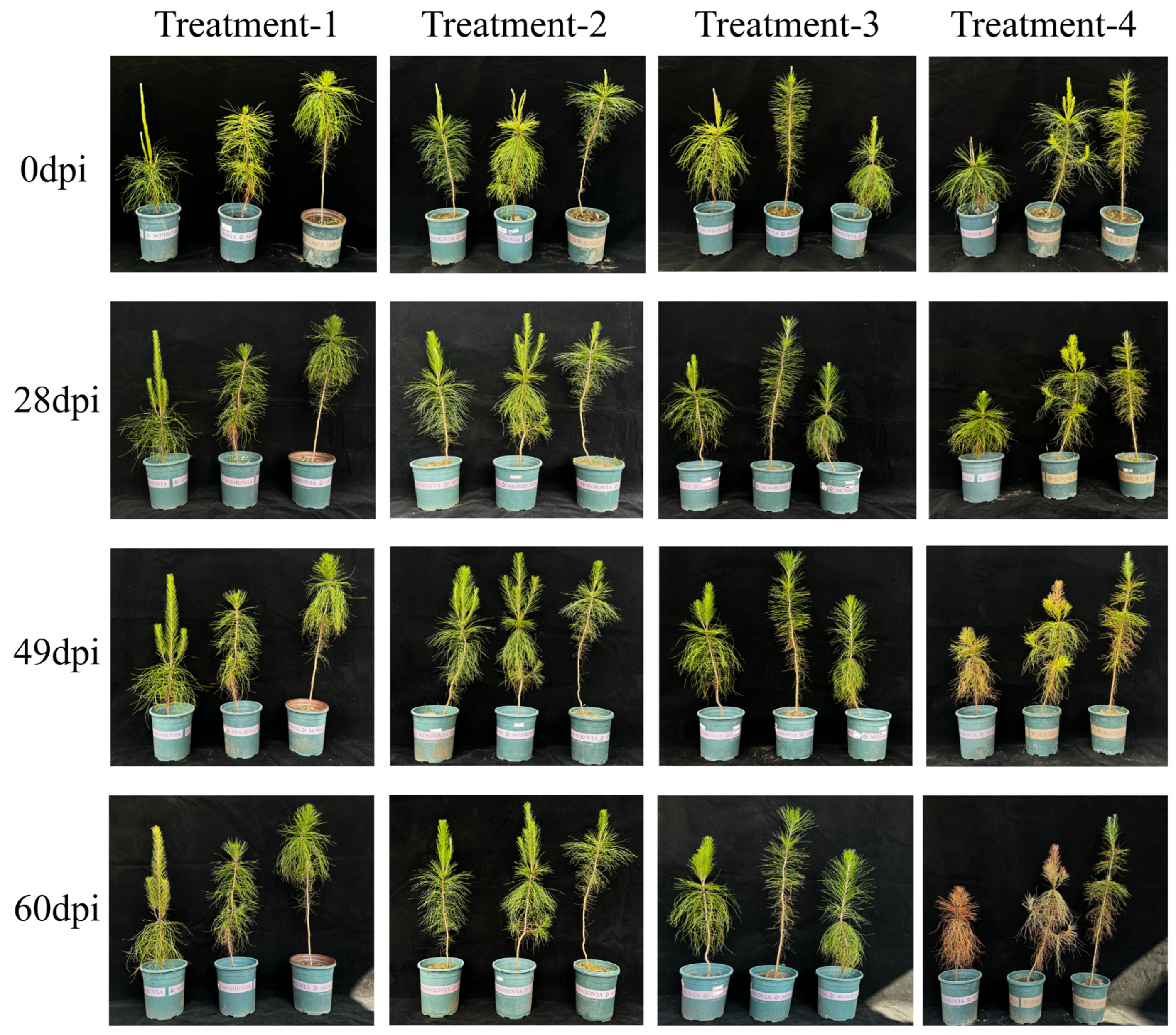
The experiment included four treatment groups. In Treatment 1, 20 mL of sterile water was sprayed onto healthy pine seedlings as a control. In Treatment 2, 20 mL of bacterial suspension (OD600 = 0.8) was sprayed onto the seedlings. In Treatment 3, the seedlings were first sprayed with 20 mL of bacterial suspension (OD600 = 0.8), followed by spraying 20 mL of the fungal spore suspension (1 × 10⁶ conidia/mL) 3 days later. Treatment 4 consisted of spraying the seedlings with 20 mL of fungal spore suspension (1 × 10⁶ conidia/mL) without prior bacterial treatment. Each treatment group consisted of six replicates. Each plant was enclosed in a plastic bag during the treatment period to prevent cross-contamination. Disease development was monitored weekly for 60 days, and the disease incidence and control efficacy were calculated and analyzed (Table 1).
S u s c e p t i b i l i t y   i n d e x = ( n u m b e r   o f   p l a n t s   i n   e a c h   d i s e a s e   g r a d e × r e p r e s e n t a t i v e   v a l u e ) t o t a l   n u m b e r   o f   p l a n t s × m a x i m u m   d i s e a s e   g r a d e   v a l u e × 100
C o n t r o l   e f f i c a c y % = c o n t r o l   s u s c e p t i b i l i t y   i n d e x c t r e a t m e n t   s u s c e p t i b i l i t y   i n d e x c o n t r o l   s u s c e p t i l i t y   i n d e x × 100

2.12. Antimicrobial Spectra of the Selected Bacteria

The broad-spectrum antimicrobial properties of the selected biocontrol bacteria were evaluated for their potential use against other plant diseases; the experimental method of plate antagonism is as described in 2.5. Nine common plant pathogenic fungi were tested as target pathogens. These included Botrytis cinerea, Fusarium graminearum, F. oxysporum, N. camelliae-oleiferae, Pythium vexans, Diplodia pinea, Alternaria sp., Verticillium dahlia, and Lecanosticta acicula. All pathogenic fungi were from the strain preservation bank of Nanjing Forestry University (Nanjing, China). These pathogens were selected for their importance in agriculture and forestry, and the results provide insights into the potential of biocontrol bacteria as broad-spectrum control agents for managing plant diseases.

2.13. Data Analysis

Microsoft Excel 2010 was used for data statistics and calculation, and IBM SPSS Statistics 26 software was used to conduct a one-way analysis of variance (one-way ANOVA) and Duncan test method to analyze the difference significance of the data (p < 0.05).

3. Results

3.1. Disease Symptoms and Fungal Isolation

In 2022, a severe outbreak of pine needle blight was reported in a P. massoniana nursery at the School of Forestry, Nanjing Forestry University. The disease affected approximately 60% of the nursery, and the individual tree infection rate was approximately 30%. Early symptoms included wilting at the needle tips and midsections of the needles, which gradually spread throughout the entire needle length. Black conidial spore disks formed at the lesion sites. The color of the lesions varied from reddish-brown to grayish-white depending on the severity of the infection. Severely infected trees exhibited widespread needle necrosis and a marked decline in overall health, ultimately leading to tree death (Figure 1A). From the infected needles, 19 fungal isolates were obtained, with one dominant strain accounting for 90% of the isolates. Molecular identification based on DNA sequencing of the ITS region and morphological characteristics confirmed that all the isolates belonged to the same fungal species. Five representative isolates were selected for further study. When cultured on PDA plates, the isolated fungus exhibited rapid growth, initially white mycelium, and density. After one week, the mycelium thickened into a cotton-like texture, and the PDA medium gradually turned light yellow (Figure 1B). The conidial spore disks were black and spherical, up to 400 μm in diameter (Figure 1C). Conidiophores are hyaline and thinly walled. Conidiogenous cells are discrete and cucullate, hyaline, or pale brown (Figure 1E). The conidia were (19.052-) 30.696 × 5.898 (-9.401) μm ( x ¯ = 21.271 × 7.382 μm, n = 30), fusiform, olive type, straight to slightly curved, and 4-septate. The basal cells were hyaline, inverted cone-shaped, and 3.233–10.000 μm ( x ¯ = 5.085 μm, n = 30). The three median cells measured 13.365–16.972 μm ( x ¯ = 14.843 μm, n = 30), with light brown to dark brown coloration. The second cell from the base measured 3.556–4.532 μm ( x ¯ = 9.943 μm, n = 30), the third cell measured 3.187–6.043 μm ( x ¯ = 5.2316 μm, n = 30), and the fourth cell measured 2.87–4.555 μm ( x ¯ = 3.527 μm, n = 30). The apical cells were 1.511–3.777 μm ( x ¯ = 3.033 μm, n = 30), inverted cone-shaped, and hyaline. There were 1–4 (mostly 3) tubular apical appendages, straight or curved, measuring 14.037–33.210 μm ( x ¯ = 19.796 μm, n = 30). A basal appendage was present, tubular, either 0 or 1, mostly 1, measuring 0–7.191 μm ( x ¯ = 3.307 μm, n = 30) (Figure 1F–J).

3.2. Pathogenicity Testing and Multilocus Phylogenetic Analyses

All five representative fungal isolates (W1, W2, W3, W4, and W5) were pathogenic to P. massoniana, with a 100% infection rate. The symptoms of the inoculated plants were observed, and samples were collected. Three days post inoculation, the pine needles began to show signs of wilting. By the fifth day, the necrotic lesions had become evident and had progressively expanded, causing the needles to yellow. After fifteen days, the lesions had spread rapidly from the inoculation site, resulting in the entire needle turning gray–brown, with the tip eventually dying. In contrast, no symptoms were observed in the control plants (Figure 1D). Pathogenic fungi were successfully reisolated from the inoculated pine needles to verify Koch’s postulates.
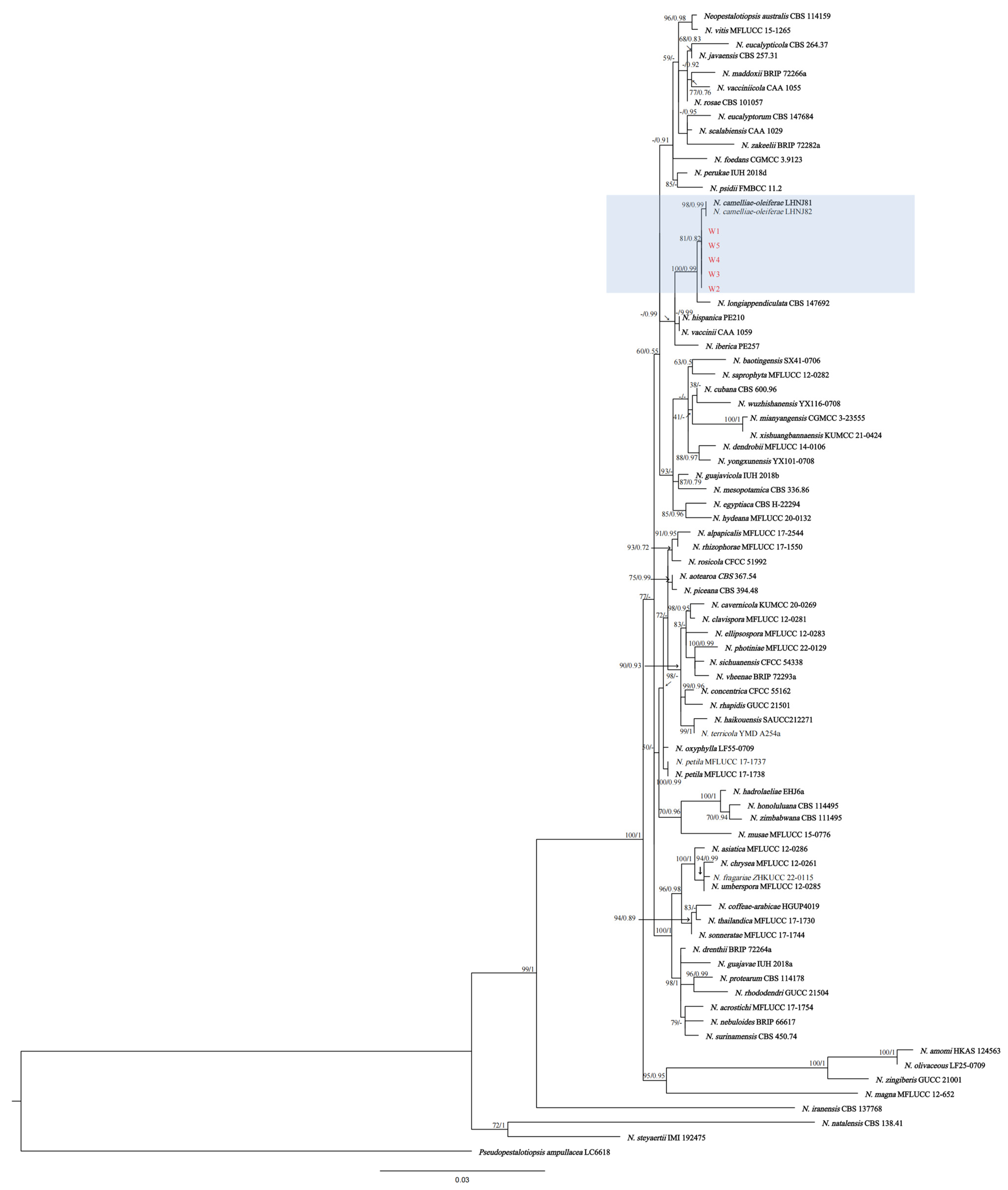
The sequences of five fungal isolates (W1, W2, W3, W4, and W5) were deposited in GenBank under accession numbers PQ803210 to PQ803214 (ITS), PQ835273 to PQ835277 (TEF1), and PQ835278 to PQ835282 (TuB2). The ITS, TuB2, and TEF1 gene regions from 73 representative strains from the same genus and one outgroup strain (Pseudopestalotiopsis ampullacea) were used to construct a phylogenetic tree. The phylogenetic analysis revealed that the W1–W5 isolates clustered together and presented a high degree of similarity with N. camelliae-oleiferae (Figure 2).

3.3. Screening and Identification of Endophytic Bacteria with Biocontrol Potential

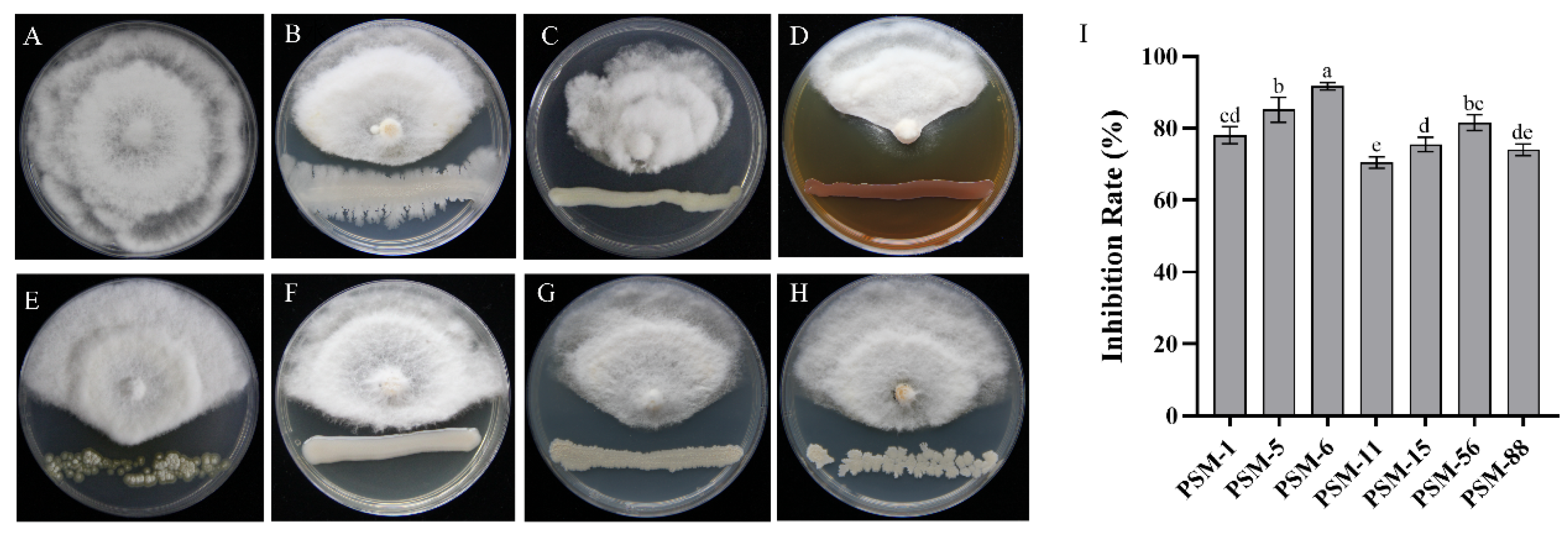
A total of 84 bacterial strains were isolated from P. massoniana needle samples. Seven strains with antagonistic activity against the pathogen responsible for pine needle blight were identified via the dual culture method. Among these strains, PSM-6 exhibited the greatest inhibitory effect, achieving an inhibition rate of over 95% (Figure 3). Therefore, PSM-6 was selected for further experiments. The remaining antagonistic and endophytic strains were stored at −80 °C for future use (Figure S1).
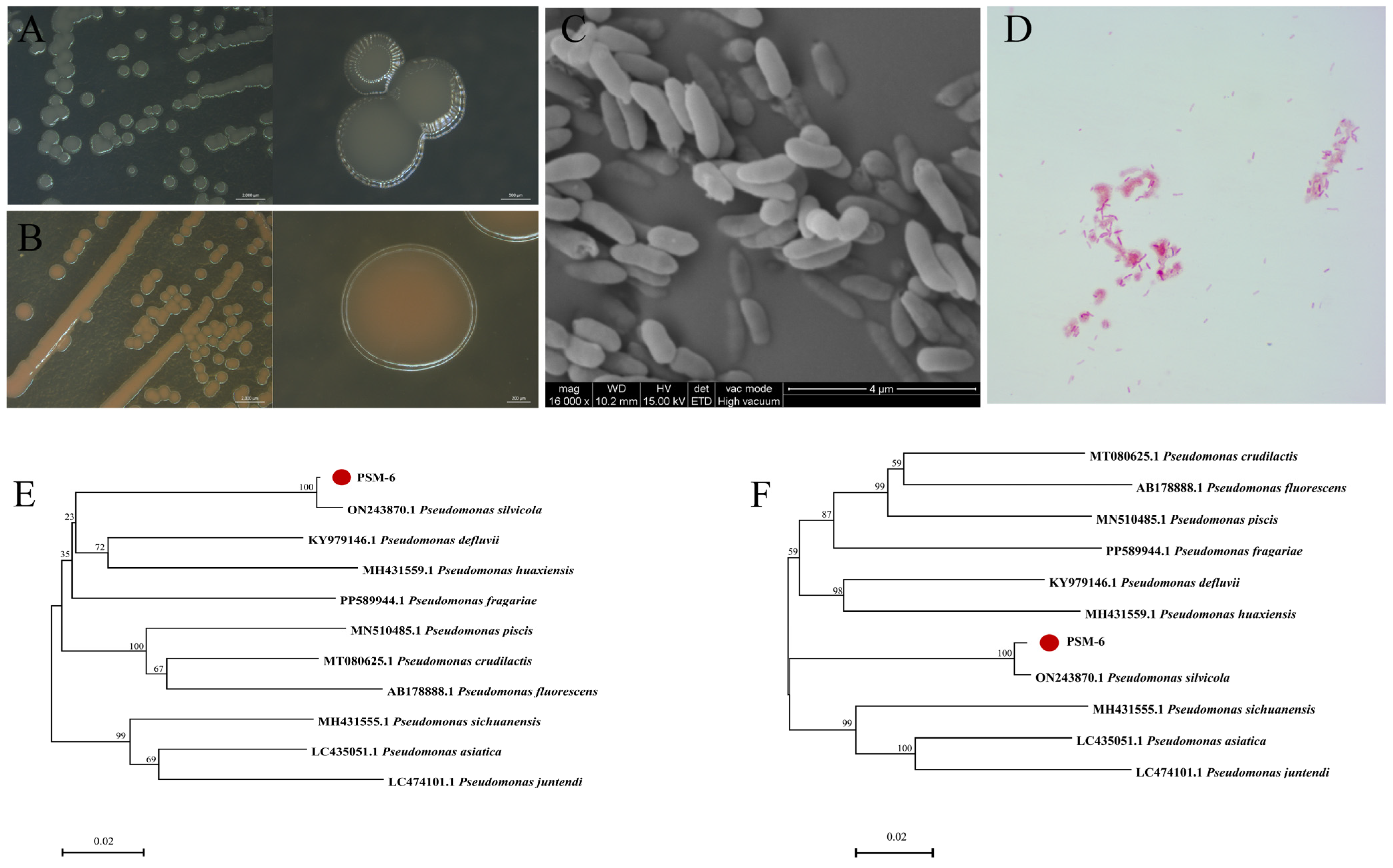
PSM-6 was inoculated onto LB and PDA media plates using the streak plate method to reveal its distinct morphological and physiological traits. On LB agar, colonies appeared creamy white with a viscous texture and a smooth surface. The individual colonies were nearly round and emitted a slight odor (Figure 4A). On PDA plates, the PSM-6 strain produced pigments that deepened in color over time, transitioning from pale yellow to orange-brown (Figure 4B). The colony morphology was similar to that observed on LB agar. Scanning electron microscopy (SEM) revealed that the PSM-6 cells were elongated and oval-shaped, with dimensions ranging from 0.8969 to 1.3710 µm in length and 0.2703–0.5828 µm in width (Figure 4C). Gram staining confirmed that PSM-6 was Gram-negative (Figure 4D), and Staphylococcus aureus was Gram-positive (Figure S2). Molecular analysis via 16S rDNA (1433 bp) and gyrB (1289 bp) sequence data, followed by phylogenetic tree construction, revealed that PSM-6 clustered within the same evolutionary branch as Pseudomonas silvicola (Figure 4E,F). Based on these results, PSM-6 shares the same branch with Pseudomonas silvicola, and phylogenetic analysis indicates that PSM-6 is a close relative of Pseudomonas silvicola. The 16S rDNA and gyrB sequences were deposited in GenBank under accession numbers PQ803218 (16S) and PQ835283 (gyrB).
The physiological and biochemical characteristics of PSM-6 are summarized in Table 2. The strain tested negative for the Voges-Proskauer (V-P) reaction, cellulase degradation, indole-3-acetic acid (IAA) production, gelatin liquefaction, inorganic phosphate solubilization, and nitrogen fixation assay. However, the strain tested positive for methyl red, catalase activity, amylase production, potassium solubilization, organic phosphorus degradation, and siderophore production. In this study, PSM-6 demonstrated plant growth-promoting effects and the ability to damage pathogenic fungal cell walls. Iron (Fe), phosphorus (P), and potassium (K) are essential nutrients for plant growth, yet their natural bioavailability for direct plant uptake is limited. PSM-6 exhibited solubilization of organophosphorus and potassium feldspar, along with siderophore production, enhancing nutrient availability. The antifungal potential of biocontrol bacteria can be assessed by their ability to disrupt pathogenic fungal cell walls. PSM-6 showed amylase and catalase activity, suggesting a possible mechanism for cell wall degradation and hydrolysis in target fungi. These findings indicate that PSM-6 is a promising candidate for further development as a dual-action biocontrol agent and biofertilizer.

3.4. Antifungal Component Testing of PSM-6

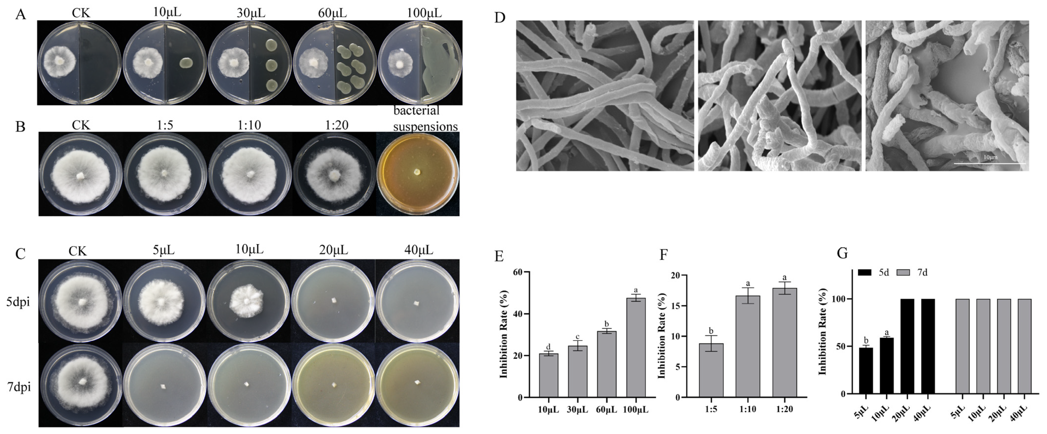
The antifungal components of microorganisms include volatile substances and non-volatile substances. In this study, PSM-6 demonstrated significant antifungal activity, with its VOCs inhibiting pathogenic fungus by 20%–50% (Figure 5A,E), and the inhibitory effect of PSM-6’s sterile supernatant was only 10%–20% (Figure 5B,F). However, the coculture experiment with bacterium suspension products resulted in a remarkable 100% inhibition rate of the pathogen (Figure 5B). These findings suggest that the inhibitory effect is primarily due to the compounds secreted by PSM-6 during its growth.
To investigate this further, we prepared the crude extracts of PSM-6 secretions by ethyl acetate. Coculture experiments with extracts at various concentrations and bacterial growth stages revealed that prolonged bacterial cultivation increased the production of inhibitory compounds. The crude extract obtained after 7 days of cultivation achieved complete inhibition (100%) at a concentration of 5 µL, whereas the extract from 5 days of cultivation required 20 µL to achieve the same effect. Furthermore, the inhibitory activity increased in parallel with the accumulation of pigments during PSM-6 growth (Figure 5C). SEM revealed that the normal hyphal (untreated) surface of the pathogen was smooth and uniform. The hyphae treated with fermentation products exhibited wrinkling and deformation. However, hyphae treated with the crude extract displayed even more severe damage, including a wrinkled and deformed surface and, in some cases, signs of breakage (Figure 5D).
These results confirmed that PSM-6 significantly inhibits fungal hyphal growth. The primary inhibitory components are extracellular secondary metabolites produced during the growth of PSM-6. These metabolites are responsible for the observed suppression of the fungal pathogen.

3.5. Biocontrol Efficacy of PSM-6 Against Pine Needle Blight in Potted Seedlings

The two-month potted seedling experiment results indicated that the biocontrol bacterium PSM-6 does not have any pathogenic effects on P. massoniana. Sixty days after inoculation with N. camelliae-oleiferae, the control group presented a disease incidence of 100%, as the disease progressed, Masson pine seedlings exhibited systemic wilting, culminating in complete mortality, resulting in a disease index of 100. However, the seedlings pretreated with PSM-6 remained relatively healthy throughout the 60-day observation period, with a disease susceptibility index of 58 and control efficacy of 42% (Figure 6). These findings highlight the excellent preventive efficacy of PSM-6 and provide a strong basis for its potential application in field trials for effective disease management.

3.6. Spectrum of the Antifungal Activity of PSM-6 Against Other Plant Pathogenic Fungi

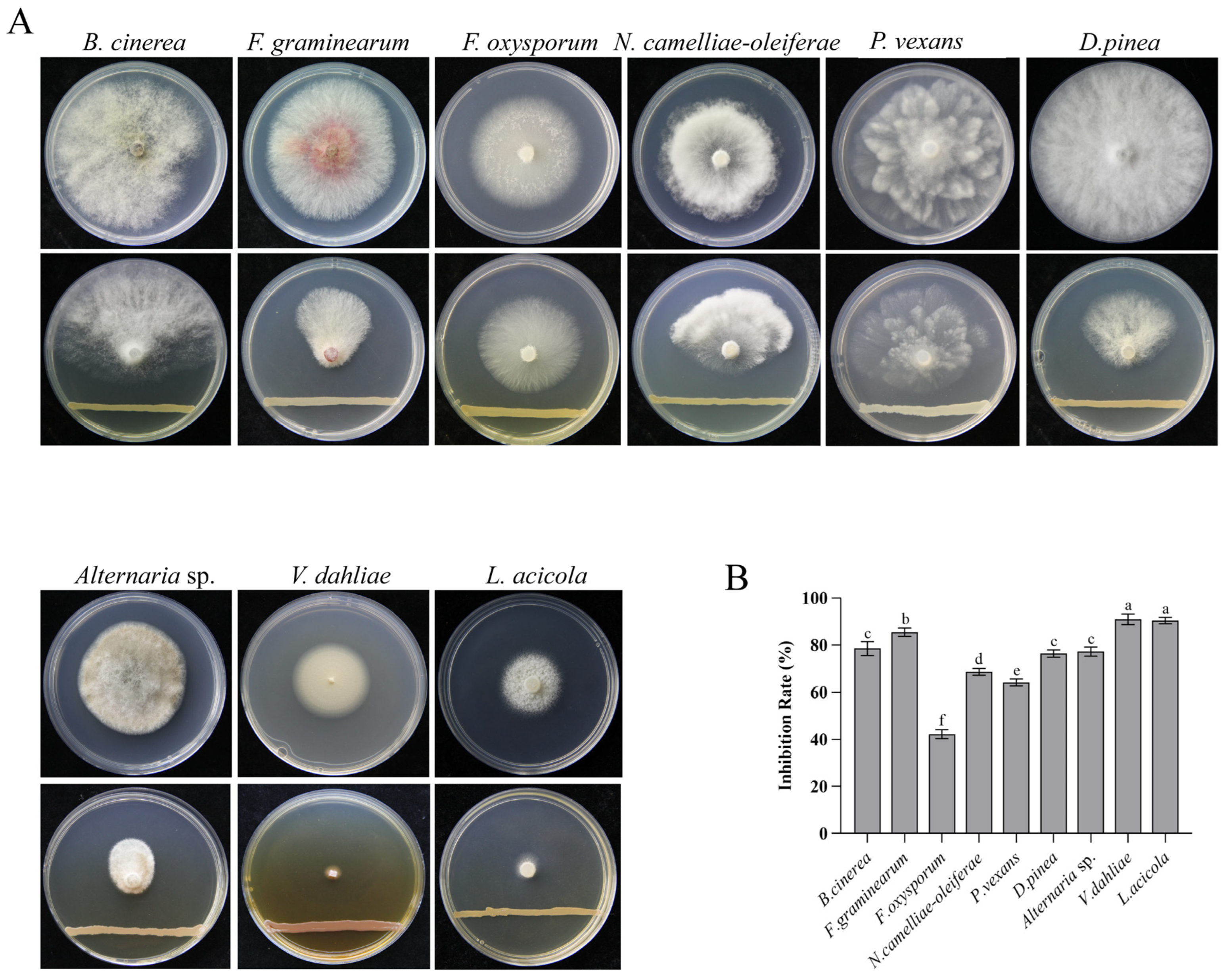
A broad-spectrum antifungal assay was conducted in this study to evaluate the effectiveness of PSM-6 against nine common and highly pathogenic plant fungal pathogens (Figure 7A). The results from the plate confrontation test revealed that PSM-6 effectively inhibited the growth of all the tested pathogens. PSM-6 achieved an inhibition rate of 91.83% against Verticillium dahliae and had strong inhibitory effects on Lecanosticta acicola, Alternaria sp., and Botrytis cinerea, with inhibition rates of 86.68%, 79.38%, and 79.34%, respectively (Figure 7B). These findings suggest that PSM-6 produces antifungal metabolic compounds capable of suppressing various plant pathogenic fungi.

4. Discussion

This study provides the first documented evidence that N. camelliae-oleiferae can cause masson pine blight. Before understanding the taxonomic status of the Neopestalotiopsis, it is essential to first understand Pestalotiopsis-like fungi. These asexual fungi, belonging to the family Amphisphaeriaceae, are widely distributed as endophytes or pathogens across diverse host plants [54]. Some scholars also believe that it belongs to the Pestalotiopsis Steyaert. The classification of Pestalotiopsis Steyaert has been controversial for a long time, and it went through four-time phases to establish its current classification system. In 1949, Steyaert distinguished Pestalotiopsis according to the number of apical appendages [55]; in 1961, Guba proposed to classify Pestalotiopsis according to the number of conidia cells; and in 1969–1980, Sutton combined the views of the two predecessors and added the type of diaphragm, a total of three characteristics to distinguish Pestalotiopsis. With the development of molecular systematics, Jeewon and Wei Jiguang et al. discovered the unique molecular biological characteristics of Pestalotiopsis and constructed a phylogenetic tree of it [56,57]. So far, the taxonomic status of Pestalotiopsis has been basically formed. Despite these advances, fungal evolutionary diversity continues to challenge classification systems. Until now, modern phylogeny divides it into three clades; they are Pestalotiopsis sensu stricto, Neopestalotiopsis, and Pseudopestalotiopsis, according to both morphological and biomolecular identification [58]. The expansion of the diversity of pathogenic fungi aggravated the blight infection of pine needles. Given the vital role of forest ecosystem preservation, developing effective biological control measures is imperative to mitigate pine disease outbreaks.
In 2023, P. silvicola was first identified as a biocontrol agent against anthracnose and was isolated from cones of Cunninghamia lanceolate [59]; this agent is highly similar to P. eucalypticola. Pseudomonas exists in soil, water, and living organisms [60,61], it can secrete many compounds; its strong survival ability and diversity of secretions make it highly regarded in the field of biological control. Previous reports have proved that its metabolites can inhibit the growth of pathogenic fungi, promote plant growth, and increase disease resistance [62,63], for example, iron (Fe) is an essential micronutrient for plant growth; however, in soil, it predominantly exists in insoluble forms that are unavailable for plant uptake; siderophores play a critical role in Fe solubilization, enhancing its bioavailability. Notably, PSM-6 exhibits both siderophore production and enzymatic activity, suggesting a dual function: not only may it suppress N. camelliae-oleiferae growth, but it could also enhance the growth potential of P. massoniana. Over the past few years, research into its potential applications has expanded, including its use in controlling diseases such as boxwood blight [64]. This study evaluated the broad-spectrum antifungal activity of P. silvicola PSM-6 against nine common plant pathogenic fungi. The results show that PSM-6 exhibited significant inhibitory effects against several filamentous fungi, particularly against Verticillium dahliae and Lecanosticta acicola, by completely suppressing their growth. These pathogens are responsible for severe annual economic losses in agriculture and forestry, leading to root rot, stem rot, and wilt diseases in host plants [65,66]. These findings indicate that PSM-6 has the potential to become a widely applicable biocontrol agent.
We also investigated the antifungal mechanisms of PSM-6. We compared the effects of volatile compounds and sterile supernatants. The secondary metabolites produced by the bacterial cells had the most effective inhibitory effect on fungal growth. Pseudomonas species are well known for their antimicrobial properties [67], producing a range of bioactive compounds such as phenazine compounds, pyrrolnitrin, pseudomonic acid, 2,4-diacetylphloroglucinol, rhizoxin, and pyoluteorin [68,69,70,71]. These compounds are secreted by pathogenic Pseudomonas strains such as Ps. aeruginosa and Ps. fluorescens and are used in biocontrol applications [72,73]. For example, phenazine-1-carboxylic acid (PCA) [74] is a nitrogen-containing heterocyclic compound produced by Pseudomonas. PCA is widely used in agricultural fungicides. Compared with other Gram-negative bacteria in the environment, Pseudomonas species have larger genome sizes ranging from 6 to 7 Mbp [75]. This enables Pseudomonas species to produce a wide range of metabolites and adapt to various ecological conditions, providing them with a competitive edge in biocontrol applications [76].
Endophytic bacteria such as PSM-6 have an advantage over soil bacteria due to their close interactions with the host plant. They can directly secrete antibiotics and growth-promoting factors within plants, effectively inhibiting pathogens or inducing systemic resistance [77,78]. Endophytic bacterial populations are typically lower than soil microbial populations are, reducing competition and enabling more efficient colonization of plant tissues. High-performing biocontrol strains of Pseudomonas are particularly adept at colonizing plant surfaces and internal tissues. In our pot experiment, PSM-6 effectively colonized the internal plant tissues of Pinus massoniana and promoted plant growth, significantly reducing the incidence of disease caused by pathogenic fungi. These findings highlight the potential of PSM-6 as an effective biocontrol agent capable of suppressing fungal diseases and enhancing plant health.

5. Conclusions

This study represents the first global report of N. camelliae-oleiferae as the causal agent of needle blight in P. massoniana, with all five fungal isolates (W1–W5) showing 100% pathogenicity, and P. silvicola strain PSM-6 demonstrates significant potential as a biocontrol agent against P. massoniana needle blight caused by N. camelliae-oleiferae. PSM-6 exhibited strong antagonistic activity both in vitro and in planta, preventing disease symptoms in seedlings over a 60-day period. The strain showed broad-spectrum antifungal activity against nine pathogenic fungi, with particularly high inhibition rates against V. dahliae (91.83%) and L. acicola (86.68%). Microscopic analysis revealed that PSM-6’s antifungal mechanism involves secondary metabolites that induce hyphal deformation. Additionally, PSM-6 demonstrated effective plant tissue colonization, promoting host growth while enhancing systemic resistance. As the first reported application of P. silvicola for pine needle blight control worldwide, these findings provide novel insights for forest pathology research and disease management. Considering PSM-6’s temperature sensitivity and short growth cycle, along with the global spread of pine diseases, we propose developing a freeze-dried powder formulation to enhance its practical application. Given its broad-spectrum activity against filamentous fungi, PSM-6 also shows promise for biological control of other phytopathogens, offering a new approach for integrated disease management strategies.

Supplementary Materials

The following supporting information can be downloaded at https://www.mdpi.com/article/10.3390/f16040650/s1: Figure S1: Ineffective antagonistic effects of the isolated endophytic bacteria against N. camelliae-oleiferae on PDA plates; Figure S2: Gram stain of Staphylococcus aureus; Table S1: Primers and PCR programs; Table S2: Information on Neopestalotiopsis fungal strains and outgroups used in the phylogenetic tree analysis.

Author Contributions

Conceptualization and methodology, X.-Y.W. and F.X.; writing—original draft, X.-Y.W. and M.L.; writing—review and editing, M.L. and M.F.L.; supervision, H.L.; preparation Project administration F.C. All authors have read and agreed to the published version of the manuscript.

Funding

This work was supported by the National Key R&D Program of China (2022YFD1401005).

Data Availability Statement

Data are contained within the article. The detailed images of the experiments are available can be provided on demand.

Acknowledgments

The authors would like to thank those who provided assistance and advice for this study, especially Mao-Jiao Zhang for her helpful suggestions and revisions.

Conflicts of Interest

The authors declare no conflicts of interest.

References

  1. Yang, Z.; Zhao, J.; Ni, H.; Wang, H.; Zhou, B. Three Subtropical Species Adapt to Drought by Reallocating Biomass and Adjusting Root Architecture. Forests 2023, 14, 806. [Google Scholar] [CrossRef]
  2. Liu, J.; Gu, Z.; Shao, H.; Zhou, F.; Peng, S. N–P Stoichiometry in Soil and Leaves of Pinus massoniana Forest at Different Stand Ages in the Subtropical Soil Erosion Area of China. Environ. Earth Sci. 2016, 75, 1091. [Google Scholar] [CrossRef]
  3. Zhou, X.; Zhao, M.; Zhou, L.; Yang, G.; Huang, L.; Yan, C.; Huang, Q.; Ye, L.; Zhang, X.; Guo, L.; et al. Regionalization of Habitat Suitability of Masson’s Pine Based on Geographic Information System and Fuzzy Matter-Element Model. Sci. Rep. 2016, 6, 34716. [Google Scholar] [CrossRef] [PubMed]
  4. Li, R.; Zhu, L.; Chen, P.; Chen, Y.; Hao, Q.; Zhu, P.; Ji, K. Functional Characterization of PmDXR, a Critical Rate-Limiting Enzyme, for Turpentine Biosynthesis in Masson Pine (Pinus massoniana Lamb.). Int. J. Mol. Sci. 2024, 25, 4415. [Google Scholar] [CrossRef] [PubMed]
  5. Vicente, C.S.L.; Soares, M.; Faria, J.M.S.; Ramos, A.P.; Inácio, M.L. Insights into the Role of Fungi in Pine Wilt Disease. J. Fungi 2021, 7, 780. [Google Scholar] [CrossRef]
  6. Seo, S.T.; Park, M.J.; Park, J.H.; Shin, H.D. First Report of Brown Spot Needle Blight on Pinus thunbergii Caused by Lecanosticta Acicola in Korea. Plant Dis. 2012, 96, 914. [Google Scholar] [CrossRef]
  7. Hu, R.; Liang, J.; Xie, X.; Zhang, Y.; Zhang, X. Incidence of Pine Needle Blight and Its Relationship with Site Factors of Japanese Red Pine Forests in the Kunyushan Mountains, East China. Glob. Ecol. Conserv. 2020, 22, e00922. [Google Scholar] [CrossRef]
  8. Li, H.; Peng, B.-Y.; Xie, J.-Y.; Bai, Y.-Q.; Li, D.-W.; Zhu, L.-H. Pestalotiopsis jiangsuensis sp. nov. Causing Needle Blight on Pinus Massoniana in China. J. Fungi 2024, 10, 230. [Google Scholar] [CrossRef]
  9. Maharachchikumbura, S.S.N.; Hyde, K.D.; Groenewald, J.Z.; Xu, J.; Crous, P.W. Pestalotiopsis Revisited. Stud. Mycol. 2014, 79, 121–186. [Google Scholar] [CrossRef]
  10. Zheng, X.; Liu, X.; Li, X.; Quan, C.; Li, P.; Chang, X.; Gu, J.; Khaskheli, M.I.; Gong, G. Pestalotiopsis Species Associated with Blueberry Leaf Spots and Stem Cankers in Sichuan Province of China. Plant Dis. 2023, 107, 149–156. [Google Scholar] [CrossRef]
  11. Bhuiyan, M.A.B.; Islam, S.M.N.; Bukhari, M.A.I.; Kader, M.A.; Chowdhury, M.Z.H.; Alam, M.Z.; Abdullah, H.M.; Jenny, F. First Report of Pestalotiopsis microspora Causing Leaf Blight of Banana in Bangladesh. Plant Dis. 2022, 106, 1518. [Google Scholar] [CrossRef] [PubMed]
  12. Fiorenza, A.; Gusella, G.; Aiello, D.; Polizzi, G.; Voglmayr, H. Neopestalotiopsis siciliana sp. nov. and N. Rosae Causing Stem Lesion and Dieback on Avocado Plants in Italy. J. Fungi 2022, 8, 562. [Google Scholar] [CrossRef] [PubMed]
  13. Li, H.; Qiu, Y.; Yao, T.; Ma, Y.; Zhang, H.; Yang, X.; Li, C. Evaluation of Seven Chemical Pesticides by Mixed Microbial Culture (PCS-1): Degradation Ability, Microbial Community, and Medicago sativa Phytotoxicity. J. Hazard. Mater. 2020, 389, 121834. [Google Scholar] [CrossRef]
  14. Ayilara, M.S.; Adeleke, B.S.; Akinola, S.A.; Fayose, C.A.; Adeyemi, U.T.; Gbadegesin, L.A.; Omole, R.K.; Johnson, R.M.; Uthman, Q.O.; Babalola, O.O. Corrigendum: Biopesticides as an Alternative to Synthetic Pesticides: A Case for Nanopesticides, Phytopesticides and Microbial Pesticides. Front. Microbiol. 2024, 14, 1258968. [Google Scholar] [CrossRef]
  15. Hufbauer, R.A.; Roderick, G.K. Microevolution in Biological Control: Mechanisms, Patterns, and Processes. Biol. Control 2005, 35, 227–239. [Google Scholar] [CrossRef]
  16. Stenberg, J.A.; Sundh, I.; Becher, P.G.; Björkman, C.; Dubey, M.; Egan, P.A.; Friberg, H.; Gil, J.F.; Jensen, D.F.; Jonsson, M.; et al. When Is It Biological Control? A Framework of Definitions, Mechanisms, and Classifications. J. Pest. Sci. 2021, 94, 665–676. [Google Scholar] [CrossRef]
  17. Lipa, J.J. Biologiczne zwalczanie pasozytow i szkodnikow a srodowisko. Wiad. Parazytol. 1991, 37, 461–470. [Google Scholar]
  18. Batra, S.W.T. Biological Control in Agroecosystems. Science 1982, 215, 134–139. [Google Scholar] [CrossRef]
  19. Naranjo, S.E.; Ellsworth, P.C.; Frisvold, G.B. Economic Value of Biological Control in Integrated Pest Management of Managed Plant Systems. Annu. Rev. Entomol. 2015, 60, 621–645. [Google Scholar] [CrossRef]
  20. Wang, Z.; Chen, X.; Zhong, T.; Li, B.; Yang, Q.; Du, M.; Zalán, Z.; Kan, J. Bioeffector Pseudomonas fluorescens ZX Elicits Biosynthesis and Accumulation of Functional Ingredients in Citrus Fruit Peel: A Promising Strategy for a More Sustainable Crop. J. Agric. Food Chem. 2021, 69, 13810–13820. [Google Scholar] [CrossRef]
  21. Sorokina, T.A.; Mishustin, E.N. Distribution of Pseudomonas Fluorescent Bacteria in Soils and in the Root Zone of Plants. Biol. Bull. Acad. Sci. USSR 1978, 5, 584–589. [Google Scholar] [PubMed]
  22. Palleroni, N.J. The Pseudomonas Story. Environ. Microbiol. 2010, 12, 1377–1383. [Google Scholar] [CrossRef] [PubMed]
  23. Mavrodi, D.V.; Parejko, J.A.; Mavrodi, O.V.; Kwak, Y.; Weller, D.M.; Blankenfeldt, W.; Thomashow, L.S. Recent Insights into the Diversity, Frequency and Ecological Roles of Phenazines in Fluorescent Pseudomonas spp. Environ. Microbiol. 2013, 15, 675–686. [Google Scholar] [CrossRef]
  24. Abdelaziz, A.A.; Kamer, A.M.A.; Al-Monofy, K.B.; Al-Madboly, L.A. Pseudomonas aeruginosa’s Greenish-Blue Pigment Pyocyanin: Its Production and Biological Activities. Microb. Cell Fact. 2023, 22, 110. [Google Scholar] [CrossRef]
  25. Gross, H.; Loper, J.E. Genomics of Secondary Metabolite Production by Pseudomonas spp. Nat. Prod. Rep. 2009, 26, 1408–1446. [Google Scholar] [CrossRef]
  26. Hashimoto, M.; Hattori, K. Oxypyrrolnitrin: A Metabolite of Pseudomonas. Chem. Pharm. Bull. 1966, 14, 1314–1316. [Google Scholar] [CrossRef]
  27. Gu, Q.; Qiao, J.; Wang, R.; Lu, J.; Wang, Z.; Li, P.; Zhang, L.; Ali, Q.; Khan, A.R.; Gao, X.; et al. The Role of Pyoluteorin from Pseudomonas protegens Pf-5 in Suppressing the Growth and Pathogenicity of Pantoea Ananatis on Maize. Int. J. Mol. Sci. 2022, 23, 6431. [Google Scholar] [CrossRef]
  28. Ma, Z.; Zhang, S.; Liang, J.; Sun, K.; Hu, J. Isolation and Characterization of a New Cyclic Lipopeptide Orfamide H from Pseudomonas protegens CHA0. J. Antibiot. 2020, 73, 179–183. [Google Scholar] [CrossRef]
  29. Guo, J.; Huang, X.; Lin, S.; Liu, Z.; Wang, H.; Gao, Q.; Zhou, H. Identification of Epicoccum latusicollum Causing Leaf Spot Disease on Bletilla striata in China. Crop Prot. 2024, 181, 106676. [Google Scholar] [CrossRef]
  30. Munakata, Y.; Spina, R.; Slezack-Deschaumes, S.; Genestier, J.; Hehn, A.; Laurain-Mattar, D. Screening of Endophytic Bacteria of Leucojum aestivum ‘Gravety Giant’ as a Potential Source of Alkaloids and as Antagonist to Some Plant Fungal Pathogens. Microorganisms 2022, 10, 2089. [Google Scholar] [CrossRef]
  31. Li, Z.; Li, H.; Zhang, J.; Zhang, S.; Zhao, Q.; Cheng, C.; Zhang, Y. Characterization of Fungal Species Isolated from Cankered Apple Barks Demonstrates the Alternaria alternata Causing Apple Canker Disease. J. Fungi 2024, 10, 536. [Google Scholar] [CrossRef] [PubMed]
  32. Morsy, A.A.; Elshahawy, I.E. Anthracnose of Lucky Bamboo Dracaena sanderiana Caused by the Fungus Colletotrichum dracaenophilum in Egypt. J. Adv. Res. 2016, 7, 327–335. [Google Scholar] [CrossRef] [PubMed]
  33. Lu, P.-K.; Liu, H.-H.; Chang, H.-X. First Report of Corynespora cassiicola Causing Target Spot on Soybean in Taiwan. Plant Dis. 2021, 105, 3753. [Google Scholar] [CrossRef] [PubMed]
  34. Ji, Y.; Wang, J.; Liu, Y.; Liu, S.; Jiang, X.; Huang, H. Isolation and Identification of Postharvest Rot Pathogens in Citrus × tangelo and Their Potential Inhibition with Acidic Electrolyzed Water. Food Environ. Virol. 2024, 16, 409–421. [Google Scholar] [CrossRef]
  35. Zhang, Y.-Y.; Zhang, T.; Li, H.-Y.; Zheng, R.; Ren, J.; Yang, Q.; Jiang, N. Nigrospora humicola (Apiosporaceae, Amphisphaeriales), a New Fungus from Soil in China. Diversity 2024, 16, 118. [Google Scholar] [CrossRef]
  36. Luo, X.-X.; Liao, M.-G.; Zhang, K.; Castaneda-Ruiz, R.F.; Ma, J.; Xu, Z.-H. Morphological and Phylogenetic Analyses Reveal Eight Novel Species of Pestalotiopsis (Sporocadaceae, Amphisphaeriales) from Southern China. MycoKeys 2024, 109, 207–238. [Google Scholar] [CrossRef]
  37. Zafri, A.S.; Muhamad, R.; Wahab, A.; Mokhtar, A.S.; Mohd Hata, E. First Report of Leaf Blight on Parthenium hysterophorus Caused by Nigrospora sphaerica in Malaysia. Plant Dis. 2021, 105, 3297. [Google Scholar] [CrossRef]
  38. Xiang, C.-Y.; Gao, F.; Jakovlić, I.; Lei, H.-P.; Hu, Y.; Zhang, H.; Zou, H.; Wang, G.-T.; Zhang, D. Using PhyloSuite for Molecular Phylogeny and Tree-Based Analyses. Imeta 2023, 2, e87. [Google Scholar] [CrossRef]
  39. Nguyen, L.-T.; Schmidt, H.A.; von Haeseler, A.; Minh, B.Q. IQ-TREE: A Fast and Effective Stochastic Algorithm for Estimating Maximum-Likelihood Phylogenies. Mol. Biol. Evol. 2015, 32, 268–274. [Google Scholar] [CrossRef]
  40. Tao, Y.; Quan, X.; Khokhar, I.; Anjum, T.; Song, H.; Mukhtar, I. First Report of Pseudopestalotiopsis theae Causing Leaf Spot of Date Palm (Phoenix dactylifera) in China. Plant Dis. 2020, 105, 508. [Google Scholar] [CrossRef]
  41. Shen, H.; Li, X.; Li, Z. Detecting and Identifying Pathogens and Antagonistic Bacteria Associated with Ginkgo biloba Leaf Spot Disease. Front. Microbiol. 2024, 15, 1346318. [Google Scholar] [CrossRef] [PubMed]
  42. Jung, B.-N.; Park, J.-H.; Lee, S.-H.; Shin, H.-D. First Report of Rhytisma sp. Associated with Tar Spots on Ilex cornuta. Korean J. Mycol. 2020, 48, 63–67. [Google Scholar] [CrossRef]
  43. Sa, R.; He, S.; Han, D.; Liu, M.; Yu, Y.; Shang, R.; Song, M. Isolation and Identification of a New Biocontrol Bacteria against Salvia miltiorrhiza Root Rot and Optimization of Culture Conditions for Antifungal Substance Production Using Response Surface Methodology. BMC Microbiol. 2022, 22, 231. [Google Scholar] [CrossRef] [PubMed]
  44. Saidi, N.; Kouki, S.; M’Hiri, F.; Hajlaoui, M.R.; Mahrouk, M.; Ou-Zari, H.; Jedidi, N.; Hasseni, A. Characterization and Selection of Bacillus sp. Strains, Effective Biocontrol Agents against Fusarium oxysporum f. sp. Radicis-lycopersici, the Causal Agent of Fusarium Crown and Root Rot in Tomato. Ann. Microbiol. 2009, 59, 191–198. [Google Scholar] [CrossRef]
  45. Panebianco, S.; Lombardo, M.F.; Anzalone, A.; Musumarra, A.; Pellegriti, M.G.; Catara, V.; Cirvilleri, G. Epiphytic and Endophytic Microorganisms Associated to Different Cultivar of Tomato Fruits in Greenhouse Environment and Characterization of Beneficial Bacterial Strains for the Control of Post-Harvest Tomato Pathogens. Int. J. Food Microbiol. 2022, 379, 109861. [Google Scholar] [CrossRef]
  46. Moyes, R.B.; Reynolds, J.; Breakwell, D.P. Differential Staining of Bacteria: Gram Stain. Curr. Protoc. Microbiol. 2009, 15, A.3C.1–A.3C.8. [Google Scholar] [CrossRef]
  47. Guerrero, R. Bergey’s Manuals and the Classification of Prokaryotes. Int. Microbiol. 2001, 4, 103–109. [Google Scholar] [CrossRef]
  48. Kuchar, K.W. Der Formenkreis von Pseudomonas punctata (Zimm.) Chester; zur Fassung der Gattung Pseudomonas in Bergey’s Manual. Zentralbl Bakteriol. Orig. 1954, 160, 511–517. [Google Scholar]
  49. Miller, D.N.; Bryant, J.E.; Madsen, E.L.; Ghiorse, W.C. Evaluation and Optimization of DNA Extraction and Purification Procedures for Soil and Sediment Samples. Appl. Environ. Microbiol. 1999, 65, 4715–4724. [Google Scholar] [CrossRef]
  50. Clarridge, J.E. Impact of 16S rRNA Gene Sequence Analysis for Identification of Bacteria on Clinical Microbiology and Infectious Diseases. Clin. Microbiol. Rev. 2004, 17, 840–862. [Google Scholar] [CrossRef]
  51. Li, X.; Wang, X.; Yang, H.; Gao, X.; Cui, Z. Application of gyrB in the Identification of Closely Related Bacteria—A Review. Wei Sheng Wu Xue Bao = Acta Microbiol. Sin. 2008, 48, 701–706. [Google Scholar]
  52. Oztas Gulmus, E.; Gormez, A. Identification and Characterization of Novel Thermophilic Bacteria from Hot Springs, Erzurum, Turkey. Curr. Microbiol. 2020, 77, 979–987. [Google Scholar] [CrossRef] [PubMed]
  53. Kai, M.; Vespermann, A.; Piechulla, B. The Growth of Fungi and Arabidopsis Thaliana Is Influenced by Bacterial Volatiles. Plant Signal Behav. 2008, 3, 482–484. [Google Scholar] [CrossRef]
  54. Jayawardena, R.S.; Zhang, W.; Liu, M.; Maharachchikumbura, S.S.N.; Zhou, Y.; Huang, J.; Nilthong, S.; Wang, Z.; Li, X.; Yan, J.; et al. Identification and Characterization of Pestalotiopsis-like Fungi Related to Grapevine Diseases in China. Fungal Biol. 2015, 119, 348–361. [Google Scholar] [CrossRef]
  55. Steyaert, R.L. Contribution à l’étude Monographique de Pestalotia de Not. et Monochaetia Sacc. (Truncatella gen. nov. et Pestalotiopsis gen. nov.). Bull. Jard. Bot. L’état A Brux. 1949, 19, 285–347. [Google Scholar] [CrossRef]
  56. Tejesvi, M.V.; Tamhankar, S.A.; Kini, K.R.; Rao, V.S.; Prakash, H.S. Phylogenetic Analysis of Endophytic Pestalotiopsis Species from Ethnopharmaceutically Important Medicinal Trees. Fungal Divers. 2009, 38, 167. [Google Scholar]
  57. Jeewon, R.; Liew, E.C.Y.; Hyde, K.D. Phylogenetic Relationships of Pestalotiopsis and Allied Genera Inferred from Ribosomal DNA Sequences and Morphological Characters. Mol. Phylogenet. Evol. 2002, 25, 378–392. [Google Scholar] [CrossRef]
  58. Ma, X.-Y.; Maharachchikumbura, S.S.N.; Chen, B.-W.; Hyde, K.D.; Mckenzie, E.H.C.; Chomnunti, P.; Kang, J.-C. Endophytic Pestalotiod Taxa in Dendrobium Orchids. Phytotaxa 2019, 419, 268–286. [Google Scholar] [CrossRef]
  59. Tian, L.; Zhang, Y.; Yang, H.; Zhao, Q.; Qiu, H.; Xu, J.; Qin, C. Taxonomic Description and Complete Genome Sequencing of Pseudomonas silvicola sp. Nov. Isolated from Cunninghamia laceolata. Forests 2023, 14, 1089. [Google Scholar] [CrossRef]
  60. Lugtenberg, B.J.; Dekkers, L.C. What Makes Pseudomonas Bacteria Rhizosphere Competent? Environ. Microbiol. 1999, 1, 9–13. [Google Scholar] [CrossRef]
  61. Goldberg, J.B. Pseudomonas: Global Bacteria. Trends Microbiol. 2000, 8, 55–57. [Google Scholar] [CrossRef] [PubMed]
  62. Preston, G.M. Plant Perceptions of Plant Growth-Promoting Pseudomonas. Philos. Trans. R. Soc. Lond. B Biol. Sci. 2004, 359, 907–918. [Google Scholar] [CrossRef] [PubMed]
  63. Meyer, J.M. Pyoverdines: Pigments, Siderophores and Potential Taxonomic Markers of Fluorescent Pseudomonas Species. Arch. Microbiol. 2000, 174, 135–142. [Google Scholar] [CrossRef]
  64. Kong, P.; Hong, C. Evaluation of 1021Bp, a Close Relative of Pseudomonas eucalypticola, for Potential of Plant Growth Promotion, Fungal Pathogen Suppression and Boxwood Blight Control. BMC Microbiol. 2024, 24, 346. [Google Scholar] [CrossRef]
  65. Mokhtari, S.; Chavez, M.; Ali, A. First Report of Fusarium oxysporum f. sp. Vasinfectum Causing Fusarium Wilt of Cotton in Kansas, U.S.A. Plant Dis. 2022, 107, 1239. [Google Scholar] [CrossRef]
  66. Tubby, K.; Adamčikova, K.; Adamson, K.; Akiba, M.; Barnes, I.; Boroń, P.; Bragança, H.; Bulgakov, T.; Burgdorf, N.; Capretti, P.; et al. The Increasing Threat to European Forests from the Invasive Foliar Pine Pathogen, Lecanosticta acicola. Ecol. Manag. 2023, 536, 120847. [Google Scholar] [CrossRef]
  67. Iakovleva, L.M.; Pastushenko, L.T.; Simonovich, I.D.; Stepaniuk, V.V. Dissotsiatsiia nekotorykh fitopatogennykh bakterii roda Pseudomonas. Mikrobiol. Zh 1978, 40, 596–599. [Google Scholar]
  68. Pascual, J.; García-López, M.; Carmona, C.; Sousa, T.d.S.; de Pedro, N.; Cautain, B.; Martín, J.; Vicente, F.; Reyes, F.; Bills, G.F.; et al. Pseudomonas soli sp. Nov., a Novel Producer of Xantholysin Congeners. Syst. Appl. Microbiol. 2014, 37, 412–416. [Google Scholar] [CrossRef]
  69. Wang, Y.-N.; He, W.-H.; He, H.; Du, X.; Jia, B.; Zeng, Z.-P.; An, M.-L.; Chen, G.-C. Pseudomonas nitritireducens sp. Nov., a Nitrite Reduction Bacterium Isolated from Wheat Soil. Arch. Microbiol. 2012, 194, 809–813. [Google Scholar] [CrossRef]
  70. Shochat, E.; Hermoni, I.; Cohen, Z.; Abeliovich, A.; Belkin, S. Bromoalkane-Degrading Pseudomonas Strains. Appl. Environ. Microbiol. 1993, 59, 1403–1409. [Google Scholar] [CrossRef]
  71. Biessy, A.; Filion, M. Phenazines in Plant-Beneficial Pseudomonas spp.: Biosynthesis, Regulation, Function and Genomics. Environ. Microbiol. 2018, 20, 3905–3917. [Google Scholar] [CrossRef] [PubMed]
  72. Löser, T.B.; Mescher, M.C.; De Moraes, C.M.; Maurhofer, M. Effects of Root-Colonizing Fluorescent Pseudomonas Strains on Arabidopsis Resistance to a Pathogen and an Herbivore. Appl. Environ. Microbiol. 2021, 87, e0283120. [Google Scholar] [CrossRef] [PubMed]
  73. Lurthy, T.; Gerin, F.; Rey, M.; Mercier, P.-E.; Comte, G.; Wisniewski-Dyé, F.; Prigent-Combaret, C. Pseudomonas Produce Various Metabolites Displaying Herbicide Activity against Broomrape. Microbiol. Res. 2025, 290, 127933. [Google Scholar] [CrossRef] [PubMed]
  74. Liu, Y.; Zhou, Y.; Qiao, J.; Yu, W.; Pan, X.; Zhang, T.; Liu, Y.; Lu, S.-E. Phenazine-1-Carboxylic Acid Produced by Pseudomonas chlororaphis YL-1 Is Effective against Acidovorax citrulli. Microorganisms 2021, 9, 2012. [Google Scholar] [CrossRef]
  75. Silby, M.W.; Winstanley, C.; Godfrey, S.A.C.; Levy, S.B.; Jackson, R.W. Pseudomonas Genomes: Diverse and Adaptable. FEMS Microbiol. Rev. 2011, 35, 652–680. [Google Scholar] [CrossRef]
  76. Espinosa-Urgel, M.; Ramos-González, M.I. Becoming Settlers: Elements and Mechanisms for Surface Colonization by Pseudomonas putida. Environ. Microbiol. 2023, 25, 1575–1593. [Google Scholar] [CrossRef]
  77. Basiru, S.; Ait Si Mhand, K.; Hijri, M. Disentangling Arbuscular Mycorrhizal Fungi and Bacteria at the Soil-Root Interface. Mycorrhiza 2023, 33, 119–137. [Google Scholar] [CrossRef]
  78. Pršić, J.; Ongena, M. Elicitors of Plant Immunity Triggered by Beneficial Bacteria. Front. Plant Sci. 2020, 11, 594530. [Google Scholar] [CrossRef]
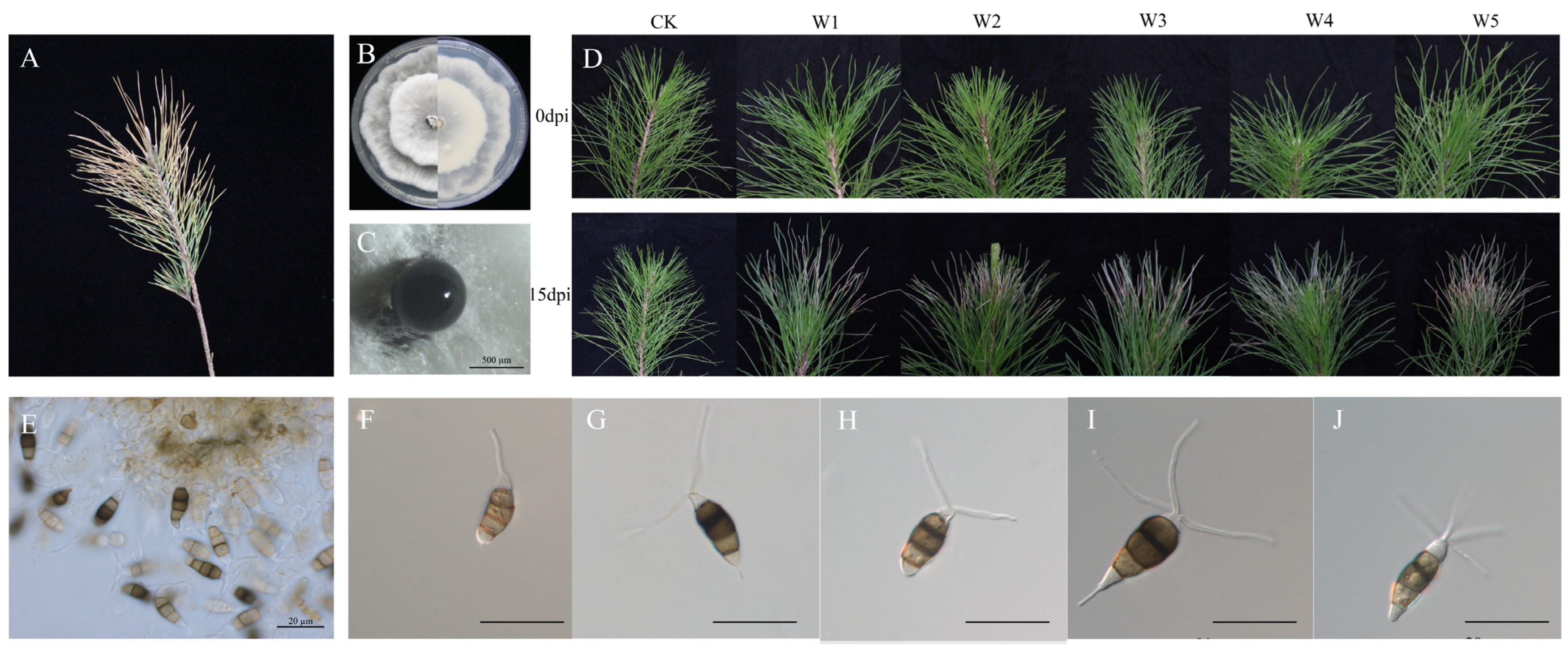
Figure 1. Symptoms of pine needle blight and the phenotypic characteristics of the fungal pathogen. (A) Symptoms of pine needle blight in the nursery. (B) Phenotypic characteristics of the fungal pathogen on the front and reverse sides of PDA plates. (C) Conidial mass of the fungal pathogen. Scale bar: 500 μm. (D) Pathogenicity of representative isolates of Neopestalotiopsis camelliae-oleiferae on Pinus massoniana. (E) Conidiophores, conidiogenous cells, and conidia. (FJ) Conidia of the fungal pathogen. Scale bar: 20 μm. dpi: days post inoculation.
Forests 16 00650 g001
Figure 2. Phylogenetic relationships of isolates and related taxa derived from concatenated sequences of the ITS, TuB2, and TEF1 genes via the maximum likelihood algorithm and Bayesian analysis. Maximum likelihood bootstrap support values ≥75% (1000 replications) and Bayesian posterior probability (PP ≥ 0.85) are shown at the nodes (ML/PP). Pseudopestalotiopsis ampullaceal (LC6618) is used as an outgroup. Bar = 0.03 substitutions per nucleotide position.
Forests 16 00650 g002
Figure 3. Antagonistic effects of the isolated endophytic bacteria against N. camelliae-oleiferae on PDA plates. (A) Control group. (B) PSM-1. (C) PSM-5. (D) PSM-6. (E) PSM-11. (F) PSM-15. (G) PSM-56. (H) PSM-88. (I) Inhibition rate of seven strains. The letters (a–e) above columns represent the significant differences at p < 0.05 according to one-way analysis of variance (ANOVA). Culture conditions: 25 °C with a 12/12 h light/dark cycle.
Forests 16 00650 g003
Figure 4. Phenotypic characterization and identification of biocontrol bacteria. (A) Colony morphology of PSM-6 on LB agar plates. (B) Colony morphology of PSM-6 on PDA plates. (C) Scanning electron microscopy image of PSM-6. (D) Gram stain of PSM-6. (E) Phylogenetic tree of PSM-6 based on the 16S rRNA gene; bootstrap value 1000, number of threads 8. (F) Phylogenetic tree of PSM-6 based on the gyrB gene.
Forests 16 00650 g004
Figure 5. Inhibitory effect of PSM-6 against N. camelliae-oleiferae. (A) Dual-culture assay in which N. camelliae-oleiferae was inoculated on the left (PDA) and 10 μL/30 μL/60 μL/100 μL bacterial fermentation broth of PSM-6 was fermented on the right (LB); no inoculated bacterial ferment was used for the control group. (B) Different proportions of sterile fermentation broth and bacterial suspensions were mixed on PDA plates and then inoculated with N. camelliae-oleiferae on PDA plates. (C) After 5 and 7 days of incubation, different concentrations of crude extract were mixed with PDA plates and then inoculated with N. camelliae-oleiferae on PDA plates. (D) SEM image of N. camelliae-oleiferae. Normal hyphal morphology of N. camelliae-oleiferae on the left, that treated with fermentation products in the middle, and that treated with crude extract on the right. Scale bar: 10 μm. (E) The inhibition rate of VOCs of PSM-6 at different volumes on the mycelial growth of N. camelliae-oleiferae. (F) The inhibition rate of sterile supernatant against N. camelliae-oleiferae. (G) The inhibition rate of the crude extract of different culture times and concentrations against N. camelliae-oleiferae. dpi: days post inoculation. Culture conditions: 25 °C with a 12/12 h light/dark cycle. The letters (a–d) above columns represent the significant differences at p < 0.05 according to one-way analysis of variance (ANOVA).
Forests 16 00650 g005
Figure 6. Effects of bacterial suspensions of PSM-6 and conidial suspensions of N. camelliae-oleiferae on the masson pine in pot test. In Treatment 1, 20 mL of sterile water was sprayed onto healthy pine seedlings as a control. In Treatment 2, 20 mL of PSM-6 bacterial suspension (OD600 = 0.8) was sprayed onto the seedlings. In Treatment 3, 20 mL of PSM-6 bacterial suspension (OD600 = 0.8) was sprayed onto the seedlings, followed by spraying 20 mL of the fungal spore suspension (1 × 10⁶ conidia/mL) 3 days later. In Treatment 4, 20 mL of fungal spore suspension (1 × 10⁶ conidia/mL) was sprayed onto the seedlings without prior bacterial treatment. Observed the disease status at 0/28/49/60 days post inoculation (dpi).
Forests 16 00650 g006
Figure 7. Spectrum of antifungal activity and the inhibition rate of fungal colony growth. (A) Antifungal activity of P. silvicola against B. cinerea, F. graminearum, F. oxysporum, N. camelliae-oleiferae, P. vexans, D. pinea, Alternaria sp., V. dahliae, and L. acicula using confrontation culture assay on PDA. Bacterial suspension: OD600 = 2.0. Culture condition: 25 °C with a 12 h light-dark cycle. (B) The inhibition rate of fungal colony growth according to a confrontation culture. The letters (a–f) above columns represent the significant differences at p < 0.05 according to one-way analysis of variance (ANOVA).
Forests 16 00650 g007
Table 1. Statistical methods for calculating disease index, susceptibility index, and control efficacy (pine needle blight).
Disease GradeClassification CriteriaRepresentative Value
1No symptoms0
2Less than 25% of leaves affected1
325%–50% of leaves affected2
450%–75% of leaves affected3
5More than 75% of leaves affected4
Table 2. Physiological test parameters of PSM-6.
Physiological CharacteristicPSM-6
Voges-Proskauer (V-P) reaction
cellulase degradation
indole-3-acetic acid (IAA) production
gelatin liquefaction
inorganic phosphate solubilization
nitrogen fixation assay
methyl red+
catalase test+
amylase production+
potassium solubilization+
organic phosphorus solubilization+
siderophore production+
+: positive; −: negative.
Disclaimer/Publisher’s Note: The statements, opinions and data contained in all publications are solely those of the individual author(s) and contributor(s) and not of MDPI and/or the editor(s). MDPI and/or the editor(s) disclaim responsibility for any injury to people or property resulting from any ideas, methods, instructions or products referred to in the content.