Article

Physiological and Molecular Insights into the Development of Single and Double Flowers in Syringa vulgaris L.

1
State Key Laboratory of Tree Genetics and Breeding, Northeast Forestry University, Harbin 150040, China
2
Key Laboratory of Fast—Growing Tree Cultivating of Heilongjiang Province, Forestry Research Institute of Heilongjiang Province, Harbin 150081, China
3
Heilongjiang Institute of Atomic Energy, Harbin 150086, China
*
Authors to whom correspondence should be addressed.
These authors contributed equally to this work.
Forests 2025, 16(3), 409; https://doi.org/10.3390/f16030409
Submission received: 14 January 2025 / Revised: 17 February 2025 / Accepted: 22 February 2025 / Published: 24 February 2025
(This article belongs to the Section Forest Ecophysiology and Biology)

Abstract

:
The double-flowering phenotype is crucial for improving the ornamental value of flowering plants; this trait substantially enhances the varieties of Syringa vulgaris L. To date, no studies have examined the transcriptomics and metabolomics of key nodes in the flower bud differentiation of the single and double flowers of Syringa. This study investigated both the single and double flowers of S. vulgaris using a comprehensive, multifaceted analytical approach, including physiological assessments, transcriptomics, and metabolomics. The floral bud differentiation process can be divided into six distinct stages. Compared with the single flowers, the double flowers of S. vulgaris presented significant developmental delays during floral bud differentiation. Although there was no significant difference in the soluble protein content between the two flower types during this process, the soluble sugar content varied during pistil primordium differentiation and as temperature increased. The antioxidant enzyme activity was significantly greater in the double flowers than in the single flowers during most differentiation stages, while the malondialdehyde (MDA) level gradually increased. The levels of endogenous hormones, such as indole-3-acetic acid (IAA), gibberellin (GA3), and abscisic acid (ABA), differed between the two flower types. The transcriptomics and metabolomics results indicated that during pistil primordium differentiation and subsequent development, the double flowers exhibited increased antioxidant enzyme activity and secondary metabolite accumulation. These secondary metabolites not only contributed to the vibrant coloration of the double flowers but also increased their cellular metabolic stability and stress tolerance through their antioxidant properties. Conversely, the rapid differentiation mechanism of the single flowers of S. vulgaris relied more on efficient primary metabolism to meet simpler structural demands. These findings not only provide scientific guidance for S. vulgaris breeding programs but also expand its potential in horticultural and landscape applications, offering a new theoretical foundation for studies on floral organ development in Oleaceae species.

1. Introduction

For ornamental plants, petals have the most ornamental and commercial value. Changes in the number and shape of petals are highly important for the development and evolution of flower patterns and are among the important factors that determine the ornamental value of plants [1,2]. Owing to the extremely high ornamental value of double flowers, the double-petal phenotype is often selected by breeders and is considered to be one of the most valuable characteristics of many species, such as Xanthoceras sorbifolium Bunge [3], Phyllostachys nuda McClure [4], Sagittaria trifolia L. [5], and Prunus mume Siebold and Zucc. [6]. The origins of double flowers in plants are diverse and complex, and different varieties and species may have different mechanisms for producing double flowers [7,8,9,10]. Multiple origins are also possible in the same variety. Homeosis has long been known in horticulture through the breeding of double flowers, in which stamens are generally replaced by petals. Accordingly, the addition of one whorl occurs in parallel with the deletion of the other whorl [11]. In 2001, Theissen et al. [12,13] further refined the ABC model, which was proposed by Coen et al. [14], and proposed the ABCDE model and the “tetramer model”. Lenhard and Lohmann [15,16] proposed the theory of the feedback regulation loop formed by the WUSCHEL (WUS) and AGAMOUS (AG) genes, which essentially reveals the molecular mechanism of the formation of double flowers.
However, simple models of flower development cannot fully explain the regulatory mechanisms of floral organ development. In addition to being regulated by genes, floral organ differentiation is also affected by external factors, such as climatic conditions, nutrients, and endogenous hormones [17,18,19,20,21]. During research on Dianthus caryophyllus L., it was discovered that a low temperature administered either continuously or at night promoted the formation of secondary growing centers within the flowers of carnations, and the number of petals increased after gibberellin A3 (GA3) or indole-3-acetic acid (IAA) was sprayed on the tip of the stem after flowering [22]. Some studies have shown that a decrease in the abscisic acid (ABA) content during flower bud differentiation in Rosa chinensis Jacq. is conducive to flower bud differentiation [23]. Endogenous hormones play important roles in the formation of flowers and inflorescences [24]. However, there have been few studies on the influence of endogenous hormones on plants of the Syringa genus. A study by Jin et al. found that applying exogenous IAA to Syringa villosa Vahl could enhance its photosynthetic capacity and increase endogenous hormone levels, thereby improving growth conditions [25]. The analysis of transcriptomes related to flower development in different plants revealed that genes related to carbohydrates, secondary metabolite synthesis, and hormone pathways are significantly differentially expressed during differentiation [26,27]. Flavonoids and phenolic acids have been identified as the primary differentially expressed metabolites of Mikania micrantha across varying altitudes [28]. Additionally, flavonoids and flavonols are present in the heads of Chrysanthemum morifolium during waterlogging stress at different stages of flower bud differentiation [29]. The contents of polyphenols, including quercetin, catechin, anthocyanins, and phenolic acids, vary during the development of flowering in Rosa rugosa Thunb. [30,31].
Syringa vulgaris L., a member of the Oleaceae family, is a deciduous shrub or small tree renowned for its strong adaptability and horticultural value [32]. This species is highly tolerant to various soil conditions, exhibiting robust resistance to both cold and heat, making it well-suited to cold climates with abundant sunlight. Native to regions spanning from the Caucasus to Afghanistan, S. vulgaris is commonly cultivated as an ornamental plant in Central and Southeastern Europe. Owing to their vibrant flower colors, these plants are important for landscape design and home gardening [33]. Morphologically, Syringa is broadly classified into single- and double-flower types. Single flowers are appreciated for their natural elegance, whereas double flowers, valued for their ornate appearance, have become a focal point of horticultural breeding efforts. Jędrzejuk et al. conducted extensive research on the development of single-flower organs in S. vulgaris from 2003 to 2008. Initially, they focused on the impact of temperature on the morphology of flower buds and the quality of panicles [34], followed by the investigation of the process of bud differentiation [35]. Their findings indicated that elevated temperatures adversely affect the normal development of male and female gametes in S. vulgaris by inhibiting tissue differentiation [36,37]. Additionally, Reza Dapour et al. performed a comparative study of floral ontogeny in the single- and double-flowered phenotypes of S. vulgaris using epi-illumination light microscopy [7]; they discovered that the traits associated with double flowers did not negatively impact the development of other floral organ whorls. Since Reza Dapour’s research, the double flowers of S. vulgaris have not been studied for many years. Rather, most studies have concentrated on analyzing the pharmaceutical value of lilac through the extract components of its flowers, bark, and leaves [38,39,40,41].
This study investigated both single and double flowers of S. vulgaris using a comprehensive, multifaceted analytical approach encompassing physiological assessments, transcriptomics, and metabolomics. Specifically, this study focused on the following aspects: (i) dynamic observation of morphological changes, identifying key morphological features during floral bud differentiation; (ii) changes in physiological and biochemical indicators, including dynamic measurements of changes in hormone levels, antioxidant enzyme activities, and metabolic products; and (iii) regulation of gene expression, using transcriptomic analysis to identify key genes and regulatory networks involved in petal development during differentiation. For the first time, this study systematically integrates genetic, metabolic, and physiological regulatory mechanisms to comprehensively elucidate the molecular basis of flower development in the single and double flowers of S. vulgaris. These findings not only provide scientific guidance for S. vulgaris breeding programs but also expand its potential in horticultural and landscape applications, offering a new theoretical foundation for studies on floral organ development in Oleaceae species.

2. Materials and Methods

2.1. Plant Materials

This study utilized the single and double flowers of S. vulgaris. All of the experimental materials were planted in 2016. Samples were collected from Qunli Syringa Park, Harbin, China. Harbin is located in Northeast China and is characterized by a temperate continental monsoon climate with distinct seasons: cold and prolonged winters, hot and brief summers, and rapid transitions in spring and autumn. Floral bud samples were collected weekly from May 2023 to April 2024, excluding the winter period. Each collection involved at least 5 g of material, with uniformly sized terminal buds selected as experimental samples.
(i)
Double flowers of the S. vulgaris cultivars (“Fengnian”) (held by Harbin land-scaping research Institute):
This experimental material originated from controlled hybridization between wild double-flowered S. vulgaris (maternal parent) and native Chinese single-flowered S. vulgaris (paternal parent) conducted during 1960–1970 (S. vulgaris (wild double-flowered) × S. vulgaris (native single-flowered)). After multigenerational phenotypic selection, a stable double-flowered progeny with superior horticultural traits was identified in 1997. This genotype was formally named as the cultivar “Fengnian” in 2021.
(ii)
Single flowers of S. vulgaris (held by Harbin landscaping research Institute):
The experimental material was clonal propagules derived from the paternal parent of the aforementioned hybridization (1).
Single-floral formula:
K 4 C 4 A 2 G 2 : 2
Double-floral formula:
K 4 C 4 + 4 + 0 2 A 0 2 G 2 : 2
where ↑ is the zygomorphic flower, K is the calyx, C is the corolla, A is the androecium, and G is the gynoecium.

2.2. Morphological Identification

The morphological characteristics of the flower buds were analyzed using the following methods:
Bud measurement: A Vernier caliper was used to measure the length and width of the flower buds, and the length-to-width ratio was calculated.
Paraffin sectioning and staining: The differentiation stages of single and double flower buds were observed through paraffin sectioning [42]. The samples were fixed with FAA fixative (a solution of 70% ethanol, acetic acid, and formaldehyde at an 18:1:1 ratio), dehydrated with a graded ethanol series, and cleared with xylene. The samples were then embedded in paraffin at 60 °C, sectioned, and stained with hematoxylin-eosin (H&E). Cellular structures were observed under a Leica microscope [43].

2.3. Physiological and Biochemical Analyses

To investigate the physiological and biochemical differences between single and double flower buds, the experiment included 14 treatments (2 flower types × 7 time points), with three biological replicates and three technical replicates for each treatment. The following parameters were measured:
(i)
Soluble sugar content (0.1 g of flower buds per run): This content was determined using the anthrone-sulfuric acid colorimetric method [44].
(ii)
Soluble protein content (0.1 g of flower buds per run): This content was measured via the Coomassie brilliant blue colorimetric method [45].
(iii)
Antioxidant enzyme activities and MDA content: The peroxidase (POD), superoxide dismutase (SOD), and catalase (CAT) activities and the malondialdehyde (MDA) content were assessed using oxidative stress detection kits. POD, SOD, and CAT activity and MDA content were measured with 0.1 g of material, using an oxidative stress series kit (microplate method) (Suzhou Grace Biotechnology Co., Ltd., Suzhou, China) [46] according to the manufacturer’s instructions.
(iv)
Endogenous hormone levels (0.1 g of flower buds per run): The levels of IAA, abscisic acid (ABA), GA3, 1-aminocyclopropane-1-carboxylic acid (ACC), salicylic acid (SA), jasmonic acid (JA), cis-ZR (cZR), and trans-ZR (tZR) were quantified via ultrahigh-performance liquid chromatography coupled with tandem mass spectrometry (UHPLC-MRM-MS/MS) [47]; the concentration ranges of these hormones were as follows [48]:
  • IAA concentration range: 10–100 ng/g;
  • GA3 concentration range: 5–50 ng/g;
  • ABA concentration range: 1–20 ng/g;
  • ACC concentration range: 5–100 ng/g;
  • SA concentration range: 500–4000 ng/g;
  • JA concentration range: 0–100 ng/g;
  • cZR concentration range: 2–30 ng/g;
  • tZR concentration range: 2–30 ng/g.
The preparation of standard solutions, calibration curve construction, and calculation of detection and quantification limits followed strict standard protocols. The accuracy and precision of the results were validated with quality control samples to ensure data reliability.

2.4. Data Analysis

Analysis of variance (ANOVA) was conducted using SPSS 27.0, and Duncan’s multiple range test was used to assess significant differences between groups (p < 0.05) [49]. Hormone concentrations (CMs) were calculated using the following formula:
C M   [ n g / g ] = C F   [ n g / m L ] × V F   [ m L ] / M S   [ g ]
where CF is the final concentration, VF is the final volume, and MS is the sample weight [47].

2.5. Transcriptome Analysis

Total RNA was extracted from single and double flower buds using TRIzol reagent (Thermo Fisher Scientific, Carlsbad, CA, USA) according to the manufacturer’s instructions. The RNA concentrations were measured with a Qubit 3.0 fluorometer (Thermo Fisher Scientific, Carlsbad, CA, USA), the purity was assessed with a NanoDrop 2000 spectrophotometer (Thermo Fisher Scientific, Wilmington, DE, USA), and the integrity was evaluated through agarose gel electrophoresis. High-quality RNA was used to construct cDNA libraries for RNA sequencing [49] (the original data were uploaded to the NCBI database at https://www.ncbi.nlm.nih.gov/ (accessed on 21 February 2025)SRA: PRJNA1220025). To ensure the sequencing quality of the RNA, the RNA integrity value (RIN) was determined using an Agilent 2100 bioanalyzer (Agilent Technologies, Santa Clara, CA, USA), with only samples exhibiting an RIN value of ≥7.0 being utilized for subsequent analysis. High-quality RNA was employed to construct cDNA libraries for RNA sequencing. RNA sequencing was conducted using the Illumina NovaSeq 6000 platform (Illumina, San Diego, CA, USA) to generate 150 bp paired-end (PE150) sequences. The library construction utilized the TruSeq RNA sample preparation kit (Illumina, San Diego, CA, USA), which encompasses mRNA enrichment, fragmentation, cDNA synthesis, linker ligation, and PCR amplification. Library quality was assessed using both Qubit 3.0 and Agilent 2100 bioanalyzers, and quantitative analysis was performed using qPCR prior to sequencing. Following quality control of the original sequencing data, de novo transcriptome assembly was executed using Trinity software (v2.15.1) [50]. Initially, Trimmomatic was employed to remove low-quality sequences and joint contamination in order to obtain clean reads. Subsequently, clean reads were aligned to the reference transcriptome using HISAT2(v2.2.1), and the data were converted to BAM format using SAMtools(v1.17) and then sorted [51]. Transcription abundance was calculated using featureCounts(v2.0.1) and normalized to transcripts per million (TPM) [52]. Differentially expressed genes (DEGs) were analyzed using DESeq2(v1.46.0), with screening criteria set at a fold-change of ≥2 and a p-value threshold of ≤0.05. Gene function annotation was enriched using Gene Ontology (GO) and the Kyoto Encyclopedia of Genes and Genomes (KEGG) to identify key biological processes, molecular functions, and cellular components. Finally, the results of the analysis were visualized using the ggplot2 software package in R (v3.5.1) to elucidate the differences between single- and double-petal transcriptomes [53].

2.6. Metabolomics Analysis

The samples were vacuum-freeze-dried using a Scientz-100F lyophilizer (Xinzhi, Ningbo, China) and ground into a fine powder using an MM400 grinding mill (Retsch GmbH, Haan, Germany). A total of 100 mg of powder was dissolved in 1.2 mL of 70% methanol extraction solution, vortexed, and incubated at 4 °C overnight. After centrifugation, the supernatant was filtered through a 0.22 μm microporous membrane and transferred to sample vials. UPLC-MS/MS analysis was performed by Wuhan MetWare Biotechnology Co., Ltd. (Wuhan, China) [54]. Principal component analysis (PCA) was used to evaluate sample dispersion, and orthogonal partial least squares discriminant analysis (OPLS-DA) was used to identify differentially expressed metabolites (DEMs) using a variable importance projection (VIP) score ≥ 1 and a fold-change ≥ 2 or ≤0.5 as the criteria [55].

2.7. Quantitative Real-Time PCR (qRT-PCR) Validation

To validate the reliability of the RNA-Seq data, nine genes were randomly selected for qRT-PCR analysis. Specific primers were designed using the INTEGRATED DNA SERVICES tool (v1.0), with 18S rRNA as the reference gene (primer sequences are listed in Table 1). Total RNA was reverse-transcribed into cDNA via a Takara kit, and qRT-PCR was performed on a 7500 Fast Real-Time PCR system. All reactions were conducted in triplicate, and the relative expression levels of each gene were calculated [54].

3. Results

3.1. Morphological Differences Between Single and Double Flower Buds

3.1.1. Flower Bud Morphological Changes

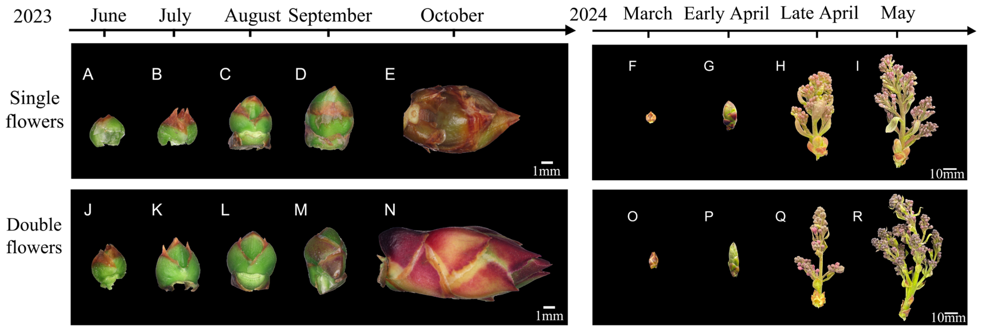
During the early stages of floral bud differentiation (inflorescence and floret primordium stages, May to June), the double flower buds presented larger longitudinal diameters and higher longitudinal-to-transverse diameter ratios than the single flower buds, whereas their transverse diameters were smaller (Figure 1 and Figure 2A–J). Around July 2, the transverse diameter of the double flower buds slightly exceeded that of the single flower buds. However, starting on July 9, the transverse diameter of the single flower buds surpassed that of the double flower buds—a trend that persisted through the flowering stage (Figure 1A and Figure 2).
During July (sepal primordium and petal primordium differentiation stages), the longitudinal diameters of both the single and double flower buds were comparable. However, beginning on July 23, the longitudinal diameters of the double flower buds gradually exceeded those of the single flower buds—a difference that persisted until flowering (Figure 1B and Figure 2). Although the longitudinal-to-transverse diameter ratio fluctuated slightly after July 2, the double flower buds consistently presented relatively high ratios, indicating that the single flower buds were relatively flat, whereas the double buds were more elongated (Figure 1C and Figure 2).

3.1.2. Dynamics of Inflorescence Development

From March to May, the flower buds gradually developed into complete inflorescences, with longitudinal growth outpacing transverse growth. For the single inflorescences, the length increased from 11.0 mm to 88.3 mm and the width increased from 8.5 mm to 38.1 mm. For the double inflorescences, the length increased from 13.2 mm to 97.6 mm and the width increased from 6.9 mm to 35.5 mm (Figure 1A,B and Figure 2F–I,O–R). In summary, the double flower buds presented elongated morphological characteristics during early growth and exhibited superior longitudinal growth compared with the single buds.

3.1.3. Anatomical Characteristics of Single and Double Flower Buds at Different Differentiation Stages

S. vulgaris floral bud differentiation follows a summer–autumn differentiation pattern, with flowering occurring the following spring after vernalization. On the basis of developmental features, the differentiation process can be divided into six stages:
(i)
Bract Primordium Differentiation Stage:
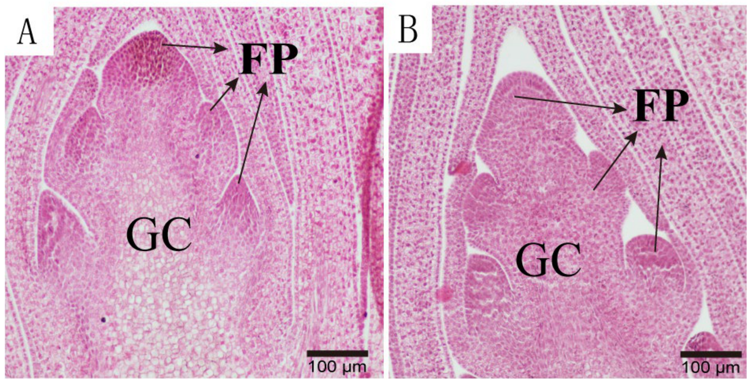
During this stage, the S. vulgaris flower buds were encased by outer scales, and the apical meristem became more pointed. The actively dividing meristematic cells were densely arranged and stained darkly (Figure 3A,B).
In terms of duration, no significant differences were observed between the single and double flower buds, with this stage lasting approximately 21 days.
(ii)
Inflorescence and Floret Primordium Differentiation Stage:
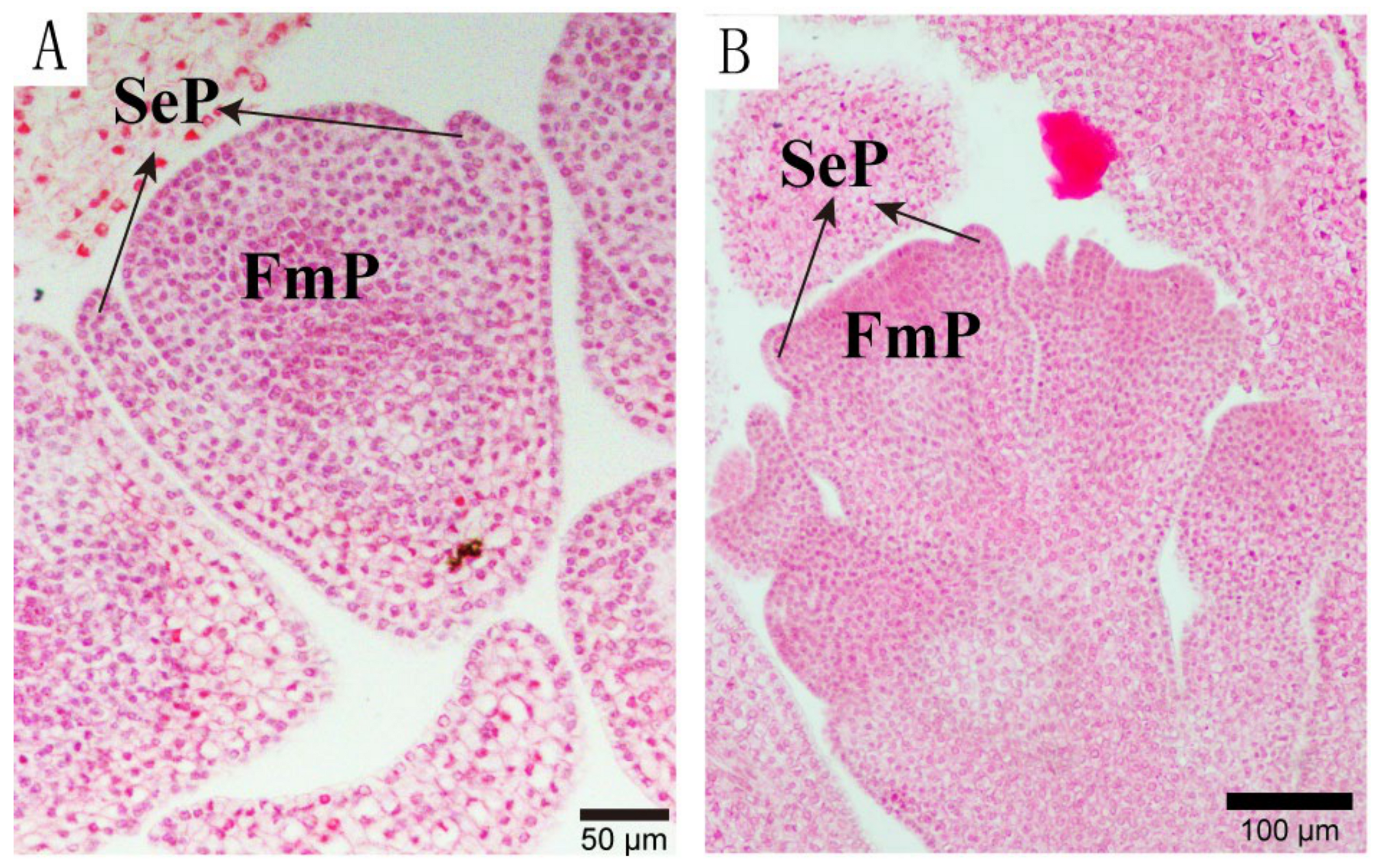
During this stage, the apical meristem flattened into a hemispherical shape, and the floret primordia differentiated symmetrically on both sides. Secondary floral pedicels developed at the base of these primordia, which further gave rise to secondary floret primordia (Figure 4A,B).
This stage lasted approximately 28 days for the single buds and 35 days for the double buds, indicating a longer duration for the double buds.
(iii)
Sepal Primordium Differentiation Stage:
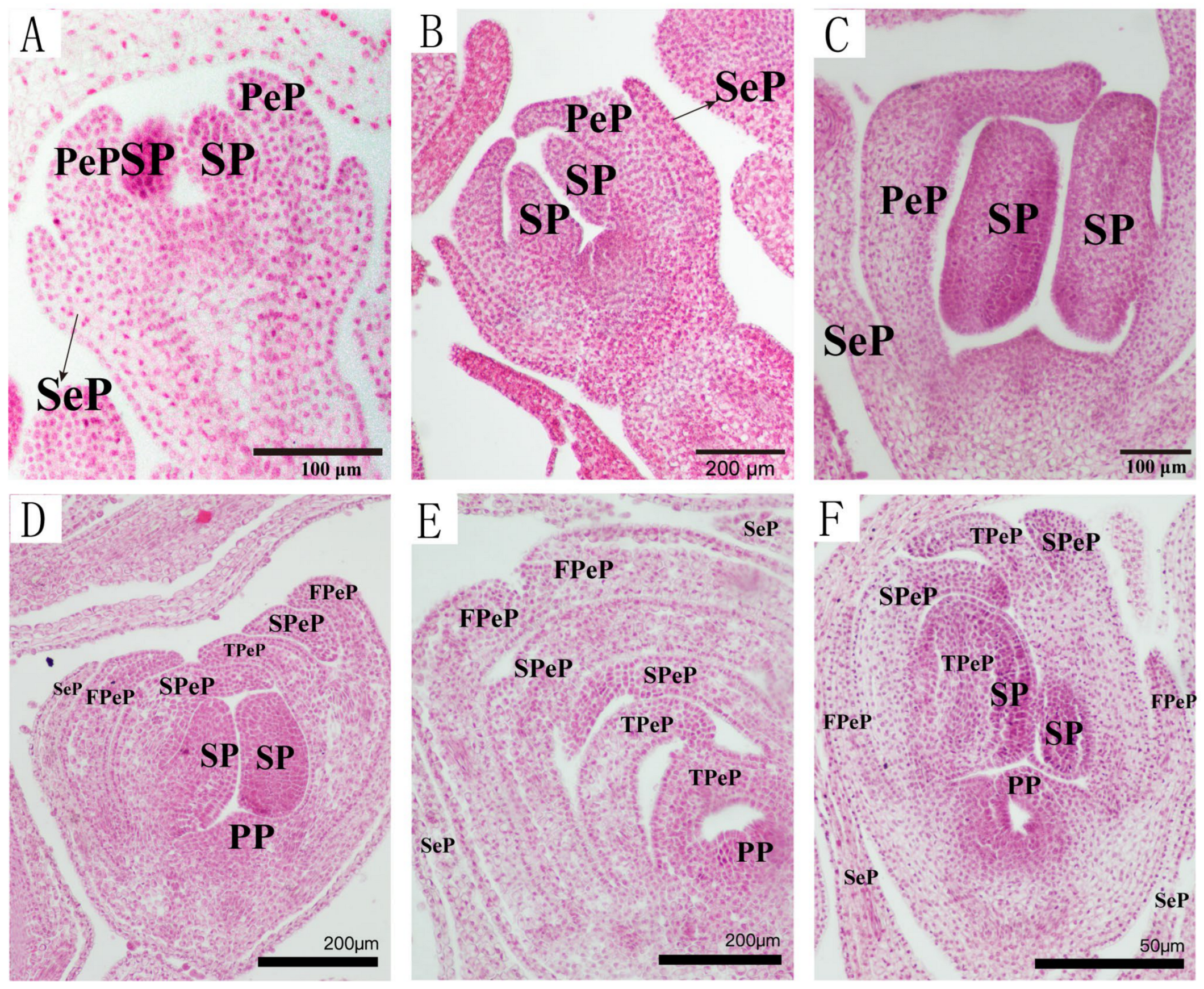
After the differentiation of the floret primordia, the first whorl of the sepal primordia emerged as lateral protrusions that curved upward to enclose the developing petal primordia. Sepal growth ceased thereafter (Figure 5A,B).
In terms of duration, this stage lasted approximately 14 days for the single flower buds but only 7 days for the double flower buds, indicating a shorter duration for the latter.
(iv)
Petal Primordium Differentiation Stage:
During this stage, spherical protrusions appeared inside the sepal primordia and gradually developed into crescent-shaped petals.
For the single flower buds, this stage lasted approximately 22 days, with relatively simple petal development (Figure 6A).
For the double flower buds, this stage lasted approximately 50 days, which was significantly longer than that for the single buds. Additional layers of petals (second and third layers) differentiated sequentially, forming overlapping structures that covered the pistils or partially transformed stamens (Figure 6B–D).
(v)
Stamen Primordium Differentiation Stage:
During this stage, the stamen primordia differentiated below the petal primordia and were arranged symmetrically.
For the single flower buds, stamen differentiation began approximately 14 days after the completion of petal primordium differentiation (Figure 7A–C).
For the double flower buds, stamen differentiation was delayed until after the prolonged petal differentiation phase. In some of the double flower buds, the stamens partially transformed into petaloid stamens, initially resembling normal stamens but elongating over time (Figure 7D–F).
(vi)
Carpel Primordium Differentiation Stage:
During this stage, carpel primordia developed immediately after stamen differentiation. The central growth point elongated and expanded at the apex, forming the stigma and style (Figure 8).
In terms of the key feature, this stage represents the final stage of floral organ differentiation and the complete development of the pistil.
A comparison of floral bud differentiation processes in the single and double flowers of S. vulgaris was conducted.
This study systematically observed the external morphology and internal anatomy of single and double flower buds from the bud stage to flowering, revealing the following key differences:
Duration: The differentiation process of the double flower buds was generally longer than that of the single flower buds, particularly during the petal and stamen differentiation stages.
Structure: The “multilayered petal” structure was a distinctive feature of the double flower buds and was closely associated with partial petaloid stamens.
Developmental patterns: Correlating external morphological changes (e.g., longitudinal-to-transverse diameter ratios) with internal anatomical features elucidated the coordinated developmental patterns of the single and double flower buds. Table 2 summarizes the differences in differentiation stages and external morphological features between the two types of flower buds.

3.2. Transcriptomic and Metabolomic Differences in the Differentiation Processes of Single and Double Flower Buds

The transcriptomic and metabolomic analyses revealed key differences between the single and double flowers of S. vulgaris during floral bud differentiation. The RNA-Seq libraries constructed for the transcriptomic analysis yielded high-quality sequencing data, identifying a total of 12,410 differentially expressed genes (DEGs), of which 7947 were upregulated and 4463 were downregulated. The Gene Ontology (GO) and Kyoto Encyclopedia of Genes and Genomes (KEGG) functional enrichment analyses revealed that these DEGs were involved primarily in critical biological functions and metabolic pathways, including the “regulation of transcription from DNA templates”, “nuclear functions”, “biosynthesis of secondary metabolites”, and “plant hormone signal transduction”.
The metabolomic analysis identified 14,282 differentially expressed metabolites (DEMs), among which 303 were successfully annotated (Appendix A Table A1). The upregulated metabolites were mainly associated with the metabolism of shikimic acid, phenylpropanoids, terpenoids, and carbohydrates, whereas the downregulated metabolites were linked predominantly to alkaloid and secondary metabolic pathways. KEGG enrichment analysis indicated that these differentially expressed metabolites were significantly enriched in pathways related to amino acid metabolism, secondary metabolite biosynthesis (e.g., flavonoids), carbohydrate metabolism, and membrane transport.
In summary, significant transcriptomic and metabolomic differences were observed between the single and double flowers of S. vulgaris during floral bud differentiation. These differences, particularly in secondary metabolism, carbohydrate metabolism, and plant hormone regulation, provide crucial insights into the molecular mechanisms underlying the formation of double flowers.

3.3. Expression Characteristics of Genes and Metabolites Related to Flower Color Formation

Flower color formation is a key manifestation of plants’ secondary metabolism and involves the precise regulation of genes and metabolites associated with flavonoid, phenylpropanoid, and aromatic amino acid metabolism.
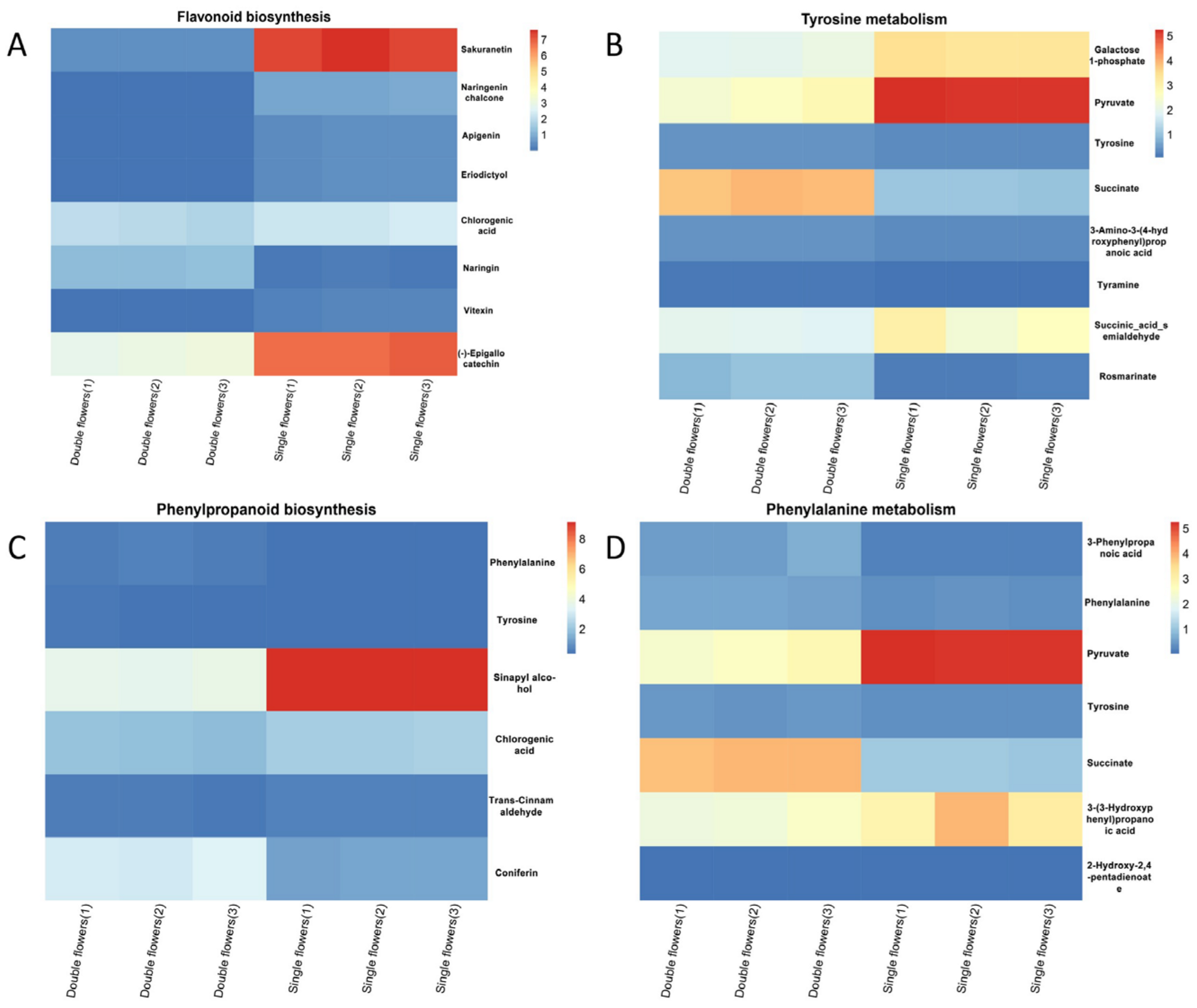
In the flavonoid metabolism pathway (Figure 9A), the metabolites sakuranetin, naringenin chalcone, and apigenin were expressed at significantly higher levels in the double flowers than in the single flowers, with sakuranetin reaching the highest expression level in the double flowers. This finding suggests that active flavonoid metabolism provides the direct material basis for the formation of flower color in double flowers. The low expression of naringin may be linked to the stability of color tones in double flowers, indicating that flowers’ color regulation depends not only on synthetic activity but also on the efficiency of intermediate metabolite conversion.
In the tyrosine metabolism pathway (Figure 9B), succinate and tyrosine were significantly upregulated in the double flowers, suggesting that tyrosine metabolism may indirectly contribute to anthocyanin formation by supplying precursor substances for phenylalanine metabolism. Additionally, the increased expression of 3-amino-3-(4-hydroxyphenyl)propanoic acid likely regulates key transformation steps in anthocyanin synthesis, contributing to the deepening of flower color in double flowers.
In the phenylpropanoid metabolism (Figure 9C) and phenylalanine metabolism (Figure 9D) pathways, key metabolites such as phenylalanine and tyrosine presented significantly higher expression levels in the double flowers than in the single flowers. Notably, the upregulation of sinapyl alcohol and pyruvate further highlights the cooperative role of phenylpropanoid and phenylalanine metabolism in promoting the accumulation of anthocyanin precursors. Additionally, the specific expression of 3-(3-hydroxyphenyl)propanoic acid and 3-(2-hydroxyphenyl)propanoic acid may be associated with the conversion and accumulation rates of pigments in double flowers, providing important molecular evidence for flowers’ color development.
By integrating the metabolic characteristics of the flavonoid, phenylpropanoid, and tyrosine metabolism pathways, the formation of flower color in double flowers is proposed to occur through the following mechanisms: (i) The accumulation of key metabolites in the flavonoid pathway directly contributes to anthocyanin synthesis. (ii) Synergistic interactions between the phenylpropanoid and phenylalanine metabolism pathways increase the supply of precursors, promoting rapid anthocyanin synthesis. (iii) Active tyrosine metabolism provides abundant substrates for related metabolic pathways, indirectly supporting flowers’ color formation.
This comprehensive analysis provides valuable insights into the molecular mechanisms underlying color formation in the double flowers of S. vulgaris.

3.4. Association Between Carbohydrate Metabolites and Petal Structure Formation During Floral Bud Differentiation in S. vulgaris

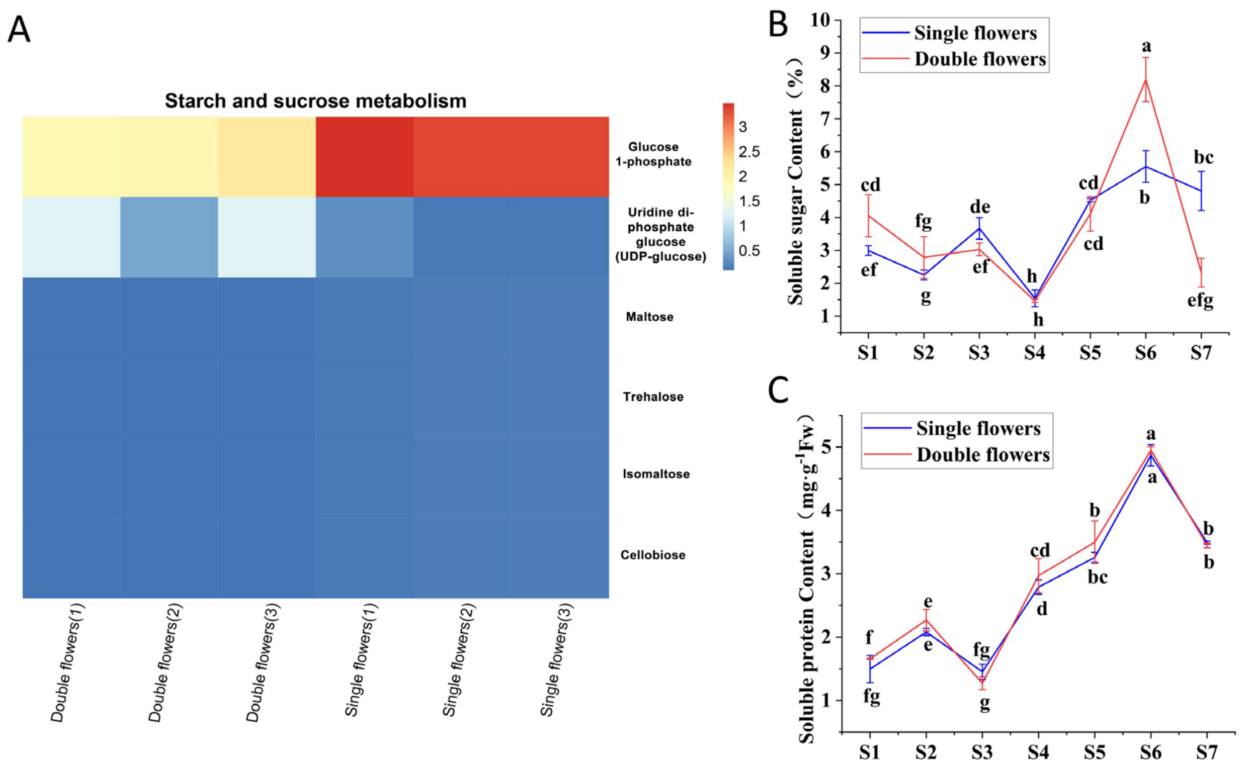
The analysis of metabolite content differences (Figure 10A) revealed significant variation in the abundance of key carbohydrate-metabolism-related metabolites, such as sucrose, glucose, and starch, during different developmental stages of the single and double flowers. Notably, the abundance of glucose 1-phosphate was significantly greater in the double flowers than in the single flowers, suggesting that elevated carbohydrate metabolism activity may be associated with an increase in the number of petals. The soluble sugar content (Figure 10B) displayed a distinct dynamic pattern during floral bud differentiation, with double flowers reaching peak soluble sugar levels during the S5 stage. This stage, which is critical for petal structure formation, resulted in significantly greater soluble sugar contents in the double flowers than in the single flowers. These findings suggest that carbohydrates may promote additional petal differentiation by regulating osmotic pressure or providing energy.
Similarly, the soluble protein content (Figure 10C) also peaked during the S5 stage, with the double flowers consistently exhibiting higher levels than the single flowers. This finding indicates that proteins closely associated with carbohydrate metabolism, such as those involved in cell division, expansion, and cell wall construction, may play important roles in petal structure formation.
Taken together, these findings suggest that carbohydrate metabolism influences lilac petal structure differentiation through the following mechanisms: (i) Metabolic products (e.g., sucrose and glucose) may act as signaling molecules to regulate the expression of genes related to petal differentiation. (ii) Energy provided by carbohydrates supports rapid cell division and expansion in petal tissues. (iii) Developmental stage-specific differences in carbohydrate metabolism directly drive the increase in the number of petals in double flowers during critical phases of floral bud differentiation.

3.5. Expression Characteristics of Genes and Metabolites Associated with Petal Layer Formation

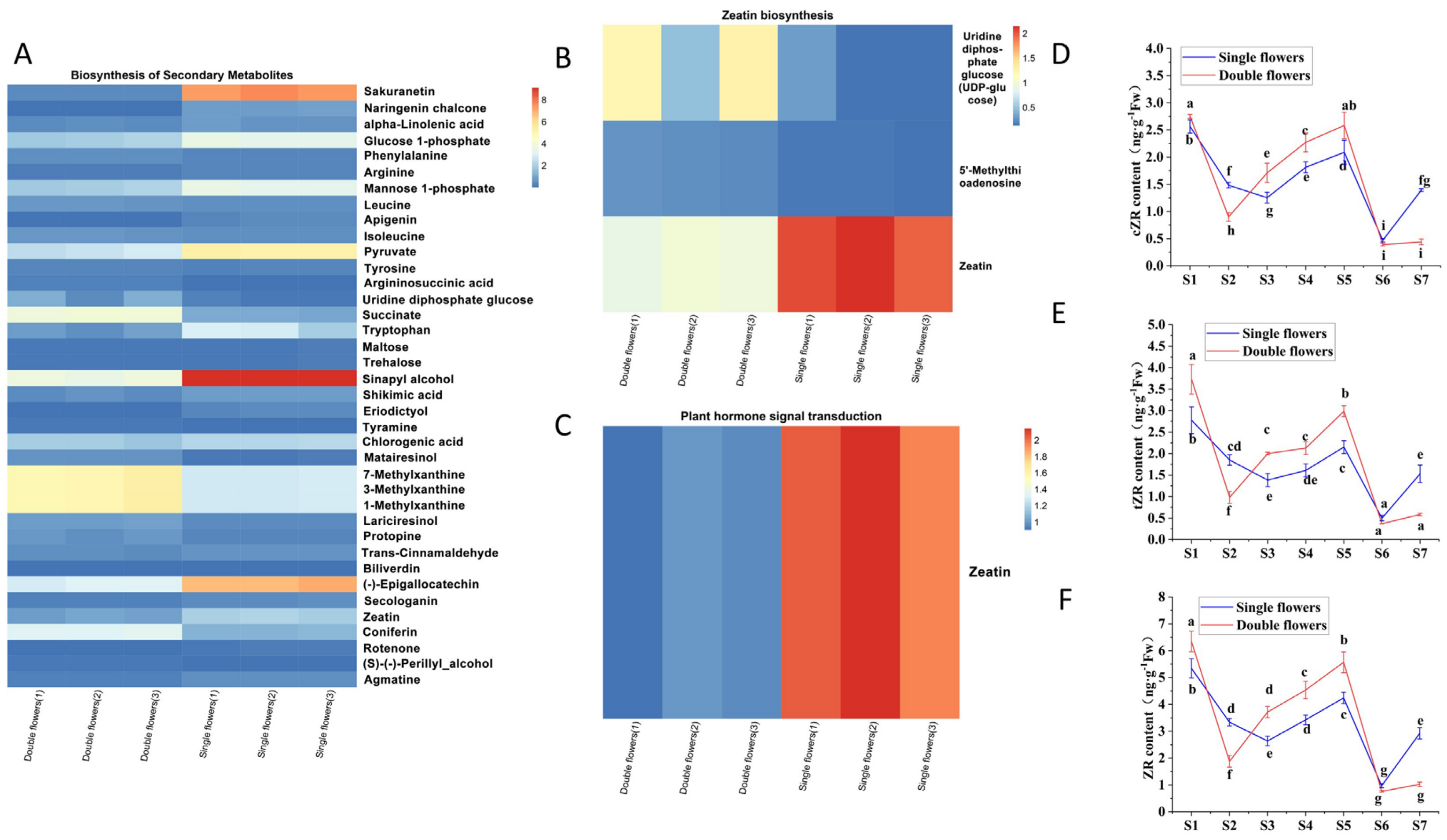
The heatmap analysis (Figure 11A) revealed significant differences in the synthesis of secondary metabolites. Metabolites such as sakuranetin and naringenin chalcone exhibited differential abundances between the single and double flowers, with markedly higher levels observed in the double flowers. Notably, the intermediate metabolite trehalose, a key product of secondary metabolism, was significantly more abundant in the double flowers, suggesting that increased secondary metabolic activity may provide both structural components and energy to support the formation of additional petal layers.
The genes related to zeatin biosynthesis (Figure 11B) presented relatively high expression levels of uridine diphosphate glucose (UDP-glucose) and 5′-methylthioadenosine in double flowers, while the signal transduction-related gene zeatin presented significantly elevated expression in the double-flower samples (Figure 11C).
These findings suggest that zeatin biosynthesis and signal transduction may play critical roles in promoting cell division and differentiation, thereby contributing to the formation of additional petal layers in double flowers.
Dynamic changes in the zeatin riboside (ZR) and cZR levels (Figure 11D,E) further supported this hypothesis. In the early stages of floral bud differentiation (S1–S2), there was little difference in the ZR content between the single and double flowers. However, during the middle differentiation stages (S3–S5), the ZR content in the double flowers increased significantly, peaking during this critical period. A similar trend was observed for cZR, indicating that zeatin-related hormones play important roles in promoting petal layer differentiation and the development of more complex floral organs. In the late differentiation stages (S6–S7), the ZR and cZR levels rapidly decreased, which may have been related to the completion of floral bud differentiation and the stabilization of developmental processes.
By combining the expression characteristics of secondary metabolites and zeatin signal transduction, it is proposed that petal layer formation may be achieved through the following mechanisms: (i) Secondary metabolites provide the energy and structural molecules necessary for the expansion and differentiation of petal layers. (ii) Enhanced zeatin biosynthesis and signal transduction pathways activate key genes associated with cell division and differentiation. (iii) Temporal regulation of ZR and cZR levels ensures precise control over the timing and layering of petal differentiation.
This integrated molecular framework offers critical insights into the regulatory mechanisms underlying the formation of petal layers in double flowers.

3.6. Differences in Endogenous Hormone Levels Between Single and Double Flowers of S. vulgaris During Floral Bud Differentiation

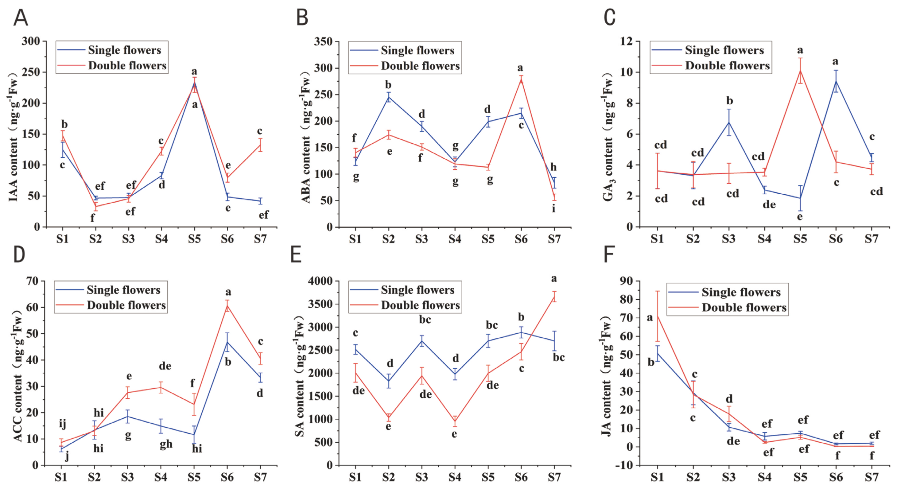
The endogenous hormone levels of the single and double flowers of S. vulgaris significantly differed during floral bud differentiation. The dynamic changes in the contents of hormones, including indole-3-acetic acid (IAA) (Figure 12A), abscisic acid (ABA) (Figure 12B), gibberellin (GA3) (Figure 12C), 1-aminocyclopropane-1-carboxylic acid (ACC) (Figure 12D), salicylic acid (SA) (Figure 12E), and jasmonic acid (JA) (Figure 12F) revealed their potential roles in regulating floral bud differentiation.
Compared with the single flowers, the double flowers presented greater levels of IAA, ACC, and GA3 during key developmental stages (e.g., S5 and S6), suggesting their roles in promoting cell division, differentiation, and petal layer formation. In contrast, the early stages (S1 to S3) of both flower types were characterized by significant decreases in ABA and JA levels, which may be critical for initiating floral bud differentiation.
Further analysis comparing the dynamic changes in various hormone ratios between the single and double S. vulgaris flowers during floral bud differentiation was conducted to elucidate the role of hormonal balance in regulating floral morphogenesis; the results were as follows:
(i)
ABA/GA3 Ratio:
The ABA/GA3 ratio in the single flowers peaked significantly during the S5 stage, where it was greater than that in the double flowers, and then decreased rapidly (Figure 13A). This finding indicates that the balance between ABA and GA3 plays a critical role in regulating cell division and differentiation during key stages of floral bud development. This balance likely contributes to inhibiting cell elongation and promoting bud maturation in single flowers.
(ii)
IAA/GA3 Ratio:
The IAA/GA3 ratio exhibited a sharp peak in the single flowers during the S5 stage, whereas it remained relatively stable in the double flowers (Figure 13B). This finding suggests that the relative levels of auxin (IAA) and gibberellin may regulate cell polarity and tissue differentiation, with the peak observed in the single flowers potentially being linked to floral organ morphogenesis.
(iii)
ABA/IAA Ratio:
The ABA/IAA ratio was greater in the double flowers during the S2 and S6 stages, while the single flowers presented a significantly greater ratio at the S5 stage (Figure 13C). This finding highlights the complex interplay between ABA and IAA at different stages, suggesting that the precise regulation of cell division and elongation mediated by these hormones contributes to multistage floral bud differentiation.
(iv)
ZR/GA3 Ratio:
The ZR/GA3 ratio in the single flowers was significantly greater than that in the double flowers during the S4 stage, after which the trends were similar for both types (Figure 13D). This finding indicates that the balance between ZR and gibberellin in early developmental stages plays a crucial role in driving floral bud differentiation, likely through promoting cell proliferation and division.
(v)
IAA/ZR Ratio:
During the S6 and S7 stages, the IAA/ZR ratio was significantly greater in the double flowers than in the single flowers (Figure 13E). This finding suggests that the relative levels of auxin and cytokinin influence cell elongation and division during late floral bud differentiation, contributing to the increased petal layers and structural complexity of double flowers.
(vi)
SA/ACC Ratio:
The SA/ACC ratio in the single flowers was significantly greater than that in the double flowers during the S1 stage, and it gradually declined thereafter (Figure 13F). This dynamic change suggests that the balance between SA and ACC may have a profound effect on floral morphogenesis during the early stages of differentiation, likely through regulating signal transduction and gene expression.
These findings highlight the importance of hormonal balance in regulating key processes of floral bud differentiation, providing insight into the molecular mechanisms underlying the development of single- and double-flowered S. vulgaris. Compared with the single flowers, the double flowers presented greater levels of IAA, ACC, and GA3 during key developmental stages (e.g., S5 and S6), suggesting their roles in promoting cell division, differentiation, and petal layer formation. In contrast, the early stages (S1 to S3) of both flower types were characterized by significant decreases in the ABA and JA levels, which may be critical for initiating floral bud differentiation.

3.7. Relationship Between the ABC Transporter System and Antioxidant Enzyme Activity During Floral Bud Differentiation in Single and Double Flowers of S. vulgaris

Transport systems play a vital role in plant development and metabolite distribution, while antioxidant enzyme activity is a key indicator of oxidative stress regulation in plant cells. By analyzing the expression of ABC transporter protein genes and antioxidant enzyme activities during floral bud differentiation in single- and double-flowered S. vulgaris, we explored the potential connections between transport systems and oxidative stress regulation.
Metabolites associated with ABC transport proteins, such as guanosine, phenylalanine, and glutamine, presented significantly higher levels in the double-flowered S. vulgaris than in the single-flowered S. vulgaris during floral bud differentiation, particularly in the early stages (S1–S3). The high abundance of nopaline may increase the transmembrane transport of metabolites and hormones, providing material support for the formation of double flowers. In contrast, the single flowers presented lower levels of related metabolites, which may limit their transport capacity and constrain the differentiation required for more complex floral structures.
Dynamic changes in antioxidant enzyme activity (Figure 14A–C) also revealed differences between flower types and stages. In the double-flowered S. vulgaris, POD activity (Figure 14A) and SOD activity (Figure 14B) peaked during the S5 stage and were significantly greater than those in the single flowers, indicating that double flowers maintain cellular homeostasis during differentiation by enhancing their ability to mitigate oxidative stress. CAT activity (Figure 14C) was significantly greater in the double flowers at the S6 stage, suggesting that the transport system may regulate hydrogen peroxide metabolism and play a role in late-stage floral bud differentiation.
In addition, the content of MDA (Figure 14D), a marker of oxidative damage, was significantly lower in the double flowers than in the single flowers during the S5 stage. This finding further supports the notion that double flowers effectively alleviate oxidative stress during differentiation through increased antioxidant enzyme activity and ABC transporter functionality.
In summary, metabolites related to ABC transport proteins may regulate antioxidant enzyme activity by facilitating the efficient distribution of secondary metabolites and hormones. For example, the high levels of guanosine and phenylalanine likely improve intracellular and extracellular transport efficiency, providing substrate support for SOD and POD activity. Additionally, the elevated levels of nopaline may promote the transmembrane transport of antioxidative metabolites, offering critical support for homeostasis regulation during the differentiation of double flowers.

4. Discussion

Flower development plays a critical role in the overall growth and development of plants, profoundly influencing their morphology and function. This study revealed that the double flowers of S. vulgaris represent a new ectopic pattern—that is, the formation of the two-layered corolla tube does not have any adverse effects on the development of other floral organs. However, the double flowers of the S. vulgaris cultivar “Fengnian” exhibited not only a two-layered corolla but also a three-layered corolla from the same plant, and the appearance of the third layer of petals affected the development of the stamen primordia. In terms of appearance, before and after the differentiation of the two-layered corolla primordium, the flower bud length of the double flowers was significantly longer than that of the single flowers, with an overall slender shape, whereas the single flowers were flat. This difference may be related to the increase in the number of petal whorls in the double flowers.
The developmental mechanisms of single and double flowers involve multiple factors, including gene expression regulation, hormonal changes, floral organ development, and environmental conditions. These factors work together to shape the final morphology and functions of flowers [56]. Specifically, the formation of double flowers often requires the development of multiple petal layers—a process that depends heavily on the coordinated operation of specific gene-regulatory networks. During the different stages of petal development, the dynamic expression of genes and their complex regulatory mechanisms play a central role in petal formation and proliferation.
Plant hormones have a significant impact on floral morphogenesis. For example, hormones such as IAA, GA3, ABA, JA, and ZR play key roles in flower development [57]. Previous studies have shown that gibberellin promotes petal elongation and expansion [58], while abscisic acid is essential for regulating petal maturation and development [59]. JA and cytokinins also have distinct functions in regulating floral quantity and quality [60,61,62]. Research indicates that the balance and interactions among these hormones are critical for determining the final morphology and physiological state of flowers [63,64]. This conclusion is consistent with the findings of Cai et al., (2024) [65], who revealed the complex interplay between hormonal regulation and gene expression, emphasizing the central role of plant hormones in flower development. Their work provides an important theoretical foundation for understanding the mechanisms underlying floral growth and development.
To elucidate the potential developmental mechanisms of the single and double flowers of S. vulgaris during floral bud differentiation, this study conducted comprehensive transcriptomic and metabolomic analyses of the two flower types. The results revealed significant changes in the expression of numerous genes and metabolites following melatonin treatment, highlighting notable differences between single and double flowers during this critical developmental stage.
The transport system plays an essential role in flower development by facilitating the movement of water, nutrients, and hormones, which directly affects floral growth and morphological changes. Efficient nutrient transport is fundamental to the proper development of petals, stamens, and pistils [66]. By comparing the transport system differences between flower types, the specific material demands of different developmental stages can be identified. We found that the single flowers of S. vulgaris rely more on the supply of carbohydrates and basic amino acids (e.g., lysine and arginine) to support rapid cell division and the formation of single-layer structures. In contrast, the double flowers of S. vulgaris, owing to their more complex multilayered petal structures, require greater amounts of cell wall materials, osmotic regulators (e.g., sorbitol and mannitol), and more complex signaling and nitrogen metabolites (e.g., glutamate and histidine). The accumulation of these substances not only supports the rapid proliferation and structural maintenance of double flowers but also significantly enhances their ability to cope with environmental stresses.
The differential accumulation of pigment-related metabolites during floral bud differentiation in the single and double flowers of S. vulgaris is highly important. These metabolites directly influence the flowers’ color intensity and vibrancy, thereby enhancing the visual appeal of S. vulgaris [67]. Moreover, pigment-related metabolites may indirectly affect the petal number and morphogenesis [68]. For example, secondary metabolites such as flavonoids not only participate in pigment deposition but may also act as signaling molecules that regulate cell division and expansion, resulting in distinct morphological differences between single and double flowers [69]. These pigment-related metabolites often possess antioxidant properties, aiding plants in resisting environmental stresses [70].
In conclusion, this study provides new insights into the regulatory mechanisms underlying the development of single and double S. vulgaris flowers. By integrating transcriptomic, metabolomic, and hormonal data, this work highlights the critical roles of gene regulation, hormonal balance, efficient nutrient transport, and secondary metabolite accumulation in shaping flowers’ morphology and function. These findings contribute to a deeper understanding of floral development mechanisms and offer valuable guidance for the breeding and cultivation of S. vulgaris with enhanced ornamental traits.
This study reveals that phenylalanine and tyrosine metabolism play critical roles in flower pigment formation. Tyrosine can be converted into phenylalanine, which serves as a key precursor in the phenylpropanoid metabolic pathway. Under the catalytic action of phenylalanine ammonia lyase (PAL), phenylalanine is converted into cinnamic acid, initiating the first step of the phenylpropanoid pathway. Cinnamic acid and its derivatives (e.g., naringenin) are subsequently transformed into anthocyanins, which directly influence flower coloration [71]. In samples of double flowers, the high expression levels of phenylalanine and naringin (the precursor of naringenin) indicate more active anthocyanin metabolism, likely contributing to the more vibrant coloration observed in double flowers [72]. Additionally, the accumulation of tyrosine and phenylpropanoid metabolites further promotes the synthesis of flavonoids and anthocyanins. Although compounds such as rosmarinic acid and chlorogenic acid do not directly participate in pigment formation, they enhance plants’ structural integrity and antioxidant capacity, thereby prolonging pigment stability (Figure 15).
In contrast, the samples of single flowers present a metabolic focus in terms of basic metabolism and energy supply. For example, the accumulation of pyruvate suggests a more stable metabolic process [73]. Although single flowers may display lower color vibrancy, the accumulation of flavonoids (e.g., apigenin and naringenin) contributes to extended pigment retention. Additionally, cinnamaldehyde and chlorogenic acid stabilize anthocyanins through enhanced antioxidant and antimicrobial properties.
Cytokinins, particularly zeatin, play a critical role in floral bud differentiation by promoting cell division and differentiation, thereby regulating floral organ formation [74]. We found that the single flowers presented relatively high rates and levels of zeatin and other cytokinins during their development, enabling rapid cell division and differentiation to form a single-layered petal structure. In contrast, the double flowers likely require sustained cytokinin signaling over a longer period to support the development of multiple petal layers.
These findings suggest that the differences in cytokinin levels and regulatory mechanisms between flower types may be key determinants of petal numbers and structural complexity. The higher cytokinin expression in single flowers drives their rapid floral development, while prolonged cytokinin activity in double flowers supports their more intricate and multilayered petal architecture (Figure 16).
The floral bud differentiation stage is crucial for determining the structure and type of floral organs. This study reveals that single-flowered S. vulgaris variants complete petal differentiation and pigment accumulation more rapidly than double-flowered variants, which accumulate more secondary metabolites. These findings provide new insights into the developmental processes of lilac flowers and establish a theoretical foundation for flower type improvement and the breeding of ornamental plants.
It should be noted that our conclusions are based on the genetic background of a single breeding lineage (wild double-flowered × native single-flowered). While this facilitates precise dissection of the molecular basis underlying petal doubling, the limited sample size necessitates caution when extrapolating these findings—the current mechanisms may predominantly explain floral morph variation within this lineage rather than representing universal patterns across S.vulgaris or Syringa. Although the sample size is limited, the stable traits of “Fengnian” (refined through 30-year selection) provide an ideal model for molecular dissection. Traditional Syringa breeding relies on phenotypic screening, whereas our study pioneers the integration of phenotypic tracking, transcriptomic profiling, and metabolomic characterization to dissect petal morphogenesis in lilacs. This multi-omics approach establishes a novel framework for deciphering the genetic and metabolic networks governing petal doubling. However, achieving genus-wide validation of these mechanisms will require comparative analyses across additional cultivars with diverse genetic backgrounds.

5. Conclusions

This study comprehensively analyzed the developmental mechanisms of the single and double flowers of S. vulgaris during the floral bud differentiation stage, revealing significant differences in gene expression, metabolite accumulation, and hormonal regulation between the two flower types. These results indicate that the single flowers of S. vulgaris rapidly complete the differentiation of single-layer petals through the rapid expression of cytokinins (e.g., zeatin) and the efficient utilization of primary metabolites. Their simpler structures enable stable growth. In contrast, the developmental process of the double flowers of S. vulgaris is more complex, characterized by the significant accumulation of secondary metabolites (e.g., anthocyanins and flavonoids) and the need for additional support from cell wall materials and osmotic regulators. This results in the formation of multilayer petal structures. Double flowers exhibit greater anthocyanin metabolism activity, contributing to their more vibrant coloration, while the accumulation of secondary metabolites further enhances their antioxidant capacity and pigment stability.
Throughout this study, we systematically monitored the entire process of floral bud differentiation in both flower types, including dynamic changes in external morphological characteristics and internal organ differentiation. The process was divided into six distinct stages, and we revealed the sequential order and approximate timing of floral organ differentiation via the use of paraffin sectioning techniques. Compared with the single flowers, the double flowers of S. vulgaris presented significant developmental delays during floral bud differentiation. During corolla development, the double flowers formed a two-layered corolla structure without affecting the development of other floral organs. The formation of a third petal layer may be linked to stamen petalization, where some stamens undergo morphological transformation into petal-like structures.
Our findings also revealed that although the soluble protein content did not significantly differ between the two flower types during floral bud differentiation, the soluble sugar content varied during pistil primordium differentiation and as the temperature rose. The antioxidant enzyme activity was significantly greater in the double flowers during most differentiation stages, while the MDA levels gradually increased. The levels of endogenous hormones, such as IAA, GA3, and ABA, differed between the two flower types, and their trends were consistent with our initial hypotheses. These changes in hormone levels and antioxidant enzyme activity further highlight the intrinsic differences between the floral bud differentiation processes of single- and double-flowered S. vulgaris.
Floral bud differentiation is a complex process influenced by environmental factors, genetic regulation, nutrient accumulation, and plant hormones. Our results revealed that during pistil primordium differentiation and subsequent development, the double flowers of S. vulgaris present increased antioxidant enzyme activity and secondary metabolite accumulation. These secondary metabolites not only contribute to the vibrant coloration of the double flowers but also increase their cellular metabolic stability and stress tolerance through their antioxidant properties. Conversely, the rapid differentiation mechanism of the single flowers of S. vulgaris relies more on efficient primary metabolism to meet simpler structural demands.
This study elucidates the physiological and molecular basis of morphological development in the single and double flowers of S. vulgaris, providing a scientific basis for enhancing their ornamental value and for performing future studies on hormonal regulation and genetic improvement. By clarifying the dynamic changes in antioxidant enzyme activity, endogenous hormone levels, and metabolites during floral organ differentiation, this study identified specific regulatory networks for further research on floral bud differentiation. These findings provide theoretical support for the control of flowering periods and breeding improvements in S. vulgaris, as well as laying a solid foundation for future research on floral bud differentiation mechanisms.

Author Contributions

Conceptualization, Z.W.; methodology, H.B. and J.L.; software, Y.M.; validation, C.L. and T.S.; formal analysis, Y.M. and P.S.; investigation, Z.W., Y.Q., Y.M. and W.H.; resources, J.L.; data curation, Z.W. and L.T.; writing—original draft preparation, Z.W. and J.L.; writing—review and editing, C.L. and J.L.; supervision, H.B. and J.L.; project administration, H.B. and J.L.; funding acquisition, H.B. and J.L. All authors have read and agreed to the published version of the manuscript.

Funding

This research was funded by the Research Fund for Provincial Research Institutes Project of Heilongjiang Province (No. CZKYF2023-1-C025, No. YB2024-02), the Natural Science Foundation of Heilongjiang Academy of Sciences (No. YZ2022YZN02), and the General Project of Youth Innovation Fund of Heilongjiang Academy of Sciences (No. CXMS2022YZN01).

Data Availability Statement

The original RNA sequencing data were uploaded to the NCBI database at https://www.ncbi.nlm.nih.gov/ (accessed on 21 February 2025). SRA: PRJNA1220025. The other original contributions presented in this study are included in this article. Further inquiries can be directed to the corresponding author.

Conflicts of Interest

The authors declare no conflicts of interest.

Appendix A

Table A1. Significantly different metabolite information.
Appendix AMS2 NameKEGG COMPOUND IDClassSubclass
SakuranetinC09833FlavonoidsFlavanones
3-Phenylpropanoic acidC05629
Naringenin chalconeC06561FlavonoidsChalcones
4-HydroxyphenethylalcoholC06044PhenylethanoidsPhenylethanoids
EsculinC09264CoumarinsSimple coumarins
gamma-Linolenic acidC06426Fatty acids and conjugatesUnsaturated fatty acids
alpha-Linolenic acidC06427Fatty acids and conjugatesUnsaturated fatty acids
Malic acidC00149Fatty acids and conjugatesDicarboxylic acids
Glucose 1-phosphateC00103SaccharidesMonosaccharides
GuanosineC00387NucleosidesPurine nucleos(t)ides
PhenylalanineC00079Small peptidesAmino acids
Galactose 1-phosphateC00446SaccharidesMonosaccharides
Dodecanedioic acidC02678Fatty acids and conjugatesDicarboxylic acids
6-Hydroxyluteolin 7-glucosideC17763FlavonoidsFlavones
ArginineC00062Small peptidesAmino acids
LPC(18:2/0:0)C04100
NorleucineC01933Fatty acids and conjugatesAmino fatty acids
9-Oxo-10(E),12(E)-octadecadienoic acidC14766Fatty acids and conjugatesUnsaturated fatty acids
Mannose 1-phosphateC00636SaccharidesMonosaccharides
LeucineC00123Small peptidesAmino acids
ApigeninC01477FlavonoidsFlavones
5,7-DihydroxychromoneC09001ChromanesChromones
Methylmalonic acidC02170Fatty acids and conjugatesDicarboxylic acids
UracilC00106PseudoalkaloidsPurine alkaloids
IsoleucineC00407Small peptidesAmino acids
2′-Deoxyguanosine 5′-monophosphate (dGMP)C00362NucleosidesPurine nucleos(t)ides
NicotinamideC00153Nicotinic acid alkaloidsPyridine alkaloids
PyruvateC00022Fatty acids and conjugatesOxo fatty acids
TyrosineC00082Small peptidesAmino acids
Argininosuccinic acidC03406Small peptidesAmino acids
Uridine diphosphate glucose (UDP-glucose)C00029Amino sugars and aminoglycosidesAmino sugars
SuccinateC00042Fatty acids and conjugatesDicarboxylic acids
UDP-galactoseC00052Amino sugars and aminoglycosidesAmino sugars
Euscaphic acidC17890TriterpenoidsUrsane and taraxastane triterpenoids
LumichromeC01727PseudoalkaloidsPteridine alkaloids
TryptophanC00078Small peptidesAmino acids
MaltoseC00208SaccharidesDisaccharides
MelibioseC05402SaccharidesDisaccharides
TrehaloseC01083SaccharidesDisaccharides
Sinapyl alcoholC02325Phenylpropanoids (C6–C3)Cinnamic acids and derivatives
Shikimic acidC00493Phenolic acids (C6–C1)Shikimic acids and derivatives
HomoarginineC01924Small peptidesAmino acids
EriodictyolC05631FlavonoidsFlavanones
Arabinono-1,4-lactoneC01114SaccharidesMonosaccharides
DaltogenC06771
3-Amino-3-(4-hydroxyphenyl)propanoic acidC04368Small peptidesAmino acids
IsomaltoseC00252SaccharidesDisaccharides
TyramineC00483Tyrosine alkaloidsPhenylethylamines
GlycerophosphocholineC00670
CellobioseC00185SaccharidesDisaccharides
2-Hydroxystearic acidC03045Fatty acids and conjugatesHydroxy fatty acids
Galactaric acidC00879Fatty acids and conjugatesHydroxy fatty acids
Chlorogenic acidC00852PhenylpropanoidsCinnamic acids and derivatives
MatairesinolC10682LignansDibenzylbutyrolactone lignans
7-MethylxanthineC16353PseudoalkaloidsPurine alkaloids
EmodinC10343Polycyclic aromatic polyketidesAnthraquinones and anthrones
Methylimidazoleacetic acidC05828Histidine alkaloidsImidazole alkaloids
Palmitic acidC00249Fatty acids and conjugatesBranched fatty acids
3-(3-Hydroxyphenyl)propanoic acidC11457
3-(2-Hydroxyphenyl)propanoic acidC01198
Dimethyl phthalateC11233Phenolic acidsSimple phenolic acids
NaringinC09789FlavonoidsFlavanones
AdenosineC00212NucleosidesPurine nucleos(t)ides
Vanillic acidC06672Phenolic acidsCinnamic acids and derivatives
GlutamineC00064Small peptidesAmino acids
5′-MethylthioadenosineC00170NucleosidesPurine nucleos(t)ides
3-MethylxanthineC16357PseudoalkaloidsPurine alkaloids
1-MethylxanthineC16358PseudoalkaloidsPurine alkaloids
PioglitazoneC07675
LariciresinolC10646LignansFuranoid lignans
PravastatinC01844Fatty acylsFatty alcohols
PsoralidinC10523IsoflavonoidsCoumestan
PiceatannolC05901StilbenoidsMonomeric stilbenes
Luteolin 7-glucosideC03951FlavonoidsFlavones
VitexinC01460FlavonoidsFlavones
Succinic_acid_semialdehydeC00232Fatty acylsFatty aldehydes
3-O-Feruloylquinic acidC02572PhenylpropanoidsCinnamic acids and derivatives
1,3-Dicaffeoylquinic acidC10445PhenylpropanoidsCinnamic acids and derivatives
SalicinC01451Phenolic acidsSimple phenolic acids
CuminaldehydeC06577MonoterpenoidsMenthane monoterpenoids
AcetophenoneC07113
Syringic acidC10833Phenolic acidsSimple phenolic acids
3,4-DihydroxybenzaldehydeC16700Phenolic acidsShikimic acids and derivatives
HyperosideC10073FlavonoidsFlavonols
QuercitrinC01750FlavonoidsFlavonols
ProtopineC05189Tyrosine alkaloidsIsoquinoline alkaloids
trans-CinnamaldehydeC00903PhenylpropanoidsCinnamic acids and derivatives
BiliverdinC00500
NandroloneC07254SteroidsEstrane steroids
(−)-EpigallocatechinC12136FlavonoidsFlavan-3-ols
3-Hydroxy-4-methoxycinnamic acidC10470PhenylpropanoidsCinnamic acids and derivatives
GlabridinC10421IsoflavonoidsPterocarpan
SecologaninC01852MonoterpenoidsSecoiridoid monoterpenoids
PitavastatinC13334
EtoposideC01576LignansArylnaphthalene and aryltetralin lignans
ZeatinC00371PseudoalkaloidsPurine alkaloids
NizatidineC07270
MorinC10105FlavonoidsFlavonols
coniferinC00761PhenylpropanoidsCinnamic acids and derivatives
NeolinustatinC08336
ArctiinC16915LignansDibenzylbutyrolactone lignans
RheinC10401Polycyclic aromatic polyketidesAnthraquinones and anthrones
RotenoneC07593IsoflavonoidsRotenoids
Procyanidin B2C17639FlavonoidsProanthocyanins
(S)-(−)-Perillyl_alcoholC02452MonoterpenoidsMenthane monoterpenoids
RosmarinateC01850PhenylpropanoidsCinnamic acids and derivatives
AgmatineC00179Ornithine alkaloidsPolyamines
Stearoyl-CoAC00412Fatty estersFatty acyl CoAs
Oleoyl-CoAC00510Fatty estersFatty acyl CoAs
NopalineC01682Small peptidesAmino acids
2-Hydroxy-2,4-pentadienoateC00596Fatty acids and conjugatesUnsaturated fatty acids
GuanineC00242PseudoalkaloidsPteridine alkaloids
2-FuroateC01546

References

  1. Eason, J.R.; Webster, D. Development and senescence of Sandersonia aurantiaca (Hook.) flowers. Sci. Hortic. 1995, 63, 113–121. [Google Scholar] [CrossRef]
  2. Önder, S.; Tonguç, M.; Önder, D.; Erbaş, S.; Mutlucan, M. Flower color and carbohydrate metabolism changes during the floral development of Rosa damascena. S. Afr. J. Bot. 2023, 156, 234–243. [Google Scholar] [CrossRef]
  3. Wang, H.; Lu, Y.; Zhang, T.; Liu, Z.; Cao, L.; Chang, Q.; Liu, Y.; Lu, X.; Yu, S.; Li, H.; et al. The double flower variant of yellowhorn is due to a LINE1 transposon-mediated insertion. Plant Physiol. 2023, 191, 1122–1137. [Google Scholar] [CrossRef]
  4. Wang, Q.; Zhang, X.; Lin, S.; Yang, S.; Yan, X.; Bendahmane, M.; Bao, M.; Fu, X. Mapping a double flower phenotype-associated gene DcAP2L in Dianthus chinensis. J. Exp. Bot. 2020, 71, 1915–1927. [Google Scholar] [CrossRef] [PubMed]
  5. Gao, M.; Jiang, W.; Lin, Z.; Lin, Q.; Ye, Q.; Wang, W.; Xie, Q.; He, X.; Luo, C.; Chen, Q. SMRT and Illumina RNA-Seq identifies potential candidate genes related to the double flower phenotype and unveils SsAP2 as a key regulator of the double-flower trait in Sagittaria sagittifolia. Int. J. Mol. Sci. 2022, 23, 2240. [Google Scholar] [CrossRef]
  6. Zhu, H.; Shi, Y.; Zhang, J.; Bao, M.; Zhang, J. Candidate genes screening based on phenotypic observation and transcriptome analysis for double flower of Prunus mume. BMC Plant Biol. 2022, 22, 499. [Google Scholar] [CrossRef] [PubMed]
  7. Dadpour, M.R.; Naghiloo, S.; Peighambardoust, S.H.; Panahirad, S.; Aliakbari, M.; Movafeghi, A. Comparison of floral ontogeny in wild-type and double-flowered phenotypes of Syringa vulgaris L. (Oleaceae). Sci. Hortic. 2011, 127, 535–541. [Google Scholar] [CrossRef]
  8. Wang, Q.; Dan, N.; Zhang, X.; Lin, S.; Bao, M.; Fu, X. Identification, Characterization and Functional Analysis of C-Class Genes Associated with Double Flower Trait in Carnation (Dianthus caryphyllus L.). Plants 2020, 9, 87. [Google Scholar] [CrossRef]
  9. Xia, Y.; Shi, M.; Chen, W.; Hu, R.; Jing, D.; Wu, D.; Wang, S.; Li, Q.; Deng, H.; Guo, Q.; et al. Expression Pattern and Functional Characterization of PISTILLATA Ortholog Associated with the Formation of Petaloid Sepals in Double-Flower Eriobotrya japonica (Rosaceae). Front. Plant Sci. 2019, 10, 1685. [Google Scholar] [CrossRef]
  10. Joan Reynolds, J.T. Double flowers a scientific study; Pembridge Press: London, UK, 1983; Volume 59. [Google Scholar]
  11. Meyerowitz, E.M.; Smyth, D.R.; Bowman, J.L. Abnormal flowers and pattern formation in floral development. Development 1989, 106, 209–217. [Google Scholar] [CrossRef]
  12. Theissen, G.; Becker, A.; Di Rosa, A.; Kanno, A.; Kim, J.T.; Münster, T.; Winter, K.U.; Saedler, H. A short history of MADS-box genes in plants. Plant Mol. Biol. 2000, 42, 115–149. [Google Scholar] [CrossRef] [PubMed]
  13. Thei, G.; Saedler, H. Plant biology: Floral quartets. Nature 2001, 409, 469–472. [Google Scholar]
  14. Coen, E.S.; Meyerowitz, E.M. The war of the whorls: Genetic interactions controlling flower development. Nature 1991, 353, 31–37. [Google Scholar] [CrossRef]
  15. Lenhard, M.; Bohnert, A.; Jürgens, G.; Laux, T. Termination of stem cell maintenance in Arabidopsis floral meristems by interactions between WUSCHEL and AGAMOUS. Cell 2001, 105, 805–814. [Google Scholar] [CrossRef] [PubMed]
  16. Lohmann, J.U.; Hong, R.L.; Hobe, M.; Busch, M.A.; Parcy, F.; Simon, R.; Weigel, D. A molecular link between stem cell regulation and floral patterning in Arabidopsis. Cell 2001, 105, 793–803. [Google Scholar] [CrossRef] [PubMed]
  17. Jackson, D.I. Effects of temperature and nutrition on growth and flower-bud initiation in apricots. N. Z. J. Agric. Res. 1970, 13, 726–734. [Google Scholar] [CrossRef]
  18. Sanyal, D.; Bangerth, F. Stress induced ethylene evolution and its possible relationship to auxin-transport, cytokinin levels, and flower bud induction in shoots of apple seedlings and bearing apple trees. Plant Growth Regul. 1998, 24, 127–134. [Google Scholar] [CrossRef]
  19. Kazan, K.; Lyons, R. The link between flowering time and stress tolerance. J. Exp. Bot. 2016, 67, 47–60. [Google Scholar] [CrossRef] [PubMed]
  20. Meijón, M.; Cañal, M.J.; Fernández, H.; Rodríguez, A.; Fernández, B.; Rodríguez, R.; Feito, I. Hormonal profile in vegetative and floral buds of azalea: Levels of polyamines, gibberellins, and cytokinins. J. Plant Growth Regul. 2011, 30, 74–82. [Google Scholar] [CrossRef]
  21. Wan, C.; Mi, L.; Chen, B.; Li, J.; Huo, H.; Xu, J.; Chen, X. Effects of nitrogen during nursery stage on flower bud differentiation and early harvest after transplanting in strawberry. Braz. J. Bot. 2018, 41, 1–10. [Google Scholar] [CrossRef]
  22. Garrod, J.F.; Harris, G.P. Studies on the glasshouse carnation: Effects of temperature and growth substances on petal number. Ann. Bot. 1974, 38, 1025–1031. [Google Scholar] [CrossRef]
  23. Guo, X.; Yu, C.; Luo, L.; Wan, H.; Zhen, N.; Xu, T.; Tan, J.; Pan, H.; Zhang, Q. Transcriptome of the floral transition in Rosa chinensis ‘Old Blush’. BMC Genom. 2017, 18, 199. [Google Scholar] [CrossRef]
  24. Alabadí, D.; Blázquez, M.A.; Carbonell, J.; Ferrandiz, C.; Perez-Amador, M.A. Instructive roles for hormones in plant development. Int. J. Dev. Biol. 2009, 53, 1597–1608. [Google Scholar] [CrossRef] [PubMed]
  25. Jin, M.; Liu, Y.; Shi, B.; Yuan, H. Exogenous IAA improves the seedling growth of Syringa villosa via regulating the endogenous hormones and enhancing the photosynthesis. Sci. Hortic. 2023, 308, 111585. [Google Scholar] [CrossRef]
  26. Singh, V.K.; Garg, R.; Jain, M. A global view of transcriptome dynamics during flower development in chickpea by deep sequencing. Plant Biotechnol. J. 2013, 11, 691–701. [Google Scholar] [CrossRef]
  27. He, C.; Liu, X.; da Silva, J.A.T.; Liu, N.; Zhang, M.; Duan, J. Transcriptome sequencing and metabolite profiling analyses provide comprehensive insight into molecular mechanisms of flower development in Dendrobium officinale (Orchidaceae). Plant Mol. Biol. 2020, 104, 529–548. [Google Scholar] [CrossRef]
  28. Pei, L.; Gao, Y.; Feng, L.; Zhang, Z.; Liu, N.; Yang, B.; Zhao, N. Phenolic acids and flavonoids play important roles in flower bud differentiation in Mikania micrantha: Transcriptomics and metabolomics. Int. J. Mol. Sci. 2023, 24, 16550. [Google Scholar] [CrossRef] [PubMed]
  29. Wang, T.; Zou, Q.; Guo, Q.; Yang, F.; Wu, L.; Zhang, W. Widely targeted metabolomics analysis reveals the effect of flooding stress on the synthesis of flavonoids in Chrysanthemum morifolium. Molecules 2019, 24, 3695. [Google Scholar] [CrossRef] [PubMed]
  30. Schmitzer, V.; Osterc, G.; Veberic, R.; Stampar, F. Correlation between chromaticity values and major anthocyanins in seven Acer palmatum Thunb. cultivars. Sci. Hortic. 2009, 119, 442–446. [Google Scholar] [CrossRef]
  31. Schmitzer, V.; Veberic, R.; Osterc, G.; Stampar, F. Changes in the phenolic concentration during flower development of rose ‘KORcrisett’. J. Am. Soc. Hortic. Sci. 2009, 134, 491–496. [Google Scholar] [CrossRef]
  32. Lewandowska, N.; Klimowicz, A. Antioxidant properties of selected parts of Syringa vulgaris L. Pomeranian J. Life Sci. 2022, 68, 64–74. [Google Scholar]
  33. Woźniak, M.; Michalak, B.; Wyszomierska, J.; Dudek, M.K.; Kiss, A.K. Effects of Phytochemically Characterized Extracts From Syringa vulgaris and Isolated Secoiridoids on Mediators of Inflammation in a Human Neutrophil Model. Front Pharmacol. 2018, 9, 349. [Google Scholar] [CrossRef] [PubMed]
  34. Jędrzejuk, A.; Szlachetka, W.I. Effect of forcing term on morphology of flower buds and quality of panicles in lilac (Syringa vulgaris L.) ‘Mme Florent Stepman’. Ann. Wars. Agric. Univ. Hortic. Landsc. Archit. 2003, 24, 41–49. [Google Scholar]
  35. Jedrzejuk, A.; Szlachetka, W. Development of flower organs in common lilac (Syringa vulgaris L.) Cv. Mme florent stepman. Acta Biol. Cracoviensia Ser. Bot. 2005, 42, 41–52. [Google Scholar]
  36. Jêdrzejuk, A. Ultrastructure of Pollen Grains from Forced and Unforced Shrubs of Common Lilac. J. Plant Growth Regul. 2005, 24, 83–92. [Google Scholar] [CrossRef]
  37. Jêdrzejuk, A.; Łukaszewska, A.J. High temperatures applied at fall forcing disturb ovule development in Syringa vulgaris L. “Mme Florent Stepman”. Acta Physiol. Plant. 2008, 30, 673–678. [Google Scholar] [CrossRef]
  38. Hanganu, D.; Niculae, M.; Ielciu, I.; Olah, N.K.; Munteanu, M.; Burtescu, R.; Ștefan, R.; Olar, L.; Pall, E.; Andrei, S.; et al. Chemical Profile, Cytotoxic Activity and Oxidative Stress Reduction of Different Syringa vulgaris L. Extracts. Mol. 2021, 26, 3104. [Google Scholar] [CrossRef] [PubMed]
  39. Gąsecka, M.; Krzymińska-Bródka, A.; Magdziak, Z.; Czuchaj, P.; Bykowska, J. Phenolic Compounds and Organic Acid Composition of Syringa vulgaris L. Flowers and Infusions. Molecules 2023, 28, 5159. [Google Scholar] [CrossRef]
  40. Varga, E.; Barabás, C.; Tóth, A.; Boldizsár, I.; Noszál, B.; Tóth, G. Phenolic composition, antioxidant and antinociceptive activities of Syringa vulgaris L. bark and leaf extracts. Nat. Prod. Res. 2019, 33, 1664–1669. [Google Scholar] [CrossRef]
  41. Tóth, G.; Barabás, C.; Tóth, A.; Kéry, Á.; Béni, S.; Boldizsár, I.; Varga, E.; Noszál, B. Characterization of antioxidant phenolics in Syringa vulgaris L. flowers and fruits by HPLC-DAD-ESI-MS. Biomed. Chromatogr. BMC 2016, 30, 923–932. [Google Scholar] [CrossRef]
  42. Shang, L.; Ma, D.; Hong, S.; Zhao, Y.; Zhang, G.; Ma, Q.; Gu, C. Observation on Flower Bud Differentiation of Crape Myrtle in Red Soil Environment. Phyton-Int. J. Exp. Bot. 2022, 91, 2607–2617. [Google Scholar] [CrossRef]
  43. Cardiff, R.D.; Miller, C.H.; Munn, R.J. Manual hematoxylin and eosin staining of mouse tissue sections. Cold Spring Harbor Protocols 2014, 2014, pdb-prot073411. [Google Scholar] [CrossRef] [PubMed]
  44. Benito, P.; Celdrán, M.; Bellón, J.; Arbona, V.; González-Guzmán, M.; Porcel, R.; Mulet, J.M. The combination of a microbial and a non-microbial biostimulant increases yield in lettuce (Lactuca sativa) under salt stress conditions by up-regulating cytokinin biosynthesis. J. Integr. Plant Biology. 2022, 66, 2140–2157. [Google Scholar] [CrossRef]
  45. Yao, Y.; Nan, L.; Wang, K.; Xia, J.; Ma, B.; Cheng, J. Integrative leaf anatomy structure, physiology, and metabolome analyses revealed the response to drought stress in sainfoin at the seedling stage. Phytochem. Analysis. 2024, 35, 1174–1185. [Google Scholar] [CrossRef] [PubMed]
  46. Qiu, P.; Li, J.; Zhang, L.; Chen, K.; Shao, J.; Zheng, B.; Zhu, L. Polyethyleneimine-coated MXene quantum dots improve cotton tolerance to Verticillium dahliae by maintaining ROS homeostasis. Nat. Commun. 2023, 14, 7392. [Google Scholar] [CrossRef]
  47. Zhou, G.; Yin, H.; Chen, F.; Wang, Y.; Gao, Q.; Yang, F.; Wan, Y. The genome of Areca catechu provides insights into sex determination of monoecious plants. New Phytol. 2022, 236, 2327–2343. [Google Scholar] [CrossRef] [PubMed]
  48. Owen, S.J.; Abrams, S.R. Measurement of plant hormones by liquid chromatography-mass spectrometry. In Plant Hormones. Methods in Molecular Biology; Humana Press: Totowa, NJ, USA, 2009; pp. 39–51. [Google Scholar]
  49. Grabherr, M.G.; Haas, B.J.; Yassour, M.; Levin, J.Z.; Thompson, D.A.; Amit, I.; Regev, A. Full-length transcriptome assembly from RNA-Seq data without a reference genome. Nat. Biotechnol. 2011, 29, 644–652. [Google Scholar] [CrossRef]
  50. Cánovas, R.; Moffat, A.; Turpin, A. CSAM: Compressed SAM format. Bioinformatics 2016, 32, 3709–3716. [Google Scholar] [CrossRef]
  51. Liao, Y.; Smyth, G.K.; Shi, W. featureCounts: An efficient general purpose program for assigning sequence reads to genomic features. Bioinformatics 2014, 30, 923–930. [Google Scholar] [CrossRef] [PubMed]
  52. Wickham, H.; Chang, W.; Henry, L.; Pedersen, T.L.; Takahashi, K.; Wilke, C.; Woo, K.; Yutani, H.; Dunnington, D.; Brand, T.v.D. Create Elegant Data Visualisations Using the Grammar of Graphics. R Package Version 3.3.2. Available online: https://cran.r-project.org/web/packages/ggplot2 (accessed on 21 February 2025).
  53. Li, X.; Cai, K.; Fan, Z.; Wang, J.; Wang, L.; Wang, Q.; Wang, L.; Pei, X.; Zhao, X. Dissection of Transcriptome and Metabolome Insights into the Isoquinoline Alkaloid Biosynthesis during stem development in Phellodendron amurense (Rupr.). Plant Sci. 2022, 325, 111461. [Google Scholar] [CrossRef] [PubMed]
  54. Li, Y.; Xu, Y.; Han, R.; Liu, L.; Pei, X.; Zhao, X. Widely targeted metabolomic profiling combined with transcriptome analysis provides new insights into lipid biosynthesis in seed kernels of Pinus koraiensis. Int. J. Mol. Sci. 2023, 24, 12887. [Google Scholar] [CrossRef] [PubMed]
  55. Xing, B.; Wan, S.; Su, L.; Riaz, M.W.; Li, L.; Ju, Y.; Shao, Q. Two polyamines-responsive WRKY transcription factors from Anoectochilus roxburghii play opposite functions on flower development. Plant Sci. 2023, 327, 111566. [Google Scholar] [CrossRef] [PubMed]
  56. Wang, H.; Lu, Y.; Zhang, Y.; Liu, G.; Yu, S.; Zheng, Z. The overall regulatory network and contributions of ABC(D)E model genes in yellowhorn flower development. BMC Plant Biol. 2024, 24, 1081. [Google Scholar] [CrossRef] [PubMed]
  57. Zhou, X.; Wang, L.; Yin, Q.; Liu, X.; Masabni, J.; Xiong, H.; Zou, F. Transcription Factor and Zeatin Co-Regulate Mixed Catkin Differentiation of Chinese Chestnut (Castanea mollissima). Forests 2023, 14, 2057. [Google Scholar] [CrossRef]
  58. Sun, T.P. Gibberellin metabolism, perception and signaling pathways in Arabidopsis. Arab. Book 2008, 6, e0103. [Google Scholar] [CrossRef] [PubMed]
  59. Meng, L.; Yang, H.; Yang, J.; Wang, Y.; Ye, T.; Xiang, L.; Chan, Z.; Wang, Y. Tulip transcription factor TgWRKY75 activates salicylic acid and abscisic acid biosynthesis to synergistically promote petal senescence. J. Exp. Bot. 2024, 75, 2435–2450. [Google Scholar] [CrossRef]
  60. Gong, F.; Jing, W.; Jin, W.; Liu, H.; Zhang, Y.; Wang, R.; Wei, Y.; Tang, K.; Jiang, Y.; Gao, J.; et al. RhMYC2 controls petal size through synergistic regulation of jasmonic acid and cytokinin signaling in rose. Plant J. Cell Mol. Biol. 2024, 120, 459–472. [Google Scholar] [CrossRef]
  61. Varaud, E.; Brioudes, F.; Szécsi, J.; Leroux, J.; Brown, S.; Perrot-Rechenmann, C.; Bendahmane, M. AUXIN RESPONSE FACTOR8 regulates Arabidopsis petal growth by interacting with the bHLH transcription factor BIGPETALp. Plant Cell 2011, 23, 973–983. [Google Scholar] [CrossRef]
  62. Brioudes, F.; Joly, C.; Szécsi, J.; Varaud, E.; Leroux, J.; Bellvert, F.; Bertrand, C.; Bendahmane, M. Jasmonate controls late development stages of petal growth in Arabidopsis thaliana. Plant J. Cell Mol. Biol. 2009, 60, 1070–1080. [Google Scholar] [CrossRef] [PubMed]
  63. Alvarez-Buylla, E.R.; Benítez, M.; Corvera-Poiré, A.; Cador, Á.C.; de Folter, S.; de Buen, A.G.; Sánchez-Corrales, Y.E. Flower development. Arab. Book/Am. Soc. Plant Biol. 2010, 8, e0127. [Google Scholar] [CrossRef]
  64. Krizek, B.A.; Anderson, J.T. Control of flower size. J. Exp. Bot. 2013, 64, 1427–1437. [Google Scholar] [CrossRef]
  65. Cai, K.; Zhu, S.; Jiang, Z.; Xu, K.; Sun, X.; Li, X. Biological macromolecules mediated by environmental signals affect flowering regulation in plants: A comprehensive review. Plant Physiol. Biochem. PPB 2024, 214, 108931. [Google Scholar] [CrossRef] [PubMed]
  66. Uchiyama, T.; Saito, S.; Yamanashi, T.; Kato, M.; Takebayashi, K.; Hamamoto, S.; Tsujii, M.; Takagi, T.; Nagata, N.; Ikeda, H.; et al. The HKT1 Na + transporter protects plant fertility by decreasing Na+ content in stamen filaments. Sci. Adv. 2023, 9, 5495. [Google Scholar] [CrossRef]
  67. Han, M.; Zhao, Y.; Meng, J.; Yin, J.; Li, H. Analysis of physicochemical and antioxidant properties of Malus spp. petals reveals factors involved in flower color change and market value. Sci. Hortic. 2023, 310, 111688. [Google Scholar] [CrossRef]
  68. Ding, B. The roles of R2R3-MYBs in regulating complex pigmentation patterns in flowers. Hortic. Plant Journal. 2023, 9, 1067–1078. [Google Scholar] [CrossRef]
  69. Taylor, L.P.; Grotewold, E. Flavonoids as developmental regulators. Curr. Opin. Plant Biol. 2005, 8, 317–323. [Google Scholar] [CrossRef] [PubMed]
  70. Wu, J.; Lv, S.; Zhao, L.; Gao, T.; Yu, C.; Hu, J.; Ma, F. Advances in the study of the function and mechanism of the action of flavonoids in plants under environmental stresses. Planta 2023, 257, 108. [Google Scholar] [CrossRef]
  71. Li, X.; Zheng, M.; Gan, Q.; Long, J.; Fan, H.; Wang, X.; Guan, Z. The formation and evolution of flower coloration in Brassica crops. Front. Genet. 2024, 15, 1396875. [Google Scholar] [CrossRef] [PubMed]
  72. Liu, J.; Wang, Y.; Zhang, M.; Wang, Y.; Deng, X.; Sun, H.; Yang, D.; Xu, L.; Song, H.; Yang, M. Color fading in lotus (Nelumbo nucifera) petals is manipulated both by anthocyanin biosynthesis reduction and active degradation. Plant Physiol. Biochem. PPB 2022, 179, 100–107. [Google Scholar] [CrossRef] [PubMed]
  73. Ishiguro, S.; Kawai-Oda, A.; Ueda, J.; Nishida, I.; Okada, K. The defective in anther dehiscence1 gene encodes a novel phospholipase A1 catalyzing the initial step of jasmonic acid biosynthesis, which synchronizes pollen maturation, anther dehiscence, and flower opening in Arabidopsis. Plant Cell 2001, 13, 2191–2209. [Google Scholar] [CrossRef]
  74. Huang, X.; Liu, L.; Qiang, X.; Meng, Y.; Li, Z.; Huang, F. Integrative Metabolomic and Transcriptomic Analysis Elucidates That the Mechanism of Phytohormones Regulates Floral Bud Development in Alfalfa. Plants 2024, 13, 1078. [Google Scholar] [CrossRef]
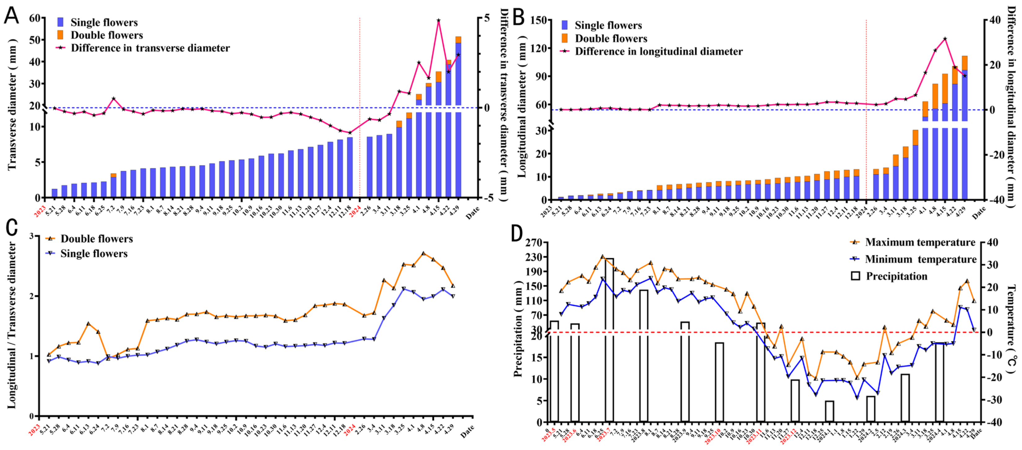
Figure 1. (A) Changes in the transverse diameter of flower buds during floral bud differentiation. (B) Changes in the longitudinal diameter of flower buds during floral bud differentiation. (C) Changes in the ratio of the longitudinal diameter to transverse diameter of flower buds during floral bud differentiation. (D) The temperature and precipitation information of Harbin during the experiment.
Forests 16 00409 g001
Figure 2. The corresponding relationship between date and morphological changes during the development of the single and double flower buds of Syringa vulgaris L. (AI) Changes in single flower bud morphology from June 2023 to May 2024. (JR) Changes in double flower bud morphology from June 2023 to May 2024.
Forests 16 00409 g002
Figure 3. Bract primordium differentiation stage of the single and double flower buds of S. vulgaris. (A) Single flower buds. (B) Double flower buds. IM: inflorescence meristem.
Forests 16 00409 g003
Figure 4. Inflorescence and floret primordium differentiation stage of the single and double flower buds of S. vulgaris. (A) Single flower buds. (B) Double flower buds. FP: floret primordium; GC: growth cone.
Forests 16 00409 g004
Figure 5. Sepal primordium differentiation stage of the single and double flower buds of S. vulgaris. (A) Single flower buds. (B) Double flower buds. SeP: sepal primordium; FmP: floral meristem primordium.
Forests 16 00409 g005
Figure 6. Petal primordium differentiation stage of the single and double flower buds in S. vulgaris. (A) Single flower buds. (BD) Double flower buds. SeP: sepal primordium; PeP: petal primordium; FPeP: first-petal whorl primordium. SPeP: second-petal whorl primordium. TPeP: third-petal whorl primordium.
Forests 16 00409 g006
Figure 7. Stamen primordium differentiation stage of the single and double flower buds of S. vulgaris. (AC) Single flower buds. (DF) Double flower buds. (E) Double flowers without stamen. (F) Double flowers with petaloid stamen. SeP: sepal primordium; PeP: petal primordium; SP: stamen primordium; PP: pistil primordium; FPeP: first-petal whorl primordium; SPeP: second-petal whorl primordium; TPeP: third-petal whorl primordium.
Forests 16 00409 g007
Figure 8. Carpel primordium differentiation stage of the single and double flower buds of S. vulgaris. (A,B) Single flower buds. (C,D) Double flower buds. (C) Double flowers with normal stamen. (D) Double flowers without stamen. SeP: sepal primordium; PeP: petal primordium; SP: stamen primordium; PP: pistil primordium; FPeP: first-petal whorl primordium; SPeP: second-petal whorl primordium; TPeP: third-petal whorl primordium.
Forests 16 00409 g008
Figure 9. Metabolic pathways associated with flower color formation in the single and double flowers of S. vulgaris. (A) Heatmap of the expression of DEGs in the flavonoid biosynthesis pathway. (B) Heatmap of the expression of DEGs in the tyrosine metabolism pathway. (C) Heatmap of the expression of DEGs in the phenylpropanoid biosynthesis pathway. (D) Heatmap of the expression of DEGs in the phenylalanine metabolism pathway.
Forests 16 00409 g009
Figure 10. Relationship between carbohydrate metabolism and petal structure formation during floral bud differentiation in S. vulgaris. (A) Heatmap of the expression of DEGs in starch and sucrose metabolism pathways. (B) Changes in the soluble sugar content of single and double flowers of S. vulgaris. (C) Changes in soluble protein content. The X-axis represents the different differentiation stages. The Y-axis is the content. The error bars indicate ± standard deviation. S1: Bract primordium differentiation stage. S2: Inflorescence and floret primordium differentiation stage. S3: Sepal primordium differentiation stage. S4: Petal primordium differentiation stage. S5: Stamen primordium differentiation stage. S6: Carpel primordium differentiation stage. S7: Rising temperature stage. Different letters indicate significant differences at the level of p < 0.05. The same applies below.
Forests 16 00409 g010
Figure 11. Expression characteristics of secondary metabolites and zeatin-related pathways during petal layer formation in S. vulgaris. (A) Heatmap of the expression of DEGs in the biosynthesis of secondary metabolites. (B) Heatmap of the expression of DEGs in zeatin biosynthesis. (C) Heatmap of the expression of DEGs in plant hormone signal transduction. (D) Changes in cis–ZR (cZR) content of the single and double flowers of S. vulgaris. (E) Changes in trans-ZR (tZR) content. (F) Changes in zeatin riboside (ZR) content. Different letters indicate significant differences at the level of p < 0.05.
Forests 16 00409 g011
Figure 12. Dynamic changes in endogenous hormone levels during floral bud differentiation in the single and double flowers of S. vulgaris. (A) Changes in indole-3-acetic acid (IAA) content. (B) Changes in abscisic acid (ABA) content. (C) Changes in gibberellin A3 (GA3) content. (D) Changes in 1-aminocyclopropane-1-carboxylic acid (ACC) content. (E) Changes in salicylic acid (SA) content. (F) Changes in jasmonic acid (JA) content. Different letters indicate significant differences at the level of p < 0.05.
Forests 16 00409 g012
Figure 13. Dynamic changes in hormone ratios during floral bud differentiation in the single and double flowers of S. vulgaris. (A) Changes in ABA/GA3 ratio. (B) Changes in IAA/GA3 ratio. (C) Changes in ABA/IAA ratio. (D) Changes in ZR/GA3 ratio. (E) Changes in IAA/ZR ratio. (F) Changes in SA/ACC ratio. Different letters indicate significant differences at the level of p < 0.05.
Forests 16 00409 g013
Figure 14. Relationship between ABC transporter-associated metabolites and antioxidant enzyme activities during floral bud differentiation in single- and double-flowered S. vulgaris. (A) Heatmap of the expression of DEGs in ABC transporters. (B) Changes in peroxidase (POD) activity. (C) Changes in superoxide dismutase (SOD) activity. (D) Changes in catalase (CAT) activity. (E) Changes in malondialdehyde (MDA) content. Different letters indicate significant differences at the level of p < 0.05.
Forests 16 00409 g014
Figure 15. Color synthesis approach of the single and double flowers of S. vulgaris.
Forests 16 00409 g015
Figure 16. Regulation mechanism of flower bud differentiation in S. vulgaris.
Forests 16 00409 g016
Table 1. Primer information.
Gene IDForward Primer (5′ to 3′)Reverse Primer (3′ to 5′)
TRINITY_DN50647_c0_g1AGCCTTCTGGTGAGCATGGATGAAGCTGTCGACAGCAGTA
TRINITY_DN36080_c0_g1CAGGCTACCGTCTTGTTGGAGTGCTAGCAGTCCTTGGTCT
TRINITY_DN34830_c0_g1TGCTGACGGTGACTGATGTTAGCTTGTGACCGTTGTAGCG
TRINITY_DN14054_c0_g1ATGCTTGGAGAGCTGTGTGCCGATGAGTCCTGAGGCTTGT
TRINITY_DN32532_c0_g1GGTGATCGTAGTGTGAGCCACTTGGCATGTTGAGGCTGAT
TRINITY_DN62324_c0_g1TGGAGCTTCTGAGGTGGTTACGTACTGCTGATCAGGATGC
TRINITY_DN42129_c0_g1CGTGAAGTGTGACTGGCTGAGCTTGAAGCTGAGCCTGATA
TRINITY_DN36412_c0_g2TGAAGCTGTCAGGACGTGTTCTTGAGTGCAGTGTCTGAGC
TRINITY_DN34320_c0_g2AGTGGTCGATCAGTGGCTGTTGACTGACTGAGGCTGTGAG
Table 2. Comparison between flower bud differentiation and external morphology in Syringa vulgaris L. (2023).
Stage of Flower
Bud Differentiation
Single FlowersDouble Flowers
Date and DurationLongitudinal
Diameter (mm)
Transverse
Diameter (mm)
Two-Layer Corolla Three-Layer CorollaLongitudinal
Diameter (mm)
Transverse
Diameter (mm)
Date and Duration
Spathe primordium
differentiation stage
21 May–
11 June
1.6403 ± 0.3231 mn1.7596 ± 0.3478 mn21 May–
11 June
21 May–
11 June
1.8328 ± 0.4437 m1.5135 ± 0.2912 n
212121
Inflorescence and small floral primordium stage11 June–
9 July
2.6529 ± 0.7669 l2.7831 ± 0.7523 kl11 June–
16 July
11 June–
16 July
3.329 ± 0.6747 j2.893 ± 0.9193 k
283535
Sepal primordium
differentiation stage
9 July–
23 July
4.0315 ± 0.4481 h4.0001 ± 0.3342 h16 July–
23 July
16 July–
23 July
4.2289 ± 0.3736 h3.75 ± 0.1971 i
1477
Petal primordium
differentiation stage
23 July–
14 August
4.4865 ± 0.4554 g4.2144 ± 0.3222 h23 July–
4 September
23 July–
11 September
6.949 ± 0.6065 c4.2258 ± 0.3898 h
224350
Stamen primordium
differentiation stage
14 August–
11 September
5.6236 ± 0.4786 e4.5532 ± 0.4696 g4 September–
11 September
8.0392 ± 0.3034 b4.6314 ± 0.2645 g
287
Pistil primordium
differentiation stage
11 September
–25 September
6.3373 ± 0.413 d5.188 ± 0.3846 f11 September–
16 October
11 September–
16 October
8.4374 ± 0.4859 a5.0727 ± 0.4019 f
143535
Note: The data in the table are the means ± standard deviations, and different letters after the data indicate significant differences at the level of p < 0.05.
Disclaimer/Publisher’s Note: The statements, opinions and data contained in all publications are solely those of the individual author(s) and contributor(s) and not of MDPI and/or the editor(s). MDPI and/or the editor(s) disclaim responsibility for any injury to people or property resulting from any ideas, methods, instructions or products referred to in the content.