Article

Genome-Wide Identification and Expression Profiling of MYC Transcription Factor Family in Toona sinensis Under Abiotic and Hormonal Stresses

1
College of Horticulture and Gardening, Yangtze University, Jingzhou 434025, China
2
Hubei Key Laboratory of Selenium Resource Research and Biological Application, Hubei Minzu University; Enshi 445000, China
3
Hubei Key Laboratory of Spices & Horticultural Plant Germplasm Innovation & Utilization, Yangtze University, Jingzhou 434025, China
4
Shashi Substation, Jingzhou Municipal Bureau of Natural Resources and Planning, Jingzhou 434025, China
*
Authors to whom correspondence should be addressed.
Forests 2025, 16(12), 1756; https://doi.org/10.3390/f16121756
Submission received: 7 October 2025 / Revised: 14 November 2025 / Accepted: 19 November 2025 / Published: 21 November 2025
(This article belongs to the Special Issue Recent Advances in Tree Genetics and Genomics)

Abstract

Toona sinensis (Chinese toon) is a valuable forest resource widely used in gastronomy, phytotherapy, and timber production. MYC transcriptional factors are critical to a variety of biological functions. However, the MYC family remains unsystematically characterized in T. sinensis. A total of 18 TsMYC genes were identified in the T. sinensis genome and grouped into six distinct subfamilies according to phylogenetic analysis in this study. This classification was further supported by analyses of gene structure and conserved motifs. Evolutionary analysis revealed that the TsMYC gene family achieved expansion via whole-genome duplication (WGD) and segmental duplication, with these genes experiencing intense purifying selection throughout the evolutionary process. Additionally, 42 cis-acting elements were detected in the promoter regions of TsMYC genes, and the protein–protein interaction (PPI) network demonstrated that MYC2 serves as a central component in the jasmonic acid (JA) signaling pathway. Expression profiling showed that all TsMYC genes except TsMYC17 were highly expressed in leaves. TsMYC genes displayed distinct expression patterns under salt stress and various phytohormone treatments, with TsMYC17 being the only gene consistently upregulated under all conditions. Subcellular localization assays confirmed that TsMYC17 is localized in the nucleus. These findings suggest that TsMYC17 may play a key role in mediating responses to multiple hormonal signals and abiotic stresses. This research lays a foundation for future investigations into the molecular characteristics and biological roles of the TsMYC17 gene.

1. Introduction

Toona sinensis (A. Juss.) Roem., commonly known as Chinese toon or Chinese mahogany, belongs to the family Meliaceae and is native to East and Southeast Asia [1]. The young buds and leaves of T. sinensis are widely consumed as a popular woody vegetable and aromatic seasoning in China [2]. It is a multifunctional tree species used for food, medicine, and timber, offering substantial prospects for development and application [3]. Furthermore, T. sinensis possesses a distinctive flavor profile primarily attributed to volatile sulfur compounds, terpenes, aldehydes, alcohols, ketones, and esters, with terpenes being the most abundant [4]. These findings suggest that terpenes play a crucial role in the flavor development of T. sinensis.
The basic helix–loop–helix (bHLH) family is the second-largest transcription factor group in plants [5,6]. myelocytomatosis (MYC) transcription factors, a subfamily of the bHLH family, possess both a HLH domain and bHLH-MYC-N domain [7]. The MYC gene family has been recognized in multiple plant species and is involved in various physiological processes, such as growth, development, flowering induction, secondary metabolism, and defense responses [8,9]. MYC2, a central transcriptional regulator in the MYC family, has been extensively characterized across multiple plant species. It constitutes the core of the JA signaling pathway, functioning through the canonical COI1–JAZ–MYC2 module to coordinate multiple genetic components. In this cascade, COI1 (CORONATINE-INSENSITIVE 1) acts as a key receptor component, while JAZ (JASMONATE ZIM-DOMAIN) proteins function as transcriptional repressors that directly inhibit MYC2 activation [10,11]. MYC transcription factors involved in the JA signaling pathway not only respond to biotic and abiotic stresses but also promote the biosynthesis of secondary metabolites, such as terpenoids [12,13]. For example, in Arabidopsis thaliana, genetic studies have identified AtMYC2, AtMYC3, and AtMYC4 as key transcriptional regulators in the jasmonic acid signaling pathway [14]. In Taxus chinensis, TcMYC2a directly regulates the biosynthesis of paclitaxel through JA signaling [15]. In Aquilaria sinensis, AsMYC2 regulates ASS1 transcription through the JA pathway, thereby modulating sesquiterpene production [16]. MYC2 also serves as a central regulator in plant hormone signaling and abiotic stress. For example, in the ethylene (ETH) signaling pathway, MdMYC2 regulates ethylene biosynthesis by binding to the G-box element in the promoter region of the ethylene response factor MdERF3 [17]. In Arabidopsis, overexpression of AtMYC2 enhances plant tolerance to salinity stress [18]. Phenotypic comparisons between myc2 and med5 mutants suggest that MYC2 negatively regulates salt tolerance through interaction with MED25. Moreover, atmyc2/3/4 triple mutants partially relieve the salt-induced inhibition of root cell elongation [19]. MYC2 also functions as a pleiotropic regulator of specialized metabolism, including anthocyanin, alkaloid, and terpenoid biosynthetic pathways. For example, overexpression of AtMYC3 and AtMYC4 in A. thaliana results in increased anthocyanin accumulation. In Nicotiana tabacum, NtMYC2 enhances the transcription of PMT, a key enzyme gene in the nicotine biosynthetic pathway, thus increasing nicotine content [20]. In Freesia hybrida, overexpression of FhMYC2 suppresses FhMYB21-mediated activation of the FhTPS1 promoter, thereby reducing linalool production [21]. In Salvia miltiorrhiza, SmMYC2 binds to the SmGGPPS1 promoter and activates its transcription, thereby promoting tanshinone biosynthesis [22].
At present, identification of the MYC gene family has been identified in multiple species, including Triticum aestivum, Oryza sativa, and Brachypodium distachyon [23]. However, a comprehensive genome-wide characterization of MYC transcription factors in T. sinensis has not yet been conducted. A total of 18 TsMYC family members were identified from the published genome sequence of T. sinensis in this study. Comprehensive bioinformatic analyses were conducted to characterize their physicochemical properties, phylogenetic relationships, conserved motifs, chromosomal distribution, synteny, promoter cis-acting elements, and potential protein–protein interactions. Expression profiling by qRT-PCR revealed distinct expression patterns of TsMYC genes across different tissues, as well as in response to NaCl and various phytohormone treatments in T. sinensis. The subcellular localization of the candidate TsMYC17 protein was confirmed by transiently expressing GFP fusion constructs in Nicotiana benthamiana epidermal cells via Agrobacterium-mediated transformation. This study provides a theoretical basis for understanding TsMYC17’s molecular regulatory mechanisms under salt and hormonal stresses.

2. Materials and Methods

2.1. Plant Material and Treatments

T. sinensis plants were cultivated in a greenhouse at Yangtze University, Jingzhou, Hubei Province, China (30.35° N, 112.15° E). Plants were maintained under natural light conditions with standardized irrigation and fertilization management. One-year-old plants with uniform growth were selected and foliar-sprayed with methyl jasmonate (MeJA, 100 µM), salicylic acid (SA, 300 µM), gibberellic acid (GA3, 100 µM), abscisic acid (ABA, 100 µM), and sodium chloride (NaCl, 150 mM), each dissolved in sterile water. The treatment concentrations were determined in advance through preliminary experiments. Samples were collected at four time points (0 h, 6 h, 12 h, and 24 h), with three biological replicates per treatment (n = 3 plants per replicate), and all samples were randomly collected. Notably, samples at each time point were obtained by repeated sampling from the same individual plant. Additionally, nine untreated one-year-old T. sinensis plants with uniform growth were selected as experimental materials, and root, stem, and leaf tissues were collected separately. All samples were stored at −80 °C until further analysis.

2.2. Identification of the TsMYC Family Members

The genomic sequence data and annotation information of T. sinensis were obtained from the CNGB Sequence Archive (https://ftp.cngb.org/pub/CNSA/data1/CNP0000958/CNS0203218/CNA0019196/ (accessed on 4 April 2024)) [24]. Hidden Markov model (HMM) profiles corresponding to the HLH domain (PF00010) and bHLH_MYC_N domain (PF14215) were retrieved from the PFAM database [25] and were employed as queries to search for potential MYC proteins in the T. sinensis proteome using HMMER3.1 (E-value ≤ 1 ×  10−5). Additionally, 14 MYC protein sequences from A. thaliana were downloaded from The Arabidopsis Information Resource (http://www.arabidopsis.org/ (accessed on 7 April 2024)). These MYC sequences were also utilized as queries for a BLAST 2.12.0 search against the T. sinensis protein database. Candidate protein sequences were further validated using multiple domain annotation tools, including NCBI-CDD and InterProScan. The physicochemical properties of the TsMYCs were assessed using the ProtParam tool (https://web.expasy.org/protparam/ (accessed on 8 April 2024)) [26]. Furthermore, the subcellular localization of T. sinensis MYC proteins was predicted using the WoLF PSORT program (https://wolfpsort.hgc.jp/ (accessed on 12 April 2024)) [27].

2.3. Multiple Sequence Alignment and Phylogenetic Analysis

Multiple sequence alignment was performed using ClustalW with default settings to compare MYC gene members from A. thaliana, O. sativa, P. tomentosa, and Camellia sinensis with the identified TsMYC members [28]. Using the Maximum Likelihood (ML) method and 1000 bootstrap replicates, the phylogenetic tree was constructed in MEGA 11.0 software [29]. The phylogenetic tree was further refined using the TVBOT (https://www.chiplot.online/tvbot.html (accessed on 18 April 2024)) [30]. Information on the MYC protein sequences from the five species is listed in Table S1.

2.4. Chromosomal Localization and Synteny Analysis

The chromosomal locations of TsMYC genes were extracted from the T. sinensis genome annotation files and visualized using TBtools V2.056 software [31]. Collinearity analysis of TsMYC genes within the T. sinensis genome was conducted using the MCScanX v1.0.0 toolkit [32,33]. Genome sequences and annotation files of A. thaliana, P. tomentosa, Z. mays, and O. sativa were obtained from the NCBI database. Synteny analysis between T. sinensis and other species, including the dicotyledonous plants A. thaliana and P. tomentosa, and the monocotyledonous plants Z. mays and O. sativa, was performed using MCScanX v1.0.0. The non-synonymous (Ka) and synonymous (Ks) substitution rates were calculated during the analysis.

2.5. Gene Structure and Conserved Motif Analysis

Conserved domains of TsMYC proteins were identified using the NCBI Batch CD-Search. The conserved motif analysis of TsMYC proteins was performed using the MEME Suite (https://meme-suite.org/meme/tools/meme (accessed on 2 May 2024)) [34]. The maximum number of motifs was set to 10, while all other parameters were maintained at their default values.

2.6. Cis-Regulatory Elements Analysis of Promoters Regions of TsMYC Genes

To investigate putative cis-acting regulatory elements, 2000 bp upstream sequences from the initiation codons of 18 TsMYC genes were extracted as promoter regions. These sequences were analyzed for cis-acting elements using the PlantCARE database (https://bioinformatics.psb.ugent.be/webtools/plantcare/html/ (accessed on 5 May 2024)) [35]. TBtools V2.056 software was used to visualize the results.

2.7. Prediction of Protein–Protein Interaction Networks

To investigate potential interactions among TsMYC proteins and their counterparts, the STRING v12.0 online tool (https://cn.string-db.org/ (accessed on 8 May 2024)) was used to construct a PPI network [36]. The amino acid sequences of TsMYCs were queried against A. thaliana in STRING, with default parameters applied for all other settings. The generated interaction network was visualized and refined using Cytoscape 3.10.2 [37], with a confidence score threshold of 0.7 applied to ensure the reliability of predicted interactions.

2.8. RNA Extraction and qRT-PCR Analysis

Leaves of T. sinensis treated with hormones or NaCl, along with untreated root, stem, and leaf samples, were thoroughly ground in liquid nitrogen. Approximately 100 mg of the resulting fine powder from each sample was accurately weighed. Total RNA was extracted from the samples following the protocol provided with the Polysaccharide Polyphenol Plant Total RNA Extraction Kit (TIANGEN, Beijing, China, DP441). The RNA concentration and integrity were assessed using an ultra-micro spectrophotometer (Thermo Fisher, Ipswich, MA, USA) and 1.0% agarose gel electrophoresis. The OD260/OD280 ratio was required to fall within the range of 1.8 to 2.1, and the RNA concentration was required to be at least 100 ng/μL. One microgram of RNA from each sample was reverse transcribed into complementary DNA (cDNA) using the reverse transcription kit. Primers were designed using the online tool provided by Integrated DNA Technologies (Supplementary Table S2), with TsActin serving as the internal reference gene [38]. Quantitative real-time PCR (qRT-PCR) was conducted using the LineGene 9600 (BioerTechnology, Hangzhou, China) Plus fluorescence quantitative PCR system. The reaction mixture contained 10 μL of 2× ChamQ Universal SYBR qPCR Master Mix (Vazyme, Nanjing, China), 0.4 μL of forward primer, 0.4 μL of reverse primer, 2 μL of cDNA template, and distilled water to a final volume of 20 μL. The thermal cycling conditions were as follows: an initial denaturation at 95 °C for 30 s; followed by 40 cycles of denaturation at 95 °C for 10 s and annealing/extension at 60 °C for 30 s; and a final melting curve analysis performed at 95 °C for 15 s, 60 °C for 60 s, and 95 °C for 15 s.

2.9. The Subcellular Localization Analysis of TsMYC17

The full-length open reading frame of TsMYC17, excluding the stop codon, was inserted into the pNC-Cam1304-SubC vector containing SfiI restriction sites to generate the recombinant vector (35S:TsMYC17-GFP) and the control vector (35S:GFP) [39]. The constructs were introduced into Agrobacterium tumefaciens strain GV3101 by electroporation and used for transient transformation of the abaxial epidermis of N. benthamiana leaves. After 24 h of dark culture and 48 h of light culture, the fluorescence signal distribution of 35S:TsMYC17-GFP and 35S:GFP was observed using a laser scanning confocal microscope (Leica TCS SP8, Wetzlar, Germany), with H2B–mCherry (nuclear localization signal, NLS) as a reference marker. The excitation wavelengths were 488 nm for GFP and 587 nm for mCherry, and emission was detected at 500–530 and 600–630 nm, respectively. The primers employed in this experiment are provided in Table S2.

2.10. Statistical Analysis

In this study, gene expression data were analyzed without prior log transformation. Statistical analyses were conducted using Origin 2024 (OriginLab Corporation, Northampton, MA, USA). One-way ANOVA was employed to assess differences in gene expression among treatments. All analyses were performed on data from three biological replicates, each with three technical replicates. The 2−ΔΔCT method was applied to quantify relative gene expression levels, with statistical significance set at a threshold of p < 0.05.

3. Results

3.1. Identification and Physicochemical Property Analysis of TsMYC Family Members

To identify the MYC protein sequences in T. sinensis, 18 TsMYC proteins were found through HMMER and BLASTP 2.14.0 searches, which were then validated using the NCBI Batch CD-Search (https://www.ncbi.nlm.nih.gov/Structure/cdd/ (accessed on 6 April 2024)). These proteins were named TsMYC1–TsMYC18 according to their chromosomal locations (Table S3). The physicochemical analysis showed that the TsMYC proteins ranged from 457 to 684 amino acids in length. Their molecular weights (MWs) varied between 51.75 and 76.38 kDa, while their theoretical isoelectric points (pIs) ranged from 5.29 to 6.49. All proteins had instability index values exceeding 40, suggesting that TsMYC proteins may exhibit relative instability in vitro. The aliphatic index varied from 66.15 to 88.77, and the GRAVY values ranged between −0.63 and −0.24, indicating that all TsMYC proteins are hydrophilic. Nuclear localization was predicted for all 18 TsMYC proteins using WoLF PSORT.

3.2. Phylogenetic and Classification Analysis of the TsMYC Gene Family

To explore the functional characteristics of TsMYC proteins, a phylogenetic tree was constructed using 68 MYC protein sequences from T. sinensis, A. thaliana, O. sativa, P. trichocarpa, and C. sinensis, employing the Maximum Likelihood (ML) method (Figure 1). The classification of these proteins followed the system established by Heim et al. for AtbHLH proteins [40]; these sequences were grouped into 26 subfamilies, designated Ia–XV. Within the bHLH transcription factor family, MYC proteins belong to subgroup III. Phylogenetic analysis revealed that the 18 TsMYC proteins clustered into six distinct branches: IIIa, IIIc, IIId(1), IIId(2), IIIe, and IIIf. Subgroup IIIa contained a single TsMYC protein, while the other subgroups included one to four TsMYC members. The TsMYC genes from various species were distributed across almost all evolutionary branches, suggesting early diversification before species divergence [41]. Further phylogenetic analysis revealed that TsMYC7, TsMYC8, and TsMYC17 from subgroup IIIe clustered with AtMYC2, which is known to play crucial roles in abiotic stress responses and secondary metabolism. This result suggests that these TsMYC proteins may have similar structural characteristics and functional properties.

3.3. Chromosomal Localization and Synteny Analysis of TsMYC Genes

Chromosomal mapping showed that the 18 identified TsMYC genes were unevenly distributed across seven chromosomes, with one to six genes per chromosome. Chromosome 23 contained the largest number of TsMYC genes, while chromosomes 13, 14, and 27 each contained only one (Figure 2).
To explore the evolutionary mechanisms underlying the expansion of the TsMYC family, gene duplication events were analyzed using MCScanX (Figure 3). The results indicated that the TsMYC gene expansion was mainly driven by whole-genome duplication (WGD) and segmental duplication events, accounting for 94.4% (17 of 18 genes), with the highest frequencies on chromosomes 23 and 24 (Table S4). Notably, only one TsMYC gene resulted from a proximal duplication event, and no tandem duplications were detected within the family. The ratio of nonsynonymous to synonymous substitutions (Ka/Ks) is critical for studying genomic evolution [42]. Synonymous substitutions refer to nucleotide mutations that do not alter amino acid sequences, whereas nonsynonymous substitutions result in amino acid changes. In brief, Ka/Ks > 1 indicates positive selection, Ka/Ks = 1 suggests neutral evolution, and Ka/Ks < 1 implies purifying selection. Ka/Ks analysis of 12 homologous TsMYC gene pairs yielded ratios ranging from 0.149 to 0.428, with a mean value of 0.244 ± 0.09 (Table S5). These findings indicate that TsMYC genes have been subjected to strong purifying selection during evolution [43].
MCScanX was further employed to analyze syntenic relationships between TsMYC genes and those from two dicots and two monocots (Figure 4). Stronger syntenic relationships were observed between TsMYC genes and MYC genes in dicotyledonous species. The greatest number of syntenic gene pairs occurred between T. sinensis and P. trichocarpa (47 pairs), followed by A. thaliana (23), Z. mays (4), and O. sativa (3). Notably, TsMYC1 showed no collinearity with the MYC genes from the other four species, suggesting that it may have originated after the divergence of these lineages. In contrast, homologous gene pairs involving TsMYC4, TsMYC5, and TsMYC8 were identified across all four species, suggesting that these orthologous relationships predated the divergence of dicots and monocots.

3.4. Gene Structure and Conserved Motifs Analysis of TsMYC Genes

To elucidate the structural diversity of TsMYC proteins and validate their subgroup classifications, gene structures, conserved motifs, and phylogenetic relationships were analyzed. The 18 TsMYC members were classified into six subgroups, with proteins within each subgroup displaying highly conserved or similar motif patterns. Motifs 1, 2, 3, 6, and 7 were universally conserved across all sequences (Figure 5A,B). Consistent with the characteristics of typical bHLH transcription factors, TsMYC proteins harbor a helix–loop–helix (HLH) domain (Motifs 1 and 2) and a conserved bHLH-MYC-N domain (Motifs 3, 4, 5, 6, and 8) (Figure 5C and Figure S1; Table S6). Members of the same subfamily exhibited analogous motif compositions, which aligned with their phylogenetic relationships and implied the conservation of their biological functions.
Gene structure analysis revealed that the number of exons in TsMYC genes varied from 1 to 8, with subgroups IIId(1) and IIId(2) lacking introns, indicating strong structural conservation within these two groups. In contrast, subgroups IIIa, IIIc, and IIIf exhibited more complex exon–intron architectures, with 7–8 exons and 6–7 introns (Figure 5D). Overall, genes belonging to the same subfamily showed high similarity in their exon–intron organization. Meanwhile, significant differences in this organizational feature between distinct subfamilies provided further support for the results of phylogenetic classification.

3.5. Cis-Acting Elements Analysis of TsMYC Promoters

Cis-acting elements within gene promoters generally regulate gene expression and function. To explore the regulatory mechanisms and expression patterns of TsMYC family genes, we extracted the 2000 bp upstream promoter sequences of each TsMYC gene from the T. sinensis genome, aiming to systematically identify potential cis-acting elements. A total of 42 cis-acting elements were predicted (Table S7). These elements were primarily associated with light responsiveness, hormonal signaling, environmental adaptation, and developmental regulation. In this study, various hormone-responsive cis-acting elements were identified, including abscisic acid–responsive elements (ABRE), auxin-responsive elements (TGA element), MeJA-responsive elements (TGACG motif and CGTCA motif), gibberellin-responsive elements (TATC-box, P-box, and GARE-motif), and salicylic acid-responsive elements (TCA element). Additionally, multiple light-responsive elements (ACE, G-box, GT1-motif, Sp1, 3-AF1 binding site, and MRE) and stress-responsive elements (ARE, MBS, LTR, and TC-rich repeats) were detected (Figure 6, Table S7). All members of the TsMYC transcription factor family contained light-responsive and anaerobic-induction elements. Among the hormone-responsive elements, those associated with abscisic acid (57) were the most abundant, followed by methyl jasmonate (MeJA)-related motifs (46). Various types and quantities of regulatory elements were identified in the promoter regions of TsMYC genes, indicating that these genes may be involved in a wide range of functions, including signal transduction, stress responses, and the regulation of secondary metabolism.

3.6. Interaction Network Prediction of the TsMYCs

To explore the potential regulatory interactions between TsMYC proteins and other proteins, a protein–protein interaction (PPI) network was constructed using the STRING database (Figure 7A). Analysis of the 18 TsMYC proteins revealed that TsMYC6, TsMYC7, TsMYC8, TsMYC14, and TsMYC17 share high structural similarity with the Arabidopsis MYC2 protein and are predicted to interact with GL3. In addition, a MYC2-centered protein–protein interaction network comprising 20 interacting proteins was constructed (Figure 7B). The results indicated that MYC2 predominantly interacts with members of the TIFY family (11 proteins) and MYB transcription factors (2 proteins). TIFY family proteins are plant-specific transcriptional regulators that play key roles in the JA signaling pathway. The interactions between MYC2 and multiple TIFY domain-containing proteins further highlight its central regulatory role in JA-mediated signal transduction.

3.7. Expression Profiling of TsMYC Genes in Different Tissue

Expression analysis of eight TsMYC genes was performed using qRT-PCR, including TsMYC7, TsMYC8, and TsMYC17 (Figure 1, Table S8), which are closely related to MYC2, and TsMYC4, TsMYC6, TsMYC9, TsMYC14, and TsMYC15 from other subgroups. For the purpose of examining tissue-specific expression patterns, quantitative analysis of transcript abundances was performed for the eight TsMYC genes in roots, stems, and leaves of T. sinensis (Figure 8). The results showed that most genes exhibited their highest transcript accumulation in leaves, whereas TsMYC17 displayed relatively low expression in this tissue.

3.8. Expression Profiles of TsMYC Genes Under MeJA Treatment

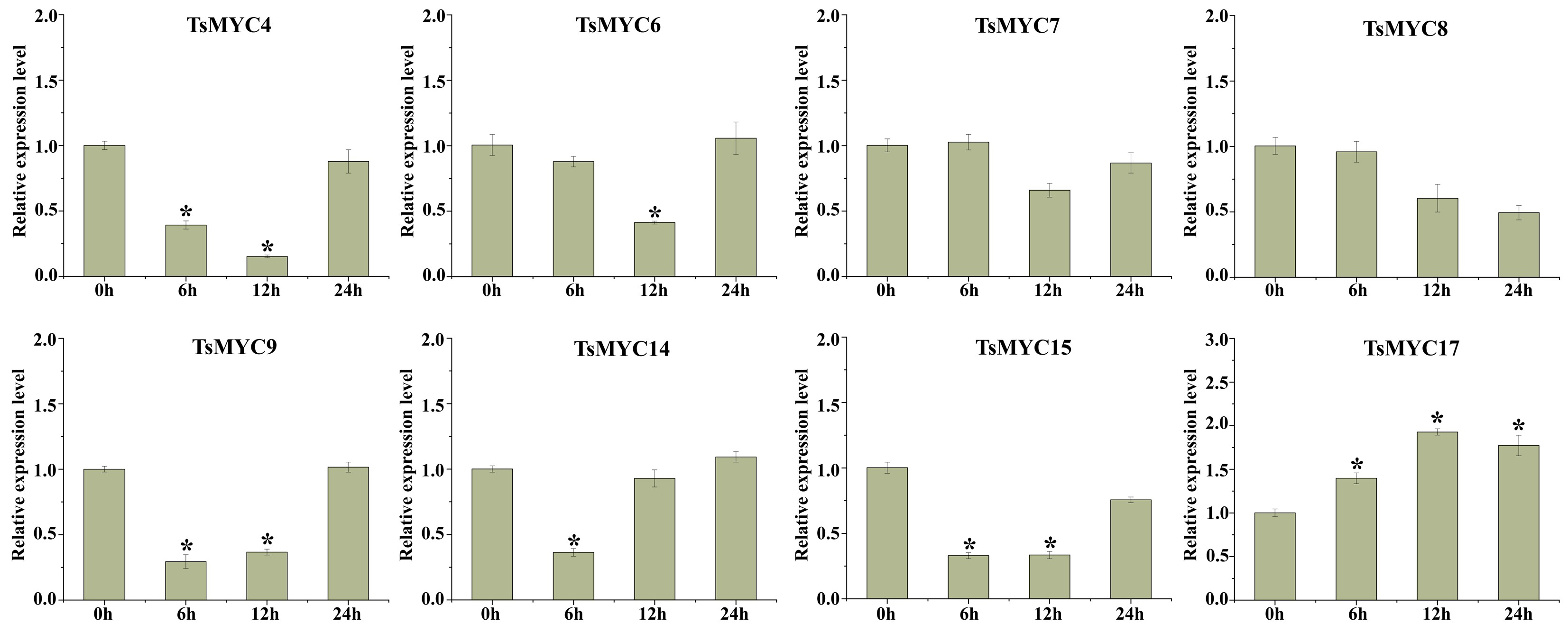
To investigate the functions of TsMYCs in terpenoid synthesis, the expression profiles of eight genes were analyzed following MeJA treatment (Figure 9). After MeJA treatment, TsMYC6 and TsMYC17 transcript levels peaked at 12 h, showing approximately 8-fold and 5-fold increases compared with pre-treatment levels, respectively. In contrast, TsMYC4, TsMYC9, TsMYC14, and TsMYC15 displayed a gradual decline in expression, with TsMYC4 showing an approximately 90% reduction at 12 h. Overall, all eight genes exhibited marked transcriptional responses to MeJA induction. Among the three homologs of AtMYC2, TsMYC17 exhibited the most significant change in expression. These findings further support the notion that MYC transcription factors are core components of the JA pathway.

3.9. The Phytohormone Response Pattern of TsMYCs

Based on the cis-acting elements identified in the promoter regions, three phytohormones (GA3, ABA, SA) were selected for foliar application, and the expression changes of eight TsMYC genes were analyzed using qRT-PCR. Generally, the expression levels of these eight TsMYCs varied with phytohormone type and treatment duration (Figure 10). After GA treatment, TsMYC4, TsMYC8, TsMYC9, and TsMYC15 expression was downregulated, whereas TsMYC6, TsMYC7, TsMYC14, and TsMYC17 were upregulated, with transcript levels peaking at 6 h. After ABA treatment, TsMYC4, TsMYC7, TsMYC8, TsMYC9, TsMYC14, and TsMYC15 showed decreased transcript abundance at 6 h. After SA treatment, TsMYC4, TsMYC8, TsMYC9, TsMYC14, and TsMYC15 exhibited a consistent downward trend, whereas TsMYC17 was markedly upregulated at 6 h, followed by a gradual decline, with the lowest expression observed at 24 h. Overall, TsMYC6 displayed a consistent and significant induction across different phytohormone treatments, whereas the remaining TsMYC genes responded in a hormone-specific manner.

3.10. Expression Profiles of TsMYC Genes Under Salt Stress

To further explore how TsMYC genes respond to NaCl stress, qRT-PCR analysis was performed, revealing differential expression patterns under salt stress. After 6 h and 12 h of NaCl treatment, the expression levels of TsMYC4, TsMYC9, and TsMYC15 were downregulated by approximately 60% compared to the control group, while only TsMYC17 maintained an upward expression trend, with its peak level nearly doubling (Figure 11).

3.11. Subcellular Localization of TsMYC17

Given the high homology between TsMYC17 and AtMYC2 (Table S8), along with the quantitative results showing an increase in TsMYC17 expression after treatment with various hormones and NaCl, this study selected TsMYC17 for further functional characterization. A GFP–TsMYC17 fusion expression vector was constructed, and a mixture containing the pNC-Cam1304-SubC-TsMYC17 construct and the mCherry vector was introduced into N. benthamiana leaves through Agrobacterium tumefaciens strain GV3101-mediated infiltration. The empty pNC-Cam1304-SubC vector served as the negative control. Green fluorescence from the TsMYC17—GFP fusion protein colocalized with red fluorescence from H2B–mCherry in the nucleus. Fluorescence signals were observed and visualized using a laser scanning confocal microscope. As predicted, fluorescence from the fusion protein was confined to the nucleus (Figure 12). These findings confirm that TsMYC17 is primarily localized in the nucleus, reinforcing its function as a nuclear-localized transcription factor.

4. Discussion

Plant growth and development are controlled by intricate gene networks [44]. Within these networks, the MYC gene family plays a critical role in regulating plant growth and development. JA and MeJA are essential signaling molecules that coordinate plant responses to both biotic and abiotic stresses, as well as regulate growth, development, and secondary metabolism. Upon sensing external MeJA stimuli, the endogenous JA level in plants increases, and JAZ proteins are bound by the E3 ubiquitin ligase SCFCOI1 complex, triggering the release of MYC2 [45]. The MYC gene family has been thoroughly studied in a range of plant species [46]. We performed a phylogenetic analysis of MYC genes in T. sinensis using the bHLH family classification system of A. thaliana. This analysis categorized the 18 TsMYC genes into six distinct subgroups. Phylogenetic analysis can uncover orthologous relationships by examining sequence similarity and protein structure, with closely related MYC genes likely having similar functions [47]. Previous studies show that CsMYC2 mediates the JA pathway by activating the G-box motif in the promoter region [48]. MYC2-mediated transcriptional suppression of CAT2 by JA reduces salt stress tolerance in Arabidopsis seedlings [49]. Sequence alignment of TsMYC7, TsMYC8, and TsMYC17 with MYC2 homologs from other plant species (Table S8) revealed that TsMYC17 shared the highest sequence similarity with AaMYC2 (77.13%), followed by TsMYC8, which exhibited 76.14% similarity with AsMYC2. Therefore, we hypothesize that TsMYC7, TsMYC8, and TsMYC17 is a homolog of MYC2, though this hypothesis requires further experimental validation.
The TsMYC genes within the same subgroup exhibit significant structural and functional similarities. The conserved motif structures of TsMYC proteins, along with their gene architectures, strongly support the classification of TsMYC genes. Previous studies have suggested that the gain or loss of exons and introns contribute to functional diversification within gene families, with introns playing a role in gene evolution [24,50]. In this study, TsMYC genes without introns were identified in subgroups IIId and IIIe. This distribution pattern is consistent with those observed in other plants, such as willows [41] and apples [51]. These results suggest that the TsMYC genes in these sub-groups may respond rapidly to various stresses [52].
Gene duplication is a key evolutionary mechanism that generates new genes and facilitates adaptation to changing environments. Recent research has suggested that T. sinensis has experienced at least two significant whole-genome duplication (WGD) events [53]. This study identified 17 TsMYC genes as resulting from WGD or segmental duplication (Table S5). Cis-acting elements are indispensable for regulating the expression of functional genes. When plants face environmental stress, these elements activate transcription factors, which in turn regulate downstream gene expression to coordinate plant stress responses [54]. In this study, the 18 TsMYC genes were found to contain various cis-acting regulatory elements in their promoter regions, suggesting that these genes have distinct regulatory functions and respond to different stresses and hormone treatments.
Plant hormones are essential regulators of growth, development, and stress responses [55], with MYC genes playing a pivotal role in mediating the interactions between these hormones. For example, AtMYC2 promotes the inactivation of gibberellins (GA) through JA signaling, thereby reducing endogenous GA levels and inhibiting plant growth [56]. During drought stress, AtMYC2 acts as a transcriptional activator of ABA-induced gene expression [57]. Furthermore, FaMYC2 levels increase significantly shortly after MeJA treatment [58]. Moreover, JA collaborates with SA to enhance heat tolerance in A. thaliana through the action of AtMYC2 [59]. In this study, the expression levels of TsMYC4, TsMYC6, TsMYC14, TsMYC15, and TsMYC17 changed significantly after MeJA treatment. These results indicate that they were involved in JA signaling pathway. TsMYC genes exhibited diverse expression patterns in response to ABA, GA3, and SA treatments, indicating functional diversity among these genes. Notably, TsMYC6 and TsMYC17 showed increased expression in response to all four hormone treatments. In general, the functions of candidate genes are often deduced by referencing the known functional information of their homologous counterparts [60]. Therefore, we aligned the protein sequence of AtMYC2 with those of TsMYC proteins. The results showed that TsMYC7, TsMYC8, and TsMYC17 shared a high degree of homology with AtMYC2. Furthermore, Studies have indicated that JA and GA both act to induce the expression of sesquiterpene synthase genes in A. thaliana, with this regulatory process relying on the key mediation of AtMYC2 [61]. After T. sinensis were separately treated with MeJA and GA3, only TsMYC17 exhibited similar expression changes. Therefore, TsMYC17 may function similarly to AtMYC2 and regulate sesquiterpene biosynthesis in T. sinensis. In this study, the expression of TsMYC17 was upregulated following NaCl treatment, suggesting that this gene contributes to the plant’s response to abiotic stress. Tissue-specific expression analysis revealed that TsMYC17 was highly expressed in the roots of T. sinensis. This finding implies that the elevated expression of TsMYC17 in the roots may regulate and contribute to enhancing the plant’s stress tolerance. Based on phylogenetic analysis, TsMYC7, TsMYC8, and TsMYC17 were identified as MYC2 homologs. PPI network analysis showed that MYC2 primarily interacts with members of the TIFY protein family, which are crucial in hormonal signaling and responses to abiotic stress, thus providing a theoretical foundation for future experiments. Based on these findings, T. sinensis plants were treated with various hormones and subjected to salt stress. Expression analysis revealed that TsMYC17 was consistently upregulated under all treatment conditions. Combined with subcellular localization and homology analyses, these results suggest that TsMYC17, as a homolog of MYC2 may be essential in regulating the growth, development, and environmental adaptation of T. sinensis. However, the precise molecular mechanisms underlying its regulatory function need further experimental validation.

5. Conclusions

This study identified 18 members of the MYC gene family in T. sinensis through phylogenetic analysis and gene structure examination. The TsMYC genes were further analyzed for chromosomal localization, cis-acting elements, and synteny relationships. Expression profiles under abiotic stress and various phytohormone treatments were evaluated, highlighting the roles of TsMYC genes in stress responses and hormone signaling pathways. Subcellular localization studies revealed that TsMYC17 is localized in the nucleus. These results lay the groundwork for future research into the biological function of TsMYC17. In subsequent research, we aim to determine whether TsMYC17 influences the formation of flavor compounds in T. sinensis through genetic transformation and terpenoid content analysis.

Supplementary Materials

The following supporting information can be downloaded at: https://www.mdpi.com/article/10.3390/f16121756/s1, Figure S1: Logo sequences for 18 TsMYC conserved motifs; Table S1: The protein sequences of MYC genes in Toona sinensis, Arabidopsis thaliana, Oryza sativa, Populus trichocarpa, Camellia sinensis; Table S2: Primers used in this study; Table S3: Basic information on the MYC genes of Toona sinensis; Table S4: Duplication Type information of TsMYC genes; Table S5: The information of gene replication type, Ka, Ks and ka/ks ratio values of TsMYC genes; Table S6: Prediction of conserved domains in 18 TsMYC protein sequences; Table S7: Information on all cis-acting elements detected in the promoter regions of TsMYC genes; Table S8: Sequence Alignment of TsMYC7, TsMYC8, TsMYC17 with MYC2 Homologs from Other Plant Species.

Author Contributions

G.Z.: Software, Data Curation, Methodology, Visualization, Writing—Original Draft. S.Z.: Methodology, Software. X.C.: Methodology. Y.L.: Software, Visualization. W.Z.: Data Curation, Project Administration, Writing—Review and Editing. F.X.: Visualization, Supervision. D.J.: Visualization, Writing—Review and Editing. All authors have read and agreed to the published version of the manuscript.

Funding

This work was supported by the Hubei Key Laboratory of Selenium Resource Research and Biological Application (No. PT10202310) and the Key Research and Development Program of Hubei Province, China (No. 2023BBB065).

Institutional Review Board Statement

This article does not contain any studies with human participants or animals performed by any of the authors.

Data Availability Statement

All analyzed data generated in this study are available within the article.

Conflicts of Interest

The authors declare no conflicts of interest.

References

  1. Peng, W.; Liu, Y.; Hu, M.; Zhang, M.; Yang, J.; Liang, F.; Wu, C. Toona. sinensis: A comprehensive review on its traditional usages, phytochemistry, pharmacology, and toxicology. Rev. Bras. Farmacogn. 2019, 29, 111–124. [Google Scholar] [CrossRef] [PubMed]
  2. Heinrich, I.; Banks, J.C. Variation in phenology, growth, and wood anatomy of Toona sinensis and Toona ciliata in relation to different environmental conditions. Int. J. Plant Sci. 2006, 167, 831–841. [Google Scholar] [CrossRef]
  3. Sun, X.; Zhang, L.; Cao, Y.; Gu, Q.; Yang, H.; Tam, J.P. Quantitative analysis and comparison of four major flavonol glycosides in the leaves of Toona sinensis (A. Juss.) Roemer (Chinese Toon) from various origins by high-performance liquid chromatography-diode array detector and hierarchical clustering analysis. Pharmacogn. Mag. 2016, 12, S270–S276. [Google Scholar] [CrossRef] [PubMed]
  4. Zhao, M.; Li, H.; Wang, R.; Lan, S.; Wang, Y.; Zhang, Y.; Li, W. Traditional Uses, Chemical Constituents and Pharmacological Activities of the Toona sinensis. Plant Mol. 2024, 29, 718. [Google Scholar] [CrossRef]
  5. Xiang, L.; Jian, D.; Zhang, F.; Yang, C.; Bai, G.; Lan, X.; Liao, Z. The cold-induced transcription factor bHLH112 promotes artemisinin biosynthesis indirectly via ERF1 in Artemisia annua. J. Exp. Bot. 2019, 70, 4835–4848. [Google Scholar] [CrossRef]
  6. Li, L.; Zhang, H.; Chai, X.; Lv, J.; Hu, L.; Wang, J.; Liu, Z. Genome-wide identification and expression analysis of the MYC transcription factor family and its response to sulfur stress in cabbage (Brassica oleracea L.). Gene 2022, 814, 146116. [Google Scholar] [CrossRef]
  7. Kazan, K.; Manners, J.M. MYC2: The master in action. Mol. Plant 2013, 6, 686–703. [Google Scholar] [CrossRef]
  8. Song, S.; Huang, H.; Wang, J.; Liu, B.; Qi, T.; Xie, D. MYC5 is involved in jasmonate-regulated plant growth, leaf senescence, and defense responses. Plant Cell Physiol. 2017, 58, 1752–1763. [Google Scholar] [CrossRef]
  9. Wang, Q.; Li, B.; Qiu, Z.; Lu, Z.; Hang, Z.; Wu, F.; Zhu, X. Genome-wide identification of MYC transcription factors and their potential functions in the growth and development regulation of tree peony (Paeonia suffruticosa). Plants 2024, 13, 437. [Google Scholar] [CrossRef]
  10. Chini, A.; Boter, M.; Solano, R. Plant oxylipins: COI1/JAZs/MYC2 as the core jasmonic acid-signaling module. FEBS J. 2009, 276, 4682–4692. [Google Scholar] [CrossRef]
  11. Pauwels, L.; Barbero, G.F.; Geerinck, J.; Tilleman, S.; Grunewald, W.; Pérez, A.C.; Goossens, A. NINJA connects the co-repressor TOPLESS to jasmonate signalling. Nature 2010, 464, 788–791. [Google Scholar] [CrossRef] [PubMed]
  12. Sohn, S.I.; Pandian, S.; Rakkammal, K.; Largia, M.J.V.; Thamilarasan, S.K.; Balaji, S.; Ramesh, M. Jasmonates in plant growth and development and elicitation of secondary metabolites: An updated overview. Front. Plant Sci. 2022, 13, 942789. [Google Scholar] [CrossRef] [PubMed]
  13. Chen, S.; Zhao, H.; Luo, T.; Liu, Y.; Nie, X.; Li, H. Characteristics and expression pattern of MYC genes in Triticum aestivum, Oryza. sativa, and Brachypodium distachyon. Plants 2019, 8, 274. [Google Scholar] [CrossRef] [PubMed]
  14. Fernández-Calvo, P.; Chini, A.; Fernández-Barbero, G.; Chico, J.M.; Gimenez-Ibanez, S.; Geerinck, J.; Solano, R. The Arabidopsis bHLH transcription factors MYC3 and MYC4 are targets of JAZ repressors and act additively with MYC2 in the activation of jasmonate responses. Plant Cell 2011, 23, 701–715. [Google Scholar] [CrossRef]
  15. Zhang, M.; Jin, X.; Chen, Y.; Wei, M.; Liao, W.; Zhao, S.; Yu, L. TcMYC2a, a basic helix-loop-helix transcription factor, transduces JA-signals and regulates taxol biosynthesis in Taxus chinensis. Front. Plant Sci. 2018, 9, 863. [Google Scholar] [CrossRef]
  16. Xu, Y.H.; Liao, Y.C.; Lv, F.F.; Zhang, Z.; Sun, P.W.; Gao, Z.H.; Wei, J.H. Transcription factor AsMYC2 controls the jasmonate-responsive expression of ASS1 regulating sesquiterpene biosynthesis in Aquilaria sinensis (Lour.) Gilg. Plant Cell Physiol. 2017, 58, 1924–1933. [Google Scholar] [CrossRef]
  17. Li, T.; Xu, Y.; Zhang, L.; Ji, Y.; Tan, D.; Yuan, H.; Wang, A. The jasmonate-activated transcription factor MdMYC2 regulates ETHYLENE RESPONSE FACTOR and ethylene biosynthetic genes to promote ethylene biosynthesis during apple fruit ripening. Plant Cell 2017, 29, 1316–1334. [Google Scholar] [CrossRef]
  18. Abe, H.; Yamaguchi-Shinozaki, K.; Urao, T.; Iwasaki, T.; Hosokawa, D.; Shinozaki, K. Role of Arabidopsis MYC and MYB Homologs in Drought and Abscisic Acid-Regulated Gene Expression. Plant Cell 1997, 9, 1859–1868. [Google Scholar] [CrossRef]
  19. Valenzuela, C.E.; Acevedo-Acevedo, O.; Miranda, G.S.; Vergara-Barros, P.; Holuigue, L.; Figueroa, C.R.; Figueroa, P.M. Salt stress response triggers activation of the jasmonate signaling pathway leading to inhibition of cell elongation in Arabidopsis primary root. J. Exp. Bot. 2016, 67, 4209–4220. [Google Scholar] [CrossRef]
  20. Shoji, T.; Hashimoto, T. Tobacco MYC2 regulates jasmonate-inducible nicotine biosynthesis genes directly and by way of the NIC2-locus ERF genes. Plant Cell Physiol. 2011, 52, 1117–1130. [Google Scholar] [CrossRef]
  21. Yang, Z.; Li, Y.; Gao, F.; Jin, W.; Li, S.; Kimani, S.; Wang, L. MYB21 interacts with MYC2 to control the expression of terpene synthase genes in flowers of Freesia hybrida and Arabidopsis thaliana. J. Exp. Bot. 2020, 71, 4140–4158. [Google Scholar] [CrossRef] [PubMed]
  22. Cao, R.; Lv, B.; Shao, S.; Zhao, Y.; Yang, M.; Zuo, A.; Ma, P. The SmMYC2-SmMYB36 complex is involved in methyl jasmonate-mediated tanshinones biosynthesis in Salvia miltiorrhiza. Plant J. 2024, 119, 746–761. [Google Scholar] [CrossRef] [PubMed]
  23. Chen, X.; Wang, D.D.; Fang, X.; Chen, X.Y.; Mao, Y.B. Plant specialized metabolism regulated by jasmonate signaling. Plant Cell Physiol. 2019, 60, 2638–2647. [Google Scholar] [CrossRef] [PubMed]
  24. Ren, X.Y.; Vorst, O.; Fiers, M.W.; Stiekema, W.J.; Nap, J.P. In plants, highly expressed genes are the least compact. Trends Genet. 2006, 22, 528–532. [Google Scholar] [CrossRef]
  25. Finn, R.D.; Coggill, P.; Eberhardt, R.Y.; Eddy, S.R.; Mistry, J.; Mitchell, A.L.; Bateman, A. The Pfam protein families database: Towards a more sustainable future. Nucleic Acids Res. 2016, 44, D279–D285. [Google Scholar] [CrossRef]
  26. Duvaud, S.; Gabella, C.; Lisacek, F.; Stockinger, H.; Ioannidis, V.; Durinx, C. Expasy, the Swiss Bioinformatics Resource Portal, as designed by its users. Nucleic Acids Res. 2021, 49, W216–W227. [Google Scholar] [CrossRef]
  27. Horton, P.; Park, K.J.; Obayashi, T.; Fujita, N.; Harada, H.; Adams-Collier, C.J.; Nakai, K. WoLF PSORT: Protein localization predictor. Nucleic Acids Res. 2007, 35, W585–W587. [Google Scholar] [CrossRef]
  28. Thompson, J.D.; Gibson, T.J.; Higgins, D.G. Multiple sequence alignment using ClustalW and ClustalX. Curr. Protoc. Bioinform. 2002, 1, 2–3. [Google Scholar] [CrossRef]
  29. Tamura, K.; Stecher, G.; Kumar, S. MEGA11: Molecular evolutionary genetics analysis version 11. Mol. Biol. Evol. 2021, 38, 3022–3027. [Google Scholar] [CrossRef]
  30. Xie, J.; Chen, Y.; Cai, G.; Cai, R.; Hu, Z.; Wang, H. Tree Visualization By One Table (tvBOT): A web application for visualizing, modifying, and annotating phylogenetic trees. Nucleic Acids Res. 2023, 51, W587–W592. [Google Scholar] [CrossRef]
  31. Chen, C.; Chen, H.; Zhang, Y.; Thomas, H.R.; Frank, M.H.; He, Y.; Xia, R. TBtools: An integrative toolkit developed for interactive analyses of big biological data. Mol. Plant 2020, 13, 1194–1202. [Google Scholar] [CrossRef] [PubMed]
  32. Wang, Y.; Tang, H.; DeBarry, J.D.; Tan, X.; Li, J.; Wang, X.; Paterson, A.H. MCScanX: A toolkit for detection and evolutionary analysis of gene synteny and collinearity. Nucleic Acids Res. 2012, 40, e49. [Google Scholar] [CrossRef]
  33. Tang, H.; Bowers, J.E.; Wang, X.; Ming, R.; Alam, M.; Paterson, A.H. Synteny and collinearity in plant genomes. Science 2008, 320, 486–488. [Google Scholar] [CrossRef] [PubMed]
  34. Bailey, T.L.; Johnson, J.; Grant, C.E.; Noble, W.S. The MEME suite. Nucleic Acids Res. 2015, 43, W39–W49. [Google Scholar] [CrossRef] [PubMed]
  35. Lescot, M.; Déhais, P.; Thijs, G.; Marchal, K.; Moreau, Y.; Van de Peer, Y.; Rombauts, S. PlantCARE, a database of plant cis-acting regulatory elements and a portal to tools for in silico analysis of promoter sequences. Nucleic Acids Res. 2002, 30, 325–327. [Google Scholar] [CrossRef]
  36. Szklarczyk, D.; Gable, A.L.; Nastou, K.C.; Lyon, D.; Kirsch, R.; Pyysalo, S.; von Mering, C. The STRING database in 2021: Customizable protein-protein networks, and functional characterization of user-uploaded gene/measurement sets. Nucleic Acids Res. 2021, 49, D605–D612. [Google Scholar] [CrossRef]
  37. Kohl, M.; Wiese, S.; Warscheid, B. Cytoscape: Software for visualization and analysis of biological networks. Methods Mol. Biol. 2011, 696, 291–303. [Google Scholar] [CrossRef]
  38. Ren, L.; Wan, W.; Yin, D.; Deng, X.; Ma, Z.; Gao, T.; Cao, X. Genome-wide analysis of WRKY transcription factor genes in Toona sinensis: An insight into evolutionary characteristics and terpene synthesis. Front. Plant Sci. 2022, 13, 1063850. [Google Scholar] [CrossRef]
  39. Yan, P.; Zeng, Y.; Shen, W.; Tuo, D.; Li, X.; Zhou, P. Nimble cloning: A simple, versatile, and efficient system for standardized molecular cloning. Front. Bioeng. Biotechnol. 2020, 7, 460. [Google Scholar] [CrossRef]
  40. Heim, M.A.; Jakoby, M.; Werber, M.; Martin, C.; Weisshaar, B.; Bailey, P.C. The basic helix-loop-helix transcription factor family in plants: A genome-wide study of protein structure and functional diversity. Mol. Biol. Evol. 2003, 20, 735–747. [Google Scholar] [CrossRef]
  41. Zhang, X.; Chen, Z.; Wang, C.; Zhou, X.; Tang, N.; Zhang, W.; Ye, J. Genome-wide identification of HD-ZIP gene family and screening of genes related to prickle development in Zanthoxylum armatum. Plant Genome 2023, 16, e20295. [Google Scholar] [CrossRef]
  42. Fay, J.C.; Wu, C.I. Sequence divergence, functional constraint, and selection in protein evolution. Annu. Rev. Genom. Hum. Genet. 2003, 4, 213–235. [Google Scholar] [CrossRef]
  43. Zhang, Z.; Li, J.; Zhao, X.Q.; Wang, J.; Wong, G.K.S.; Yu, J. KaKs_Calculator: Calculating Ka and Ks Through Model Selection and Model Averaging. Genom. Proteom. Bioinform. 2006, 4, 259–263. [Google Scholar] [CrossRef]
  44. Muiño, J.M.; de Bruijn, S.; Pajoro, A.; Geuten, K.; Vingron, M.; Angenent, G.C.; Kaufmann, K. Evolution of DNA-binding sites of a floral master regulatory transcription factor. Mol. Biol. Evol. 2016, 33, 185–200. [Google Scholar] [CrossRef]
  45. Chini, A.; Fonseca, S.G.D.C.; Fernandez, G.; Adie, B.; Chico, J.M.; Lorenzo, O.; Solano, R. The JAZ family of repressors is the missing link in jasmonate signalling. Nature 2007, 448, 666–671. [Google Scholar] [CrossRef]
  46. Niu, Y.; Figueroa, P.; Browse, J. Characterization of JAZ-interacting bHLH transcription factors that regulate jasmonate responses in Arabidopsis. J. Exp. Bot. 2011, 62, 2143–2154. [Google Scholar] [CrossRef] [PubMed]
  47. Wang, Y.; Zhang, Y.; Fan, C.; Wei, Y.; Meng, J.; Li, Z.; Zhong, C. Genome-wide analysis of MYB transcription factors and their responses to salt stress in Casuarina equisetifolia. BMC Plant Biol. 2021, 21, 328. [Google Scholar] [CrossRef] [PubMed]
  48. Chen, S.; Kong, Y.; Zhang, X.; Liao, Z.; He, Y.; Li, L.; Hong, G. Structural and functional organization of the MYC transcriptional factors in Camellia sinensis. Planta 2021, 253, 93. [Google Scholar] [CrossRef] [PubMed]
  49. Song, R.F.; Li, T.T.; Liu, W.C. Jasmonic acid impairs Arabidopsis seedling salt stress tolerance through MYC2-mediated repression of CAT2 expression. Front. Plant Sci. 2021, 12, 730228. [Google Scholar] [CrossRef]
  50. Chung, B.Y.; Simons, C.; Firth, A.E.; Brown, C.M.; Hellens, R.P. Effect of 5’UTR introns on gene expression in Arabidopsis thaliana. BMC Genom. 2006, 7, 120. [Google Scholar] [CrossRef]
  51. Yang, J.; Gao, M.; Huang, L.; Wang, Y.; van Nocker, S.; Wan, R.; Gao, H. Identification and expression analysis of the apple (Malus × domestica) basic helix-loop-helix transcription factor family. Sci. Rep. 2017, 7, 28. [Google Scholar] [CrossRef]
  52. Jeffares, D.C.; Penkett, C.J.; Bähler, J. Rapidly regulated genes are intron poor. Trends Genet. 2008, 24, 375–378. [Google Scholar] [CrossRef] [PubMed]
  53. Wang, J.; Yang, Z.; Lei, T.; Zhang, Y.; Xiao, Q.; Yu, Z.; Wang, J. A likely autotetraploidization event shaped the Chinese mahogany (Toona. sinensis) genome. Hortic. Plant J. 2023, 9, 306–320. [Google Scholar] [CrossRef]
  54. Wittkopp, P.J.; Kalay, G. Cis-regulatory elements: Molecular mechanisms and evolutionary processes underlying divergence. Nat. Rev. Genet. 2011, 13, 59–69. [Google Scholar] [CrossRef] [PubMed]
  55. Kazan, K.; Manners, J.M. The interplay between light and jasmonate signaling during defence and development. J. Exp. Bot. 2011, 62, 4087–4100. [Google Scholar] [CrossRef]
  56. Fukazawa, J.; Mori, K.; Ando, H.; Mori, R.; Kanno, Y.; Seo, M.; Takahashi, Y. Jasmonate inhibits plant growth and reduces gibberellin levels via microRNA5998 and transcription factor MYC2. Plant Physiol. 2023, 193, 2197–2214. [Google Scholar] [CrossRef]
  57. Abe, H.; Urao, T.; Ito, T.; Seki, M.; Shinozaki, K.; Yamaguchi-Shinozaki, K. Arabidopsis AtMYC2 (bHLH) and AtMYB2 (MYB) function as transcriptional activators in abscisic acid signaling. Plant Cell 2003, 15, 63–78. [Google Scholar] [CrossRef]
  58. Garrido-Bigotes, A.; Figueroa, N.E.; Figueroa, P.M.; Figueroa, C.R. Jasmonate signalling pathway in strawberry: Genome-wide identification, molecular characterization and expression of JAZs and MYCs during fruit development and ripening. PLoS ONE 2018, 13, e0197118. [Google Scholar] [CrossRef]
  59. Clarke, S.M.; Cristescu, S.M.; Miersch, O.; Harren, F.J.; Wasternack, C.; Mur, L.A. Jasmonates act with salicylic acid to confer basal thermotolerance in Arabidopsis thaliana. New Phytol. 2009, 182, 175–187. [Google Scholar] [CrossRef]
  60. Yue, H.; Wang, M.; Liu, S.; Du, X.; Song, W.; Nie, X. Transcriptome-wide identification and expression profiles of the WRKY transcription factor family in Broomcorn millet (Panicum miliaceum L.). BMC Genom. 2016, 17, 343. [Google Scholar] [CrossRef]
  61. Hong, G.J.; Xue, X.Y.; Mao, Y.B.; Wang, L.J.; Chen, X.Y. Arabidopsis MYC2 interacts with DELLA proteins in regulating sesquiterpene synthase gene expression. Plant Cell 2012, 24, 2635–2648. [Google Scholar] [CrossRef]
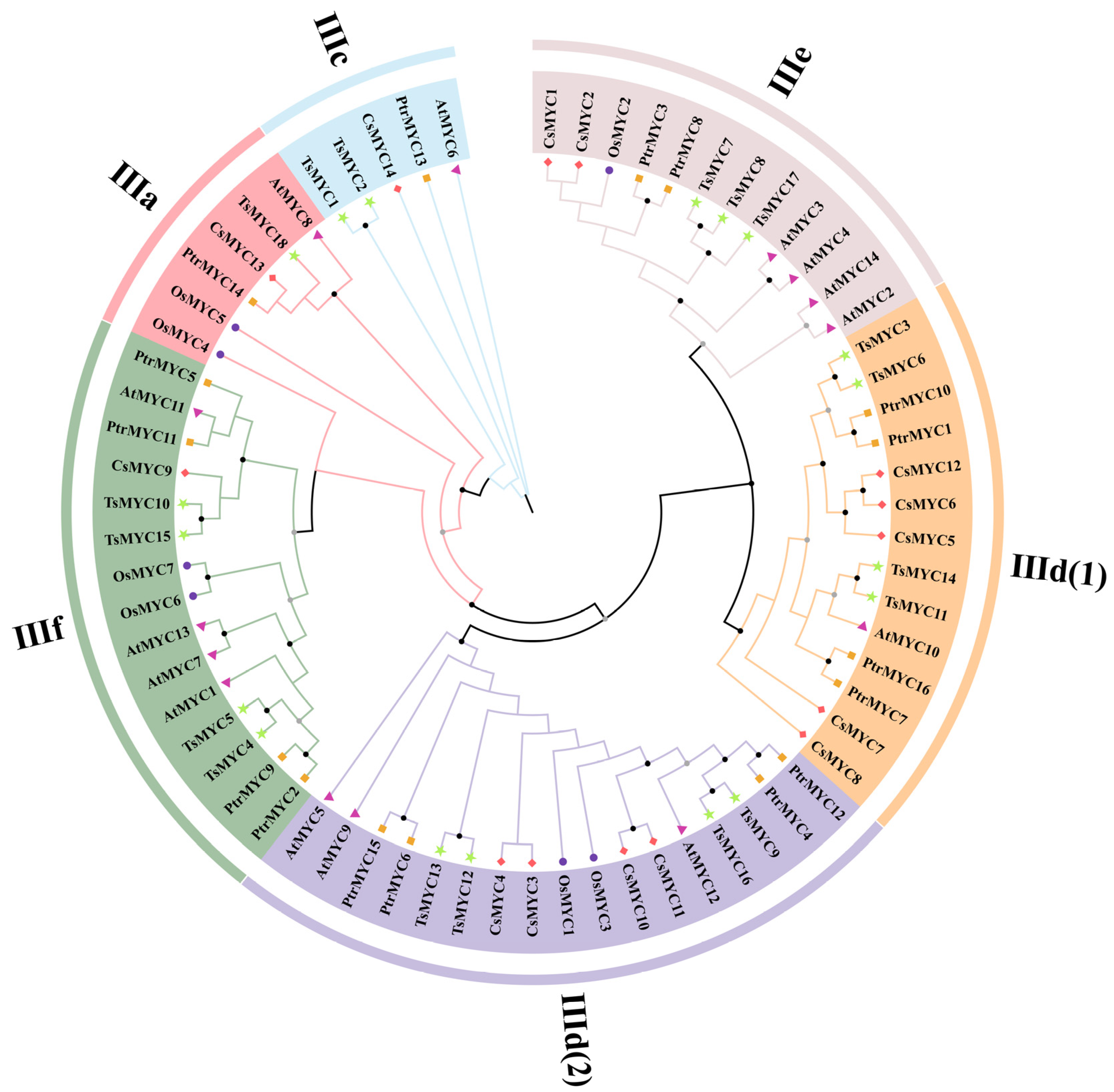
Figure 1. Phylogenetic tree of MYC proteins from T. sinensis, A. thaliana, O. sativa, P. trichocarpa, and C. sinensis. Black dots indicate bootstrap values greater than 90%, and gray dots indicate values greater than 70%. All T. sinensis MYC proteins were assigned to subgroups according to the classification criteria established for A. thaliana MYC proteins. Different colors represent different species: green stars for T. sinensis, pink triangles for A. thaliana, orange squares for O. sativa, red diamonds for P. trichocarpa, and purple circles for C. sinensis.
Forests 16 01756 g001
Figure 2. Chromosomal distribution of TsMYC genes. Changes in color on the chromosomes represent differences in gene density.
Forests 16 01756 g002
Figure 3. Synteny relationships of TsMYC genes in T. sinensis. The gray lines within the circle represent all syntenic blocks in the T. sinensis genome, while red lines indicate syntenic relationships between TsMYC genes. The heatmap inside the circle indicates gene density, and the outer blue boxes represent the chromosomes.
Forests 16 01756 g003
Figure 4. Synteny relationships between T. sinensis and two dicotyledonous species (Arabidopsis thaliana and Populus trichocarpa) and two monocotyledonous species (Oryza sativa and Zea mays). Chromosome numbers are displayed above each chromosome. Blue lines indicate syntenic relationships between the genomes of T. sinensis and A. thaliana, P. trichocarpa, O. sativa, and Z. mays.
Forests 16 01756 g004
Figure 5. Phylogenetic relationships, conserved motifs, domains, and gene structures of the 18 TsMYC proteins. (A) Phylogenetic tree of TsMYC proteins. Different colors represent different subclades of the TsMYC protein. (B) Conserved motifs in TsMYC proteins. Ten different colors represent ten unique motifs. (C) Conserved domains of TsMYC proteins. Green and yellow denote different conserved domains. (D) Exon–intron structures of TsMYC genes. Green indicates coding sequences (CDS) and yellow indicates untranslated regions (UTRs).
Forests 16 01756 g005
Figure 6. Cis-acting elements identified in the promoter regions of TsMYC genes are depicted, with different colors representing distinct types of cis-acting elements. The size of the circles indicates the number of each cis-acting element. The lengths of the colored bars correspond to the number of each cis-acting element.
Forests 16 01756 g006
Figure 7. Predicted protein–protein interaction networks of MYC proteins. (A) Protein–protein interaction network of the 18 TsMYC proteins. (B) Protein–protein interaction network of TsMYC6/7/8/14/17(AtMYC2).
Forests 16 01756 g007
Figure 8. qRT-PCR determination of relative expression of eight TsMYC genes in T. sinensis tissues. Data represent the means of three biological replicates ±SD. Relative gene expression was analyzed via the 2−ΔΔCT method, with the value in the Root set as 1.
Forests 16 01756 g008
Figure 9. qRT-PCR analysis of relative expression levels of eight TsMYC genes in T. sinensis leaves under MeJA Treatment. Asterisks indicating statistically significant differences (p < 0.05). The 0 h sample served as the untreated control, with its relative gene expression set to 1. Samples at 6 h, 12 h, and 24 h were compared against the baseline.
Forests 16 01756 g009
Figure 10. Relative expression levels of eight TsMYC genes in T. sinensis leaves under phytohormone treatments analyzed by qRT-PCR. Different colors represent different phytohormone: red for GA3, blue for ABA, orange for SA. Asterisks indicating statistically significant differences (p < 0.05). The 0 h sample served as the untreated control, with its relative gene expression set to 1. Samples at 6 h, 12 h, and 24 h were compared against the baseline.
Forests 16 01756 g010
Figure 11. qRT-PCR analysis of relative expression levels of eight TsMYC genes in T. sinensis leaves under NaCl Treatment. Asterisks indicating statistically significant differences (p < 0.05). The 0 h sample served as the untreated control, with its relative gene expression set to 1. Samples at 6 h, 12 h, and 24 h were compared against the baseline.
Forests 16 01756 g011
Figure 12. Subcellular localization of TsMYC17. The TsMYC17-GFP fusion protein expressed from the pNC-Cam1304-SubC-TsMYC17 represents the experimental group, while the GFP from the empty pNC-Cam1304-SubC vector serves as the control. The scale bar represents 50 μm and 10 μm. The green color represents GFP fluorescence, the red color represents the nucleus-specific marker fluorescence, and the yellow color represents the overlapping signal of the two fluorescences.
Forests 16 01756 g012
Disclaimer/Publisher’s Note: The statements, opinions and data contained in all publications are solely those of the individual author(s) and contributor(s) and not of MDPI and/or the editor(s). MDPI and/or the editor(s) disclaim responsibility for any injury to people or property resulting from any ideas, methods, instructions or products referred to in the content.