Article

Systematic Identification and Functional Study of Vitamin B6-Related PDX2 Genes in the Ginkgo biloba Genome

1
Key Laboratory of Forestry Biotechnology of Hunan Province, Central South University of Forestry and Technology, Changsha 410004, China
2
Yuelushan Laboratory Carbon Sinks Forests Variety Innovation Center, Central South University of Forestry and Technology, Changsha 410012, China
3
Huaihua Forestry Science Research Institute, Huaihua 418000, China
4
Administration Bureau of Swan Mountain National Forest Park, Zixing 423406, China
*
Authors to whom correspondence should be addressed.
These authors contributed equally to this work.
Forests 2025, 16(10), 1562; https://doi.org/10.3390/f16101562
Submission received: 15 September 2025 / Revised: 1 October 2025 / Accepted: 7 October 2025 / Published: 10 October 2025
(This article belongs to the Section Genetics and Molecular Biology)

Abstract

Vitamin B6 is an essential coenzyme involved in various metabolic processes critical for plant growth and development. However, its biosynthesis and regulatory mechanisms remain poorly understood in the ancient gymnosperm Ginkgo biloba. In this study, we identified two members of the PDX2 gene family (Gb_34755 and Gb_34990) through genome-wide analysis and characterized their molecular and functional properties. Bioinformatic analysis revealed distinct physicochemical traits and subcellular localizations, with Gb_34755 predicted in the cytoplasm and Gb_34990 in both chloroplasts and cytoplasm. Both proteins contain the glutaminase-related PLN02832 domain, indicating involvement in VB6 biosynthesis. Chromosomal mapping placed the genes in transcriptionally active regions on chromosomes 6 and 9. Phylogenetic analysis showed close evolutionary relationships between Ginkgo PDX2 genes and those in ferns and gymnosperms, distinct from angiosperms. Promoter analysis revealed differential enrichment of cis-elements: Gb_34990 harbored low-temperature and salicylic acid-responsive elements, while Gb_34755 showed motifs related to development. Gene expression profiling indicated significant upregulation (p < 0.05) of both genes during the late developmental stages of Ginkgo kernels, coinciding with peak VB6 content. Functional validation via transient overexpression in Nicotiana benthamiana confirmed a positive regulatory role, with VB6 levels increasing from 3.38 μg/g to 12.17 μg/g (p < 0.05). This study provides the first comprehensive functional analysis of the PDX2 gene family in Ginkgo and confirms their critical role in VB6 biosynthesis. These findings enhance our understanding of vitamin metabolism in gymnosperms and present promising targets for metabolic engineering in plants.

1. Introduction

Vitamin B6 (VB6) is an essential water-soluble micronutrient required by all living organisms [1]. In plants, its active form, pyridoxal 5′-phosphate (PLP) [2], functions as a ubiquitous enzymatic cofactor involved in amino acid metabolism [3], reactive oxygen species detoxification [4], and the synthesis of diverse secondary metabolites. VB6 levels strongly influence plant development, stress tolerance, and nutritional quality [4]. De novo biosynthesis of VB6 occurs through a DXP-independent pathway catalyzed by pyridoxal 5′-phosphate synthase (PDX1) and the glutaminase PDX2 [5,6,7], with PDX2 initiating the pathway by deaminating glutamine to generate ammonia, which is subsequently utilized by PDX1 to form PLP [7,8]. Although PDX genes have been characterized in several model angiosperms such as Arabidopsis thaliana and Oryza sativa [9,10,11], the functional roles of PDX2 remain poorly understood in evolutionarily ancient lineages.
G. biloba, the only extant member of Ginkgoaceae, represents a “living fossil” with more than 270 million years of evolutionary history [12]. Beyond its phylogenetic importance, ginkgo kernels are widely consumed as both medicine and food, in part due to their relatively high VB6 content, which has been associated with beneficial effects on cognition and antioxidant capacity [13,14,15,16,17]. Previous analyses have demonstrated that VB6 levels in ginkgo kernels surpass those in many common nuts and seeds, suggesting the presence of unique genetic or regulatory mechanisms [18,19,20]. However, the molecular regulation of VB6 biosynthesis in ginkgo remains largely unexplored, and no systematic investigation of PDX2 genes in gymnosperms has been reported. This gap limits our understanding of whether VB6 metabolism in ginkgo is conserved with angiosperms or follows distinct strategies.
Therefore, the present study was designed with three primary objectives: (i) to perform a genome-wide identification and characterization of PDX2 genes in G. biloba; (ii) to investigate their molecular properties, evolutionary relationships, and expression patterns during kernel development; and (iii) to validate their functional roles in VB6 biosynthesis through heterologous expression in N. benthamiana. Collectively, this work provides the first functional insights into PDX2 genes in ginkgo, enriches current understanding of vitamin metabolism in gymnosperms, and offers potential molecular targets for nutritional enhancement through metabolic engineering.

2. Materials and Methods

2.1. Plant Materials

Experimental samples were collected from the G. biloba cultivar ‘Foshou’ (Germplasm accession number: ZNL-2), cultivated in the botanical garden of Central South University of Forestry and Technology (China, geographic coordinates: 28.13° N, 112.98° E).
The N. benthamiana ecotype used in this study was N. benthamiana. Seeds were sown in potting soil and covered with plastic film to maintain appropriate moisture conditions. The soil is a loamy type consisting of approximately 50% sand, 30% silt, and 20% clay, with organic matter content of about 2.8%, pH 6.5. The site has not been exposed to any synthetic fertilizers or pesticides in the past five years and has a history of rotational planting, including legumes, vegetables, and medicinal herbs. Soil samples were collected at a depth of 0–20 cm, air-dried, and sieved before use in pots. A total of one plant per pot was grown for uniformity. Each pot had dimensions of 25 cm in height and 20 cm in diameter, with drainage holes at the bottom.
Plants were cultivated in a precisely controlled climate chamber under the following conditions: temperature of 22 °C/18 °C (day/night), with a photoperiod of 16 h light/8 h dark. N. benthamiana seedlings with 4–6 true leaves, approximately one month after sowing, were selected as experimental materials for transient transformation experiments.

2.2. Methods

2.2.1. PDX2 Gene Family Analysis

The PDX2 gene family in G. biloba was identified using the published genome data (https://ginkgo.zju.edu.cn/genome/, accessed on 25 December 2024). Candidate PDX2 genes were screened by BLAST searches against the Ginkgo genome using annotations. Open reading frames were predicted with NCBI ORF Finder, and protein sequences were analyzed for conserved domains using Pfam and HMMER. Only sequences containing the characteristic R domain were retained.
Multiple sequence alignments of Ginkgo and reference PDX2 proteins were performed with ClustalX [21]. Phylogenetic analysis was conducted in MEGA 12.0 using the Neighbor-Joining method with 1000 bootstrap replicates [22].
For promoter analysis, 2000 bp upstream regions of PDX2 genes were extracted and analyzed for cis-acting elements using PlantCARE [23], with visualization in TBtools. Physicochemical properties of PDX2 proteins were calculated using ExPASy ProtParam. Subcellular localization was predicted with PSORT. Conserved motifs were identified with MEME. Use NCBI to determine the conserved domains of proteins.

2.2.2. Construction of Overexpression Vector for the Target Gene

An overexpression vector was constructed via homologous recombination based on the target gene sequence [24]. Primers (Table 1) were designed with homologous arms matching the XhoI-linearized sites of the 3302-ruby-kan vector (a modified pCAMBIA1300-Ruby binary vector), as shown in Table 1. The ruby reporter gene and the target gene (Gb_34755) were each driven by independent CaMV 35S promoters, enabling separate expression within the same T-DNA region and allowing visual selection of positive transformants by red pigmentation.
The vector was linearized with XhoI, and the target gene fragment was amplified with homologous arms. Homologous recombination was performed using a commercial recombinase kit (Gene Master Pro Kit, item number BG0017, Baoguang Technology, Chongqing, China), following the manufacturer’s protocol. The ligation product was transformed into E. coli, and positive clones were screened by colony PCR (CT006487 American Bole) and confirmed by sequencing. Verified plasmids were extracted for subsequent Agrobacterium transformation.

2.2.3. Agrobacterium-Mediated Transient Overexpression in N. benthamiana

Competent Agrobacterium tumefaciens strain GV3101 cells were thawed on ice and transformed with 0.01–1 μg of recombinant plasmid DNA via the freeze–thaw method. After recovery in LB medium at 28 °C with shaking for 3–4 h, cells were plated on selective LB agar and incubated for 40–50 h. Positive colonies were cultured overnight in LB with kanamycin and rifampicin.
The overnight culture was subcultured into fresh LB supplemented with antibiotics, 20 mM acetosyringone, and 10 mM MES buffer, and incubated overnight. Cells were harvested by centrifugation, washed, and resuspended in infiltration buffer to an OD600 of ~1.0, then incubated in the dark for 5 h before infiltration.
The bacterial suspension (OD600 ≈ 0.6–1.0) was infiltrated into the abaxial side of N. benthamiana leaves using a 5 mL needleless syringe, with approximately 2–3 mL per infiltration site. Excess solution was removed, and infiltration sites were marked.
Post-infiltration, plants were kept in low-light or dark conditions for 1 day, followed by 2 days under normal light. Monitor the color of the leaves, and conduct testing once they turn red. Care was taken to avoid over-infiltration to prevent leaf damage.

2.2.4. Validation of Target Gene Overexpression

Total RNA was extracted from infiltrated N. benthamiana leaves using the E.Z.N.A. Plant RNA Kit (Omega Bio-Tek, Norcross, GA, USA), and integrity was checked by 1% agarose gel electrophoresis. First-strand cDNA was synthesized using a commercial reverse transcription kit (ExonScipt RT Mix (with dsDNase), item number BG0070, Baoguang Technology, China). Specific primers for GbPDX2 and the reference gene Actin (N. benthamiana Actin-7, NM_001425880.1) were designed with Primer Premier 5.0 (Table 1).
qRT-PCR was performed using 2× Universal Blue SYBR Green qPCR Master Mix. The amplification program was: 95 °C for 30 s; 40 cycles of 95 °C for 15 s and 60 °C for 30 s; followed by melting curve analysis. Relative expression was calculated using the 2−ΔΔCt method. Each sample included three biological and three technical replicates. Statistical analysis was performed using one-way ANOVA with Tukey’s test; p < 0.05 was considered significant.

2.2.5. Determination of VB6 Content in Transiently Transformed N. benthamiana Leaves

N. benthamiana leaf samples from different biological replicates were rapidly frozen in liquid nitrogen and ground into a uniform powder using a tissue grinder. Three biological replicates were set for each developmental stage. A 5 g aliquot of each powdered sample was accurately weighed and transferred into a 2 mL pre-chilled centrifuge tube, then stored at −80 °C for no longer than 72 h.
Total VB6 content was measured using the Solarbio VB6 Detection Kit (item number BC2114, Beijing Soleibao Technology Co., Ltd., Beijing, China), strictly following the manufacturer’s instructions. The response peak area of the reaction solution was determined using high-performance liquid chromatography, and the total VB6 content was calculated accordingly.
Samples (5 g, fresh) were extracted with 0.6 mL of extraction buffer at a material-to-liquid ratio of 1:6 (w/v). Fresh samples were homogenized before extraction. The mixture was sealed, incubated at 60 °C for 30 min in a water bath, then cooled to room temperature. Reagent I (0.1 mL) and distilled water (0.3 mL) were added, mixed, and left to stand for 2 min. The solution was centrifuged at 10,000 rpm for 10 min, the centrifuge used is the 5424R Germany and the supernatant was filtered through a water-based syringe filter into brown sample vials for analysis. If necessary, sample dilutions were performed to ensure clarity.
VB6 quantification was performed using HPLC (LC-15C Shimadzu Corporation, Kyoto, Japan) with fluorescence detection (Ex = 293 nm, Em = 395 nm) [25]. Chromatographic conditions included an injection volume of 10 µL, a flow rate of 1 mL/min, a column temperature of 30 °C, and a total run time of 10 min. The B6 standard was used to determine retention time (approximately 7.8 min) and establish peak identification. Peak areas from sample injections were measured to quantify VB6 content.

2.2.6. Data Analysis

Correlation heatmaps were generated using TBtools software and the online heatmap generation platform ChiPlot (https://www.chiplot.online/heatmap.html, accessed on 14 September 2025). Bar charts were created with GraphPad Prism10 software. Each sample comprised three biological replicates and three technical replicates. Statistical analysis employed one-way ANOVA followed by Tukey’s test; p < 0.05 was considered statistically significant [26].

3. Results

3.1. Identification of PDX2 Gene Family Members and Analysis of Protein Physicochemical Properties

This study combined genome-wide identification of PDX2 homologous genes in G. biloba with functional validation of Gb_34755 through heterologous overexpression and biochemical assays, HMM (Hidden Markov Model) profiling was employed to validate results from the NCBI Batch Conserved Domain Database (CDD) and Pfam. The analysis identified two PDX2 gene family members. Table 2 presents the physicochemical characteristics and predicted subcellular localization of the two PDX2 proteins, Gb_34755 and Gb_34990.
Physicochemical analysis revealed that the protein encoded by Gb_34755 consists of 254 amino acids, with a molecular weight of 27.66 kDa and a theoretical isoelectric point (pI) of 7.82, suggesting a slightly basic nature under physiological conditions. The instability index of this protein is 45.86, which exceeds the threshold of 40, indicating it may be an unstable protein. Its grand average of hydropathicity (GRAVY) score is –0.13, with a hydropathy index of 91.42, suggesting that this protein is generally hydrophilic and may function in aqueous cellular environments.
In contrast, Gb_34990 encodes a shorter protein of 213 amino acids, with a molecular weight of 22.60 kDa and a pI of 6.59, indicating a mildly acidic profile. The instability index of 35.62 classifies it as a stable protein. Its GRAVY score is slightly higher (0.006), yet still indicative of a predominantly hydrophilic protein.
Subcellular localization predictions suggest that Gb_34755 is localized in the cytoplasm, whereas Gb_34990 is predicted to localize in both the chloroplast and cytoplasm. This divergence indicates that members of the PDX2 gene family may participate in distinct metabolic pathways of VB6-related metabolic pathways through different subcellular compartments. For instance, the chloroplast-localized Gb_34990 may be directly involved in coenzyme synthesis related to photosynthesis, while the cytoplasmic Gb_34755 may play a role in primary metabolic processes.
In summary, the variation in physicochemical properties and subcellular localization among the PDX2 gene family members in G. biloba reflects their potential functional differentiation. These findings provide important clues for further exploration of the molecular mechanisms underlying their roles in different physiological contexts.

3.2. Conserved Motif and Gene Structure Analysis of the PDX2 Gene Family

In the genome-wide identification and functional analysis of the PDX2 gene family in G. biloba, the identification of conserved motifs and domains provides critical insights into the functional divergence and evolutionary relationships within this gene family (Figure 1). Sequence alignment of Gb_34755 and Gb_34990 revealed that both genes contain multiple conserved motifs (Figure 1A,C). Although the biological functions of some motifs remain to be experimentally validated, their conservation among PDX2 family members suggests that these regions are essential for protein function.
PDX2 proteins are known to function as part of the VB6 biosynthetic complex, typically interacting with PDX1 proteins to carry out PLP biosynthesis. Using the Conserved Domain Database (CDD) from NCBI, conserved domain prediction was conducted for PDX2 protein sequences. The results indicated that PDX2 proteins belong to the GAT-1 superfamily and are annotated as type I glutamine amidotransferases (Figure 1B). Specifically, the region spanning amino acids 1–244 of the PDX2 protein contains the PLN02832 domain (glutamine amidotransferase subunit of the pyridoxal 5′-phosphate synthase complex), which is a characteristic domain of the trimeric glutamine amidotransferase family and plays a crucial role in the biosynthesis pathway of VB6.
In this study, Gb_34755 and Gb_34990 exhibited similar distributions of conserved motifs, although sequence variations in certain motifs may reflect functional specialization. For instance, variations observed in some motifs of Gb_34990 may be associated with its subcellular localization in chloroplasts and its adaptation to photosynthesis-related metabolic pathways. The conservation and divergence of these motifs suggest that the PDX2 gene family has undergone functional differentiation during evolution. The differences in motif composition between Gb_34755 and Gb_34990 may be related to their regulatory roles in distinct subcellular environments, providing a basis for further investigation into their expression patterns across different tissues or under various stress conditions.
In summary, the analysis of conserved motifs not only supports the functional conservation of the PDX2 gene family but also lays a molecular foundation for elucidating the VB6-related metabolic pathways in G. biloba. Future studies may validate the functions of key motifs through site-directed mutagenesis or transgenic approaches.

3.3. Phylogenetic Analysis of the PDX2 Gene Family

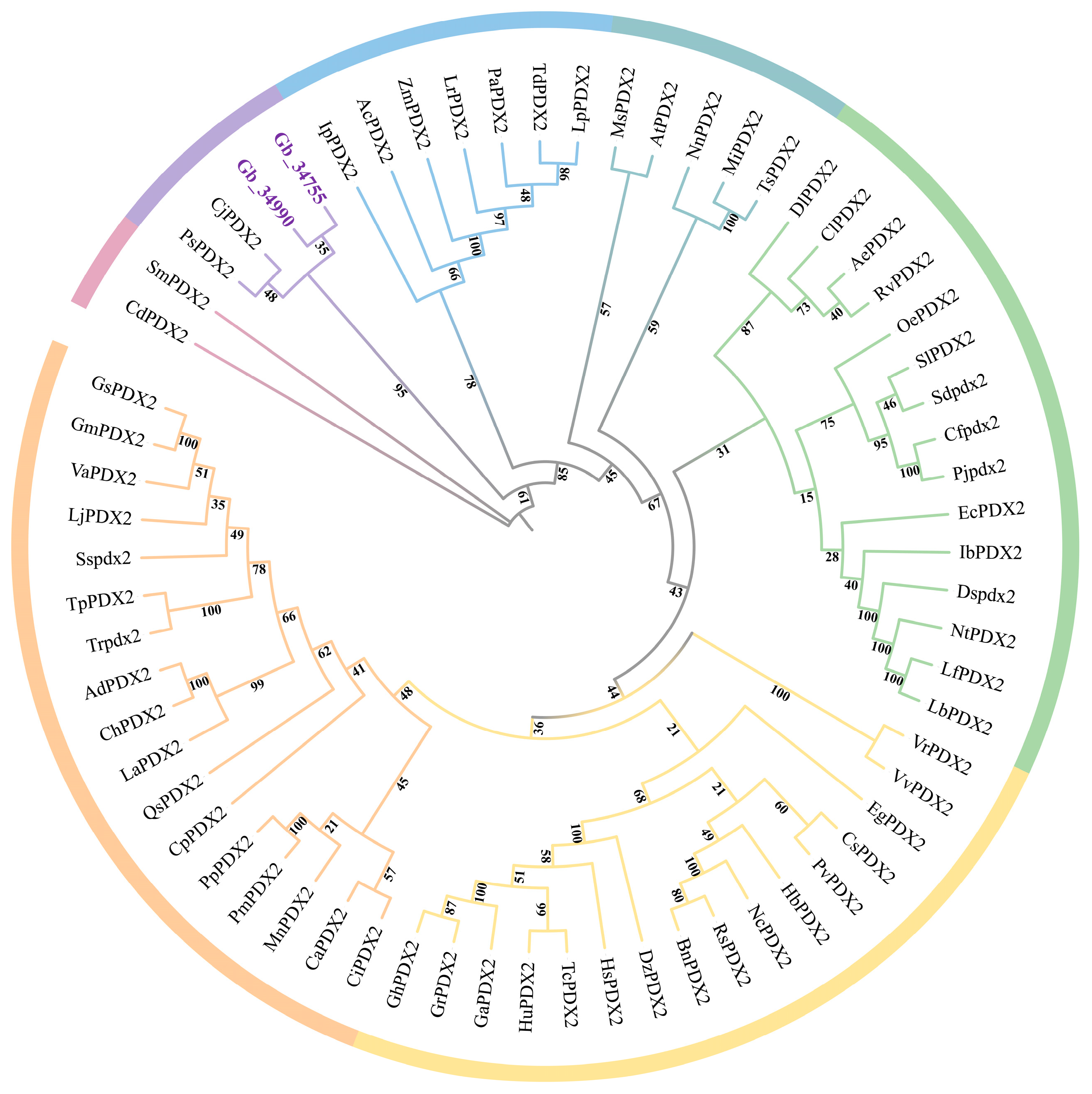
Phylogenetic tree analysis revealed that the PDX2 gene family in G. biloba (GbPDX2) comprises two members: Gb_34755 and Gb_34990 (Figure 2). Together with PDX2 genes from other species, these genes form several evolutionary clades, reflecting both the evolutionary conservation and functional divergence of the gene family. The clustering of Ginkgo PDX2 genes with those of Selaginella moellendorffii (SmPDX2) and Ceratopteris richardii (CdPDX2) indicates a high degree of conservation of PDX2 genes between Ginkgo and early land plants such as ferns. In addition, the grouping of GbPDX2 genes with PDX2 genes from gymnosperms, such as Picea sitchensis (PsPDX2) and Cryptomeria japonica (CjPDX2), suggests that Ginkgo, as one of the most ancient extant gymnosperms, possesses unique evolutionary features within the gymnosperm lineage. This pattern reflects adaptive modifications of the VB6–related metabolism in Ginkgo during evolution to meet distinct physiological requirements or environmental stresses.
Notably, the GbPDX2 genes of Ginkgo do not cluster directly with the PDX2 genes of most angiosperms, such as A. thaliana (AtPDX2), O. sativa (OsPDX2), or Zea mays (ZmPDX2), indicating that the PDX2 genes in Ginkgo diverged from those of angiosperms at an early stage of evolution. Angiosperm PDX2 genes mainly group into several independent clades (e.g., monocots and dicots), while GbPDX2 genes are more closely related to those of gymnosperms and ferns, which is consistent with the phylogenetic position of Ginkgo. This study provides important clues for understanding the evolutionary history of PDX2 genes in Ginkgo and their roles in VB6-related metabolic pathways. Future research involving gene expression and functional experiments will be essential to further verify their biological functions.

3.4. Analysis of Cis-Acting Elements in the Promoters of PDX2 Gene Family Members

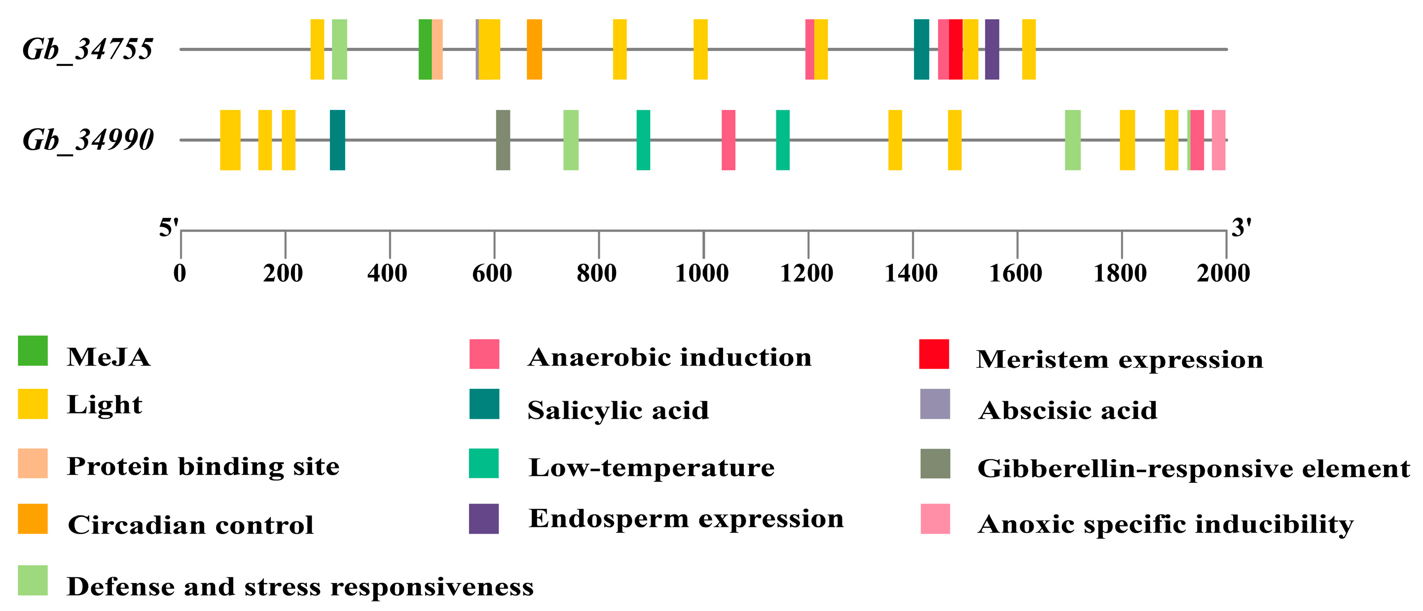
Promoters are regulatory regions of genes that contain various cis-acting elements capable of responding to environmental stimuli and modulating gene expression. Based on the analysis of cis-acting elements in the promoter regions of G. biloba PDX2 gene family members (Figure 3), we systematically identified the distribution patterns of multiple functional cis-elements in the promoters of two genes (Gb_34755 and Gb_34990). The results revealed that light-responsive elements are highly enriched in the promoter regions of both genes, suggesting that light signaling may play a significant role in regulating the expression of Ginkgo PDX2 genes.
In addition, elements associated with defense and stress responsiveness, as well as methyl jasmonate (MeJA) responsive elements, are widely present, indicating the potential involvement of these genes in plant stress response mechanisms. Notably, the promoter of Gb_34990 is enriched in low-temperature responsive elements and salicylic acid-responsive elements, implying that this gene may function in cold stress responses and immune defense signaling pathways.
In contrast, the promoter of Gb_34755 contains a higher abundance of meristem expression elements and endosperm expression elements, suggesting its specific regulatory role during plant growth and developmental stages. Furthermore, both promoters harbor multiple elements related to anaerobic induction and anoxic-specific inducibility, indicating that Ginkgo PDX2 genes may be activated under hypoxic stress conditions. The presence of abscisic acid (ABA)-responsive elements further supports the potential involvement of these genes in environmental adaptation and endogenous hormone-regulated pathways.
In summary, the enrichment of diverse cis-acting elements in the promoters of Ginkgo PDX2 gene family members suggests that their expression is regulated by a variety of internal and external environmental factors. These include light signals and hormonal responses, as well as multiple stress conditions, reflecting their important roles in Ginkgo’s adaptation to complex environments and in the regulation of growth and development. These findings provide a theoretical basis for subsequent functional validation and regulatory mechanism studies.

3.5. Function Annotation Display of the PDX2 Family Genes in G. biloba

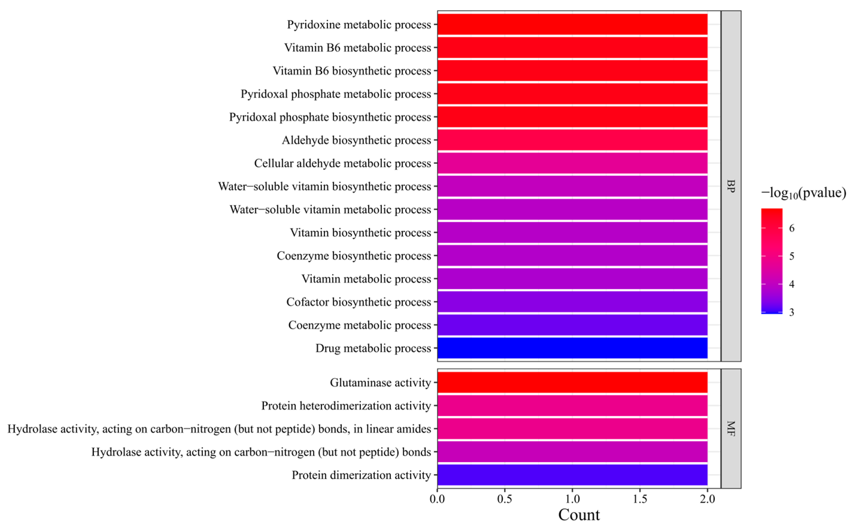
A comprehensive functional characterization of the G. biloba PDX2 gene family was conducted through systematic Gene Ontology (GO) function annotation display. According to the function annotation results, members of the PDX2 gene family exhibit distinct functional features at both the Biological Process and Molecular Function levels. The analysis showed that Ginkgo PDX2 genes are significantly enriched in pathways related to VB6 metabolism (Figure 4). Among these, the pyridoxine metabolic process and VB6 biosynthetic process displayed the highest enrichment levels (represented by red bars), indicating that this gene family plays a central role in the VB6-related metabolic pathways.
In addition, these genes are notably involved in the pyridoxal phosphate metabolic process, water-soluble vitamin biosynthetic process, and coenzyme biosynthetic process. Interestingly, the cellular aldehyde metabolic process also showed significant enrichment, suggesting that the PDX2 gene family may participate in the regulation of secondary metabolic pathways that are specific to Ginkgo.
At the Molecular Function level, the PDX2 gene family is primarily associated with the following activities: Glutaminase activity: directly related to the catalytic function of the VB6 synthase complex; Protein heterodimerization activity: forming the molecular basis for functional complexes with PDX1 proteins; Hydrolase activity acting on C–N bonds: involved in the hydrolysis of amide bonds during VB6-related metabolic pathways.
Of particular interest is the significant enrichment of protein dimerization activity, which aligns well with the known molecular mechanism by which PDX2 proteins function through homo- or heterodimer formation.
Comparative analysis revealed that the enrichment pattern of Ginkgo PDX2 genes in the VB6 synthesis pathway is conserved with that observed in angiosperms such as A. thaliana. However, the specific enrichment in aldehyde metabolic processes may reflect a unique adaptation of secondary metabolism in gymnosperms. Moreover, the pronounced enrichment of glutaminase activity (highlighted in red bars) supports a potential novel role of PDX2 genes in nitrogen metabolism in Ginkgo.

3.6. Expression Analysis of the PDX2 Gene Family

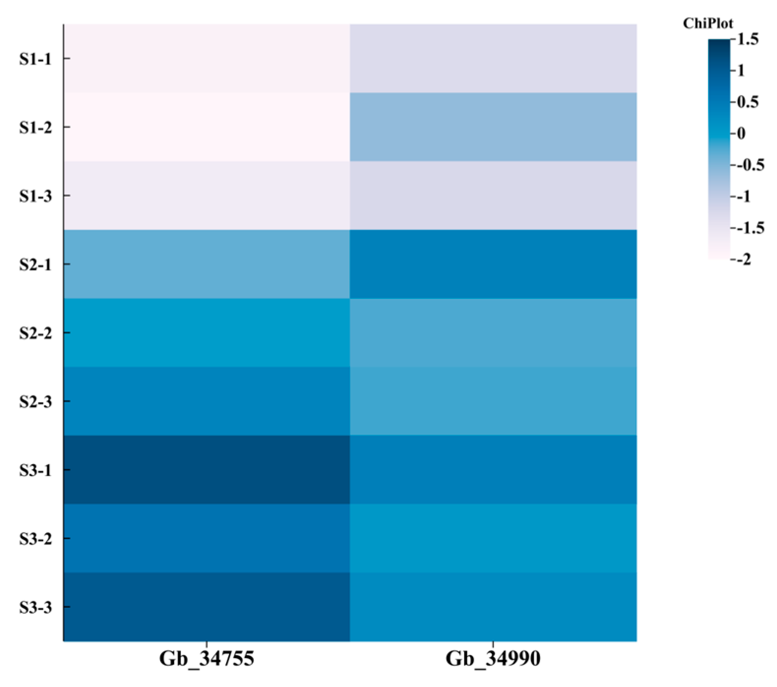
All kernel samples were collected from a single healthy and mature G. biloba tree (cultivar ‘Foshou’) at three different developmental stages (June, July, and August). At each stage, seeds were harvested from multiple fruits and pooled to form a biological sample representing that timepoint. Based on the heatmap analysis results (Figure 5), the G. biloba PDX2 gene family members (Gb_34755 and Gb_34990) exhibit distinct expression patterns across three developmental stages of the kernel (S1—early developmental stage (June), S2—mid-transition stage (July), and S3—late/mature stage (August)). Early developmental stage (June–S1): Both genes show the lowest expression levels across all biological replicates (S1-1 to S1-3). Mid-transition stage (July–S2): Expression levels approach the baseline. Maturation and accumulation stage (August–S3): Expression levels increase significantly, showing high expression in this phase.
A comparative analysis across the three stages revealed a monotonic upward trend in expression from S1 to S3, with Gb_34755 and Gb_34990 displaying highly synchronized expression patterns. This expression dynamic likely reflects the period of high VB6 accumulation in Ginkgo seed kernels during August, suggesting that PDX2 genes may be involved in vitamin synthesis during the maturation stage. The co-expression of these two genes implies potential functional complementarity within the metabolic pathway.
Using a threshold of |log2FC| ≥ 1 to identify candidate genes, Gb_34755 was ultimately selected as the key candidate gene.

3.7. Construction of the PDX2 Gene Overexpression Vector

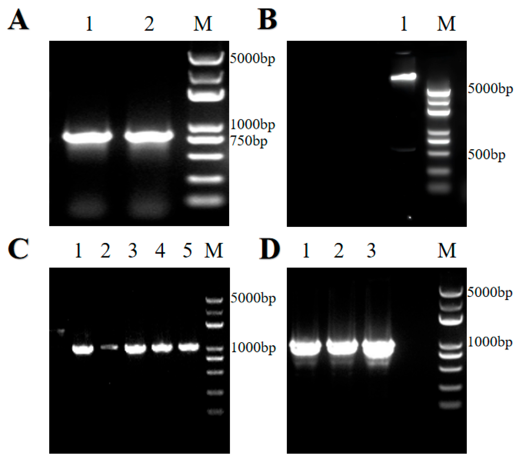
The length of the PCR amplified product was 805 bp, which corresponds to the sum of the target gene size obtained from genomic sequencing (765 bp) and the homologous arm sequences (40 bp) (Figure 6A). The vector was linearized by restriction enzyme digestion, producing a faint fragment of approximately 550 bp, indicating successful vector digestion (Figure 6B).
After positive clones were identified, plasmids were extracted from expanded cultures and sequenced. Agarose gel electrophoresis of the PCR product showed a final band length of 1063 bp, confirming the successful construction of the overexpression vector containing the target gene (Figure 6C).
The validated recombinant plasmid was then introduced into Agrobacterium tumefaciens. Single colonies were randomly selected from LB plates containing the appropriate antibiotics and cultured for 4–6 h. PCR was performed on Agrobacterium tumefaciens bacterial suspensions to identify positive clones. Gel electrophoresis revealed bands of the expected length, consistent with the size of the target gene, indicating successful transformation (Figure 6D).
The Agrobacterium cultures identified as positive by electrophoresis were further amplified and mixed with an appropriate amount of glycerol, then stored at ultra-low temperatures for subsequent genetic transformation experiments.

3.8. Transient Overexpression Functional Validation in N. benthamiana Leaves

In this study, a N. benthamiana transformation system based on the ruby reporter gene was successfully established (Figure 7), serving as a heterologous expression platform for functional studies of G. biloba PDX2 genes. Experimental results showed that wild-type control (WT) leaves exhibited the typical green phenotype (Figure 7A), while the overexpression transgenic lines (OE), due to stable transformation of the ruby gene (containing the PDX2 candidate gene), displayed a distinct red coloration in the leaves (Figure 7B).
This system confirmed the visual detectability of the ruby reporter gene in N. benthamiana and provides a reliable platform for subsequent functional validation of Ginkgo PDX2 genes.
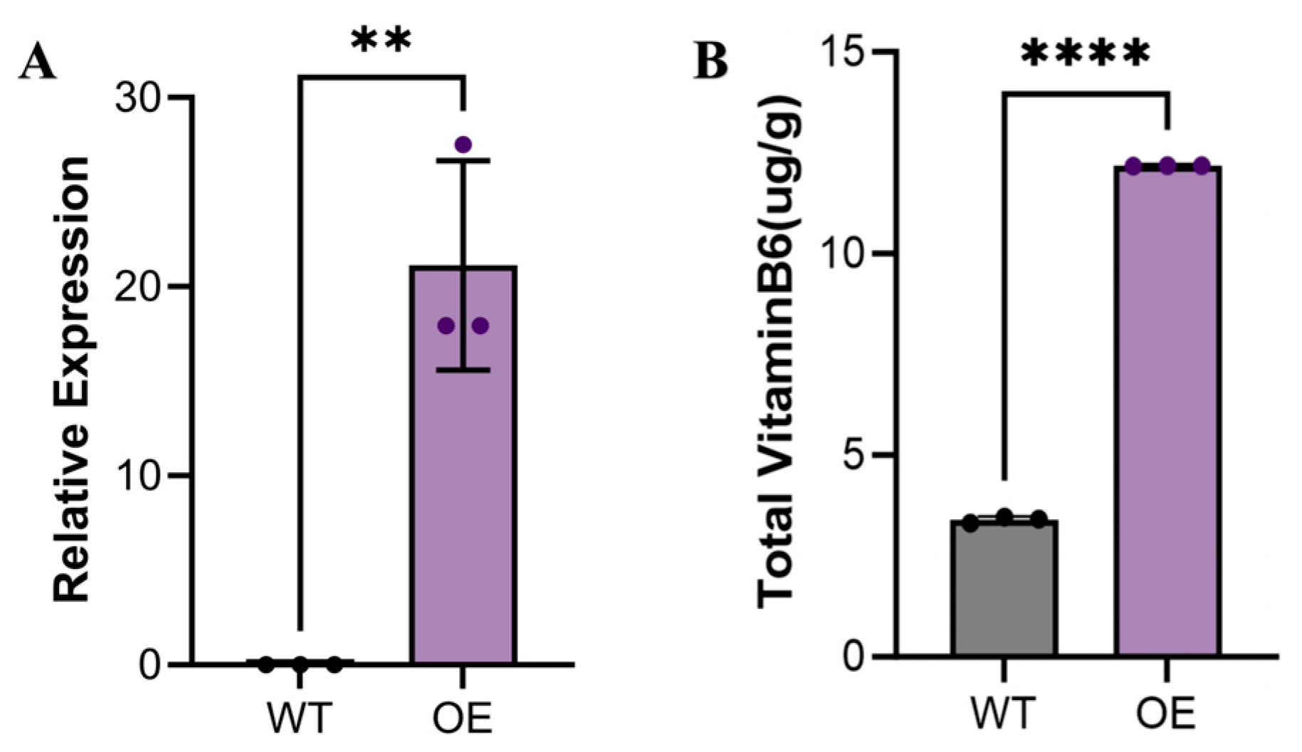
Using Agrobacterium-mediated transient transformation, we successfully achieved overexpression of the G. biloba PDX2 gene in N. benthamiana leaves. qRT-PCR analysis (Figure 8A) revealed that the mRNA expression level of the PDX2 gene in overexpression lines (OE) was significantly upregulated compared to the wild-type (WT) (p < 0.05), indicating that the exogenous gene was successfully integrated and efficiently expressed. This result validates the reliability of the experimental system and establishes a foundation for subsequent functional studies.
To investigate the biological function of the PDX2 gene, we measured the VB6 content in both WT and OE N. benthamiana leaves (Figure 8B). The results demonstrated that VB6 levels in the OE lines were significantly higher than those in the WT (p < 0.05). Specifically, the VB6 content in WT leaves was 3.38 ± 0.08 μg/g, while in OE lines it increased significantly to 12.172 ± 0.006 μg/g—representing an approximately four-fold increase (p < 0.05). This substantial enhancement strongly supports the positive regulatory role of the Ginkgo PDX2 gene in VB6-related metabolic pathways.
This finding is consistent with the known function of PDX2 genes in the VB6 metabolic pathway and further underscores their critical role in coenzyme biosynthesis and metabolic regulation in plants. All data are presented as the mean ± standard deviation (SD) of three independent biological replicates, and statistical significance was confirmed (p < 0.05), ensuring the reliability and reproducibility of the results.
The increased VB6 content in OE lines not only confirms the functional role of the PDX2 gene but also highlights its potential application in enhancing VB6 levels in plants. Through this transient overexpression system, we preliminarily elucidated the role of the G. biloba PDX2 gene in VB6-related metabolic pathways and demonstrated its capacity to significantly enhance VB6 accumulation in N. benthamiana. These findings provide important evidence for further elucidating the molecular mechanisms and application potential of Ginkgo PDX2 genes.

4. Discussion

This study presents the first systematic identification and functional analysis of PDX2 genes in G. biloba, an evolutionarily ancient gymnosperm species. Two homologs, Gb_34755 and Gb_34990, were identified, both containing the conserved glutaminase domain (PLN02832), and their expression patterns were shown to differ across developmental stages of kernels, suggesting functional divergence under spatial and temporal regulation. Notably, transient overexpression of these genes in N. benthamiana significantly enhanced VB6 (pyridoxal 5′-phosphate, PLP) content, providing supporting evidence for their active role in VB6 biosynthesis. These results extend current understanding of plant vitamin metabolism, which has so far primarily focused on angiosperms such as Arabidopsis thaliana and Oryza sativa [9,27], by introducing gymnosperms into the comparative framework.
Mechanistically, the functional divergence of the two PDX2 homologs can be linked to differences in predicted subcellular localization and cis-regulatory architectures. Gb_34755 is localized to the cytoplasm, whereas Gb_34990 is predicted to be dual-localized in both the cytoplasm and the chloroplast. The presence of chloroplast localization is unusual for PDX2 proteins, which are predominantly cytosolic in angiosperms [6], and may reflect evolutionary adaptations in Ginkgo that compartmentalize VB6 biosynthesis for more efficient metabolic flux or plastid-specific functions. This is consistent with studies showing that the subcellular localization of vitamin biosynthetic enzymes can impact metabolic regulation and cofactor availability [28].
Promoter analysis revealed that Gb_34990 is enriched with stress- and hormone-responsive elements, including those responsive to salicylic acid, abscisic acid, and low temperature, indicating possible induction under abiotic stress. In contrast, Gb_34755 harbors more elements associated with growth and developmental processes, particularly auxin and gibberellin responsiveness, suggesting a role in basal metabolic function during seed development. This partitioning supports a model of subfunctionalization following gene duplication, where duplicated genes acquire distinct regulatory contexts to diversify function [29]. Similar differential regulation and specialization of PDX family members have also been reported in rice and tomato under abiotic stress [5]. Moreover, the comparatively high protein instability index predicted for Gb_34755 may suggest transient or fine-tuned expression during specific stages, whereas Gb_34990 appears to maintain more stable expression over developmental stages, reflected by its expression peak in late kernel development.
Phylogenetic analysis placed the Ginkgo PDX2 genes closer to those in ferns and gymnosperms than to angiosperm counterparts, which aligns with Ginkgo’s long-diverged evolutionary history [30]. The evolutionary conservation of the GAT-1 domain through different plant lineages suggests that this core enzymatic function has been maintained since the early land plant ancestors. However, the regulatory divergence observed in our study may represent an early evolutionary shift in functional specialization within gymnosperms [31], potentially shaped by adaptation to unique photoperiods, longevity, and stress resistance characteristics of woody perennials [32].
While the current study provides functional insights, several limitations remain. Firstly, subcellular localization was predicted computationally and not validated experimentally; future work involving fluorescent tagging, yeast two-hybrid assays, or co-immunoprecipitation is warranted. Secondly, although transient expression in N. benthamiana offers a rapid tool for initial functional assessment, it does not fully recapitulate Ginkgo’s native physiological context. Differences in metabolic pathways and expression networks between the two species may affect the interpretation of heterologous overexpression results. Finally, detailed analysis of VB6 biosynthesis flux, expression under different stress conditions, and gene regulation in diverse tissues will be necessary to further delineate the roles of PDX2 genes in vivo.
In conclusion, this research expands the current understanding of vitamin B6 metabolism by demonstrating for the first time the expression, localization, and functional characteristics of PDX2 homologs in a gymnosperm species. The novel insights into subcellular compartmentalization, promoter responsiveness, and evolutionary divergence underscore the biological complexity and specialization of VB6 biosynthetic regulation in ancient plant lineages. These findings pave the way for future research into metabolic engineering of VB6 pathways in woody plants and may contribute to the development of micronutrient-fortified crops with enhanced stress tolerance and health-promoting properties.

5. Conclusions

This study provides the first comprehensive characterization of the PDX2 gene family in G. biloba, revealing their potential roles in vitamin B6 (VB6) biosynthesis. Two members, Gb_34755 and Gb_34990, were identified and shown to contain conserved glutaminase domains characteristic of key enzymes in VB6 metabolism. Their predicted differential subcellular localization suggests functional divergence between developmental and stress-responsive pathways. Expression profiling demonstrated strong upregulation of both genes during late seed development, coinciding with VB6 accumulation, and heterologous overexpression in N. benthamiana confirmed that Gb_34755 enhances VB6 content. Promoter analyses revealed distinct cis-regulatory elements, indicating specialized regulatory roles in growth and environmental adaptation.
While this work expands our understanding of VB6 metabolic pathways in gymnosperms, several limitations should be acknowledged. Functional interpretations were partly based on computational predictions that require in planta validation, and the lack of stable genetic transformation systems in ginkgo currently restricts direct functional testing. Future efforts should therefore focus on developing transformation techniques for gymnosperms, performing tissue- and organ-specific expression analyses, and dissecting upstream transcriptional regulation and protein–protein interactions within the PDX complex. The application of advanced tools such as CRISPR/Cas genome editing and multi-omics integration will further enable precise functional dissection and metabolic engineering of VB6 pathways.
Taken together, this study not only provides the first insights into PDX2 gene function in an ancient gymnosperm but also highlights the novelty and evolutionary significance of ginkgo as a model for studying metabolic adaptations. The resources and knowledge generated here offer a foundation for future functional genomics investigations and provide promising targets for improving nutritional quality and stress resilience in woody crops through metabolic engineering.

Author Contributions

Conceptualization, H.J., Y.X., C.Y., Z.F., J.L., S.Z., M.L. and Y.W.; methodology, H.J., Y.X., C.Y., Z.F., Z.Y., M.L. and Y.W.; software, H.J. and Y.X.; validation, H.J. and Y.X.; formal analysis, H.J. and Y.X.; investigation, H.J., Y.X., C.Y., J.L., S.Z., M.L. and Y.W.; writing—original draft preparation, H.J., Y.X. and Z.F.; writing—review and editing, H.J., Y.X., Z.F., M.L. and Y.W.; supervision, M.L. and Y.W.; project administration, Y.W.; funding acquisition, Y.W. All authors have read and agreed to the published version of the manuscript.

Funding

This research was funded by the Natural Science Foundation of China (32171842), Innovation Project of Central South University of Forestry and Technology (90102-63223068), and the National Key Research and Development Project of China (2019YFD1100403).

Data Availability Statement

The original contributions presented in this study are included in the article. Further inquiries can be directed to the corresponding author.

Conflicts of Interest

The authors declare no conflicts of interest.

References

  1. Gorelova, V.; Colinas, M.; Dell’aGlio, E.; Flis, P.; Salt, D.E.; Fitzpatrick, T.B. Phosphorylated B6 vitamer deficiency in SALT OVERLY SENSITIVE 4 mutants compromises shoot and root development. Plant Physiol. 2021, 188, 220–240. [Google Scholar] [CrossRef]
  2. Parra, M.; Stahl, S.; Hellmann, H. Vitamin B6 and Its Role in Cell Metabolism and Physiology. Cells 2018, 7, 84. [Google Scholar] [CrossRef]
  3. Mooney, S.; Leuendorf, J.-E.; Hendrickson, C.; Hellmann, H. Vitamin B6: A Long Known Compound of Surprising Complexity. Mol. Plant 2009, 2, 329–351. [Google Scholar] [CrossRef]
  4. Vanderschuren, H.; Boycheva, S.; Li, K.-T.; Szydlowski, N.; Gruissem, W.; Fitzpatrick, T.B. Strategies for Vitamin B6 biofortification of plants: A dual role as a micronutrient and a stress protectant. Front. Plant Sci. 2013, 4, 143. [Google Scholar] [CrossRef]
  5. Fitzpatrick, T.B.; Amrhein, N.; Kappes, B.; Macheroux, P.; Tews, I.; Raschle, T. Two independent routes of de novo Vitamin B6 biosynthesis: Not that different after all. Biochem. J. 2007, 407, 1–13. [Google Scholar] [CrossRef] [PubMed]
  6. Tambasco-Studart, M.; Titiz, O.; Raschle, T.; Forster, G.; Amrhein, N.; Fitzpatrick, T.B. Vitamin B6 biosynthesis in higher plants. Proc. Natl. Acad. Sci. USA 2005, 102, 13687–13692. [Google Scholar] [CrossRef]
  7. Raschle, T.; Amrhein, N.; Fitzpatrick, T.B. On the two components of pyridoxal 5′-phosphate synthase from Bacillus subtilis. J. Biol. Chem. 2005, 280, 32291–32300. [Google Scholar] [CrossRef] [PubMed]
  8. Dong, Y.; Sueda, S.; Nikawa, J.; Kondo, H. Characterization of the products of the genes SNO1 and SNZ1 involved in pyridoxine synthesis in Saccharomyces cerevisiae. Eur. J. Biochem. 2004, 271, 745–752. [Google Scholar] [CrossRef]
  9. Wagner, S.; Bernhardt, A.; Leuendorf, J.E.; Drewke, C.; Lytovchenko, A.; Mujahed, N.; Gurgui, C.; Frommer, W.B.; Leistner, E.; Fernie, A.R.; et al. Analysis of the Arabidopsis rsr4-1/PDX1-3 mutant reveals the critical function of the PDX1 protein family in metabolism, development, and Vitamin B6 biosynthesis. Plant Cell 2006, 18, 1722–1735. [Google Scholar] [CrossRef]
  10. Mangel, N.; Fudge, J.B.; Li, K.T.; Wu, T.Y.; Tohge, T.; Fernie, A.R.; Szurek, B.; Fitzpatrick, T.B.; Gruissem, W.; Vanderschuren, H. Enhancement of Vitamin B6 levels in rice expressing Arabidopsis Vitamin B6 biosynthesis de novo genes. Plant J. 2019, 17, 1047–1065. [Google Scholar] [CrossRef] [PubMed]
  11. Yao, M.; Hong, B.; Ji, H.; Guan, C.; Guan, M. Genome-wide identification of PDX and expression analysis under waterlogging stress exhibit stronger waterlogging tolerance in transgenic Brassica napus plants overexpressing the BnaPDX1.3 gene compared to wild-type plants. Front. Plant Sci. 2025, 16, 1533219. [Google Scholar] [CrossRef] [PubMed]
  12. Singh, B.; Kaur, P.; Singh, R.D.; Ahuja, P.S. Biology and chemistry of Ginkgo biloba. Fitoterapia 2008, 69, 401–418. [Google Scholar] [CrossRef]
  13. Kisvarga, S.; Hamar-Farkas, D.; Horotán, K.; Gyuricza, C.; Ražná, K.; Kučka, M.; Harenčár, Ľ.; Neményi, A.; Lantos, C.; Pauk, J.; et al. Investigation of a Perspective Urban Tree Species, Ginkgo biloba L., by Scientific Analysis of Historical Old Specimens. Plants 2024, 13, 1470. [Google Scholar] [CrossRef] [PubMed]
  14. Chen, Y.; Fu, C.; Wu, Z.; Xu, H.; Liu, H.; Schneider, H.; Lin, J. Ginkgo biloba. Trends Genet. 2021, 37, 488–489. [Google Scholar] [CrossRef]
  15. Boateng, I.D.; Yang, X.-M. Ginkgo biloba L. seed; A comprehensive review of bioactives, toxicants, and processing effects. Ind. Crops Prod. 2022, 176, 114281. [Google Scholar] [CrossRef]
  16. Šamec, D.; Karalija, E.; Dahija, S.; Hassan, S.T.S. Biflavonoids: Important Contributions to the Health Benefits of Ginkgo (Ginkgo biloba L.). Plants 2022, 11, 1381. [Google Scholar] [CrossRef]
  17. Liu, W.; Zou, M.; Wang, Y.; Cao, F.; Su, E. Ginkgo seed proteins: Characteristics, functional properties and bioactivities. Plant Foods Hum. Nutr. 2021, 76, 281–291. [Google Scholar] [CrossRef]
  18. Sado, T.; Nakata, S.; Tsuno, T.; Sato, M.; Misawa, Y.; Yamauchi, S.; Inaba, Y.; Kobayashi, D.; Wada, K. Concentrations of various forms of Vitamin B6 in ginkgo seed poisoning. Brain Dev. 2019, 41, 292–295. [Google Scholar] [CrossRef]
  19. Malouf, R.; Grimley Evans, J. Vitamin B6 for cognition. Cochrane Database Syst. Rev. 2003, 4, CD004393. [Google Scholar] [CrossRef]
  20. Stach, K.; Stach, W.; Augoff, K. Vitamin B6 in health and disease. Nutrients 2021, 13, 3229. [Google Scholar] [CrossRef] [PubMed]
  21. Chatzou, M.; Magis, C.; Chang, J.-M.; Kemena, C.; Bussotti, G.; Erb, I.; Notredame, C. Multiple sequence alignment modeling: Methods and applications. Brief. Bioinform. 2016, 17, 1009–1023. [Google Scholar] [CrossRef] [PubMed]
  22. Huelsenbeck, J.P.; Bull, J.J.; Cunningham, C.W. Combining data in phylogenetic analysis. Trends in Ecol. Evol. 1996, 11, 152–158. [Google Scholar] [CrossRef] [PubMed]
  23. Hernandez-Garcia, C.M.; Finer, J.J. Identification and validation of promoters and cis-acting regulatory elements. Plant Sci. 2014, 217, 109–119. [Google Scholar] [CrossRef]
  24. Krejci, L.; Altmannova, V.; Spirek, M.; Zhao, X. Homologous recombination and its regulation. Nucleic Acids Res. 2012, 40, 5795–5818. [Google Scholar] [CrossRef]
  25. Bisp, M.R.; Bor, M.V.; Heinsvig, E.M.; Kall, M.A.; Nexø, E. Determination of vitamin B6 vitamers and pyridoxic acid in plasma: Development and evaluation of a high-performance liquid chromatographic assay. Anal. Biochem. 2002, 305, 82–89. [Google Scholar] [CrossRef]
  26. Mishra, P.; Pandey, C.M.; Singh, U.; Keshri, A.; Sabaretnam, M. Selection of appropriate statistical methods for data analysis. Ann. Card. Anaesth. 2019, 22, 297–301. [Google Scholar] [CrossRef] [PubMed]
  27. Tambasco-Studart, M.; Tews, I.; Amrhein, N.; Fitzpatrick, T.B. Functional analysis of PDX2 from Arabidopsis, a glutaminase involved in Vitamin B6 biosynthesis. Plant Physiol. 2007, 144, 915–925. [Google Scholar] [CrossRef]
  28. Hanson, A.D.; Beaudoin, G.A.; McCarty, D.R.; Gregory, J.F. Does abiotic stress cause functional B vitamin deficiencies in plants? Plant Physiol. 2016, 172, 2082–2097. [Google Scholar] [CrossRef]
  29. Wang, H.; Zhang, J.; Yao, S.; Cheng, X.; Ji, K.; Yu, Q. Genome-Wide Identification and Expression Analysis of Members in the YT521-B Homology Domain-Containing RNA Binding Protein Family in Ginkgo biloba. Plants 2024, 13, 3589. [Google Scholar] [CrossRef]
  30. Zhao, Y.-P.; Fan, G.; Yin, P.-P.; Sun, S.; Li, N.; Hong, X.; Hu, G.; Zhang, H.; Zhang, F.-M.; Han, J.-D.; et al. Resequencing 545 ginkgo genomes across the world reveals the evolutionary history of the living fossil. Nat. Commun. 2019, 10, 4201. [Google Scholar] [CrossRef]
  31. Hu, Y.; Yu, Z.; Gao, X.; Liu, G.; Zhang, Y.; Šmarda, P.; Guo, Q. Genetic diversity, population structure, and genome-wide association analysis of ginkgo cultivars. Hortic. Res. 2023, 10, uhad136. [Google Scholar] [CrossRef] [PubMed]
  32. Tian, Y.; Han, X.; Qu, Y.; Zhang, Y.; Rong, H.; Wu, K.; Xu, L. Genome-wide identification of the ginkgo (Ginkgo biloba L.) LBD transcription factor gene and characterization of its expression. Int. J. Mol. Sci. 2022, 23, 5474. [Google Scholar] [CrossRef] [PubMed]
Figure 1. Analysis of the conserved motifs and predicted functional domains in the PDX2 gene family proteins of G. biloba. (A) Distribution of conserved protein motifs identified from amino acid sequences using MEME. (B) Predicted functional domains (SNO-glutamine amidotransferase family) from protein sequences using SMART. (C) Logo representation of conserved motifs based on amino acid sequences (Different colors in Motif 1–5 represent different motif sequences).
Forests 16 01562 g001
Figure 2. Phylogenetic analysis of PDX2 protein sequences from G. biloba and other plant species. The analysis was conducted using the Neighbor-Joining (NJ) method with 1000 bootstrap replications. The two G. biloba PDX2 sequences analyzed in this study are shown in purple and correspond to Gb_34755 and Gb_34990. Sequences from other species were obtained from NCBI databases, and their gene names are represented by species-specific abbreviations (e.g., AtPDX2 for Arabidopsis thaliana, OsPDX2 for Oryza sativa).
Forests 16 01562 g002
Figure 3. Cis-acting element analysis of the 2000 bp promoter regions upstream of Gb_34755 and Gb_34990 in G. biloba. Colored boxes represent specific categories of cis-elements related to hormone responsiveness (e.g., abscisic acid, gibberellin, salicylic acid), stress signals (e.g., low temperature, anoxia, anaerobic induction), light responsiveness, and tissue-specific expression (e.g., meristem, endosperm).
Forests 16 01562 g003
Figure 4. Function Annotation Display of the PDX2 family genes in G. biloba. The x-axis shows the number of genes involved (Count), and the color gradient represents statistical significance (−log10p-value). BP (Biological Process)—processes and pathways in which the genes are involved (e.g., VB6 biosynthetic process, metabolic process); MF (Molecular Function)—the biochemical activities of gene products (e.g., glutaminase activity, hydrolase activity).
Forests 16 01562 g004
Figure 5. Expression analysis of Gb_34755 and Gb_34990 in G. biloba kernels at three developmental stages. Gene expression values were normalized, and heatmap intensity corresponds to relative expression. The stages are defined as follows: S1—early developmental stage (June), S2—mid-transition stage (July), S3—late/mature stage (August). Each stage includes three independent biological replicates (S1-1 to S1-3). Use one-way analysis of variance (ANOVA) to compare whether the differences among the three periods (S1, S2, S3) are significant. (Gb_34755: p ≈ 1.13 × 10⁻⁵, Gb_34990: p ≈ 0.0012).
Forests 16 01562 g005
Figure 6. Verification of the construction of the Gb_34755 overexpression vector and transformation validation. (A) PCR amplification of the target gene showing a product of ~805 bp in Lane 1 and 2. (B) Enzyme digestion of the vector plasmid. Lane 1 shows two expected bands: >5000 bp (vector backbone, 3302-ruby-kan) and ~550 bp (small fragment). (C) Colony PCR of E. coli transformants: Lanes 1–5 show successful amplification of the target gene (~1063 bp), indicating the presence of the insert. (D) PCR verification of recombinant plasmid in Agrobacterium tumefaciens: Lanes 1–3 display clear bands of ~1063 bp. M: DNA marker (DL5000, TaKaRa Biotechnology), with band sizes indicated on the side (500 bp–5000 bp).
Forests 16 01562 g006
Figure 7. Phenotypic comparison of N. benthamiana leaves under transient expression. (A) Wild-type (non-transformed, labeled as WT) N. benthamiana plants show fully green leaves. (B) Leaves of plants overexpressing the Gb_34755 gene (labeled as OE) exhibit significant purple-red pigmentation. WT = wild-type; OE = overexpression. Transient transformation was performed using Agrobacterium-mediated infiltration.
Forests 16 01562 g007
Figure 8. Verification of overexpression in the transient transformation of N. benthamiana leaves. (A) qRT-PCR analysis of the expression level of the PDX2 gene in wild-type and overexpression (B) Analysis of VB6 content in wild-type and overexpression. (Bars are means ± SD of three independent experiments, with indicating a statistically significant difference; p < 0.05). ** indicate p ≤ 0.01, while **** indicate p ≤ 0.0001.
Forests 16 01562 g008
Table 1. Primer sequence for PCR amplification.
Primer NamePrimer Sequence (5′→3′)
Gb_34755-FTCTCTACAAATCTATCTCTCATGTCTGTAGGAGTTTTGGCC
Gb_34755-RACACATTATTATGGAGAAACTCATGGCTCAAAACTTAAGTTTCT
Gb_34755-FTCTCTACAAATCTATCTCTCATGTCTGTAGGAGTTTTGGCC
LB-TDNA-RTGGCAGGATATATTGTGGTGTAAAC
NtActin-FTTTCCTAGCATTGTGGGTCG
NtActin-RCCCCTCTTGGATTGAGCTTC
q Gb_34755-FTGAAGTAAGGAAACCCGAGCAA
q Gb_34755-RTTGGACTGTACAATCAAGCCCTC
Table 2. Physicochemical properties and subcellular localization of PDX2 genes.
Sequence IDAmino Acid NumberMolecular Weight (Da)Theoretical pIInstability IndexHydropathy IndexAverage
Hydropathicity (GRAVY)
Subcellular
Localization
Gb_3475525427,664.77.8245.8691.42−0.13Cytoplasm
Gb_3499021322,595.96.5935.6289.390.006Chloroplast, Cytoplasm
Disclaimer/Publisher’s Note: The statements, opinions and data contained in all publications are solely those of the individual author(s) and contributor(s) and not of MDPI and/or the editor(s). MDPI and/or the editor(s) disclaim responsibility for any injury to people or property resulting from any ideas, methods, instructions or products referred to in the content.