Article

Synergistic Ability of Chitosan and Trichoderma harzianum to Control the Growth and Discolouration of Common Sapstain Fungi of Pinus radiata

Wood Science Design Scion, 49 Sala Street, Rotorua 3010, New Zealand
*
Author to whom correspondence should be addressed.
Forests 2021, 12(5), 542; https://doi.org/10.3390/f12050542
Submission received: 8 April 2021 / Revised: 22 April 2021 / Accepted: 24 April 2021 / Published: 27 April 2021
(This article belongs to the Special Issue Designed Wood Products for Aesthetic and Built Environment)

Abstract

:
An environmentally compatible method for controlling sapstain fungi in wood was evaluated, using a combination of chitosan and an albino strain of Trichoderma harzianum, a biological control agent (BCA). The growth and penetration into the wood of the sapstain fungi Ophiostoma piceae, Leptographium procerum, and Sphaeropsis sapinea were assessed in radiata pine wafers treated with chitosan and BCA, both alone and in combination. Several mycological and microscopic techniques were used, including a gfp (green fluorescent protein) transformed strain of O. piceae for assessing the depth of penetration in the wood samples. The synergy between the chitosan and BCA was evident, and for two tested fungi, only the combination of chitosan and BCA afforded protection. The synnemata (recognized by erect conidiogenous cells bearing conidia) was observed on the surface of the wafers inoculated with L. procerum and O. piceae, but the hyphae were unable to penetrate and melanise. The results suggest that the limited ability of chitosan to penetrate deeply into the wood was compensated by the fast growth of T. harzianum in the inner wood.

1. Introduction

Radiata pine (Pinus radiata, D. Don) is the most important plantation forest species in New Zealand and many other countries. After harvest, logs of radiata become susceptible to invasion by a variety of microorganisms including wood discolouring fungi [1,2]. The discolouration caused by sapstain fungi significantly reduces the grade and monetary value of logs and lumber [3]. The discolouring fungi or sapstaining fungi do not affect the strength properties of the wood, but cause a blueish or greyish discolouration. Sapstain (or blue stain) fungi are characterised as a particular group of fungi that usually infect the sapwood of freshly felled logs, and utilise simple sugars and starch as nutrients [4,5]. Apart from causing sapstain in wood, several species are pathogenic; for example, S. sapinea, one of the most common sapstain fungi, is also an endophytic pathogen of pine [6]. To prevent fungal discolouration and the consequential economic losses, wood is traditionally treated with antifungal chemicals [7]. However, most of the chemicals currently used for this purpose are toxic and are increasingly under scrutiny for adverse environment and health reasons.
The quest for an alternative to toxic wood preservatives [8] has prompted several investigations on biological control agents (BCAs). These investigations have included bacteria, such as Bacillus spp. and Pseudomonas spp., and fungi, such as Ophiostoma spp., Trichoderma spp., and Gliocladium spp. [9,10,11,12,13]. However, the field performance of BCAs has not yet fully been proven as a satisfactory system [14,15,16]. The inconsistent results imply either an inability to control some microbial strains or difficulties in growing the BCAs under commercial conditions on certain wood substrates or under specific environmental conditions [17]. For example, in a field test, CartapipTM, an albino strain of Ophiostoma piliferum, was successfully used to protect heat-treated Canadian softwoods; however, the results showed a poor reproducibility [10,14]. Yang and Rossignol [13] evaluated Gliocladium roseum as an antagonistic fungus to sapstain. The treatment showed promising results as a potential biological agent for inhibiting sapstain in logs and green lumber, and a U.S. patent was granted [18]. However, its effectiveness is inconsistent in different wood species and the treatment is unable to protect pre-colonised wood. These previous studies suggest that the poor performance of BCAs in wood under natural conditions could be caused by a lack of a comprehensive competitive ability against other wood-degrading fungi. To improve the effectiveness of biological agents, one possible approach could be to combine a BCA with an environmentally friendly bioactive molecule, which acts in synergy with the BCA to provide an enhanced competitive advantage during fungal interactions [4].
Chitosan, a derivative of chitin, has been shown to have an antifungal activity [19,20]. Chitosan is a polymer of D-glucosamine, and can be derived from chitin upon de-acetylation of the N-acetyl glucosamine monomer component. Despite the fact that fungicidal and fungistatic activities have been reported for chitosan in the literature, its application is limited because of its very low solubility in water [21,22,23]. Therefore, the implementation of a method to control sapstain fungi based entirely on chitosan is currently impractical.
In the present study, we investigated the potential application of an integrated approach to the control of commonly occurring sapstain fungi in wood. This integrated approach involved the use of an albino strain of Trichoderma harzianum in conjunction with chitosan. Previous work from our group at Scion (New Zealand Forest Research Institute Ltd., Rotorua, New Zealand) suggested that various factors, including the molecular weight (MW) and degree of deacetylation, affect the bioactivity of chitosan [19,24]. For example, lower molecular weight (LMW) chitosan performs better against sapstain fungi than higher molecular weight chitosan [24]. Furthermore, in our preliminary in vitro study, the chitosan concentration tested showed fungistatic activity and hence delayed colony formation of two sapstain fungi, but the growth of T. harzianum was not affected at the same concentration [20].
The tolerance of T. harzianum to LMW chitosan is intriguing. Several Trichoderma species, including T. harzianum, have been recognized as potential biocontrol agents for wood degrading fungi [15,25,26,27]. This prompted us to test an integrated approach for controlling sapstain, whereby T. harzianum and chitosan can act synergistically.
The aim of this research is to develop a benign antisapstain system that can be applied in the forest as soon as logs are harvested. During harvesting, the use of heavy machinery causes damage to log surfaces, which initiates sapstain infection [28]. Ideally, there is a need for a fungal control agent that has a broad spectrum of activity against fungi and could be readily applied in a forest. An albino strain of T. harzianum was evaluated as a BCA, keeping in mind that when growing on logs albino fungus there is a minimal visual effect.

2. Materials and Methods

2.1. Preparation of Fungal Cultures and Wood Material

An albino strain of Trichoderma harzianum Rifai isolate #CBS 597.91 was used throughout the study as the biological control agent (BCA). The strain was screened for the purpose of biocontrol [26] and was kindly provided by Dr. Kurt Messner, Vienna University of Technology. Three sapstain fungi, Sphaeropsis sapinea (Fr:Fr) Dyko and Sutton, isolate # 2/94; Leptographium procerum (Kendrick) M.J. Wingfield, isolate # 281; and Ophiostoma piceae (Münch) H. and P. Syd, isolate # 8/00 were used as the challenge fungi (obtained from Forest Research Institute, New Zealand Culture Collection). All of the fungi were grown on 90 mm vented Petri dishes containing 25 mL malt agar (MA; 2% malt extract, 1% agar) and incubated in an environmentally controlled growth room (at 25 °C and 75% relative humidity in the dark) for 5 days. The isolate of O. piceae was OPGF-I, which was gfp (green fluorescent protein), transformed and kindly provided by Dr. J. J. Morrell (Dept. of Wood and Science Engineering, Oregon State Univeristy, U.S.A.). This gfp transformed fungus exhibits green fluorescence when exposed to blue light.
Radiata pine sapwood was cut into flat-sawn (tangential) wafers (50 × 35 × 7 mm3), Gamma irradiated (27.6 kGy, by S.P. Animal Health Limited, Lower Hutt, New Zealand), and then stored in a freezer until use.

2.2. Wood Treatment and Infection

Low molecular weight chitosan (Sigma-Aldrich Ltd., Milwaukee, WI, USA) was solubilised in 1% acetic acid and blended for 20 min at a high speed in a blender (Waring, commercial blender) to provide a chitosan stock solution (pH 4.6–4.7) as described previously [24]. Working solutions were obtained by further dilution with sterile deionised water (the final solution pH 5.0) prior to the treatment of the wood wafers.
Fifty sapwood wafers were thawed out overnight at room temperature and randomly assigned to each treatment, so that twenty-five wafers were treated by dipping in the chitosan solution and 25 wafers in sterile deionised water. Following treatment, each wafer was placed into individual plastic containers measuring 90 mm in diameter and 60 mm deep. To maintain the relative humidity during incubation, these containers had a layer of sterile damp vermiculite overlaid by moisten sterilised filter paper discs. The wafers were placed onto two plastic support rods to prevent contact with the filter paper. The wafers were immediately inoculated centrally with a 5 mm malt agar plug from the growing margin of the colony of either S. sapinea, L. procerum, or O. piceae. Each wafer was also inoculated with T. harzianum in the same manner, either simultaneously with the challenge fungi or 1, 2, or 3 days after pre-infection with the challenge fungus. The wafers inoculated with only the challenge fungi were used as the control. Therefore, a total of 10 treatments were included in each experiment (testing either S. sapinea, L. procerum, or O. picea) and each treatment was carried out in five replicates. The experiments were repeated three times.

2.3. Assessments and Statistical Analysis
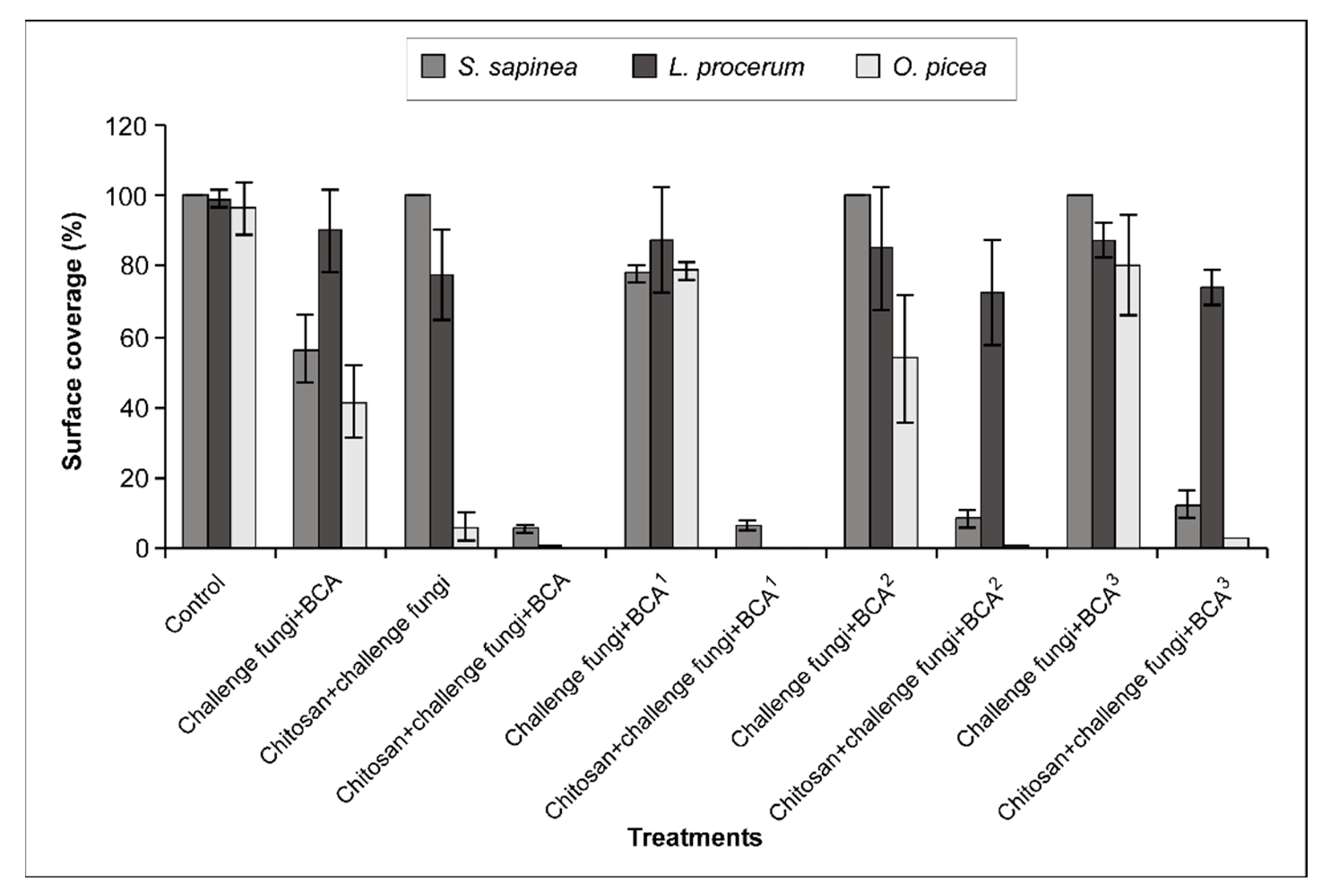
The inoculated surface of the wafers was assessed after 12 and 24 days of incubation. At each assessment time, the fungal growth and pigmentation of the challenge fungi over the surface of wafer was rated between 0 to 5, corresponding to a percentage of fungal surface cover (Table 1). The growth of S. sapinea was confirmed based on melanisation (usually S. sapinea melanised within a week on wood). The growth of L. procerum and O. piceae was confirmed based on melanisation and synnemata state (recognized by erect conidiogenous cells bearing conidia). For gfp transformed O. piceae, wafers were also observed using a Leica MZFL 111 fluorescence stereo microscope equipped with a green fluorescence filter set (excitation: 465–495, emission: 515–555 nm). Images were recorded using a digital camera (Power Shot S70, Axio Cam HRc, Jena, Germany Zeiss). The presence and/or absence of T. harzianum was confirmed by white spores (T. harzianum usually sporulate within 5 days after inoculation).
A statistical analysis of the data was carried out using the SAS PROC GLM (SAS Institute Inc. 1989, Cary, NC, USA). As the surface coverage data were recoded using visual scores, the distribution properties of the data were analysed. It was found that the data were not normally distributed, and thus transformation (square root) was used to normalize the data before using one way analysis of variance (ANOVA) and LSD (least significant differences) tests to compare the mean percentage coverage of each sapstain fungus under different treatment conditions. A two way ANOVA was conducted with LSD test to examine differences between fungi at different treatment conditions. Differences were considered to be significant at p ≤ 0.05.
For each of the three test fungi, a destructive assessment was also carried out after 24 days of incubation. The wood wafers were cut in half at the middle of the radial face to expose the middle tangential face. Microtome sections were cut along the tangential face and the fungal presence was observed. Furthermore, three 90 µm surface radial subsequent sections were cut to see the presence of challenge fungi within the wood (surface to 270 µm depth of wood). One 90 µm thick section contained three cell layers; therefore, about nine cell layers were monitored. The presence of sapstain fungi was ascertained depending on fungal discolouration, shape of fungal spores, and re-isolation on MA and on an Ophiostoma-selective medium [29], in which MA was amended with cycloheximide and streptomycin (MCS).
For the gfp transformed O. piceae, wood sections were examined using a Zeiss fluorescence microscope with a green filter set (excitation: 450–490, emission: 515). All of the images were recorded using AxioCam MRC with Axio Vision 4.3 (Zeiss).
For the statistical analysis, five mean depths from surface were categorised as follows: 1 = 0 µm depth, 2 = up to 90 µm, 3 = 91–180 µm, 4 = 181–270 µm, and 5 = more than > 270 µm. One way ANOVA was conducted and LSD tests were used to compare the mean depth penetration for the different treatment/species. Differences were considered to be significant at p ≤ 0.05.

3. Results

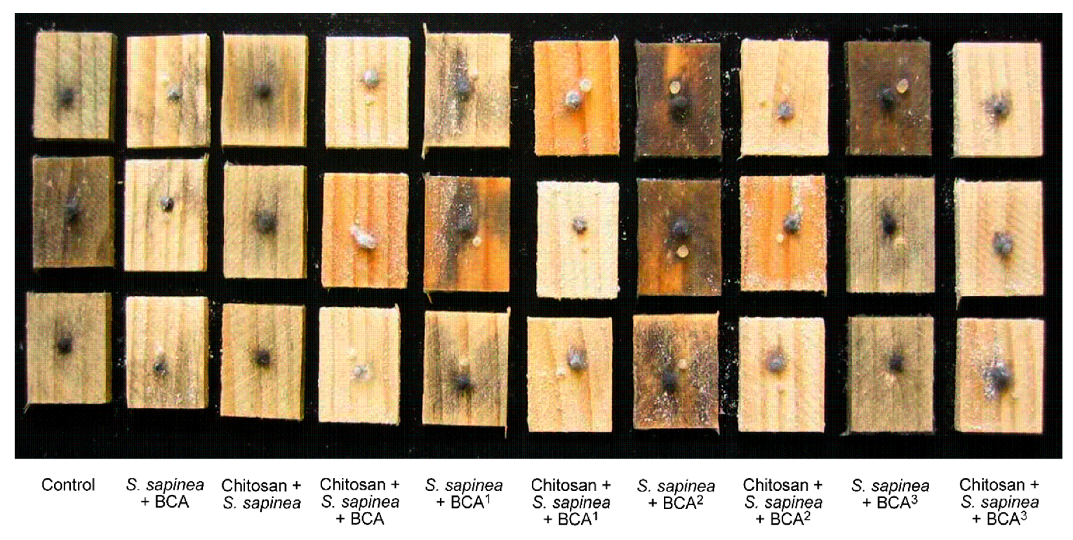
3.1. Observation of Fungal Growth on the Surface of Wafers

3.1.1. Sphaeropsis sapinea

After 24 days of incubation, the wood wafer surface was covered with S. sapinea in both the control (untreated) and wafers treated with chitosan (Figure 1 and Figure 2). However, when untreated wafers were simultaneously inoculated with the challenge fungus (S. sapinea) and T. harzianum, the growth of S. sapinea was restricted; only 50% surface coverage was observed at the end of the incubation period. The growth of S. sapinea was almost completely inhibited when the BCA was used along with chitosan. The synergy between the chitosan and BCA was evident; the growth of S. sapinea was significantly lower on the wafers treated with the combined treatment (BCA along with chitosan) when compared with treatment with either chitosan or BCA alone (p ≤ 0.05). When the BCA was inoculated after a delay of 1, 2, and 3 days, chitosan was essential to restrict the growth of S. sapinea. For instance, when untreated wafers were inoculated with S. sapinea and BCA was introduced after a 2 day delay, 100% coverage of S. sapinea was observed, but only 8% coverage was observed when it was treated with chitosan prior to BCA application (Figure 1 and Figure 2).

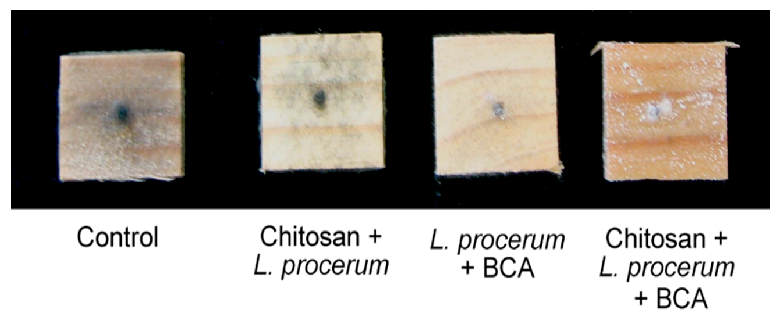
3.1.2. Leptographium procerum

Compared with the growth on the untreated wafers, the chitosan treated wafers had some inhibitory effect on the growth of L. procerum, whereas the wafers treated with BCA alone had very little effect on fungal growth (p ≤ 0.05). However, no growth of L. procerum was observed when the chitosan and the BCA were both used at the same time (Figure 1). For L. procerum on the treated wafers, the time of introduction of the BCA was a key prerequisite to the inhibition of fungal growth. For example, where treated wafers were inoculated with L. procerum and the BCA was introduced after 1 day delay, no growth of L. procerum was observed on the surface, but when the delay for BCA introduction was more than one day, the surface was covered with the growth of L. procerum. Once again, as for S. sapinea, the synergy between chitosan and BCA was apparent; without chitosan, BCA alone was unable to restrict the growth of L. procerum. However, unlike S. sapinea, where melanisation was observed when the fungus was growing on wafers (Figure 2) with L. procerum, melanisation was only noticed on the chitosan treated wafers along with the control. The wafers inoculated with BCA showed no melanisation (Figure 3). On those wafers, only synnemata covered the surface and no mycelium was observed after 24 days of incubation.

3.1.3. Ophiostoma piceae

Unlike the two previous challenge fungi, the treatment of chitosan almost entirely inhibited the growth of O. piceae on the wafer surface; only 5–10% coverage was observed on the wafers treated with chitosan alone. When BCA alone was introduced at the same time as O. piceae on the untreated wafers, it significantly reduced the growth of O. piceae compared with the controls (p ≤ 0.05), but still allowed for about 50% coverage (Figure 1). However, like L. procerum, no melanisation was noticed on the BCA inoculated wafers; only synnemata growth was observed for O. piceae—mycelium masses were lacking. No growth of O. piceae was observed on the wafers treated with chitosan and inoculated with BCA at the same time or even after the delayed inoculation of BCA. However, when BCA was introduced on untreated wafers after a time delay, irrespective of whether it was 1, 2, or 3 days, more than 50% coverage of synnemata was observed on the wood wafers (Figure 1).
Fluorescence microscopy showed that the gfp transformed O. piceae mycelium mass covered the control wood wafers with well-developed black synnemata with spore heads (Figure 4A). Untreated wafers inoculated with BCA alone showed some fluorescence of O. piceae around the inoculum (Figure 4B), which was largely non-pigmented synnemata. However, the BCA covered the wafer surface, as detected under the stereo microscope through the auto-fluorescence of the mycelium of T. harzianum (Figure 4B).

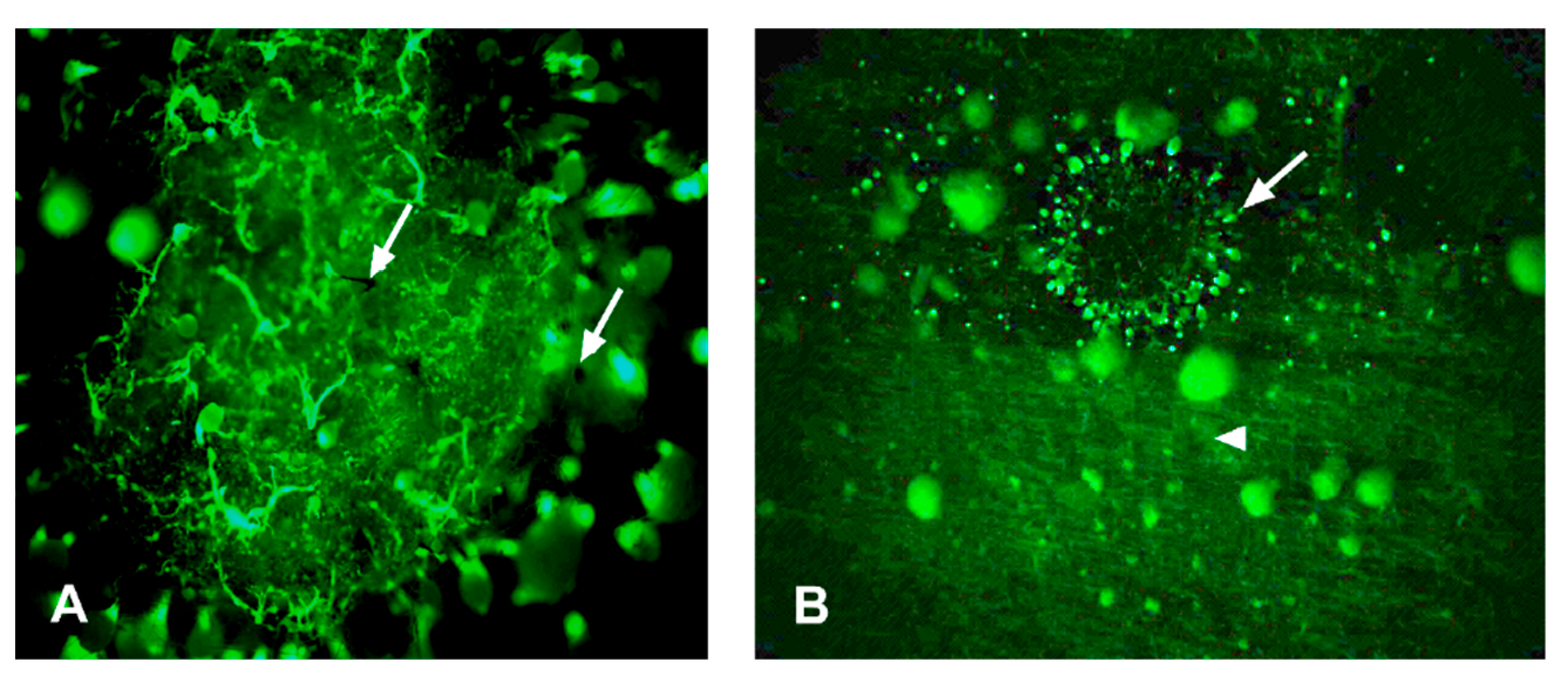
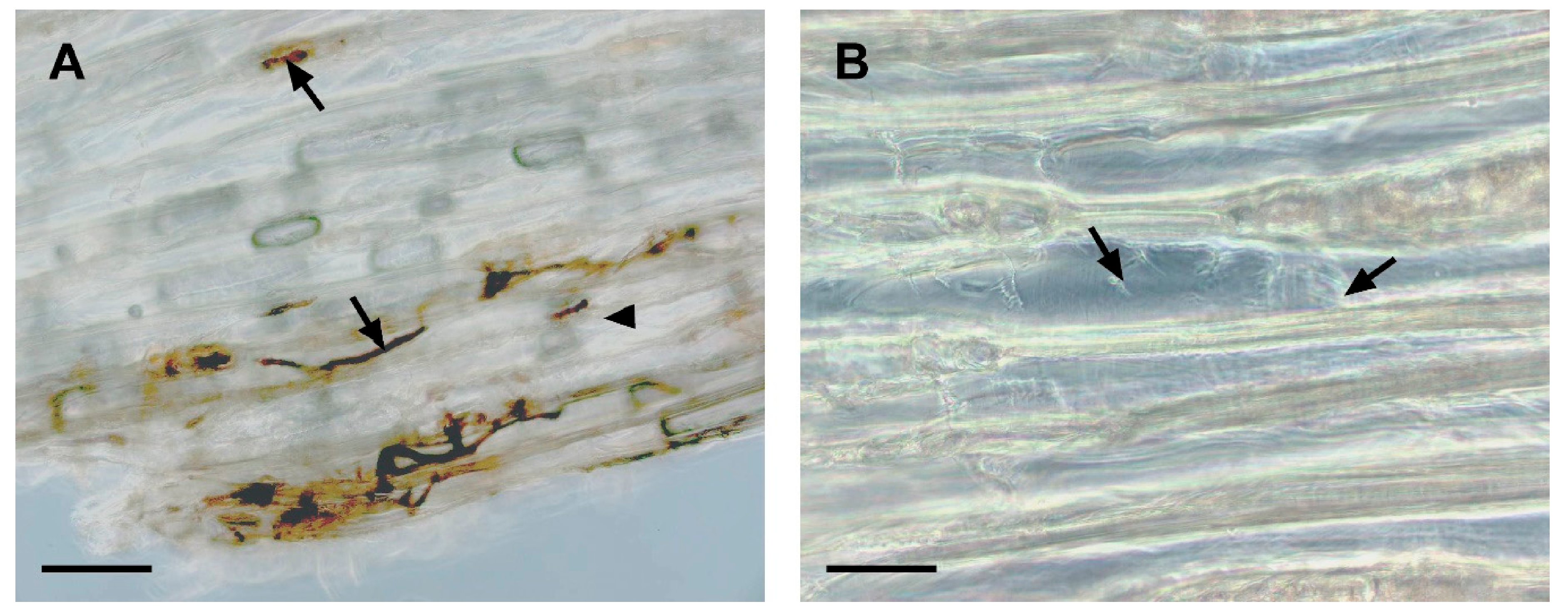
3.2. Microscopic Observations of Sapstain Penetration into Wafers

The depth of penetration by the challenge fungi was examined microscopically after 24 days of incubation (Table 2). The data presented in Table 2 are a summary of the observations based on the melanisation, shape of spores, and re-culturing hand cut sections on an MA and Ophiostoma selective medium (MCS agar medium). Unlike those of the challenge fungi, the spores of T. harzianum are subglobose, and therefore were easily differentiated.

3.2.1. Sphaeropsis sapinea

The untreated, chitosan treated, and untreated wafer inoculated with BCA had complete penetration (>270 µm or more than nine cell layers deep) of S. sapinea growth in the wood layers (Figure 5A). However, the chitosan treated wafers with BCA almost entirely stopped the internal colonisation of S. sapinea. Thus, S. sapinea was only observed in cell layers up to 90 µm from the surface. Below a 90 µm depth, only the BCA was observed (Figure 5B). In both the treated and untreated wafers, the extent of internal colonisation by S. sapinea tended to be inversely related to the delay time of BCA inoculation.

3.2.2. Leptographium procerum

There was no penetration of L. procerum in the wafers that were treated with chitosan and simultaneously inoculated with L. procerum and BCA, which was also confirmed by re-culturing wood sections on an Ophiostoma selected medium. After the final assessment, sections taken from a ≥270 µm depth showed no growth on an MCS agar medium, but replicate sections had growth of T. harzianum on an MA medium, suggesting that L. procerum was not present below a 270 µm depth from surface, but T. harzianum had completely penetrated.

3.2.3. Ophiostoma piceae

Under UV light, gfp transformed O. piceae fluoresced distinctively. Untreated wafers inoculated with BCA restricted the growth; fluorescing hyphae of O. piceae were localised only up to 5–6 cell layers deep. This result was also confirmed using an MSC agar medium. No growth was observed when sections (taken from >180 µm depth) were re-cultured on an MSC agar media, but replicate sections showed T. harzianum growth on an MA medium. In untreated wafers, the internal growth of the O. piceae was deeper when the BCA was inoculated with a time delay of 1, 2, or 3 days. However, the trend was not inversely related to the delay time like the two other test fungi (Table 2).

4. Discussions

The present study was successful in identifying an integrated system for the protection of fresh wood against sapstain. The chitosan/BCA treatment system protected radiata pine sapwood wafers more effectively than the chitosan or BCA used alone. In this integrated approach, the concentration of chitosan used seemed to be delaying the growth of sapstain fungus, and this delay gave the BCA time to establish and grow normally on the wood. Once established, BCA deprived nutrients to the sapstain fungus and hence colonisation. Once established, as T. harzianum is a fast growing fungus, it penetrated deeply into the wood [30]. Our in-vitro study showed that T. harzianum is very tolerant against chitosan, while chitosan has a fungistatic effect against sapstain fungi, depending on the concentration used [4,20]. At a cellular level, it is possible that in the chitosan/T. harzianum combination, chitosan may be acting fungistatically to delay the growth of sapstain fungi by inducing morphological changes in the cell wall and cell membrane [31], and the introduction of T. harzianum can degrade cell walls through the production of lytic enzymes [32]. Further work is needed to understand more clearly the mechanisms of the synergistic effects at a cellular level.
Despite providing encouraging results, this study also highlighted the limitation of this integrated approach. For L. procerum, the time of introduction of BCA is key to inhibiting its growth. When chitosan treated wafers were inoculated with BCA after more than a one day delay, with respect to the inoculation with the challenge fungus, the inhibition of L. procerum was not observed. The reason for this might be twofold: either the fast germination of L. procerum captured the substrate a day before BCA introduction and therefore the growth of BCA was restricted, or BCA was less effective against L. procerum compared with the other test fungi, which warrants further investigation.
The fact that chitosan alone seemed to have a greater inhibitory effect against O. piceae than L. procerum and S. sapinea is in general agreement with another study, where the antifungal activity of chitosan was shown to be limited to certain fungal species and not others. A Japanese Patent to Takashi and Mansato [33] describes the use of very low molecular weight chitosan to control Alternaria alternata fungus in pears, but this material was not effective in pears against other fungi. Chitosan consists of one–four linked heterogeneous polymers of glucosamine and N-acetyl glucosamine derivatives of chitin produced through the deacetylation of the nitrogen. The antifungal activity of the oligomers obtained from the hydrolysis of chitosan is dependent on the molecular weight of the product [19,34].
Trichoderma harzianum alone showed some inhibitory effect on the growth of the challenge fungi. Trichoderma spp. are known to have several antagonistic mechanisms [35,36,37] that contributes to its success as a BCA [38,39]. These include the production of antibiotics [40,41], lytic enzymes [35], siderophores [42,43], and volatile organic compounds [44,45]. In this study, the growth inhibition of sapstain fungi caused by T. harzianum was probably because of both its competitive and combative abilities, as Trichoderma spp. are in fact fast growing and produce many extracellular enzymes [46]. The extracellular enyzyme produced by Trichoderma has been used for various applications, including as a food additive [47]. Furthermore, it has been shown that enzyme mixtures from Trichoderma are strongly synergistic in their ability to degrade fungal cell walls and control pathogenic fungi [45].
Trichoderma harzianum also seemed to limit melanisation in two of the test fungi. Ophiostoma piceae and its anamorph, L. procerum, showed no melanisation when grown in the presence of BCA. The wood surface was only covered with synnemata (Graphium state) and no mycelial mass was observed. These observations are in agreement with those of Xiao et al. [30], where the interaction between O. piceae and T. harzianum was studied. Synnemata is a group of erect, sometimes fused conidiophores, that produce conidia at its apex. It is likely that T. harzianum is restricting the maturity and/or budding of conidia from synnema, and thus spore germination, or it could simply be due to nutrient unavailability. As T. harzianum is a rapid coloniser by utilising the available nutrients from wood, it deprives nutrients to other fungi. The impact of nutrients is considered to be a major factor for the pigmentation of sapstain fungi, both in nutrient mediums [48] and on wood [49]. In this study, the visual surface coverage for both O. piceae and L. procerum was significantly higher on the wafers treated only with BCA compared with the wafers treated with chitosan. However, the BCA inoculated wafers had no melanisation and less penetration in the wood layers. A similar conclusion was drawn from other studies, where prior establishment of Trichoderma spp. restricted the penetration [30] and growth [37] of wood inhabiting fungi in wood.
The use of gfp transformed O. piceae proved to be an invaluable tool throughout this study, as it fluoresces distinctively in mixed cultures. In wood, it is desirable to visualise fungal growth in the wood in order to ascertain the effect of the BCA on the growth of the target organism. Biological control agents differ fundamentally from chemical fungicides, in that they must grow and proliferate to be effective. For maximum effectiveness, fungal BCA’s should become established and remain active against the target organism during infection period [50]. Future work in this direction should be aimed at quantifying the growth of different fungal species in wood in order to obtain a more complete picture of the effect of BCA on the growth of the target organisms. The application of molecular biology techniques is currently being investigated for this purpose.

5. Conclusions

In the area of wood protection, a benign and effective antisapstain system that can be applied in forests as soon as logs are harvested is needed because pre-infection or pre-colonisation is a particular problem for export logs. This laboratory study has identified positive effects between the use of chitosan and BCA against three tested challenge (sapstain) fungi. Our initial field trial produced encouraging results (data not shown), and this approach could potentially be the next generation of wood protection, which will be based on an environmentally sound technology.

Author Contributions

Conceptualization, T.S.; methodology, T.S. and C.C.; software, T.S.; validation, T.S. and C.C.; formal analysis, T.S. and C.C.; investigation, T.S. and C.C.; resources, T.S.; data curation, C.C.; writing—original draft preparation, T.S. and C.C.; writing—review and editing, T.S.; visualization, T.S.; supervision, T.S.; project administration, T.S.; funding acquisition, T.S. All authors have read and agreed to the published version of the manuscript.

Funding

This research was funded by the Ministry of Business, Innovation, and Employment (MBIE), Wellington, New Zealand.

Acknowledgments

The authors acknowledge the Scion Wood Protection staff members for their contribution in assisting in trials.

Conflicts of Interest

The authors declare no conflict of interest.

References

  1. McCarthy, J.K.; Brockerhoff, E.G.; Didham, R.K. An Experimental Test of Insect-Mediated Colonisation of Damaged Pinus radiata Trees by Sapstain Fungi. PLoS ONE 2013, 8, e55692. [Google Scholar] [CrossRef] [PubMed]
  2. Thwaites, J.M.; Farrell, R.L.; Duncan, S.M.; Reay, S.D.; Blanchette, R.A.; Hadar, E.; Hadar, Y.; Harrington, T.C.; McNew, D. Survey of potential sapstain fungi on pinus radiata in New Zealand. N. Z. J. Bot. 2005, 43, 653–663. [Google Scholar] [CrossRef]
  3. Horgan, G. Cost–benefits of antisapstain treatment of New Zealand radiata pine for present and future markets. In Proceedings of the 2nd New Zealand Sapstain Symposium, Rotorua, New Zealand, 18–19 November 1999. [Google Scholar]
  4. Chittenden, C.; Singh, T. In vitro evaluation of combination of Trichoderma harzianum and chitosan for the control of sapstain fungi. Biol. Control 2009, 50, 262–266. [Google Scholar] [CrossRef]
  5. Hernandez, V.A.; Galleguillos, F.; Robinson, S. Fungal pigments from spalting fungi attenuating blue stain in Pinus spp. Int. Biodeter. Biodegr. 2016, 107, 154–157. [Google Scholar] [CrossRef]
  6. Bihon, W.; Slippers, B.; Burgess, T.; Wingfield, M.J.; Wingfield, B.D. Sources of Diplodia pinea endophytic infections in Pinus patula and P. radiata seedlings in South Africa. For. Pathol. 2011, 41, 370–375. [Google Scholar] [CrossRef] [Green Version]
  7. Kim, J.-J.; Ra, J.-B.; Kim, H.-J.; Kim, G.-H. Sapstain and mold control on radiata pine lumber: Laboratory and field tests of selected fungicides. Mycobiology 2002, 30, 37–40. [Google Scholar] [CrossRef]
  8. Singh, T.; Singh, A.P. A review on natural products as wood protectant. Wood Sci. Technol. 2012, 46, 851–870. [Google Scholar] [CrossRef]
  9. Benko, R.; Highley, T.L. Selection of media for screening interaction of wood-attacking fungi and antagonistic bacteria. Mater. Org. 1990, 25, 173–180. [Google Scholar]
  10. Gradinger, C.; Boisselet, T.; Stratev, D.; Ters, T.; Messner, K.; Fackler, K. Biological control of sapstain fungi: From laboratory experiments to field trials. Holzforschung 2009, 63, 751–759. [Google Scholar] [CrossRef]
  11. O’Callahan, D.R.; Singh, T.; McDonald, I.R. Evaluation of lactic acid bacterium from chilli waste as a potential antifungal agent for wood products. J. Appl. Microbiol. 2012, 112, 436–442. [Google Scholar] [CrossRef]
  12. Singh, T.; Chittenden, C. In-vitro antifungal activity of chilli extracts in combination with Lactobacillus casei against common sapstain fungi. Int. Biodeterior. Biodegrad. 2008, 62, 364–367. [Google Scholar] [CrossRef]
  13. Yang, D.Q.; Rossignol, L. Evaluation of Gliocladium roseum against wood-degrading fungi in vitro and on major Canadian wood species. Biocontrol Sci. Technol. 1999, 9, 409–420. [Google Scholar] [CrossRef]
  14. Byrne, A. Chemical control of biological stain: Past, present and future. In Proceedings of the Biology and Prevention of Sapstain; Forest Products Society: Madison, WI, USA, 1998; pp. 63–69. [Google Scholar]
  15. Ribera, J.; Fink, S.; Bas, M.D.C.; Schwarze, F.W.M.R. Integrated control of wood destroying basidiomycetes combining Cu-based wood preservatives and Trichoderma spp. PLoS ONE 2017, 12. [Google Scholar] [CrossRef]
  16. Vanneste, J.L.; Hill, R.A.; Kay, S.J.; Farrell, R.L.; Holland, P.T. Biological control of sapstain fungi with natural products and biological control agents: A review of the work carried out in New Zealand. Mycol. Res. 2002, 106, 228–232. [Google Scholar] [CrossRef]
  17. Held, B.W.; Thwaites, J.M.; Farrell, R.L.; Blanchette, R.A. Albino strains of Ophiostoma species for biological control of sapstaining fungi. Holzforschung 2003, 57, 237–242. [Google Scholar] [CrossRef]
  18. Yang, D.Q. Integrated Method for Protecting Logs and Green Lumber from Sapstain and Mold. U.S. Patent 6083537, 4 July 2000. [Google Scholar]
  19. Hussain, I.; Chittenden, C.; Singh, T. Scoping antifungal activities of various forms of chitosan oligomers and their potential fixation in wood. J. Chem. Chem. Eng. 2013, 7, 1175. [Google Scholar]
  20. Singh, T.; Vesentini, D.; Singh, A.P.; Daniel, G. Effect of chitosan on physiological, morphological, and ultrastructural characteristics of wood-degrading fungi. Int. Biodeterior. Biodegrad. 2008, 62, 116–124. [Google Scholar] [CrossRef]
  21. Xuan Du, D.; Xuan Vuong, B. Study on Preparation of Water-Soluble Chitosan with Varying Molecular Weights and Its Antioxidant Activity. Adv. Mater. Sci. Eng. 2019, 2019, 8781013. [Google Scholar] [CrossRef] [Green Version]
  22. Vårum, K.M.; Ottøy, M.H.; Smidsrød, O. Water-solubility of partially N-acetylated chitosans as a function of pH: Effect of chemical composition and depolymerisation. Carbohydr. Polym. 1994, 25, 65–70. [Google Scholar] [CrossRef]
  23. Minh, N.C.; Nguyen, V.H.; Schwarz, S.; Stevens, W.F.; Trung, T.S. Preparation of water soluble hydrochloric chitosan from low molecular weight chitosan in the solid state. Int. J. Biol. Macromol. 2019, 121, 718–726. [Google Scholar] [CrossRef]
  24. Hussain, I.; Singh, T.; Chittenden, C. Preparation of chitosan oligomers and characterization: Their antifungal activities and decay resistance. Holzforschung. 2012, 66, 119–125. [Google Scholar] [CrossRef]
  25. Liu, J.; Morrell, J. The Role of Chitinase in Bioprotectant Activity against Wood Staining Fungi; The International Research Group on Wood Preservation: Stockholm, Sweden, 1996; p. 11. [Google Scholar]
  26. Messner, K.; Fleck, V.; Buergel, J. Strains of the Trichoderma Fungus, Fungicide Derived Therefrom and Process for Using the Same. International Application WO 9308694, 13 May 1993. [Google Scholar]
  27. Roco, A.; Pérez, L.M. In vitro biocontrol activity of Trichoderma harzianum on Alternaria alternata in the presence of growth regulators. Electron. J. Biotechnol. 2001, 4, 68–73. [Google Scholar]
  28. Uzunovic, A.; O’Callahan, D.; Kreber, B. Mechanical tree havesters spread fungal inoculum onto freshly felled Canadian and New Zealand pine logs. For. Prod. J. 2004, 54, 34–40. [Google Scholar]
  29. Harrington, T. Cycloheximide sensitivity as a taxonomic character in Ceratocystis. Mycologia 1981, 73, 1123–1129. [Google Scholar] [CrossRef]
  30. Xiao, Y.; Morrell, J.; Ciuffetti, L. Effects of Prior Establishment of Trichoderma Harzianum on Ophiostoma Picea Growth in Freshly Sawn Douglas Fir Sapwood; International Research Group on Wood Preservation: Brisbane, Australia, 2003; p. 03-10476. [Google Scholar]
  31. Vesentini, D.; Steward, D.; Singh, A.P.; Ball, R.; Daniel, G.; Franich, R. Chitosan-mediated changes in cell wall composition, morphology and ultrastructure in two wood-inhabiting fungi. Mycol. Res. 2007, 111, 875–890. [Google Scholar] [CrossRef]
  32. Witkowska, D.; Maj, A. Production of lytic enzymes by Trichoderma spp. and their effect on the growth of phytopathogenic fungi. Folia Microbiol. 2002, 47, 279–282. [Google Scholar] [CrossRef]
  33. Takashi, K.; Masato, I. Agent for Controlling Black Spot of Pear. Japanese Patent 62198604, 14 September 1987. [Google Scholar]
  34. Silva-Castro, I.; Casados-Sanz, M.; Alonso-Cortés, A.L.; Martín-Ramos, P.; Martín-Gil, J.; Acuña-Rello, L. Chitosan-based coatings to prevent the decay of Populus spp. Wood caused by trametes versicolor. Coatings 2018, 8, 415. [Google Scholar] [CrossRef] [Green Version]
  35. da Silva, J.A.T.; de Medeiros, E.V.; da Silva, J.M.; Tenório, D.A.; Moreira, K.A.; da Silva Nascimento, T.C.E.; Souza-Motta, C. Antagonistic activity of Trichoderma spp. against Scytalidium lignicola CMM 1098 and antioxidant enzymatic activity in cassava. Phytoparasitica 2017, 45, 219–225. [Google Scholar] [CrossRef]
  36. Mazrou, Y.S.A.; Makhlouf, A.H.; Elseehy, M.M.; Awad, M.F.; Hassan, M.M. Antagonistic activity and molecular characterization of biological control agent Trichoderma harzianum from Saudi Arabia. Egypt. J. Biol. Pest Control 2020, 30. [Google Scholar] [CrossRef] [Green Version]
  37. Schubert, M.; Fink, S.; Schwarze, F.W.M.R. Evaluation of Trichoderma spp. as a biocontrol agent against wood decay fungi in urban trees. Biol. Control 2008, 45, 111–123. [Google Scholar] [CrossRef]
  38. Li, Y.; Sun, R.; Yu, J.; Saravanakumar, K.; Chen, J. Antagonistic and Biocontrol Potential of Trichoderma asperellum ZJSX5003 against the Maize Stalk Rot Pathogen Fusarium graminearum. Indian J. Microbiol. 2016, 56, 318–327. [Google Scholar] [CrossRef] [Green Version]
  39. Sood, M.; Kapoor, D.; Kumar, V.; Sheteiwy, M.S.; Ramakrishnan, M.; Landi, M.; Araniti, F.; Sharma, A. Trichoderma: The “secrets” of a multitalented biocontrol agent. Plants 2020, 9, 762. [Google Scholar] [CrossRef]
  40. Dennis, C.; Webster, J. Antagonistic properties of species groups of Trichoderma I: Production of non-volatile antibiotics. J. Agric. Food Chem. 1971, 20, 437–438. [Google Scholar]
  41. Dotson, B.R.; Soltan, D.; Schmidt, J.; Areskoug, M.; Rabe, K.; Swart, C.; Widell, S.; Rasmusson, A.G. The antibiotic peptaibol alamethicin from Trichoderma permeabilises Arabidopsis root apical meristem and epidermis but is antagonised by cellulase-induced resistance to alamethicin. BMC Plant Biol. 2018, 18, 1–11. [Google Scholar] [CrossRef]
  42. Ahmed, E.; Holmström, S.J.M. Siderophores in environmental research: Roles and applications. Microb. Biotechnol. 2014, 7, 196–208. [Google Scholar] [CrossRef]
  43. Zhao, L.; Wang, Y.; Kong, S. Effects of Trichoderma asperellum and its siderophores on endogenous auxin in Arabidopsis thaliana under iron-deficiency stress. Int. Microbiol. 2020, 23, 501–509. [Google Scholar] [CrossRef]
  44. Humphris, S.N.; Wheatley, R.E.; Bruce, A. The effects of specific volatile organic compounds produced by Trichoderma spp. on the growth of wood decay basidiomycetes. Holzforschung 2001, 55, 233–237. [Google Scholar] [CrossRef]
  45. Vinale, F.; Sivasithamparam, K. Beneficial effects of Trichoderma secondary metabolites on crops. Phytother. Res. 2020, 34, 2835–2842. [Google Scholar] [CrossRef]
  46. Kredics, L.; Antal, Z.; Szekeres, A.; Hatvani, L.; Manczinger, L.; Vágvölgyi, C.; Nagy, E. Extracellular proteases of Trichoderma species: A review. Acta Microbiol. Immunol. Hung. 2005, 52, 169–184. [Google Scholar] [CrossRef]
  47. Raveendran, S.; Parameswaran, B.; Ummalyma, S.B.; Abraham, A.; Mathew, A.K.; Madhavan, A.; Rebello, S.; Pandey, A. Applications of microbial enzymes in food industry. Food Technol. Biotechnol. 2018, 56, 16–30. [Google Scholar] [CrossRef]
  48. Eagen, R.; Brisson, A.; Breuil, C. The sap-staining fungus Ophiostoma piceae synthesizes different types of melanin in different growth media. Can. J. Microbiol. 1997, 43, 592–595. [Google Scholar] [CrossRef]
  49. Jankowiak, R.; Bilański, P.; Chyrzyński, Ł.; Strzałka, B. Identification of sapstain fungi from Scots pine pallets and assessment of their staining ability. Eur. J. Plant Pathol. 2018, 150, 307–322. [Google Scholar] [CrossRef]
  50. Sajitha, K.L.; Dev, S.A.; Maria Florence, E.J. Biocontrol potential of Bacillus subtilis B1 against sapstain fungus in rubber wood. Eur. J. Plant Pathol. 2018, 150, 237–244. [Google Scholar] [CrossRef]
Figure 1. Surface coverage (%) of the sapstain fungal growth observed on radiata pine wood wafers after different treatments (n = 5). Error bars refer to standard error of means. Biological control agent (BCA)1Trichoderma was inoculated after a one day delay; BCA2Trichoderma was inoculated after a two day delay; BCA3Trichoderma was inoculated after a three day delay).
Forests 12 00542 g001
Figure 2. Effect of chitosan and BCA on S. sapinea after 24 days of incubation. Photograph showing three replicates per treatment. BCA1Trichoderma was inoculated after a one day delay; BCA2Trichoderma was inoculated after a two day delay; BCA3Trichoderma was inoculated after a three day delay.
Forests 12 00542 g002
Figure 3. Effect of chitosan and BCA on L. procerum after 24 days of incubation.
Forests 12 00542 g003
Figure 4. Stereo light micrographs showing the fluorescence of O. piceae on wood wafers: (A) Mycelium mass and well developed synnemata (arrows) over untreated wafers; (B) non pigmented synnemata of O. piceae fluorescing (arrow) on wafers inoculated with BCA. Mycelium of BCA (T. harzianum) auto-fluorescing (arrow-head).
Forests 12 00542 g004
Figure 5. (A). Wood sections (270 µm from surface) taken from untreated, but BCA inoculated, wafers before S. sapinea inoculation. Arrows indicate melanised hyphae of S. sapinea. Bar = 20 µm. (B) Wood section (90–180 µm from surface) taken from wafers treated with BCA before inoculation with S. sapinea. Arrows indicate hyphae. Bar = 20 µm.
Forests 12 00542 g005
Table 1. Assessment system used to determine the fungal surface growth on wafers.
RatingSurface Cover (%)
00 (no growth)
11–5
26–25
326–50
451–75
576–100
Table 2. Presence of challenge fungus in the wood cell depth (µm) after 24 days of incubation (n = 5).
TreatmentsS. sapineaL. procerumO. picea
Control>270 a*>270 a>270 a
Challenge fungus + BCA>270 abUp to 270 abUp to 180 de
Chitosan + Challenge fungus>270 a>270 abUp to 90 gh
Chitosan + Challenge fungus + BCAUp to 90 gh0 i0 i
Challenge fungus + BCA1Up to 270 bUp to 270 b>270 ab
Chitosan + Challenge fungus + BCA1Up to 180 de0 hi0 i
Challenge fungus + BCA2>270 a>270 a>270 a
Chitosan + Challenge fungus + BCA2Up to 180 dUp to 90 fg0 i
Challenge fungus + BCA3>270 a>270 a>270 a
Chitosan + Challenge fungus + BCA3Up to 270 cUp to 90 ef0 hi
Notes: * means the same letters are not significantly different. BCA1Trichoderma was inoculated after a one day delay; BCA2Trichoderma was inoculated after a two day delay; BCA3Trichoderma was inoculated after a three day delay.
Publisher’s Note: MDPI stays neutral with regard to jurisdictional claims in published maps and institutional affiliations.

