Article

Interactions between Phytophthora cactorum, Armillaria gallica and Betula pendula Roth. Seedlings Subjected to Defoliation

1
Institute of Biological Sciences, Faculty of Biology and Environmental Sciences, Cardinal Stefan Wyszynski University in Warsaw, Wóycickiego 1/3 Street, 01-938 Warsaw, Poland
2
Institute of Forest Sciences, Faculty of Civil Engineering and Environmental Sciences, Białystok University of Technology, Wiejska 45E, 15-351 Białystok, Poland
3
Institute of Environmental Engineering and Energy, Faculty of Civil Engineering and Environmental Sciences, Białystok University of Technology, Wiejska 45E, 15-351 Białystok, Poland
4
Forest Protection Department, Forest Research Institute, Braci Leśnej 3, 05-090 Sękocin Stary, Poland
5
Department of Biology, University of Aveiro, 3810-193 Aveiro, Portugal
6
Department of Forest Botany, Warsaw University of Life Sciences, Nowoursynowska 159, 02-776 Warsaw, Poland
7
Environmental Sciences, University of Guelph, Guelph, ON N1G 2W1, Canada
*
Author to whom correspondence should be addressed.
Forests 2020, 11(10), 1107; https://doi.org/10.3390/f11101107
Submission received: 29 July 2020 / Revised: 12 October 2020 / Accepted: 15 October 2020 / Published: 19 October 2020
(This article belongs to the Special Issue Abiotic and Biotic Stress in Forest and Plantation Trees)

Abstract

:
The purpose of this study was to better understand the interactive impact of two soil-borne pathogens, Phytophthora cactorum and Armillaria gallica, on seedlings of silver birch (Betula pendula Roth.) subjected to stress caused by mechanical defoliation, simulating primary insect feeding. This is the first experimental confirmation of silver birch seedling root damage (and in consequence shoot mortality) caused by the additive effect of defoliation stress and P. cactorum inoculation via soil. However, the most severe damage to roots occurred after A. gallica inoculation. One year after treatments, chlorophyll fluorescence measurement, and gas chromatography coupled with mass spectrometry (GC-MS) were used to analyze the photosynthetic activity in leaves, the volatile organic compounds (VOCs) emitted by the birch leaves, and chemical compounds from the roots. The cumulative effect of the two pathogens and partial defoliation reduced photosynthetic activity, suggesting dysfunction of photosystem PSII due to the applied stresses. In summary, it seems that the main differences in photosynthetic performance could be attributed to Armillaria infection. The birch leaves in seedlings exposed to 50% defoliation, and inoculation with P. cactorum and A. gallica, emitted more aromatic carbonyls and alcohols, as well as half as much aliphatic esters, compared to controls. In infected birch roots, the production of phenols, triterpenes, and fatty alcohols increased, but fatty acids decreased. Higher levels of aromatic carbonyls and alcohols in leaves, as well as phenolic compounds in the roots of stressed birches (compared to control) suggest an activation of plant systemic acquired resistance (SAR).

1. Introduction

Silver birch (Betula pendula Roth.) is common throughout the lowlands and lower mountainous regions in Europe, and is subject to attack by Phytophthora cactorum (Lebert and Cohn) J. Schröt (as a primary pathogen) and Armillaria gallica Marxm. and Romagn. (as a secondary one) [1,2]. To date, over 150 Phytophthora species (Oomycetes) have been described from a broad range of hosts, including forest tree species and ornamental plants [3,4]. These groups of pathogens have not only led to extensive mortality of different forest tree species, but are also responsible for negative ecological impacts in many countries across the world. Moreover, climate warming and increased mean precipitation in the growing season have often been associated with increased invasiveness of phytophthoras in many plants [3,5]. In the last few decades, interest in the Phytophthora species as plant pathogens has been growing in Europe and on other continents, since these pathogens are causing emerging diseases of many forest tree species [6,7,8,9]. Phytophthora ramorum Werres, De Cock and Man in’t Veld is an example of a harmful pathogen, which is responsible for sudden oak death (SOD) in North America and sudden larch death in Europe (SLD) [10,11,12,13]. Generally, the risk of transferring phytophthoras from nurseries to forest plantations is considered high, as P. ramorum was transmitted in seedlings from nurseries in California, and it affected production in over 20 US states [14].
In Europe, P. cactorum and a lot of other Phytophthora spp. are abundant in many local forest environments, enduring different climatic conditions [15,16]. P. cactorum was isolated by Jung and Blaschke [15] from declining oaks stands (Quercus robur and Q. petraea), and by Lilja et al. [17] and Hantula et al. [18] from strawberry (Fragaria x ananassa) fruits and silver birch (B. pendula) roots. So far in Poland, this fungus has only been reported in nurseries on sycamore maple (Acer pseudoplatanus L.), black alder (Alnus glutinosa Gaertn.), European beech (Fagus sylvatica L.), and silver birch (B. pendula Roth.) [19].
In the 1980s birch stand decline was observed in Poland, but it was then considered as a complex disease involving water issues caused by drought. It is now known that species of Phytophthora causing damage to fine roots can mimic drought symptoms [20,21]. Some experiments demonstrated the possible susceptibility of one-year-old silver birch seedlings of Polish provenance to three species of Phytophthora, i.e., P. cinnamomi, P. citrophthora, and P. plurivora [22,23]. Nevertheless, the only published records of P. cactorum associated with birch in Poland are also of young birch seedling tissues [19], but not on adult trees [4].
Generally, tree damage and eventual mortality are caused, not by a single pathogen, but a whole suite of disturbance agents, like harmful fungi and defoliator insects, or unfavorable abiotic conditions. In Europe, among the root and butt rot fungi (Armillaria spp.), A. gallica is considered to be a weak pathogen of many forest tree species, increasing its activity after severe drought or defoliation [24,25,26]. However, Armillaria spp. are known to have a growing destructive impact in European ecosystems due to climate changing conditions, both in forest and in urban environments [27,28].
During the last few years, abundant research has been devoted to the combined effects of pests and pathogens on forest ecosystems [29]. Additionally, along with phytopathogens, birch trees are often subjected to attack by several insect defoliators, such as Deporaus betulae (L.), Phyllobius betulae F., and Ph. arborator (Herbst.), leading to severe damage of the crown [30,31]. A tree’s response to direct damage (e.g., defoliation) consists not only of a consecutive production of new leaves from dormant buds, but also of physiological and biochemical reactions. Many plants, including forest tree species, change their photosynthetic activity and the level of volatile organic compounds (VOC) secretion in response to damage caused by external stress factors [32,33,34]. Plants weakened by partial defoliation very often become vulnerable to multi-pathogen infections [35].
The main purpose of this work was to better understand interactions between organisms which could be associated with birch decline syndrome. The interactions between soil borne pathogens on the production of secondary metabolites (such as priming) by birch have not been sufficiently studied, and an objective was to examine the effects of fungus (A. gallica) and oomycete (P. cactorum) on the chemical composition of birch volatiles, secreted by young birch seedlings. To our knowledge there are no scientific reports in Poland about interactions between P. cactorum and A. gallica, which cohabitate in local forests in the same ecological niche, the soil [36]. Furthermore, since defoliation may predispose trees to infection, we hypothesized that combinations of partial defoliation and infection by P. cactorum and A. gallica would cause more mortality than each stress alone. We attempted to mimic insect defoliation by the gradual cutting of leaves over time to generate a less abrupt physiological stress. We also analyzed photosynthetic activity, via chlorophyll fluorescence, and secondary metabolite content in leaves and roots to probe at the mechanism of physiological and biochemical plant response to separate, and combined, abiotic and biotic stresses. The main purpose of our investigation was to explore the effects of two soil borne pathogens (P. cactorum and A. gallica) on B. pendula seedlings subjected to stress caused by partial mechanical defoliation, simulating insect attack. We tried to determine the following: (i) whether P. cactorum and A. gallica can affect the health of birch seedlings; and (ii) whether stress from partial defoliation can stimulate enhanced root infection. We also attempted to figure out how the combined effects, caused by the aforementioned potential pathogens and partial defoliation, influenced the photosynthetic activity of the treated plants, and what chemical compounds were produced in the stressed leaves and roots of the birch seedlings.

2. Material and Methods

2.1. Growth of Plant Material and General Experiment Design

In March 2017, two-year-old silver birch (B. pendula Roth.) plants were collected in Chojnów Forest District, Poland (52°02′20.3″N, 21°05′21.1″E), and re-planted singly in 15 L pots filled with a peat/perlite mixture 1:1 (v/v). Sixty-four plants were grown in controlled greenhouse conditions (ca. 22 °C, and relative humidity 65% ± 5%) for a year, and then were used as 3-year-old seedlings in experiments in spring 2018. They were irrigated with tap water once a day, and were not fertilized. Some pots were treated with inoculum of P. cactorum (in April 2018) or A. gallica (at the beginning of June 2018) or defoliated (in June 2018). For each treatment, eight replicate pots were used and the experiment was designed as follows: (1) control; (2) Armillaria gallica; (3) Phytophthora cactorum; (4) defoliation 50%; (5) Armillaria gallica + defoliation 50%; (6) Phytophthora cactorum + defoliation 50%; (7) Armillaria gallica + Phytophthora cactorum + defoliation 50%; (8) Armillaria gallica + Phytophthora cactorum.

2.2. Inoculation of Plants with Pathogen Isolates

2.2.1. Phytophthora Cactorum

Liquid medium, containing 250 cm3 of vermiculite, 20 cm3 of millet, and 175 cm3 of V8 medium (100 mL of multi vegetable juice, 2 g of calcium carbonate CaCO3, and 900 mL of distilled water), was dispensed into 1 L Erlenmeyer flasks (890 cm3 of medium per flask) and autoclaved for 15 min at 121 °C. An isolate of P. cactorum, from the IBL collection (GenBank accession number NCBI KX242303), was placed into the flasks using hyphae grown on V8 agar (ten pieces of agar about 0.5 × 0.5 cm covered with mycelium). The cultures had been incubated for 7 days at room temperature [37]. They were also used to inoculate soil samples (prepared from a mixture of peat, sand and perlite in a 1:1:1 ratio) and added to each individual 1 L pot in a volume equal to 2% of the volume of the soil. The untreated check did not contain P. cactorum inoculum.

2.2.2. Armillaria Gallica

To prepare the A. gallica inoculum, 1.5–2 cm thick stems of hazelnut (Corylus avellana L.) were first cut into 10 cm long sections, placed in a 630 cm3 metal container, wrapped with aluminum, and autoclaved for 80 min at 121 °C. After cooling, hyphal plugs from 3 week old cultures of A. gallica (GenBank accession number NCBI DQ115578) on 2% malt agar were added to the sterilized stem containers, with the full contents of a 9 cm diameter petri plate per container of hazelnut cuttings. The inoculated stem segments were then incubated at 25 °C for 3 months until segments were completely overgrown by mycelium. After this, the stem segments were placed in the appropriate birch seedling pots, with 2 segments per pot. Autoclaved hazelnut segments were placed in pots as controls.

2.3. Verification of the Seedling Infection

The occurrence of inoculated pathogens in rhizosphere soil was checked as follows: (i) visually for Armillaria rhizomorphs attached to roots, following Oszako et al. [38], or (ii) by baiting with leaves, or (iii) culturing (P. cactorum) [39]. Attempts were made to isolate P. cactorum from baited leaves and from rhizosphere soil containing fine roots in order to test if the introduced pathogen was still present, and vital. For baiting, soil samples were taken from several pots of birch seedlings and were put into plastic boxes and watered (1 cm above soil level). On the surface of water, previously washed and dried 7–14 day-old oak and beech leaves were placed, and incubated at room temperature. They were observed for discoloration daily for a week. When brownish spots appeared on the leaf surfaces (generally after 3–7 days of incubation at 20 °C), leaves were cut into small pieces (approx. 5 × 5 mm) and transferred to a selective medium: PARPNH (agar containing V8) agar with various antibiotics, incubated at 20 °C in the dark, and the hyphae emerging after 48 h were transferred to V8, agar medium. Cultures were checked for axenicity over the following days. Initial identification, following morphological descriptions in Erwin and Ribeiro et al. [6], was confirmed molecularly (described below in the in-planta section).
To assess the presence of P. cactorum in planta, roots were collected a year after inoculation and washed thoroughly. Genomic DNA was extracted and purified using a NucleoSpin® Plant II Midi kit (Macherey-Nagel, Düren, Germany). PCR was done using species-specific primers designed for P. cactorum isolate, JF300214.1 from GenBank, following Nowakowska et al. [40]. The TaqMan probe was labeled with the reporter dyes, i.e., JOE (6-carboxy-4,5-dichloro-2,7-dimethoxyfluorescein) at the 5’-end and HBQ1 quencher at the 3′-end (Sigma-Aldrich, St. Louis, MO, USA). PCR amplifications were performed in a total volume of 15 µL containing 1 × Master Mix (Sigma-Aldrich), 2 μM of each primer, 0.2 µM of probe, and 2 µL of diluted genomic DNA. PCR amplification included one cycle of denaturation at 95 °C for 3 min, 40 cycles of 95 °C for 30 s, 55–61 °C for 30 s, and 72 °C for 30 s. Amplifications were performed in a RotorGene 6000 (Qiagen, Hilden, Germany) apparatus, and fluorescence of newly synthetized amplicons was monitored in each PCR cycle during the annealing phase, following the manufacturer’s instructions.

2.4. Defoliation of Birch Seedlings

To mimic progressive defoliation, such as that caused by foliar insects and possibly foliar pathogens, 32 seedlings were partially and progressively defoliated over four weeks. In the first week 50% of the leaves had 25% of their area removed. In the successive three weeks, 25% more each week was removed from these leaves, resulting in 50% defoliation after four weeks.

2.5. Evaluation of Birch Health Status

The last year of the experiment, in July 2019 the health status of all 64 birch seedlings was visually assessed on the following scale: 1, healthy with 30–50% of leaf and shoot loss; 2, diseased with 51–75% shoot loss; 3, dying with more than 76% of shoot loss [41]. We assumed that birch damage up to 50% can be scored as healthy, and those showing higher levels of damage (51–75%) are definitely diseased, while those damaged more than 75% are rated as dying, or are dead. Since the defoliation treatment occurred only in the first year, it was reasonable to assess defoliation in the succeeding years.

2.6. Measurement of Plant Response to Stress

2.6.1. Biometric Parameters

After the 2-year-old seedlings had 13 months more growth, plant growth parameters were assessed. As soon as he plants were removed from their pots and cleaned, height and root collar diameter were measured, and fine roots were scanned on an EPSON Perfection V700 Photo Scanner (Regent Instruments Inc. Ltd., Quebec, QC, Canada), and the images processed using WinRhizo® v. 2009 c 32-bit software (Regent Instruments Inc. Ltd., Quebec, QC, Canada). The number of living fine roots (<2 mm) per length of mother roots (2–5 mm) was calculated, and transferred to Excel for calculation of the following representative parameters: number of tips, fine root length, mother root length, and fine root length per major root length, total root length, fine root surface area, and fine root tips. After drying at 60 °C for 72 h (after which weight did not change), the dry biomass weight was measured in 8 replicates per treatment.

2.6.2. Fluorometric Analysis of Chlorophyll a

Fluorescence emitted by chlorophyll a is quantified by exposing a leaf (first kept in the dark) to light of a defined wavelength, and measuring the amount of light re-emitted at longer wavelength [42]. Twelve months after the start of the experiment, chlorophyll fluorescence of the leaves was measured directly with a Handy PEA (Handy Plant Efficiency Analyser) fluorimeter, following instructions from Hansatech Instruments Ltd. (King’s Lynn, Norfolk, UK). In total, 10 measurements were made on each of three randomly chosen leaves for each of the eight treatments. Prior to measurements, leaves of 32 randomly chosen seedlings (half from each treatment) were kept for 30 min in the dark, where to the surface of each leaf, special clips were attached providing darkness, in order to slow down photosynthetic activity. The measurements were done on the central part of each leaf blade, on 4 mm2 of each sample, i.e., in three replications per pot (from the top, middle, and lower part of the crown). The measurement conditions consisted of 1 s pulses of 3500 µmol m−2s−1 light. To measure the physiological stress at the level of photosystem II (PSII), several photosynthetic parameters were assessed: F0, initial fluorescence; Fm, maximal intensity of fluorescence; F0/Fm, maximal photosynthetic activity; DI0/CS0, photosynthetic efficiency measured as energy dissipation in form of heat; and PI total, total performance index of photosystem II (PSII) [43].

2.7. Secondary Metabolites Contained in Birch Leaves and Roots

The leaf sample (1 g) was harvested from three birch seedlings. For each experimental treatment (1 to 8), one research sample was investigated. Similarly, eight samples of birch root were prepared. The volatile secondary metabolites emitted by birch leaves were analyzed using headspace solid phase microextraction and gas chromatography-mass spectrometry (HS-SPME/GC-MS) methods, following Isidorov et al. [44] and Oszako et al. [38]. The detection was performed in a full scan mode from 29 to 600 atomic mass units (amu).
Metabolites extracted from birch roots were analyzed with gas chromatography-mass spectrometry (GC-MS), a method previously developed by Stocki et al. [45,46], using GC-MS apparatus (Agilent Technologies Inc., Santa Clara, CA, USA). The samples were processed through an Agilent 7890A gas chromatograph with an Agilent 5975C mass spectrometer. Injection of each 1 µL sample was done using an Agilent 7693A autosampler. Chromatographic separation was performed on a capillary column HP-5MS (30 m × 0.25 mm × 0.25 μm) at a helium flow rate of 1 mL min−1. The injector worked in a split (1:10) mode at a temperature of 300 °C. The initial column temperature was 50 °C, rising to 325 °C, at 3 °C min−1, and the final temperature was held for 10 min. The detection was performed in a full scan mode from 41 to 800 amu. For identification of extracted components, both mass spectra and retention indices were used. After integration, the content (%) of each component in the total ion current (TIC) was calculated. All measurements for leaves and root extracts were performed in 3 replicates, to determine the detection error.

2.8. Statistical Analysis

For health data, growth data, and fluorescence data, normality of distribution was tested using the Shapiro–Wilk test, and homogeneity of variance with Levene’s test. For data which did not satisfy the assumptions of ANOVA, and to enable parametric testing, data were subjected to Box–Cox transformation [47]. In order to assess which treatments differed significantly, two-way ANOVA (analysis of variance) and Tukey post-hoc tests were used. All analyses were performed in the STATISTICA 13.1 package (StatSoft Polska, Kraków, Poland) for α = 0.05.

3. Results

In 2018, the 64 three-year-old potted birch seedlings were subjected to eight different treatments involving partial defoliation, inoculation with two different pathogens, and combinations thereof. In 2019, after a year of growth in the greenhouse, the 64 four-year-old seedlings were subjected to the following analyses: (i) baiting-based, and molecular, detection of the P. cactorum and A. gallica in the soil and/or in plants; (ii) health state assessment of seedlings; (iii) evaluation of the response to stress factors, via biometric and photosynthetic measurements; and (iv) detection and measurement of volatiles emitted by leaves and compounds extracted from roots. The two-way ANOVA revealed no statistically significant interactions at p = 0.05.

3.1. Detection of P. cactorum and A. gallica

P. cactorum was detected from the rhizosphere soil of all inoculated plants via baiting techniques. The isolates obtained were identified as P. cactorum, following descriptions in Erwin and Ribeiro [6]. Real-time PCR reactions, performed with the specific P. cactorum probes and primers, yielded positive results for this pathogen presence in all roots taken from inoculated silver birch (Ct values ranged from 21.5 to 24.1), as was the case in isolation through baiting technique.
Successful infection was observed a year after inoculation with A. gallica. Typical rhizomorphs of the fungus, attached to the root system of birch seedlings, were observed in all infected potted plants (i.e., in treatments 5–8 of the experiment).

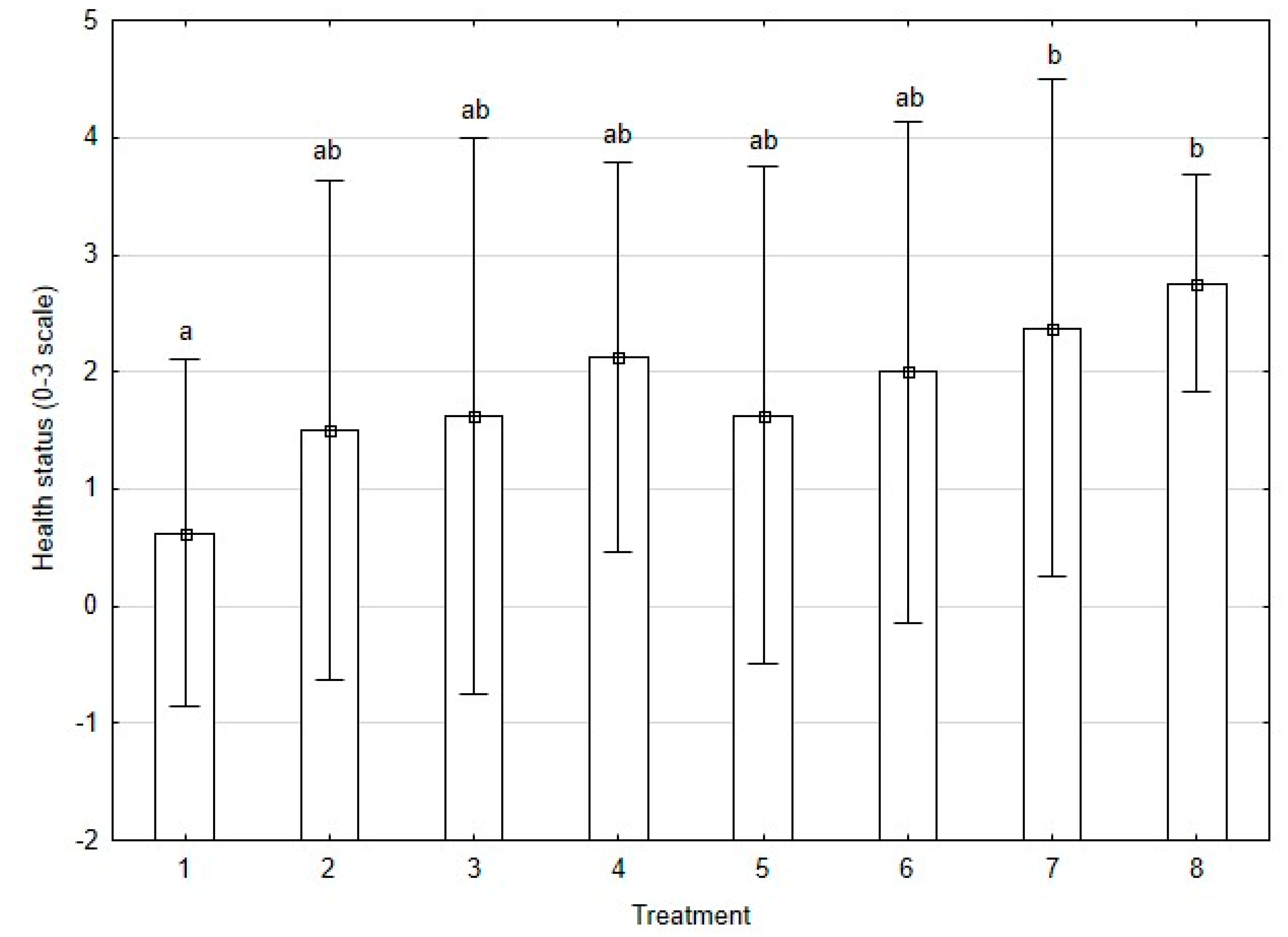
3.2. Health Status Assessment of Plants

One year after the beginning of the experiment, the highest negative influence on birch health was noted in the treatment with both Phytophthora and Armillaria (Figure 1). The shoot damage and defoliation were minor in the control, while the highest damage to seedlings occurred in treatments 7 and 8 (cumulative effect of two pathogens and/or defoliation) compared with treatments 2, 3, and 4 (the influence of each pathogen separately and defoliation, respectively). A. gallica + P. cactorum (8) caused the most severe damage (nearly reaching 75% leaf fall), finally leading to mortality of shoots and seedlings.

3.3. Plants Response to Stress

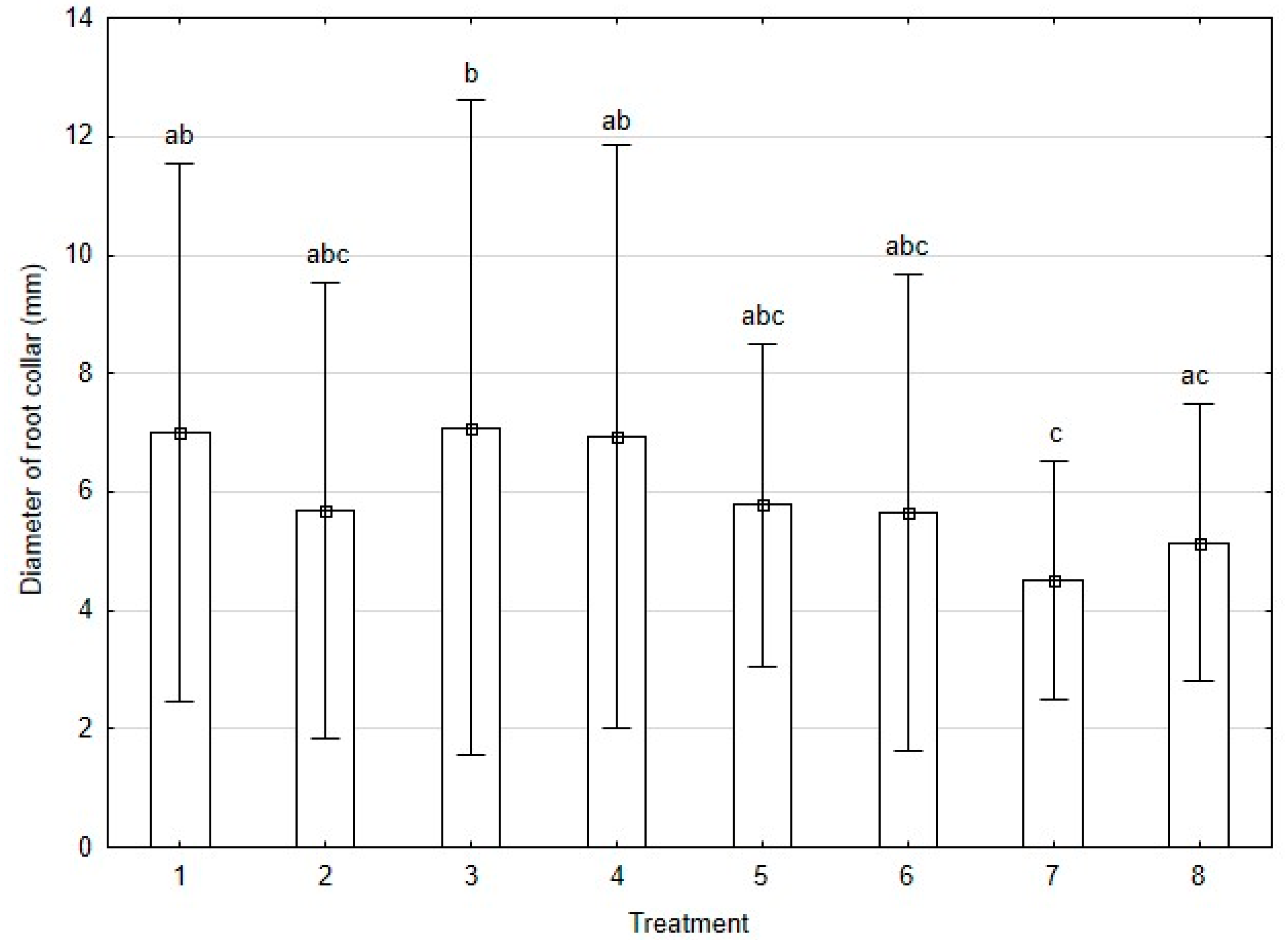
3.3.1. Growth Parameters

The comparison of seedling height between different experimental treatments showed no statistically significant differences (data not shown). However, the comparison of root collar thickness indicated some differences between treatments, especially with the additive effects of two pathogens and defoliation (Figure 2).
Figure 2 shows that neither P. cactorum nor A. gallica alone had effects on collar root diameter, and that the combined treatment had a greater effect. If those results are correct, the only plausible explanation is that there is a cumulative effect between both factors and defoliation.
Analysis of other growth parameters, such as the number of root tips, fine root length, fine root surface area, and fine root tips showed significant decreases in treatment (8) Armillaria gallica + Phytophthora cactorum, compared to the control (Table 1). The most pronounced decrease of most of the parameters was observed for treatment 8 (A. gallica + P. cactorum), when seedlings were subjected to inoculation with both pathogens. In treatment 8, fine root length decreased by 70%, total root length by 69%, and fine root surface area by 58%. In total, five of eight root growth parameters significantly decreased in treatment 8, compared to the control.

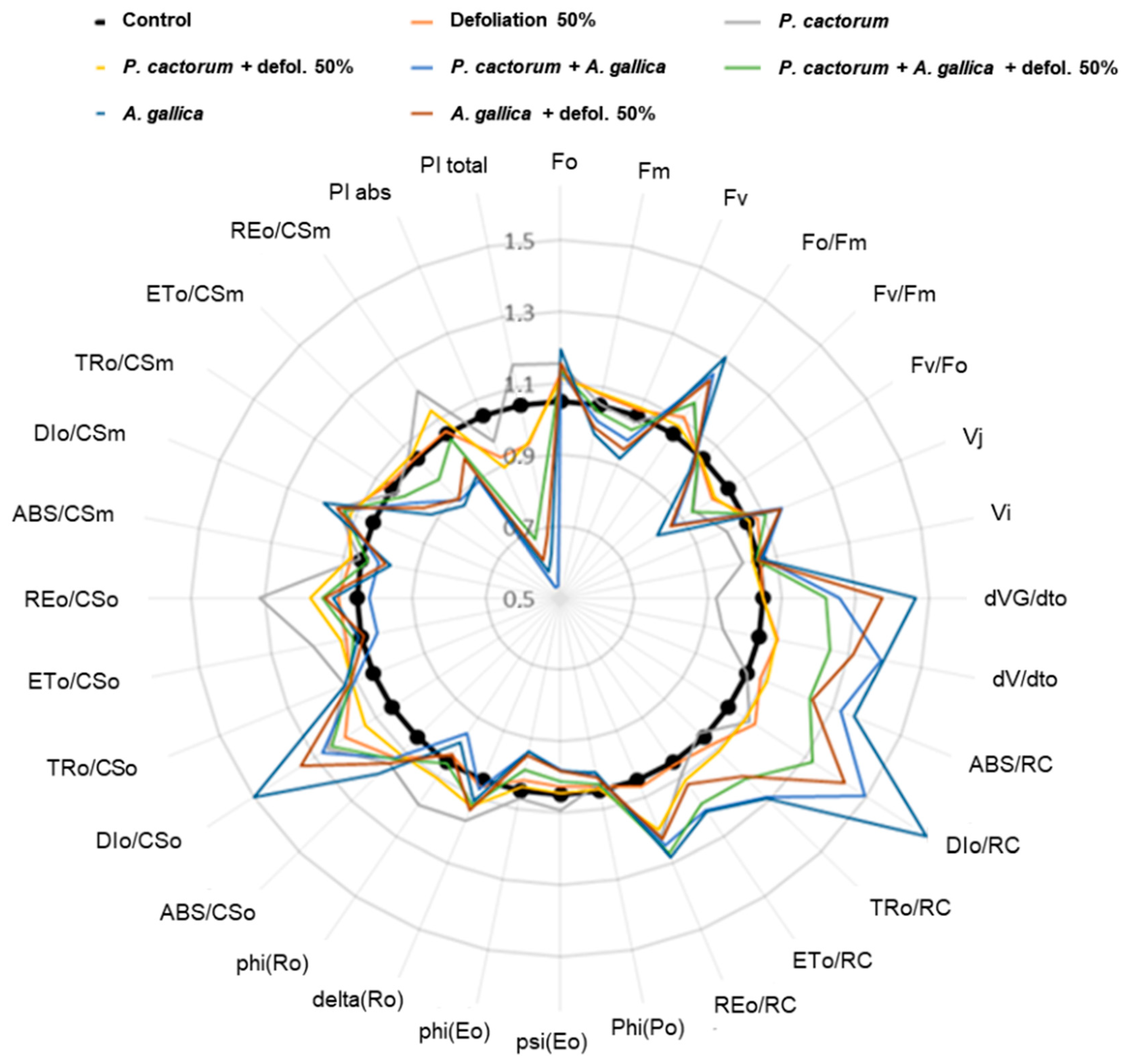
3.3.2. Chlorophyll Fluorescence

The physiological status of the birch seedlings was estimated based on selected chlorophyll fluorescence parameters. The initial fluorescence (F0), in contrast to maximal fluorescence (Fm), was higher in all treatments compared to the control (Figure 3, Table S2). The highest ratios F0/Fm were observed in A. gallica alone and in combination with P. cactorum, and this indicated that the highest level of chlorophyll fluorescence was inversely proportional to photosynthetic activity. This was in concordance with the highest ratios, DI0/CS0, denoted in seedlings subjected to A. gallica infections, showing the highest amount of dissipated energy in the form of heat (i.e., the highest loss of absorbed light energy). Defoliation, 50%, alone did not heavily affect the photosynthetic activity (F0/Fm ~ 1.0 and DI0/CS0 ~ 1.2).
In fact, all treatments showed reduced total photosynthetic activity (PI total <1.0) in comparison to the control plants (PI total = 1.0) except the treatment with P. cactorum alone, which did not adversely affect the photosynthetic activity, as PI total increased by 10% and F0/Fm ~ 1.0 (Figure 3). Other combinations showed low photosynthetic efficiency, and among all treatments, defoliation combined with P. cactorum resulted in slightly higher values of PI total (0.9) compared to the other treatments (Figure 3, Table S2). The interaction between the two pathogens and partial defoliation reduced PI total to 0.7, suggesting some compensation of the loss of activity in PSII due to the defoliation. In summary, it seems that the main differences in photosynthetic performance were given by the presence of Armillaria, confirmed by Tukey’s post-hoc test. P. cactorum alone did not seem to have effects on the photosynthetic parameters different than that of the control (Table S2).

3.4. Secondary Metabolites of Leaves and Roots

Defoliation combined with inoculation with the two pathogens affected the chemical composition of the metabolites produced by leaves and roots of B. pendula, although no statistical differences were found except for sesquiterpenes (Table 2, Table 3, Tables S3 and S4). Nevertheless, qualitative and quantitative assessments of VOC emission are provided in supplementary materials (Tables S5 and S6). The amount of monoterpenes, e.g., citronellol, neral, (E)-geraniol, α-citral, and eugenol was the highest in leaves under the influence of stress caused by P. cactorum + defoliation (treatment 6) (Table 2, Table S5). Birch leaves infected by both pathogens (treatments 7, 8) had significantly less sesquiterpenes compared to the control, including α-copaene, β-bourbonene, or β-caryophyllene (Table 2 and Table S5). The amount of aromatic esters (especially methyl salicylate) increased after infection by P. cactorum (3) and A. gallica + defoliation (5), compared to the control (Table 2 and Table S5). The highest content of aromatic carbonyl compounds (7.12%) was found in the treatment with P. cactorum + defoliation (6), and the highest content of aromatic alcohols (12.47%) in the treatment with both A. gallica and P. cactorum (8) (Table 2). Stressed birch leaves in treatments 7 and 8 emitted more aliphatic acids and aliphatic alcohols, but remarkably reduced the emission of alkanes and alkenes (Table 2).
The content of chemical compounds in root extracts was also affected for the experimental treatments, but no statistical differences were noticed between any treatment regarding their chemical composition (Table 3 and Table S4). Quantitatively, the control roots (treatment 1) produced lower amounts of phenolic compounds and triterpenes, compared to treatments 2–8 (Table 3 and Table S6). In particular, the defoliation treatment (4) and A. gallica + P. cactorum treatment (8) showed, on average, levels of phenolic compounds up to 15 fold higher than the control treatment (Table 3).

4. Discussion

4.1. Birch Damage Caused by P. cactorum and A. gallica

As far as we know, this is the first report in Poland concerning interactions between the oomycete P. cactorum and the fungus A. gallica, inoculated on silver birch seedlings. The pathogen P. cactorum was already previously recorded on gooseberry [48]. However, in Finland [49] it was known as a primary agent responsible for destroying birch fine roots, but in our experiment A. gallica appeared to be more harmful than P. cactorum. Conventionally, it is presumed that Armillaria species follows primary infection as an opportunistic pathogen. The later pathogens are often stimulated by root wounds and biochemical changes caused by defoliation [50]. Our previous investigations confirmed that A. gallica prefers to attack roots, first affected by Phytophthora infection [38]. However, in this study we did not find any significant interactions between those organisms (Table 1 and Table S1), although we found interactions in treatment 6 (Table S1). This is an interesting result, because defoliation should affect the normal functioning of the hydraulic system of the tree. In our experiment, it seems that the joint effect of both stresses is then capable of causing greater damage to roots. In our experiment P. cactorum, inoculated via soil, caused more severe damage than when plants were weakened by defoliation (behaving as an opportunistic pathogen). For the moment we are not able to demonstrate by statistical analysis that P. cactorum significantly alters the physiology or the behavior of birch seedlings, nevertheless we confirmed its activity (by baiting) and presence (by isolations and qPCR) in the rhizosphere and root tissues of B. pendula. This is in line with other experiments performed on ash seedlings [51]. Inoculation of potted ash seedlings, Fraxinus excelsior, by P. plurivora via soil two months before shoot inoculation via wounds by Hymenoscyphus fraxineus, demonstrated induction of plant resistance and less mortality (by 40%) than expected, and achieved in H. fraxineus treatment alone (100%). However, P. cactorum is known as a primary pathogen, causing disease in silver birch seedlings, and can often be isolated in forest nurseries, e.g., using baiting (plant traps) from irrigation water ponds, or directly from necrotic lesions on stems [52].
Silver birch decline in Poland became more prevalent starting in the 1980s, but phytophthorosis was not detected, although cankers on their stems and reddish exudates were commonly noticed at the time [53,54]. The mortality of birches was instead connected at that time with severe drought and water uptake problems by roots damaged by Armillaria fungi. Armillaria root disease caused by A. ostoyae (Romagn.) Herink is a common disease in many parts of the Northern hemisphere, including western North America, and Europe [27]; and with changing climate the incidence of root disease and butt rot caused by A. ostoyae is likely to increase, causing direct and indirect tree mortality [55]. Nevertheless, most Armillaria fungi existing in broad-leaved forests are considered to be opportunistic secondary pathogens (like A. gallica), so they usually act after primary pathogens such as Phytophthora species. The combination of oomycetes (P. alni, P. plurivora, P. cambivora, and P. quercina) as primary pathogens and fungi (Armillaria spp.) as secondary ones was already noticed in alder, ash, beech, and oak stands in the UK and Serbia [2,56]. It was confirmed that A. gallica mainly attacks fine roots, which start to rot and separate from the main roots, while at the separation point, elongated wounds are followed by callus formation and characteristic scars [2]. Subsequently, the substances secreted from the scars stimulate the growth of rhizomorphs from the inoculum of A. gallica. The rhizomorphs usually attach to the surface of thicker roots, feeding saprophytically, but when plants are weakened, the fungus begins infection [57,58]. Based on our observations, we suggest that similar interactions take place in Polish birch stands. Although many birches died rapidly once the crown symptoms appeared (die-back), in other stands the disease took a chronic form with the loss of foliage and branch dieback increasing over time, suggesting the activity of some resistance or tolerance mechanism [59]. B. pendula can be infected by P. cactorum [48] and A. gallica in inoculation tests, but a clear pathogenic relationship needs to be demonstrated in the field. Our future work will focus on the additive effects between pathogen infections and defoliation of birch seedlings in lab and field studies.

4.2. Birch Damage (Defoliation) Caused by Insect Pests

In general, insects responsible for the defoliation of birch generally have one generation in June, such as Scolionera betuleti (Zdd.) [60], Messa nana (Kl.), which can also feed on silver birch [61], and Agromyza alnibetulae (Hend.) [62]. In our experimental design, the defoliation of birch seedlings was performed in June (similarly to insect attack in the forest), and aimed to mimic the defoliation caused by endophagous and folivore insects of birch, with defoliation of 50% of the leaves gradually occurring over a four-week period (25% of each leaf cut off per week). The endophagous and folivorous species very often weaken trees, but rarely cause mortality of deciduous trees species [63,64], and this was also observed in our experiments.
In our study, the partial defoliation of the host (B. pendula), probably simulated damage by an aphid group of folivorous insects, often occurring in broad-leaved forests, which do not cause direct total defoliation or immediate death of the host, but provoke weakening of the host through gradual and partial defoliation.

4.3. Effects of Stress Factors on Physiological Response of Plants

The impact of harmful biotic factors on forest tree species is often assessed by visual estimation of the general health condition of the crown [65,66]. P. cactorum and A. gallica directly destroy the plant root system, and in consequence influence the condition of foliage. After one year of our experiment, the influence of defoliation stress on plant health status was more pronounced than the influence of each pathogen alone. The lowest health status estimation was found with the treatment of both pathogens (Phytophthora and Armillaria) combined with defoliation. Perhaps the damage to roots caused by pathogens was masked by partial defoliation, which caused less visual disease symptoms. Since all investigated stress factors can affect birch root or foliage, we first attempted to examine the plant general response at the level of fine root damage, and later, at the level of photosynthetic activity. We discuss the results in the following sections.

4.3.1. Root Status of Treated Plants

In our study, we placed P. cactorum directly into the soil, to simulate infections in the natural conditions in forests, without wounding, because that is a very invasive method. After inoculation and observation of disease symptoms, and through baiting techniques and observing infection of leaves (oak and beech), this demonstrated the viability of P. cactorum inoculum. By re-isolation on artificial media (PARPH), and obtaining pure cultures, and comparing them with the original P. cactorum inoculum we showed that the pathogen was present and alive. The observed increase of phenolic compounds in birch roots, and the development of rhizomorphs of A. gallica attached to roots, demonstrated that the infection method we had chosen was successful [50].
The combination of the stress factors (two soil borne pathogens and defoliation), decreased significantly the development of root collar thickness in birch seedlings. Surprisingly, P. cactorum alone seemed not to play an important role in the development of this growth parameter. Statistically significant decrease of birch fine root length by ca. 75% and fine root surface areas by ca. 73% took place only in the cases of P. cactorum + A. gallica + 50% defoliation, and P. cactorum + A. gallica. Lower level damage (15%) of the root system caused by rot has been observed by Mauer and Palátová [25] on root systems of 20-year-old birches affected by defoliation, and on root systems of 15-year-old birches in the Ore Mountains (Czech Republic). In the case of the Ore Mountains study, the dominant fungi developing on roots were A. gallica (like in the Polish case) and A. ostoyae (also very common in Polish forests). Defoliated trees showed a clear loss of small roots, deteriorated longevity of small roots, and a switch from ectomycorrhizae to ectendomycorrhizae composition [25].

4.3.2. Photosynthetic Activity

The only observed increase of total PI was in plants infected by P. cactorum, and this was probably due to a triggered natural mechanism of compensation, as a response of birch leaves to the primary agent of fine root infection. Figure 3 shows average the change regarding control treatment, which was supported by statistical analysis (Table S1). Defoliation caused by pests and pathogens are often reflected in changes in fluorescence parameters [67,68,69]. In our experiments, the interaction with the two pathogens and partial defoliation diminished by 30% the value of the total performance index (PI total), compared to untreated plants. The only observed increase of total PI was in plants infected by P. cactorum, and this was probably due to the triggered natural mechanism of compensation, as a response of the birch leaves to the primary agent of fine root infection. At least at the beginning, the strategy of plants fighting against a recognized pathogen can be increasing the efficiency of photosynthesis [70,71]. Similar decrease of the PI parameter was observed in wheat, after infection by Fusarium sp. Due to the high sensitivity of the photosynthetic apparatus, chlorophyll fluorescence has been used for early detection of plant disease in asymptomatic plants, enabling prevention measures, e.g., against fusarioses in crops [72]. Physiological stress assessed by photosynthesis effectiveness, resulting in PI total <1, can be informative about weakened plants, susceptible to natural infections by nearby pathogens.

4.4. Effects of Stress Factors on Host Chemical Compound Production

Plants respond to harmful biotic factors by multiple mechanisms of defense, including production of specific defense compounds, such as aromatic esters (benzyl acetate, methyl salicylate), aromatic alcohols (p-cresol), and monoterpenes (neral, geraniol, eugenol), as well as sesquiterpenes (α–copaene, β-bourbonene), and oxygen derivatives of these two groups of chemicals [73,74,75]. Generally, the secondary metabolites have two major roles in stressed plants: (i) the development of induced systemic resistance (ISR), or systemic acquired resistance (SAR) mechanisms, and (ii) the direct protection of plants against harmful pests and pathogens [76].
In our experiment, the signaling molecules secreted by birch leaves comprised molecules involved in SAR development, i.e., monoterpenes (geraniol and eugenol), triterpenes (lupeol, betulin) as well as aromatic esters, including methyl salicylate. The amount of compounds known as important signaling molecules in SAR increased substantially in birch leaves and roots subjected to defoliation, or pathogen infection coupled with defoliation treatment. Insect attack is known to trigger SAR responses [77] and ISR responses [78], which presumably involve the production of volatile compounds that may deter or enhance further insect attack. Defoliation-associated induction of resistance has been found for birch [79,80].
The effects of interactions between soil borne pathogens on the production of secondary metabolites by birch (such as those leading to SAR development) have not been sufficiently studied, and therefore we focused on the effects of the fungus (A. gallica) and the oomycete (P. cactorum) on the chemical composition of compounds secreted by leaves and roots. Although statistical differences between treatments were found only for sesquiterpenes, qualitative and quantitative assessment of VOC emission is provided in supplementary material, and discussed in a similar way to other related work [44]. The significantly lower content of sesquiterpenes in the leaves of the treated birches (especially during A. gallica infection) compared to the control, may have resulted from the redirection of these substances from leaves to the roots, where they can directly interact with the pathogen. Due to their lipophilic properties, sesquiterpenes and phyto-flavonoids produced after pathogen penetration into plant cells, take part in the direct inhibition of harmful microorganisms [81]. We contend that A. gallica benefited from the weakening of the plants (after plant defoliation), and this was reflected in the level of defensive substances (phenolic compounds and triterpenes), which were reduced (compared to defoliation alone, or Armillaria treatment alone), hence the infection process. Phytophthora could also mask its presence in plants, because the birch seedlings did not react like in the case of root infection by Armillaria, as the level of phenolic compounds in the roots rose only by about 5–6 times (not 15 times like in the case of the fungus). Keča et al. [51] showed that P. plurivora, introduced into the soil two months before stem inoculation of ash seedlings with Hymenoscyphus fraxineus (T. Kowalski, Baral, Queloz, Hesoya, comb. nov.), increased plant tolerance to fungal disease.
Qualitative and quantitative compositions of extracts of naturally and artificially defoliated birch leaves in Russia revealed that the content of free sterols, triterpene compounds, and flavones decreased one year after defoliation, but the amount of flavanones and flavanonols increased [82]. The increased production of such secondary compounds has protective functions, as a part of the plant’s strategy in the fight against harmful insects [83,84]. The concentration of antimicrobial compounds increased in the leaves of defoliated red oaks, in comparison to the non-defoliated plants, but the total amounts of nitrogen, sugars, proteins, starch, lignin, and hemicellulose were lower after one year of defoliation [85,86]. The defoliated trees had reduced amounts of nutrients, i.e., monosaccharides, amino acids, or fatty acids, in order to reduce their attractiveness for herbivorous insects [87].
The content of secondary metabolites in birch tissues may be influenced by many other factors, such as: species, age, or health condition [88]. We only dealt with young B. pendula, growing under different stress conditions, and probably cannot fully extrapolate these results directly to adult trees. Nevertheless, defoliation can invoke the SAR mechanism of defense, especially if it occurs for the first time and is accompanied by simultaneous root infection [89]. Another aspect worth studying in the future is coordinated defense mechanisms by distance, e.g., several VOCs or terpenoids may be involved in signaling among neighboring poplar species attacked by insects or herbivorous animals [33].

5. Summing up and Conclusions

In our study we focused only on the species of Armillaria as fungi, and these often associate with trees (rhizomorphs are superficially attached to the external bark at the root collar); and when host plants are weakened, the infection process usually starts. However, the role of other fungi in this process needs more investigation. They may accelerate the speed of tree death (synergy) or slow it down if fungi show antagonistic interactions, such as Trichoderma spp. killing rhizomorphs of Armillaria [90]. The occurrence of P. cactorum in a forest nursery usually causes problems with plant health because it can be pathogenic to many forest tree species. In our research we wanted to demonstrate changes in the physiology or the chemical composition of the plant due to the effect of P. cactorum. We were wondering if it were possible that, even if P. cactorum colonizes the root, the mere presence of the pathogen does not produce changes in the hydric balance and the physiological activity of the tree, providing that there is enough water in the soil. Therefore, we stressed plants (defoliation, inoculation) to recognize changes in the physiology and in the chemical composition of birch seedlings, due to the effect of P. cactorum alone, or in combination with Armillaria. In our experimental conditions we confirmed the damage to roots and as a consequence, to shoots, but the photosynthetic efficiency did not differ between the control and P. cactorum treatment. However, inoculation of birch seedlings with P. cactorum increased the amount of phenolic compounds compared to the non-inoculated control, indicating a plant response to infection. The heavily stressed plants also showed changes in triterpenes in roots and leaves. Similar observations were made for A. gallica, inoculation alone or in combination with artificial defoliation. In forests, these factors, among many others, can appear together in the same place and time, leading to the death of a tree or whole ecosystem [91].
Finally, the combination of stress factors in our experiment diminished the health of the investigated birch seedlings, including decreased root collar diameter, decreased number of tips (including fine roots), and decreased root length for fine roots and mother roots, among other effects. Taken together, the cumulative effect of different stress factors in a forest are major determinants of health. We hypothesized that P. cactorum is not more aggressive in combination with A. gallica, because A. gallica does not enhance the effect of the P. cactorum, or vice versa, nor does P. cactorum infection make the host more sensitive to the action of the Armillaria. After statistical analysis we can affirm that the evaluated parameters (the number of root tips, fine root length, fine root surface area, and fine root tips) in treatment (8), Armillaria gallica + Phytophthora cactorum, significantly diminished compared to the control (cumulative effect). On the other hand, two-way ANOVA revealed no significant interactions, which implied that effects were additive, meaning that the presence of P. cactorum in addition to other stress factors, such as defoliating insects and Armillaria gallica would have increasingly aggravating effects on plant health.
In conclusion, this research on birch seedlings subjected to partial defoliation, fungal infection by A. gallica, oomycete infection by P. cactorum, and combination thereof, demonstrated the following: (i) the oomycete P. cactorum can worsen health of silver birch (B. pendula) seedlings, (ii) the mortality of birch seedlings may occur after one growing season due to an interaction of P. cactorum with A. gallica, (iii) the fungus A. gallica caused significant loss of the photosynthetic apparatus efficiency compared to the control, (iv) infection by A. gallica caused a decrease of sesquiterpenes content in the leaves of the treated birches, revealing possible redirection of these groups of chemical compounds from the leaves to the roots, where they can directly interact with the pathogen during SAR.

Supplementary Materials

The following are available online at https://www.mdpi.com/1999-4907/11/10/1107/s1. Table S1: Result of two-way ANOVA (p-value) for all roots parameters. Table S2: Measurement values for selected parameters of chlorophyll fluorescence. Table S3: Statistical significance by two-way ANOVA of the chemical composition of VOCs emitted by birch seedlings leaves in the treatments, (1)—Control; (2)–Armillaria gallica; (3)—Phytophthora cactorum; (4)—Defoliation 50%; 5—Armillaria gallica + defoliation 50%; (6)—Phytophthora cactorum + defoliation 50%; (7)—Armillaria gallica + Phytophthora cactorum + defoliation 50%; (8)—Armillaria gallica + Phytophthora cactorum. More details are given in Table S5. Table S4: Statistical significance by two-way ANOVA of the chemical composition of VOCs emitted by birch roots during treatments, (1)—Control; (2)—Armillaria gallica; (3)—Phytophthora cactorum; (4)—Defoliation 50%; (5)—Armillaria gallica + defoliation 50%; (6)—Phytophthora cactorum + defoliation 50%; (7)—Armillaria gallica + Phytophthora cactorum + defoliation 50%; (8)—Armillaria gallica + Phytophthora cactorum. More details are given in Table S6. Table S5: Detailed chemical composition of extracts from birch seedling leaves in the following experimental treatments: (1)—Control; (2)—Armillaria gallica; (3)—Phytophthora cactorum; (4)—Defoliation 50%; (5)—Armillaria gallica + defoliation 50%; (6)—Phytophthora cactorum + defoliation 50%; (7)—Armillaria gallica + Phytophthora cactorum + defoliation 50%; (8)—Armillaria gallica + Phytophthora cactorum. Table S6: The detailed chemical composition of extracts from birch seedling roots in the following experimental treatments: (1)—Control; (2)—Armillaria gallica; (3)—Phytophthora cactorum; (4)—Defoliation 50%; (5)—Armillaria gallica + defoliation 50%; (6)—Phytophthora cactorum + defoliation 50%; (7)—Armillaria gallica + Phytophthora cactorum + defoliation 50%; (8)—Armillaria gallica + Phytophthora cactorum.

Author Contributions

Conceptualization, J.A.N., T.O. and S.Ś.; methodology, M.S., N.S., J.A.N., J.M.C.; software, M.T. (Miłosz Tkaczyk); validation, M.S., J.A.N., and T.H.; formal analysis, M.S., N.S., J.A.N., J.M.C.; writing—original draft preparation, J.A.N., T.O., M.T. (Mirela Tulik), S.Ś.; writing—review and editing, J.A.N., T.O., T.H.; supervision, J.A.N., T.H.; project administration, T.O.; funding acquisition, T.O. All authors have read and agreed to the published version of the manuscript.

Funding

The research was carried out as part of the work No. WZ/WB-INL/1/2019 at the Białystok University of Technology and financed from a research subsidy provided by the Minister of Science and Higher Education. Part of the study was financed by the Forest Research Institute as a scholarship to Joao Maria Caetano who contributed to this paper during his three-month stay in the Institute as its fellow.

Acknowledgments

We express our gratitude to Anna Żółciak for valuable comments and discussion during manuscript preparation. Thank you, Daria Berezovska student of the Cardinal Stefan Wyszyński University in Warsaw (UKSW) for the help with graphical performance of chlorophyll fluorescence and Sylwia Puchalska student of the Forest Sciences Institute of the Bialystok Technological University in Hajnówka for the help with biometric measurements.

Conflicts of Interest

The authors declare no conflict of interest. The funders had no role in the design of the study; in the collection, analyses, or interpretation of data; in the writing of the manuscript, or in the decision to publish the results.

References

  1. Rishbeth, J. Armillaria in an ancient broadleaved woodland. Eur. J. Plant. Pathol. 1991, 21, 238–249. [Google Scholar] [CrossRef]
  2. Keča, N.; Koufakis, I.; Dietershagen, J.; Nowakowska, J.A.; Oszako, T. European oak decline phenomenon in relation to climatic changes. Folia For. Pol. Series A 2016, 58, 170–177. [Google Scholar] [CrossRef] [Green Version]
  3. Brasier, C.M. Phytophthora Biodiversity: How Many Phytophthora Species Are There? In Phytophthoras in Forests and Natural Ecosystems, Proceedings of the Fourth Meeting of the International Union of Forest Research Organizations (IUFRO) Working Party S07.02.09; Monterey: Forest Service, CA, USA, 2009; pp. 101–115. [Google Scholar]
  4. Jung, T.; Orlikowski, L.; Henricot, B.; Abad-Campos, P.; Aday, A.G.; Aguín Casal, O.; Bakonyi, J.; Cacciola, S.O.; Cech, T.; Chavarriaga, D.; et al. Widespread Phytophthora infestations in European nurseries put forest, semi-natural and horticultural ecosystems at high risk of Phytophthora diseases. For. Pathol. 2016, 46, 134–163. [Google Scholar] [CrossRef] [Green Version]
  5. Aguayo, J.; Elegbede, F.; Husson, C.; Saintonge, F.X.; Marçais, B. Modeling climate impact on an emerging disease, the Phytophthora alni-induced alder decline. Glob. Chang. Biol. 2014, 20, 3209–3221. [Google Scholar] [CrossRef] [PubMed] [Green Version]
  6. Erwin, D.C.; Ribeiro, O.K. Phytophthora Diseases Worldwide; APS Press: St. Paul, MN, USA, 1996; 562p. [Google Scholar]
  7. Brasier, C.M.; Robredo, F.; Ferraz, J.F.P. Evidence for Phytophthora cinnamomi involvement in Iberian oak decline. Plant Pathol. 1993, 42, 140–145. [Google Scholar] [CrossRef]
  8. Jung, T. Beech decline in Central Europe driven by the interaction between Phytophthora infections and climatic extremes. For. Pathol. 2009, 39, 73–94. [Google Scholar] [CrossRef]
  9. Orlikowski, L.B.; Oszako, T. Phytophthorosis in Nurseries and Forest Stands; CILP: Warszawa, Polska, 2009. [Google Scholar]
  10. Rizzo, D.M.; Garbelotto, M.; Davidson, J.M.; Slaughter, G.W.; Koike, S.T. Phytophthora ramorum as the cause of extensive mortality of Quercus spp. and Lithocarpus densi-florus in California. Plant Dis. 2002, 86, 205–214. [Google Scholar] [CrossRef] [Green Version]
  11. Brasier, C.M. Sudden oak death: Phytophthora ramorum exhibits transatlantic differences. Mycol. Res. 2003, 107, 257–259. [Google Scholar] [CrossRef]
  12. Brasier, C.; Webber, J. Sudden larch death. Nature 2010, 466, 824–825. [Google Scholar] [CrossRef]
  13. Grűnwald, N.J.; LeBoldus, J.M.; Hamelin, R.C. Ecology and evolution of the sudden oak death pathogen Phytophthora ramorum. Annu. Rev. Phytopathol. 2019, 57, 301–321. [Google Scholar] [CrossRef]
  14. Ivors, K.; Garbelloto, M.; Vries, D.E.; Ruyter-Spira, C.; Hekkert, B.T.; Rosenzweig, N.; Bonants, P. Microsatellite markers identify three lineages of Phytophthora ramorum in US nurseries, yet single lineages in US forest and European nursery populations. Mol. Ecol. 2006, 15, 1493–1505. [Google Scholar] [CrossRef] [PubMed]
  15. Jung, T.; Blaschke, H. Phytophthora root rot in declining forest trees. Phyton 1996, 36, 95–102. [Google Scholar]
  16. Hantula, J.; Lilja, A.; Nuorteva, H.; Parikka, P.; Werres, S. Pathogenicity, morphology and genetic variation of Phytophthora cactorum from strawberry, apple, rhododendron, and silver birch. Mycol. Res. 2000, 104, 1062–1068. [Google Scholar] [CrossRef]
  17. Lilja, A.; Rikala, R.; Hietala, A.; Heinonen, R. Stem lesions on Betula pendula seedlings in Finnish forest nurseries and the pathogenicity of Phytophthora cactorum. Eur. J. For. Pathol. 1996, 26, 89–96. [Google Scholar] [CrossRef]
  18. Hantula, J.; Lilja, A.; Parikka, P. Genetic variation and host specificity of Phytophthora cactorum isolated in Europe. Mycol. Res. 1997, 101, 565–572. [Google Scholar] [CrossRef]
  19. Stępniewska, H. Occurrence of Phytophthora cactorum on tree seedlings with damping-off symptoms in some forest nurseries in south of Poland. Phytopathol. Pol. 2003, 29, 53–67. [Google Scholar]
  20. Brasier, C.M. Phytophthora cinnamomi and oak decline in southern Europe. Environmental constraints including climate change. Ann. For. Sci. 1996, 53, 347–358. [Google Scholar] [CrossRef] [Green Version]
  21. Ruiz Gomez, F.J.; Pérez-de-Luque, A.; Sánchez-Cuesta, R.; Quero, J.L.; Navarro Cerrillo, R.M. Differences in the response to acute drought and Phytophthora cinnamomi rands infection in Quercus ilex L. seedlings. Forests 2018, 9, 634. [Google Scholar] [CrossRef] [Green Version]
  22. Orlikowski, L.B.; Ptaszek, M.; Trzewik, A.; Orlikowska, T. Relationship between source of water, surveying time and occurrence of Phytophthora spp. Prog. Plant Prot. 2008, 48, 246–251. [Google Scholar]
  23. Ptaszek, M.; Orlikowski, L.B. Occurrence of Phytophthora species in watercourses and reservoirs in Poland and the threat to cultivated plants by this genera. Prog. Plant Prot. 2015, 55, 64–70. [Google Scholar] [CrossRef] [Green Version]
  24. Guillaumin, J.J.; Mohammed, C.; Anselmi, N.; Courtecuisse, R.; Gregory, S.C.; Holdenrieder, O.; Intini, M.; Lung, B.; Marxmüller, H.; Morrison, D.; et al. Geographical distribution and ecology of the Armillaria species in western Europe. Eur. J. For. Pathol. 1993, 23, 321–341. [Google Scholar] [CrossRef]
  25. Mauer, O.; Palátová, E. The role of root system in silver birch (Betula pendula Roth) dieback in the air-polluted area of Krušné hory Mts. J. For. Sci. 2003, 49, 191–199. [Google Scholar] [CrossRef] [Green Version]
  26. Desprez-Loustau, M.L.; Marçais, B.; Nageleisen, L.M.; Piou, D.; Vannini, A. Interactive effects of drought and pathogens in forest trees. Ann. For. Sci. 2006, 63, 597–612. [Google Scholar] [CrossRef] [Green Version]
  27. La Porta, N.; Capretti, P.; Thomsen, I.M.; Kasanen, R.; Hietala, A.M.; Von Weissenberg, K. Forest pathogens with higher damage potential due to climate change in Europe. Can. J. Plant Pathol. 2008, 30, 177–195. [Google Scholar] [CrossRef]
  28. Chandelier, A.; Gerarts, F.; San Martin, G.; Herman, M.; Delahaye, L. Temporal evolution of collar lesions associated with ash dieback and the occurrence of Armillaria in Belgian forests. For. Pathol. 2016, 46, 289–297. [Google Scholar] [CrossRef]
  29. Jactel, H.; Desprez-Loustau, M.L.; Battisti, A.; Brockerhoff, E.; Santini, A.; Stenlid, J.; Björkman, C.; Branco, M.; Dehnen-Schmutz, K.; Douma, J.C.; et al. Pathologists and entomologists must join forces against forest pest and pathogen invasions. NeoBiota 2020, 58, 107–127. [Google Scholar] [CrossRef]
  30. Kula, E.; Pešlová, A.; Buchtová, D. Effects of nitrogen on the selection of food by Phyllobius arborator (Herbst). J. For. Sci. 2008, 54, 17–23. [Google Scholar] [CrossRef] [Green Version]
  31. Urban, J. A contribution to the knowledge of biology and harmfulness of Deporaus betulae (L.) (Coleoptera, Attelabidae). Acta Univ. Agric. Silvic. Mendel. Brun. 2012, 60, 317–338. [Google Scholar] [CrossRef] [Green Version]
  32. Šimpraga, M.; Ghimire, R.P.; Van Der Straeten, D.; Blande, J.D.; Kasurinen, A.; Sorvari, J.; Holopainen, T.; Adriaenssens, S.; Holopainen, J.K.; Kivimäenpää, M. Unravelling the functions of biogenic volatiles in boreal and temperate forest ecosystems. Eur. J. For. Res. 2019, 138, 763–787. [Google Scholar] [CrossRef] [Green Version]
  33. Li, T.; Grauer-Gray, K.; Holopainen, J.K.; Blande, J.D. Herbivore gender effects on volatile induction in Aspen and on olfactory responses in leaf beetles. Forests 2020, 11, 638. [Google Scholar] [CrossRef]
  34. Peghaire, E.; Hamdache, S.; Galien, A.; Sleiman, M.; ter Halle, A.; El Alaoui, H.; Kocer, A.; Richard, C.; Goupil, P. Inducing plant defense reactions in tobacco plants with phenolic-rich extracts from red maple leaves: A characterization of main active ingredients. Forests 2020, 11, 705. [Google Scholar] [CrossRef]
  35. Schoeneweiss, D.F. The influence of stress on diseases of nursery and landscape plants. J. Aboric. 1978, 4, 217–225. [Google Scholar]
  36. Oszako, T.; Sikora, K.; Belbahri, L.; Nowakowska, J.A. Molecular analysis of Phytophthora species found in Poland. Folia For. Pol. Series A 2017, 59, 321–328. [Google Scholar] [CrossRef]
  37. Tkaczyk, M.; Milenković, I.; Nowakowska, J.A.; Borys, M.; Kałuski, T.; Gawlak, M.; Czyż, M.; Oszako, T. Morphological and molecular identification of Phytophthora species isolated from the rhizosphere of declining oak trees in Krotoszyn Plateau. Genetika 2017, 49, 203–215. [Google Scholar] [CrossRef]
  38. Oszako, T.; Żółciak, A.; Tulik, M.; Tkaczyk, M.; Stocki, M.; Nowakowska, J.A. Influence of Bacillus subtilis and Trichoderma asperellum on the development of birch seedlings infected with fine root pathogen. Phytophthora Plurivora Sylwan 2019, 163, 1006–1015. [Google Scholar] [CrossRef]
  39. Jung, T.; Blaschke, H.; Neumann, P. Isolation, identification and pathogenicity of Phytophthora species from declining oak stands. Eur. J. For. Pathol. 1996, 26, 253–272. [Google Scholar] [CrossRef]
  40. Nowakowska, J.A.; Malewski, T.; Tereba, A.; Oszako, T. Rapid diagnosis of pathogenic Phytophthora species in soil by real-time PCR. For. Pathol. 2017, 47, e12303. [Google Scholar] [CrossRef]
  41. Oszako, T.; Voitka, D.; Tkaczyk, M.; Keča, N.; Belbahri, L.; Nowakowska, J.A. Assessment of interactions between defoliation and Phytophthora plurivora stem infections of birch seedlings. For. Chron. 2018, 94, 140–146. [Google Scholar] [CrossRef]
  42. Maxwell, K.; Johnson, G.N. Chlorophyll fluorescence—A practical guide. J. Exp. Bot. 2000, 51, 659–668. [Google Scholar] [CrossRef]
  43. Goltsev, V.N.; Kalaji, H.M.; Paunov, M.; Bąba, W.; Horaczek, T.; Mojski, J.; Kociel, H.; Allakhverdiev, S.I. Variable chlorophyll fluorescence and its use for assessing physiological condition of plant photosynthetic apparatus. Russ. J. Plant Physol. 2016, 63, 869–893. [Google Scholar] [CrossRef]
  44. Isidorov, V.A.; Stocki, M.; Vetchinikova, L. Inheritance of specific secondary volatile metabolites in buds of white birch Betula pendula and Betula pubescens hybrids. Trees Struct. Funct. 2019, 33, 1329–1344. [Google Scholar] [CrossRef] [Green Version]
  45. Stocki, M.; Zapora, E.; Rój, E.; Bakier, S. Recovering biologically active compounds from logging residue of birch (Betula spp.) with supercritical carbon dioxide. Przem. Chem. 2018, 97, 774–778. [Google Scholar]
  46. Stocki, M.; Bakier, S.; Isidorov, V. Study on extraction of biologically active compounds from birch (Betula) buds with supercritical carbon dioxide. Przem. Chem. 2019, 98, 1988–1991. [Google Scholar]
  47. Box, G.E.P.; Cox, D.R. An analysis of transformations. J. R. Stat. Soc. Series B 1964, 26, 211–243. [Google Scholar] [CrossRef]
  48. Meszka, B.; Bielenin, A. Gooesberry—A new host of Phytophthora cactorum. Prog. Plant Prot. 2011, 51, 1184–1187. [Google Scholar]
  49. Lilja, A.; Karjalainen, R.; Parikka, P.; Kammiovirta, K.; Nuorteva, H. Pathogenicity and genetic variation of Phytophthora cactorum from silver birch and strawberry. Eur. J. Plant Pathol. 1998, 104, 529–535. [Google Scholar] [CrossRef]
  50. Wargo, P.M. Defoliation-induced chemical changes in sugar maple roots stimulate growth of Armillaria mellea. Phytopathology 1972, 62, 1278–1283. [Google Scholar] [CrossRef]
  51. Keča, N.; Tkaczyk, M.; Żółciak, A.; Stocki, M.; Kalaji, H.M.; Nowakowska, J.A.; Oszako, T. Survival of European ash seedlings treated with phosphite after infection with the Hymenoscyphus fraxineus and Phytophthora species. Forests 2018, 9, 442. [Google Scholar] [CrossRef] [Green Version]
  52. Rytkönen, A.; Lilja, A.; Petäistö, R.L.; Hantula, J. Irrigation water and Phytophthora cactorum in a forest nursery. Scand. J. For. Res. 2008, 23, 404–411. [Google Scholar] [CrossRef]
  53. Bernadzki, E.; Kowalski, M. Birch in formely arable land. Sylwan 1983, 12, 33–42. [Google Scholar]
  54. Ajchler, M.; Lobocka, M.; Oszako, T. Pathogenic oomycetes of Phytophthora genus—A new threat to forests in Europe. Sylwan 2017, 161, 870–880. [Google Scholar]
  55. Sturrock, R.N. Climate change and forest diseases: Using today’s knowledge to address future challenges. For. Syst. 2012, 21, 329–336. [Google Scholar] [CrossRef] [Green Version]
  56. Denman, S.; Kirk, S.A.; Brasier, C.M.; Webber, J.F. In vitro leaf inoculation studies as an indication of tree foliage susceptibility to Phytophthora ramorum in the UK. Plant Pathol. 2005, 54, 512–521. [Google Scholar] [CrossRef]
  57. Wargo, P.M. Lysis of the cell wall of Armillaria mellea by enzymes from forest trees. Physiol. Plant Pathol. 1975, 5, 99–105. [Google Scholar] [CrossRef]
  58. Lamoure, D.; Guillaumin, J.J. The life cycle of the Armillaria mellea complex. Eur. J. Plant Pathol. 1985, 15, 288–293. [Google Scholar]
  59. Hoogesteger, J. Tree Ring Dynamics in Mountain Birch; SLU: Uppsala, Sweden, 2006. [Google Scholar]
  60. Beiger, M. Harmfulness of leaf mining insects to forest trees and shrubs. Sylwan 1985, 129, 83–92. [Google Scholar]
  61. Soika, G.; Labanowski, G. The most dangerous pests of birch and their control. Szkółkarstwo 2000, 4, 36–40. [Google Scholar]
  62. Spencer, K.A. The Agromyzidae (Diptera) of Fennoscandia and Denmark; Scandinavian Science Press: Vinderup, Denmark, 1976; 606p. [Google Scholar]
  63. Sznaider, Z. Atlas of Damage to Trees and Shrubs Caused by Insects and Arachnids, 2nd ed.; Wydawnictwo Naukowe PWN: Warszawa, Polska, 1991. [Google Scholar]
  64. Grodzki, W. Leafy admixture species and the threat of mountain stands caused by insect pests. Pr. IBL Series A 2001, 3, 917–922. [Google Scholar]
  65. Bussotti, F.; Pollastrini, M. Traditional and novel indicators of climate change impacts on European forest trees. Forests 2017, 8, 137. [Google Scholar] [CrossRef] [Green Version]
  66. Sierota, Z.; Grodzki, W.; Szczepkowski, A. Abiotic and biotic disturbances affecting forest health in Poland over the past 30 years: Impacts of climate and forest management. Forests 2019, 10, 75. [Google Scholar] [CrossRef] [Green Version]
  67. Oleksyn, J.; Karolewski, P.; Giertych, M.J.; Zytkowiak, R.; Reich, P.B.; Tjoelker, M.G. Primary and secondary host plants differ in leaf-level photosynthetic response to herbivory: Evidence from Alnus and Betula grazed by the alder beetle, Agelastica Alni. New Phytol. 1998, 140, 239–249. [Google Scholar] [CrossRef]
  68. Aldea, M.; Hamilton, J.G.; Resti, J.P.; Zangerl, A.R.; Berenbaum, M.R.; Frank, T.D.; DeLucia, E.H. Comparison of photosynthetic damage from arthropod herbivory and pathogen infection in understory hardwood saplings. Oecologia 2006, 149, 221–232. [Google Scholar] [CrossRef]
  69. Barry, K.M.; Pinkard, E.A. Growth and photosynthetic responses following defoliation and bud removal in Eucalypts. For. Ecol. Manag. 2013, 293, 9–16. [Google Scholar] [CrossRef]
  70. Kalaji, H.M.; Jajoo, A.; Oukarroum, A.; Brestic, M.; Zivcak, M.; Samborska, I.A.; Cetner, M.D.; Łukasik, I.; Goltsev, V.; Ladle, R.J. Chlorophyll a fluorescence as a tool to monitor physiological status of plants under abiotic stress conditions. Acta Physiol. Plant. 2016, 38, 101–112. [Google Scholar] [CrossRef] [Green Version]
  71. Kalaji, M.H.; Goltsev, V.N.; Żuk-Gołaszewska, K.; Zivcak, M.; Brestic, M. Chlorophyll Fluorescence: Understanding Crop Performance—Basics and Applications; CRC Press: Boca Raton, FL, USA, 2017. [Google Scholar]
  72. Bauriegel, E.; Herppich, W.B. Hyperspectral and chlorophyll fluorescence imaging for early detection of plant diseases, with special reference to Fusarium spec. infections on wheat. Agriculture 2014, 4, 32–57. [Google Scholar] [CrossRef] [Green Version]
  73. Fraenkel, G.S. The raison d’être of secondary plant substances. Science 1959, 129, 1466–1470. [Google Scholar] [CrossRef]
  74. Telford, A.; Cavers, S.; Ennos, R.A.; Cottrell, J.E. Can we protect forests by harnessing variation in resistance to pests and pathogens? Forestry 2015, 88, 3–12. [Google Scholar] [CrossRef] [Green Version]
  75. Voda, K.; Boh, B.; Vrtačnik, M.; Pohleven, F. Effect of the antifungal activity of oxygenated aromatic essential oil compounds on the white-rot Trametes versicolor and the brown-rot Coniophora puteana. Int. Biodeter. Biodegr. 2003, 51, 51–59. [Google Scholar] [CrossRef]
  76. Haukioja, E. Tree defenses against insects. In Multigenic and Induced Systemic Resistance in Plants; Tuzun, S., Bent, E., Eds.; Springer: Boston, MA, USA, 2006; pp. 279–295. [Google Scholar] [CrossRef]
  77. Bodenhausen, N.; Reymond, P. Signaling pathways controlling induced resistance to insect herbivores in Arabidopsis. MPMI 2007, 20, 1406–1420. [Google Scholar] [CrossRef] [Green Version]
  78. Stotz, H.U.; Pieetendrigh, B.R.; Kroymann, J.; Weniger, K.; Fritsche, J.; Bauke, A.; Mitchell-Olds, T. Induced plant defense responses against chewing insects. Ethylene signaling reduces resistance of Arabidopsis against Egyptian cotton worm but not diamondback moth. Plant Physiol. 2000, 124, 1007–1017. [Google Scholar] [CrossRef] [Green Version]
  79. Haukioja, E. Inducible defences of white birch to a geometrid defoliator, Epirrita autumnata. In Proceedings of the 5th International Symposium on Insect–Plant Relationships; Visser, J.H., Minks, A.K., Eds.; Pudoc: Wageningen, The Netherlands, 1982; pp. 199–203. [Google Scholar]
  80. Ruuhola, T.; Yang, S.Y.; Ossipov, V.; Haukioja, E. Foliar oxidases as mediators of the rapidly induced resistance of mountain birch against Epirrita autumnata. Oecologia 2008, 154, 725–730. [Google Scholar] [CrossRef]
  81. Jyothilakshmi, M.; Jyothis, M.; Narayanan, G.N.H.; Latha, M.S. Antidermatophytic and protease-inhibiting activities of zerumbone: A natural sesquiterpene from the rhizome of Zingiber zerumbet (L.) Roscoe ex JE.; Smith. Pharmacogn. Mag. 2017, 13, 2–6. [Google Scholar] [CrossRef] [PubMed]
  82. Shul’ts, E.E.; Bakhvalov, S.A.; Martem’yanov, V.V.; Petrova, T.N.; Syromyatnikova, I.N.; Shakirov, M.M.; Tolstikov, G.A. Effects of natural and artificial defoliation on the content and composition of extractive substances in birch leaves. Appl. Biochem. Microbiol. 2005, 41, 94–98. [Google Scholar] [CrossRef]
  83. Lahtinen, M.; Kapari, L.; Kenttä, J. Newly hatched neonate larvae can glycosylate: The fate of Betula pubescens bud flavonoids in first instar Epirrita autumnata. J. Chem. Ecol. 2006, 32, 537–546. [Google Scholar] [CrossRef] [PubMed]
  84. Ádám, A.L.; Nagy, Z.Á.; Kátay, G.; Mergenthaler, E.; Viczián, O. Signals of systemic immunity in plants: Progress and open questions. Int. J. Mol. Sci. 2018, 19, 1146. [Google Scholar] [CrossRef] [Green Version]
  85. Haukioja, E.; Neuvonen, S. Induced long-term resistance of birch foliage against defoliators: Defensive or incidental? Ecology 1985, 66, 1303–1308. [Google Scholar] [CrossRef]
  86. Rossiter, M.; Schulz, J.C.; Baldwin, I.T. Relationships among defoliation, red oak phenolics, and gypsy moth growth and reproduction. Ecology 1988, 69, 267–277. [Google Scholar] [CrossRef]
  87. Lappalainen, J.H.; Koricheva, J.; Helander, M.L.; Haukioja, E. Densities of endophytic fungi and performance of leafminers (Lepidoptera: Eriocraniidae) on birch along a pollution gradient. Environ. Poll. 1999, 104, 99–105. [Google Scholar] [CrossRef]
  88. Liimatainen, J.; Karonen, M.; Sinkkonen, J.; Helander, M.; Salminen, J.-P. Characterization of phenolic compounds from inner bark of Betula pendula. Holzforschung 2012, 66, 171–181. [Google Scholar] [CrossRef]
  89. Pastor, V.; Balmer, A.; Gamir, J.; Flors, V.; Mauch-Mani, B. Preparing to fight back: Generation and storage of priming compounds. Front. Plant Sci. 2014, 5, 295. [Google Scholar] [CrossRef] [Green Version]
  90. Manion, P.D. Tree Disease Concepts; Prentice-Hall, Inc.: Upper Saddle River, NJ, USA, 1981. [Google Scholar]
  91. Woo, S.L.; Ruocco, M.; Vinale, F.; Nigro, M.; Marra, R.; Lombardi, N.; Pascale, A.; Lunzuise, S.; Manganiello, G.; Lorito, M. Trichoderma-based products and their widespread use in agriculture. Open Mycol. J. 2014, 8, 71–125. [Google Scholar] [CrossRef] [Green Version]
Figure 1. Health status of the 64 birch seedlings in combination with seven different stress conditions compared to the control. Scale of damage comprises: (1) healthy plants with 30–50% defoliation; (2) diseased with 51–75% defoliation; and (3) dying with more than 75% defoliation. Each bar represents the mean of eight independent observations. Treatments: (1) control; (2) Armillaria gallica; (3) Phytophthora cactorum; (4) defoliation 50%; (5) Armillaria gallica + defoliation 50%; (6) Phytophthora cactorum + defoliation 50%; (7) Armillaria gallica + Phytophthora cactorum + defoliation 50%; (8) Armillaria gallica + Phytophthora cactorum. Letters above each standard deviation bar are based on Tukey post-hoc tests, and letters in common indicate no significance difference (p < 0.05).
Forests 11 01107 g001
Figure 2. Comparison of root collar diameter of birch seedlings between treatments: (1) control; (2) A. gallica; (3) P. cactorum; (4) defoliation 50%; (5) A. gallica + defoliation 50%; (6) P. cactorum + defoliation 50%; (7) A. gallica + P. cactorum + defoliation 50%; (8) A. gallica + P. cactorum. Each treatment included eight repetitions (n = 8). Letters above each standard deviation bar are based on Tukey post-hoc tests, and letters in common indicate no significance difference; p = 0.043.
Forests 11 01107 g002
Figure 3. Comparison of chlorophyll fluorescence parameters in different treatments of the experiment (colored lines, values in %). Explanation of the most important parameters (describing Clf and PI total) and their significance is given in the text and in Table S2.
Forests 11 01107 g003
Table 1. Influence of investigated effects on root growth parameters assessed by two-way ANOVA (n = 64, mean values).
T 1InteractionsRoot Parameters
A. g.2P. c.3Def. 4NoT 5,*FRL 6,*MRL 7FRL/MRL 8TRL 9,*FRSA 9,*FRT 10,*Dry Mass
1---4891.37b1485.10ab63.6031.801577.01b166.94b4887.75b23.20
2+--3362.87ab905.21ab48.8218.06988.57ab122.61ab3359.12ab16.78
3-+-2459.87ab725.40ab36.3014.62798.95ab96.60ab2456.50ab9.45
4--+3740.37ab985.43ab49.6019.691045.35ab134.58ab3738.12ab17.37
5+-+3770.87ab1164.54ab57.3631.691226.70ab155.43b3767.62ab23.12
6-++2659.87ab890.58ab41.4515.32965.20ab115.09ab2656.75ab14.26
7+++3621.25ab1105.68ab51.7321.081153.46ab124.79ab3618.75ab19.29
8++-1670.87a446.61a26.2514.05483.77a70.74a1668.37a9.28
1 Treatment: (1) control; (2) Armillaria gallica; (3) Phytophthora cactorum; (4) defoliation 50%; (5) Armillaria gallica + defoliation 50%; (6) Phytophthora cactorum + defoliation 50%; (7) Armillaria gallica + Phytophthora cactorum + defoliation 50%; (8) Armillaria gallica + Phytophthora cactorum. Interactions: 2 Armillaria gallica; 3 Phytophthora cactorum; 4 defoliation. Root parameters: 5 number of tips; 6 fine root length (<2 mm); 7 mother root length (2–5 mm); 8 total root length (mm); 9 fine root surface area; 10 fine root tips. * Results within a column with the same letter(s) are not significantly different at p = 0.05, using Tukey post-hoc test.
Table 2. The chemical composition of volatile organic compounds (VOCs) emitted by birch seedlings leaves in the treatments, (1) control; (2) Armillaria gallica; (3) Phytophthora cactorum; (4) defoliation 50%; (5) Armillaria gallica + defoliation 50%; (6) Phytophthora cactorum + defoliation 50%; (7) Armillaria gallica + Phytophthora cactorum + defoliation 50%; (8) Armillaria gallica + Phytophthora cactorum. More details are given in Table S2. Detection error does not exceed 5% of a given value.
Group of CompoundsChemical Content (%) by TreatmentLSD 1)
(p = 0.05)
12345678
Monoterpenes15.9519.2317.4218.5015.1921.5915.4614.220.333
Sesquiterpenes7.576.1618.3419.2014.9511.311.491.480.004 **
Aromatic Esters5.525.918.053.967.126.684.984.660.998
Aromatic Carbonyls1.323.874.032.474.377.125.466.980.805
Aromatic Alcohols5.648.358.827.607.0210.6211.7512.470.986
Aliphatic Esters14.416.273.756.975.113.862.853.310.154
Aliphatic Acids1.12--0.38-0.1711.859.680.154
Aliphatic Carbonyls21.5822.4717.1217.2120.7918.3220.6618.300.711
Aliphatic Alcohols15.3615.3112.7613.3815.788.4317.1321.410.873
Alkanes and Alkenes7.128.657.047.446.629.622.731.830.189
Other Compounds1.781.621.271.531.561.383.884.310.522
Unidentified Compounds2.632.171.401.381.490.891.761.350.917
1) least significant difference; ** p < 0.01.
Table 3. The chemical composition of extracts in birch seedlings roots under treatments, (1) control; (2) Armillaria gallica; (3) Phytophthora cactorum; (4) defoliation 50%; (5) Armillaria gallica + defoliation 50%; (6) Phytophthora cactorum + defoliation 50%; (7) Armillaria gallica + Phytophthora cactorum + defoliation 50%; (8) Armillaria gallica + Phytophthora cactorum. More details are given in Table S2. Detection error does not exceed 5% of a given value.
Group of CompoundsChemical Content (%) by TreatmentLSD
(p = 0.05)
12345678
Phenolic Compounds0.8715.355.0211.914.834.367.8513.320.711
Triterpenes1.253.632.738.644.463.694.8210.250.864
Sterols33.5339.5828.3741.8328.9429.6123.3035.800.998
Fatty Acids44.4721.4732.3015.6532.1033.9335.8215.500.086
Fatty Alcohols4.235.456.436.299.276.157.285.670.954
Other Compounds8.338.1712.018.318.7915.009.037.830.479
Unidentified Compounds7.306.3513.147.3911.607.2611.9011.640.751
Publisher’s Note: MDPI stays neutral with regard to jurisdictional claims in published maps and institutional affiliations.

