Article

Winter Embolism and Recovery in the Conifer Shrub Pinus mugo L.

1
Department of Botany, University of Innsbruck, Sternwartestr. 15, 6020 Innsbruck, Austria
2
Institute of Botany, University of Natural Resources and Life Sciences, BOKU Vienna, Gregor-Mendel-Straße 33, 1180 Vienna, Austria
*
Author to whom correspondence should be addressed.
Forests 2019, 10(11), 941; https://doi.org/10.3390/f10110941
Submission received: 30 September 2019 / Revised: 21 October 2019 / Accepted: 22 October 2019 / Published: 24 October 2019
(This article belongs to the Special Issue Alpine and Polar Treelines in a Changing Environment)

Abstract

:
Research Highlights: Pronounced winter embolism and recovery were observed in the Alpine conifer shrub Pinus mugo L. Data indicated that the hydraulic courses and underlying mechanism were similar to timberline trees. Background and Objectives: At high elevation, plants above the snow cover are exposed to frost drought and temperature stress during winter. Previous studies demonstrated winter stress to induce low water potentials (Ψ) and significant xylem embolism (loss of conductivity, or LC) in evergreen conifer trees, and recovery from embolism in late winter. Here, we analyzed xylem hydraulics and related structural and cellular changes in a conifer shrub species. Materials and Methods: The uppermost branches of Pinus mugo shrubs growing at the Alpine timberline were harvested over one year, and the Ψ, water content, LC, proportion of aspirated pits, and carbohydrate contents were analyzed. Results: Minimum Ψ (−1.82 ± 0.04 MPa) and maximum LC (39.9% ± 14.5%) values were observed in mid and late winter, followed by a recovery phase. The proportion of aspirated pits was also highest in winter (64.7% ± 6.9% in earlywood, 27.0% ± 1.4% in latewood), and decreased in parallel with hydraulic recovery in late winter and spring. Glucose and fructose contents gradually decreased over the year, while starch contents (also microscopically visible as starch grains in needle and stem tissues) increased from May to July. Conclusions: The formation and recovery of embolism in Pinus mugo were similar to those of timberline trees, as were the underlying mechanisms, with pit aspiration enabling the isolation of embolized tracheids, and changes in carbohydrate contents indicating adjustments of osmotic driving forces for water re-distribution. The effects of future changes in snow cover regimes may have pronounced and complex effects on shrub-like growth forms, because a reduced snow cover may shorten the duration of frost drought, but expose the plants to increased temperature stress and impair recovery processes.

1. Introduction

At high elevation, plants, unless protected by the snow cover, are exposed to high stress intensities during winter. They have to cope with temperature stress (i.e., low minimum temperatures and high temperature fluctuations) as well as winter drought. Especially evergreen trees were demonstrated to experience frost drought, because water is lost via (cuticular) transpiration over the crown, while the frozen soil blocks water uptake for months [1]. Consequently, low water potentials (Ψ) develop, which, in combination with frequent freeze–thaw events, can lead to the formation of xylem embolism.
Embolism blocks the water transport and thus the water supply of distal tissues. On drought, low Ψ causes air entry into xylem conduits via pits (“air seeding”; [2]), and disruption of the metastable water columns. The Ψ thresholds for embolism are species-specific and depend on xylem structures [3,4,5]. Embolism can also be caused by freeze–thaw events [6] when bubbles, which are formed in conduits during freezing, expand on thawing. This occurs when bubbles entrapped in the ice are large and Ψ of the melting sap is low. At high elevation, tree crowns are exposed to numerous freeze–thaw events, and more than 100 frost cycles per winter were reported at the European Alp´s timberline [7,8], causing embolism even in conifers with small tracheids. Accordingly, winter embolism was found in several conifer trees growing at high elevation: At the Rocky Mountains´ treeline, ca. 35% loss of conductivity (LC) was observed in Pinus albicaulis Engelm. and more than 25% LC was observed in Larix lyallii Parl. [9]. At the European alpine timberline, conifers (Picea abies L. Karst., Pinus mugo L., and Juniperus communis L.) can exhibit up to 100% LC, which is induced by the combination of drought and freeze–thaw stress [10,11].
However, how can heavily embolized trees survive, considering that more than 50% LC [12,13] was supposed to be critical or even lethal for conifers? Interestingly, studies reported a stepwise decrease in the LC of several conifers (Abies, Juniperus, Larix, Picea, Pinus, Thuja and Tsuga) during the end of winter and spring [7,9,10,11,14]. Ref. [15] found recovery from 60% LC in Pseudotsuga menziesii (Mirb.) Franco, and [1] reported the recovery process of Picea abies to be based on active, cellular processes. The decrease in LC corresponded to changes in aquaporin density in the needle mesophyll as well as to changes in starch contents in mesophyll, xylem, and phloem. Aquaporins enable water transport between cells [16,17,18], and may therefore be responsible for water shifts towards embolized xylem. Carbohydrates might be important to create the osmotic driving force for water shifts [19,20,21,22]. Ref. [1] also assumed that recovery was based on water uptake over branches, similar to the needle uptake of water demonstrated by [23] on Picea glauca (Moench) Voss and according to the findings of other authors e.g., [24,25,26,27]. Thereby, snow, covering branches in late winter and melting upon increasing temperatures, may be an important source for water uptake and recovery. According to [21], hydraulic recovery under negative Ψ also requires an isolation of embolized conduits from functional xylem areas. In conifers, this isolation might be based on the special pit architecture [28,29,30], as the proportion of aspirated pits corresponded well to winter embolism in Picea abies [1].
Thus, the snow cover plays a dual role for tree hydraulics: It impairs tree water relations when ice formed in roots and the stem base under/in the snow layer blocks the uptake of soil water for months. In contrast, snow melting from branches may help trees recover from winter embolism. While these processes and respective dynamics (i.e., increasing frost drought until late winter followed by recovery) are relatively well understood in tall, adult trees e.g., [1,9,15], the situation of small trees or shrubs is more complex. As demonstrated in [31], snow cover protection has a major influence on the development of Ψ and embolism in small trees. The same is probably true for shrubs e.g., [32,33] but respective knowledge is scarce.
In the present study, we analyzed the formation and recovery of embolism over one year in Pinus mugo, which is a coniferous shrub of the Alpine “Krummholz” zone. This species is well adapted to snow loads (as well as avalanches) as the branches are very flexible and, consequently, often temporarily buried under the snow cover. The complex and unknown snow cover “history” makes the interpretation of winter measurements unavoidably difficult, but interesting from a hydraulic point of view, as embolism formation and recovery periods might alternate. We measured Ψ, water content, LC, and the proportion of aspirated pits in branch and needle xylem as well as carbohydrate contents of needles, stem wood (secondary xylem), and secondary phloem (Figure 1). We hypothesized an overall weaker annual course of embolism formation and recovery compared to trees as well as recovery periods already in winter. Recovery was expected to correspond, as in trees, to changes in carbohydrate contents and the proportion of aspirated pits.

2. Materials and Methods

2.1. Plant Material

This study was performed on knee pine (Pinus mugo Turra) growing at Birgitz Köpfl, Tyrol, Austria (2035 m, 47°11’N/11°19’E). For measurements, the uppermost, sun-exposed branches of shrubs (height up to 2.5 m) growing in a northwest exposed slope were used. For logistic reasons, samples harvested at the timberline were sometimes stored at −20 °C until taking measurements of sugar/starch contents and anatomy.
Measurements during winter and spring were performed in parallel with a study on Picea abies [1]. Climate data (daily minimum, mean and maximum air temperatures, daily sum of precipitation) of a nearby weather station (Mt. Patscherkofel, 2.252 m) were provided by ZAMG Zentralanstalt für Meteorologie und Geodynamik, Austria.

2.2. Seasonal Course

Samples were harvested from November 2010 to November 2011 in ca. monthly intervals between 11:00 and 14:00, respectively. Three shrubs were randomly selected and one branch per shrub was harvested, whereby in winter, branches above the snow cover were used. First, end twigs (ca. 10 cm) of branches were sampled and enclosed in plastic bags for transport to the laboratory and analysis of water potential (Ψ; see Section 2.3) and needle sugar/starch content. Second, branches (up to 1 m in length, basal diameter up to 1.5 cm, age up to 10 years) were cut and enclosed in plastic bags. Main stems of these branches were used for analysis of the loss of conductivity (LC), proportion of aspirated pits, and xylem and phloem starch content. In addition, needles were sampled for microscopic analyses (sugar/starch, proportion of aspirated pits).

2.3. Water Potential (Ψ)

Ψ measurements were made directly after field trips. For the analysis of Ψ, terminal segments of three side twigs (length up to 10 cm) per selected branch were cut, tightly wrapped in plastic bags, and transported to the laboratory within 1–2 h. Then, the Ψ of samples was measured with a Scholander apparatus (Model 1000, PMS, USA), whereby the entire end twig was sealed into the pressure chamber before the pressure was slowly increased until the water front was visible at the cut stem surface. The discrimination of water and resin was possible due to different viscosities and bubble sizes. Ψ values were averaged per branch.

2.4. Loss of Conductivity (LC) and Water Content (WC)

Samples for LC measurements were stored and wrapped in plastic bags in a fridge at ca. 10 °C until the following day at a maximum. LC was measured with a Xyl’em system (Bronkhorst, France) on three subsamples and averaged per branch. Segments (length ~ 4 cm, xylem diameter ~ 0.8 cm) of the main stem were cut under water, and LC was determined by measuring the increase in hydraulic conductivity after the removal of xylem embolism by repeated high-pressure flushes [34]. The sample preparation and measurement procedure followed a previously described protocol [11]: Flushing (at 80 kPa) and conductivity measurements (at 4 kPa) were done with distilled, filtered (0.22 µm), and degassed water containing 0.005% (v/v) "Micropur" (Katadyn Products Inc., Wallisellen, Switzerland) to prevent microbial growth. Flushing was repeated until measurements showed no further increase in conductivity, and the loss of conductivity was calculated as LC = (1 − Kmin/Kmax) × 100, where Kmin is the initial conductivity, and Kmax is the maximal conductivity.
WC was measured gravimetrically on additional segments (length ca. 2 cm) cut from the main stem of samples (ME2355, Sartorius, Götingen, Germany). Bark and wood were separated, and the fresh weight was determined before samples were dried at 80 °C in and oven (48 hours) and re-weighted.

2.5. Anatomical Analysis

Fresh branch segments, and needles were placed directly into 100% ethanol in order to avoid pit aspiration [35,36]. Samples remained in 100% ethanol for 48 h, and were thereafter embedded in Technovit 7100 (Heraeus Kulzer, Germany). Semi-thin transverse sections (1–2 μm) were made on a rotary microtome (RM2235 Leica, Germany). Sections were thereafter stained for 3 min with Toluidine blue (Merck, Germany) solution (0.03 g/100 mL tap water (pH 7)), washed in distilled water, dehydrated in 99% alcohol, and mounted in Euparal (Carl Roth, Germany). The staining of starch grains was performed with Lugol´s solution (Fluka, Sigma-Aldrich, USA). Lugol´s solution is a solution of 2 g of potassium iodide with 1 g of iodine in distilled water. Samples were stained for 2min, washed with distilled water, and embedded in glycerol.
Starch grains and bordered pits were observed with a Leica CTR6000 microscope equipped with a digital camera (DMC2900 Leica, Germany). Pits in the xylem of needles and in the sapwood were classified as “closed” if the pit membrane was aspirated (Figure 2a,b) or as “open” if the pit membrane was positioned in the middle of the pit chamber (Figure 2c,d). Toluidine blue stains the torus of the pit in dark purple-dark blue, which gives a strong contrast to the turquoise-stained lignified pit chamber walls and secondary cell walls of the tracheids [1]. The classification of pit aspiration in order to assess the proportion of aspirated pits in sapwood was done for the earlywood and latewood of the latest three annual rings within a tangential window of 2–3 mm on 2–3 specimens from each tree. Regarding latewood, only pits in the last formed five cell rows close to the ring border were analyzed (Figure 2a). The screening of pit aspiration in earlywood was done mainly in the first formed 10 cell rows. In total, 7163 bordered pits were analyzed: on average, 512 pits per tree.

2.6. Glucose, Fructose, and Starch Content

Analyses were performed for all samples harvested between November 2010 and August 2011. The needles were cut from branches, and from the main stem, and xylem and phloem samples were separated. Dried and finely ground plant material was incubated two times with 80% ethanol (v/v) at 75 °C with polyvinylpyrrolidon (PVP 40, Sigma Chemicals, USA) added. The supernatants containing the low molecular weight sugars were collected, the solvent was evaporated, and the residue was dissolved in citrate buffer pH 4.0. After dissociating the sucrose with invertase (β-fructosidase), the content of glucose was determined photometrically [37]. Fructose was converted to glucose with isomerase (phosphoglucose isomerase) and quantified using a standard test (Enzytec E1247, r-biopharm, Germany) measuring the absorption of NADPH [38] at 340 nm. The precipitate containing the starch was incubated with sodium hydroxide (0.5 M, 1 h at 60 °C), neutralized with hydrochloric acid, treated with amylase (amyloglucosidase) to break down the starch to glucose, and measured as described (Enzytec E1210, r-biopharm, Germany).

2.7. Statistics

Values were averaged per branch, which is the statistical unit throughout the study, and are given as the mean ± SE. Differences were tested with Student’s t test and correlations with Pearson’s coefficient were determined at p = 0.05.

3. Results

3.1. Water Potential and Embolism

During summer months (May–September; Figure 3b), Ψ varied between −1 and −1.5 MPa. Note that these measurements were taken around midday on sunny days and thus reflect values during transpiration. In winter, a decrease to −1.82 ± 0.04 MPa (22 February), followed by a steep increase to −0.34 ± 0.04 MPa (4 April), was observed. Values before (22 February) and after (19 May) this phase of intense winter drought significantly differed from peak Ψ in midwinter (22 March). The course of Ψ followed the course of the water content of the bark and (less) to that of the xylem, although no significant correlation was observed. LC was 15% at a maximum from November 2010 to the end of 2010 and from April 2011 to November 2011 (Figure 3c). In winter months, a maximum of 39.9% ± 14.5% was reached at 22 March, and significant differences between midwinter (22 February) and late winter values (19 May) were found.

3.2. Proportion of Aspirated Pits

The proportion of aspirated pits was overall higher in earlywood than in latewood (Figure 3d). The highest proportion of aspirated pits was observed on 22 February (64.7% ± 6.9% in earlywood (Figure 2b), 27.0% ± 1.4% in latewood), after which pit aspiration gradually decreased (17.5% ± 4.1% in earlywood, 3.6% ± 3.2% in latewood on 19 May). The strongest decrease was observed between 4 April and 19 May (significant differences in earlywood). A high proportion of latewood pits did not aspirate, even during winter time (Figure 2a). The lowest proportion of aspirated pits could be found from May to July (Figure 2c,d, Figure 3d). Although Figure 3 indicates a parallel decrease in LC and proportion of aspirated pits, due to the limited number of data points, no significant correlation was observed.
In needles, no screening for the proportion of aspirated pits could be made, because in each transverse section of the vascular bundles, only a few number of bordered pits could be found. However, we have hints that the bordered pits of the needle xylem are as well closed in winter (Figure 4a,b) and open in summer (Figure 4c,d) until—at least—early September (Figure 4e).

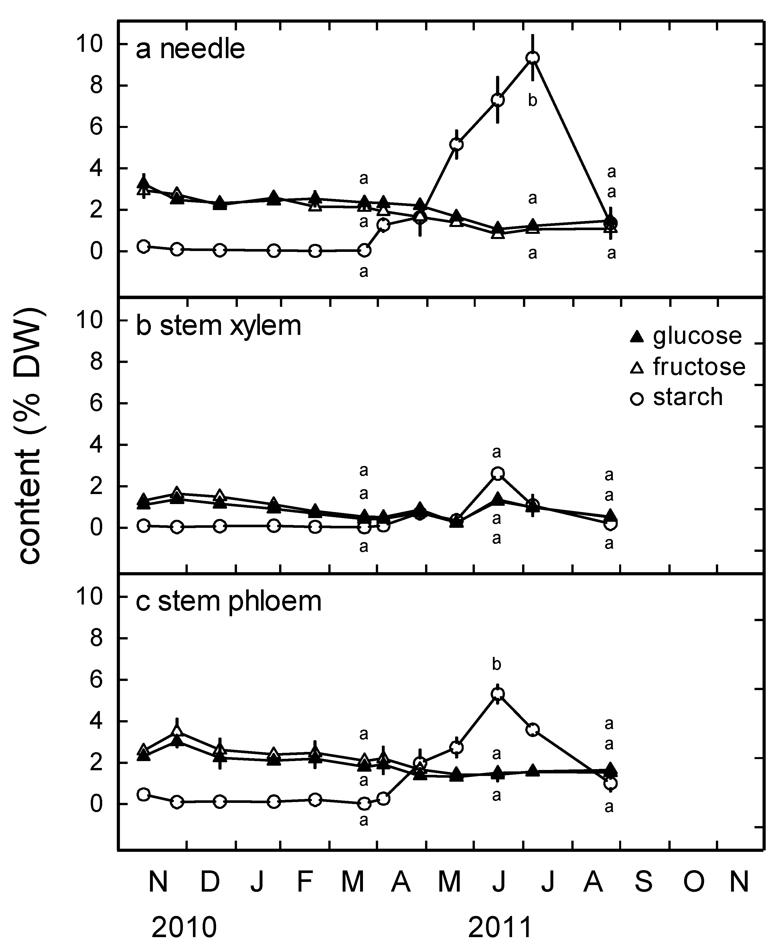
3.3. Glucose, Fructose and Starch Content

Most pronounced courses in carbohydrate contents were found in needles (Figure 5a): While glucose and fructose concentrations gradually decreased from November 2010 (3.3% ± 0.5% DW glucose, 2.9% ± 0.4% DW fructose) to August 2011 (1.5% ± 0.3% DW glucose, 1.1% ± 0.2% DW fructose), starch contents were low until May 2010, peaked in July 2011 (9.3% ± 1.1% DW), and then decreased to 1.4% ± 0.8% DW (August 2011). July values were significantly higher than those in March and August. Annual course patterns were similar but less clear in stem sapwood and secondary phloem, with the highest starch contents reached in July (Figure 5b,c).
Histochemical observation of the starch grains (Figure 6) in needles and secondary phloem confirmed the results of the chemical analyses. Firstly, a lower number of starch grains were found in the living cells of the secondary phloem, i.e., parenchyma cells of the rays and of the tangential parenchyma bands between the sieve cells (Figure 6a), compared to the needles (Figure 6b,c) and secondly, the maximum amount of starch was present in July 2011 in both organs. Starch grains were detectable until November 2011, but not in the period from 21 December 2010 until 22 March 2011. In midwinter and early spring, when starch was absent in the needles (Figure 7a), several white-colored vesicles were found in the mesophyll cells, and the cells of the endodermis contained small translucent (hydrophobic) droplets. During late spring and summer, no such droplets were found in the endodermis (Figure 7b); instead, the cells were filled with quite big starch grains. Starch grains in the endodermis were much bigger than those in the parenchyma cells of the mesophyll or near the vascular bundles (Figure 6b). The Casparian strip of the endodermis is impregnated with lignin, as indicated by its turquoise-greenish color when stained with toluidine blue.

4. Discussion

Winter stress induced embolism in Pinus mugo, as observed in other conifers growing at high elevation. Pinus mugo also showed recovery during late winter and spring, and findings indicate that underlying mechanisms were similar to those found in timberline trees: pit aspiration probably enabled the isolation of embolized tracheids until their functionality was re-established (Figure 3d), and changes in carbohydrate contents demonstrate cellular activity during late winter, which might enable osmotic adjustment to create driving forces for water re-distribution. However, in contrast to our hypothesis, the annual course in water potential (Ψ), loss of conductivity (LC), and related structural parameters were not more complex than that in trees, despite the shrub habit of Pinus mugo and respective more snow coverage.
The maximum LC in Pinus mugo reached during the study period was about 40% (Figure 3c), which is remarkably similar to the LC in Picea abies (43.4% ± 1.6%; [1]) observed in the same winter season and at the same study site. Accordingly, the minimum Ψ was about −1.8 MPa in Pinus mugo (Figure 3b) and −1.69 ± 0.11 MPa in Picea abies. This indicates that conifer shrubs and trees were exposed to identical winter drought stress, and that the more frequent cover by snow did not protect shrubs from dehydration during winter months. This may be due to the following two explanations: First, note that uppermost branches were harvested during winter. These were probably the branches experiencing the most stress and being protected by snow for relatively short periods only. [33] already demonstrated that branches permanently covered by snow develop only moderate Ψ and LC. Second, trees can store a lot of water in the trunk, which may buffer water losses induced by frost drought. [31] found water storage to increase and drought effects to decrease with tree size. The small branches of Pinus mugo cannot store larger amounts of water and thus Ψ decreased, even when drought stress was lower than in neighboring trees. In the branches harvested for this study, we could also show that even the bordered pits of the xylem from the needle vascular bundles can be aspirated during winter time (Figure 4a,b) in order to prevent the further embolism of adjacent tracheids. Relatively short-term snow covers on upper branches may also explain why we did not observe complex sequences of embolism formation and recovery but, as in trees [7,9,10,11,14,15], a period of embolism formation during midwinter and of recovery during late winter and spring. Similar to findings in Picea abies [1], embolism formation and repair thereby corresponded to changes in pit aspiration and carbohydrate contents.
The course of the proportion of aspirated pits followed the course of LC during the recovery period with decreasing LC and the opening of pits from late winter onwards (Figure 3c,d). In contrast, the proportion of aspirated pits was already high from the beginning of winter, while LC progressively increased. This indicates that some pits closed independently of embolism formation, maybe due to early frost events. Accordingly, the correlation (earlywood pits r2 = 0.25) of the proportion of aspirated pits versus LC was weaker than that observed in Picea abies [1]. Overall, earlywood tracheids showed a higher proportion of aspirated pits, while latewood pits remained open more frequently. It remains to be studied whether this is due to lower embolism or less efficient pit aspiration in latewood [35]. As earlywood tracheids and their pits are hydraulically much more relevant than latewood tracheids, the findings again underline that the special conifer pit architecture [28,39] plays a central role for conifer hydraulic safety and pit aspiration, as well as probably for embolism recovery in secondary xylem (Figure 2), and even in the xylem of the vascular bundles of the needles (Figure 4).
Observed recovery also corresponded to changes in carbohydrate contents (Figure 5). Similar to Picea abies [1], a first peak in starch concentrations was observed in late winter (4 April), indicating relevant redistributions of carbohydrates within the plants. However, starch was not present year-round in Pinus mugo secondary phloem and needles. In the period from December until March, lipid droplets were found, especially in the endodermis cells (Figure 7a). According to [40], fats or lipid droplets are present the whole year; in some species, they are the only visible winter food reserve. Such species, including mainly diffuse porous angiosperm, but also the genus Pinus, are termed as “fat trees” [41,42]. In our study, starch grains were not present until the end of March (Figure 6). It has to be considered that the first changes in starch contents were observed when the snow cover was still present and the plants did not have access to soil water. However, the link between changes in carbohydrate contents and xylem refilling has yet to be demonstrated. According to [22], it is likely that carbohydrates are required to create an osmotic force, which enables pulling water into water films on the cell walls of embolized conduits also; see [43,44,45]. However, we did not observe relevant changes in glucose or fructose during recovery, probably because the formation of osmotic active sugars occurs locally and in comparably low overall concentrations. Unfortunately, a separate analysis of tracheid contents, as in the much bigger Populus [44] vessels, is hardly possible in conifers. The remarkable drop in starch content in late summer (Figure 5 and Figure 6) indicates the beginning of transition to lipid pools and the displacement of storage pools to stem and roots.
Overall, embolism and refilling in Pinus mugo were surprisingly similar to the seasonal patterns observed in other conifers. The relevance of snow cover protection for both embolism formation and repair complicates predictions of the winter stress situation under future climate: in the Alps, the temperature increase was above the global average during the past decades [46,47], so that reductions in snow cover heights and durations have to be expected in future. An estimation of future precipitation changes is difficult, as the mountain terrain is complex. However, some studies for instance indicate an increase of spring droughts [48,49,50]. A reduced snow cover may reduce the time of blocked access to soil water and thus reduce frost drought. At the same time, the lack of snow protection may expose the plants to increased temperature amplitudes and freeze–thaw cycles, which can also induce embolism (see introduction). Furthermore, a lack of snow cover in late winter may hinder refilling processes, when water cannot be taken up over the branch surface. Shrub-like growth forms, such as Pinus mugo, might be especially affected by changes in snow cover regimes, but more studies are required to unravel the significance and dynamics of processes during embolism formation and recovery.

Author Contributions

Conceptualization, S.M. and S.R.; methodology, S.M., P.S., and S.R.; formal analysis, S.M., P.S., and S.R.; investigation, S.M., P.S., and S.R.; resources, S.M. and S.R.; data curation, S.M., P.S., and S.R.; writing—original draft preparation, S.M. and S.R.; writing—review and editing, S.M. and S.R.; visualization, S.M. and S.R.; supervision, S.M.; project administration, S.M. and S.R.; funding acquisition, S.M. and S.R.

Funding

This research was funded by the Austrian Science Fund, grant number I826–B25 and P32203_B.

Acknowledgments

We thank Birgit Dämon for his excellent assistance. Climate data were provided by ZAMG Zentralanstalt für Meteorologie und Geodynamik, Austria.

Conflicts of Interest

The authors declare no conflict of interest.

References

  1. Mayr, S.; Schmid, P.; Laur, J.; Rosner, S.; Charra-Vaskou, K.; Dämon, B.; Hacke, U.G. Uptake of water via branches helps timberline conifers refill embolized xylem in late winter. Plant Physiol. 2014, 164, 1731–1740. [Google Scholar] [CrossRef] [PubMed]
  2. Tyree, M.T.; Zimmermann, M.H. Xylem Structure and the Ascent of Sap, 2nd ed.; Springer: Berlin, Germany, 2002; p. 284. [Google Scholar] [CrossRef]
  3. Hacke, U.G.; Sperry, J.S. Functional and ecological xylem anatomy. Perspect. Plant Ecol. Evol. Syst. 2001, 4, 97–115. [Google Scholar] [CrossRef] [Green Version]
  4. Sperry, J.S. Evolution of water transport and xylem structure. Int. J. Plant Sci. 2003, 164, S115–S127. [Google Scholar] [CrossRef]
  5. Choat, B.; Jansen, S.; Brodribb, T.J.; Cochard, H.; Delzon, S.; Bhaskar, R.; Bucci, S.J.; Field, T.S.; Gleason, S.M.; Hacke, U.G.; et al. Global convergence in the vulnerability of forests to drought. Nature 2012, 491, 752–755. [Google Scholar] [CrossRef] [Green Version]
  6. Pittermann, J.; Sperry, J.S. Analysis of freeze-thaw embolism in conifers. The interaction between cavitation pressure and tracheid size. Plant Physiol. 2006, 140, 374–382. [Google Scholar] [CrossRef]
  7. Mayr, S.; Schwienbacher, F.; Bauer, H. Winter at the alpine timberline. Why does embolism occur in Norway spruce but not in stone pine? Plant Physiol. 2003, 131, 780–792. [Google Scholar] [CrossRef]
  8. Mayr, S.; Wieser, G.; Bauer, H. Xylem temperatures during winter in conifers at the alpine timberline. Agric. For. Meteorol. 2006, 137, 81–88. [Google Scholar] [CrossRef]
  9. Sparks, J.P.; Black, R.A. Winter hydraulic conductivity end xylem cavitation in coniferous trees from upper and lower treeline. Arct. Antarct. Alp. Res. 2000, 32, 397–403. [Google Scholar] [CrossRef]
  10. Mayr, S.; Wolfschwenger, M.; Bauer, H. Winter-drought induced embolism in Norway spruce (Picea abies) at the Alpine timberline. Physiol. Plant. 2002, 115, 74–80. [Google Scholar] [CrossRef]
  11. Mayr, S.; Hacke, U.; Schmid, P.; Schwienbacher, F.; Gruber, A. Frost drought in conifers at the alpine timberline: Xylem dysfunction and adaptations. Ecology 2006, 87, 3175–3185. [Google Scholar] [CrossRef]
  12. Brodribb, T.; Cochard, H. Hydraulic failure defines the recovery and point of no return in water-stressed conifers. Plant Physiol. 2009, 149, 575–584. [Google Scholar] [CrossRef] [PubMed]
  13. Hammond, W.M.; Yu, K.L.; Wilson, L.A.; Will, R.E.; Anderegg, W.R.; Adams, H.D. Dead or dying? Quantifying the point of no return from hydraulic failure in drought-induced tree mortality. New Phytol. 2019, 223, 1834–1843. [Google Scholar] [CrossRef] [PubMed]
  14. Sperry, J.S.; Nichols, K.L.; Sullivan, J.E.M.; Eastlack, S.E. Xylem embolism in ring-porous, diffuse-porous, and coniferous trees of northern Utah and interior Alaska. Ecology 1994, 75, 1736–1752. [Google Scholar] [CrossRef]
  15. McCulloh, K.A.; Johnson, D.M.; Meinzer, F.C.; Lachenbruch, B. An annual pattern of native embolism in upper branches of four tall conifer species. Am. J. Bot. 2011, 98, 1007–1015. [Google Scholar] [CrossRef]
  16. Maurel, C.; Verdoucq, L.; Luu, D.T.; Santoni, V. Plant aquaporins: Membrane channels with multiple integrated functions. Annu. Rev. Plant Biol. 2008, 59, 595–624. [Google Scholar] [CrossRef]
  17. Gomes, D.; Agasse, A.; Thiébaud, P.; Delrot, S.; Gerós, H.; Chaumont, F. Aquaporins are multifunctional water and solute transporters highly divergent in living organisms. Biochim. Biophys. Acta 2009, 1788, 1213–1228. [Google Scholar] [CrossRef] [Green Version]
  18. Heinen, R.B.; Ye, Q.; Chaumont, F. Role of aquaporins in leaf physiology. J. Exp. Bot. 2009, 60, 2971–2985. [Google Scholar] [CrossRef] [Green Version]
  19. Holbrook, N.M.; Zwieniecki, M.A. Embolism repair and xylem tension: Do we need a miracle? Plant Physiol. 1999, 120, 7–10. [Google Scholar] [CrossRef]
  20. Hacke, U.G.; Sperry, J.S. Limits to xylem refilling under negative pressure in Laurus nobilis and Acer negundo. Plant Cell Environ. 2003, 26, 303–311. [Google Scholar] [CrossRef]
  21. Zwieniecki, M.A.; Holbrook, N.M. Confronting Maxwell’s demon: Biophysics of xylem embolism repair. Trends Plant Sci. 2009, 14, 530–534. [Google Scholar] [CrossRef]
  22. Nardini, A.; Lo Gullo, M.A.; Salleo, S. Refilling embolized xylem conduits: Is it a matter of phloem unloading? Plant Sci. 2011, 180, 604–611. [Google Scholar] [CrossRef] [PubMed]
  23. Laur, J.; Hacke, U.G. Exploring Picea glauca aquaporins in the context of needle water uptake and xylem refilling. New Phytol. 2014, 203, 388–400. [Google Scholar] [CrossRef] [PubMed]
  24. Katz, C.; Oren, R.; Schulze, E.-D.; Milburn, J.A. Uptake of water and solutes through twigs of Picea abies (L.) Karst. Trees 1989, 3, 33–37. [Google Scholar] [CrossRef]
  25. Sparks, J.P.; Campbell, G.S.; Black, R.A. Water content, hydraulic conductivity, and ice formation in winter stems of Pinus contorta: A TDR case study. Oecologia 2001, 127, 468–475. [Google Scholar] [CrossRef]
  26. Burgess, S.S.O.; Dawson, T.E. The contribution of fog to the water relations of Sequoia sempervirens (D. Don): Foliar uptake and prevention of dehydration. Plant Cell Environ. 2004, 27, 1023–1034. [Google Scholar] [CrossRef]
  27. Limm, E.B.; Simonin, K.A.; Bothman, A.G.; Dawson, T.E. Foliar water uptake: A common water acquisition strategy for plants of the redwood forest. Oecologia 2009, 161, 449–459. [Google Scholar] [CrossRef]
  28. Pittermann, J.; Sperry, J.S.; Hacke, U.G.; Wheeler, J.K.; Sikkema, E.H. Torus-margo pits help conifers compete with angiosperms. Science 2005, 310, 1924. [Google Scholar] [CrossRef]
  29. Hacke, U.G.; Jansen, S. Embolism resistance of three boreal conifer species varies with pit structure. New Phytol. 2009, 182, 675–686. [Google Scholar] [CrossRef]
  30. Plavcová, L.; Jansen, S.; Klepsch, M.; Hacke, U.G. Nobody’s perfect: Can irregularities in pit structure influence vulnerability to cavitation? Front. Plant Sci. 2013, 12, 453. [Google Scholar] [CrossRef]
  31. Mayr, S.; Charra-Vaskou, K. Winter at the alpine timberline causes complex within-tree patterns of water potential and embolism in Picea abies. Physiol. Plant. 2007, 131, 131–139. [Google Scholar] [CrossRef]
  32. Mayr, S.; Gruber, A.; Bauer, H. Repeated freeze-thaw cycles induce embolism in drought stressed conifers (Norway spruce, stone pine). Planta 2003, 217, 436–441. [Google Scholar] [CrossRef] [PubMed]
  33. Mayr, S.; Gruber, A.; Schweinbacher, F.; Dämon, B. Winter-embolism in a “Krummholz”-Shrub (Pinus mugo) growing at the alpine timberline. Austrian J. For. Sci. 2003, 1, 29–38. [Google Scholar]
  34. Sperry, J.S.; Donnelly, J.R.; Tyree, M.T. A method for measuring hydraulic conductivity and embolism in xylem. Plant Cell Environ. 1988, 11, 35–40. [Google Scholar] [CrossRef]
  35. Liese, W.; Bauch, J. On the closure of bordered pits in conifers. Wood Sci. Technol. 1967, 1, 1–13. [Google Scholar] [CrossRef]
  36. Rosner, S.; Gierlinger, N.; Klepsch, M.; Karlsson, B.; Evans, R.; Lundqvist, S.-O.; Světlík, J.; Børja, I.; Dalsgaard, L.; Andreassen, K.; et al. Hydraulic and mechanical dysfunction of Norway spruce sapwood due to extreme summer drought in Scandinavia. For. Ecol. Manag. 2018, 409, 527–540. [Google Scholar] [CrossRef]
  37. Bauer, H.; Plattner, K.; Volgger, W. Photosynthesis in Norway spruce seedlings infected by the needle rust Chrysomyxa rhododendri. Tree Physiol. 2000, 20, 211–216. [Google Scholar] [CrossRef]
  38. Bergmeyer, H.U.; Bernt, E. Sucrose. In Methods of Enzymatic Analysis; Bergmeyer, H.U., Ed.; Academic Press: New York, NY, USA, 1974; Volume 3, pp. 1176–1179. [Google Scholar]
  39. Hacke, U.G.; Sperry, J.S.; Pittermann, J. Analysis of circular bordered pit function–II. Gymnosperm tracheids with torus-margo pit membranes. Am. J. Bot. 2004, 91, 386–400. [Google Scholar] [CrossRef]
  40. Murmanis, L.; Evert, R.F. Parenchyma cells of secondary phloem in Pinus strobus. Planta 1967, 73, 301–318. [Google Scholar] [CrossRef]
  41. Ziegler, H. Storage, Mobilization and Distribution of Reserve Material in Trees. In The Formation of Wood in Forest Trees; Zimmermann, M.H., Ed.; Academic Press: New York, NY, USA, 1964; pp. 303–320. [Google Scholar]
  42. Höll, W. Storage and mobilization of carbohydrates and lipids. In Trees-Contributions to Modern Tree Physiology; Rennenberg, H., Eschrich, W., Ziegler, H., Eds.; Blackhuhys Publishers: Leiden, The Netherlands, 1997; pp. 197–211. [Google Scholar]
  43. Bucci, S.J.; Scholz, F.G.; Goldstein, G.; Meinzer, F.C.; Sternberg, L.D.L. Dynamic changes in hydraulic conductivity in petioles of two savanna tree species: Factors and mechanisms contributing to the refilling of embolized vessels. Plant Cell Environ. 2003, 26, 1633–1645. [Google Scholar] [CrossRef]
  44. Secchi, F.; Zwieniecki, M.A. Analysis of xylem sap from functional (nonembolized) and nonfunctional (embolized) vessels of Populus nigra: Chemistry of refilling. Plant Physiol. 2012, 160, 955–964. [Google Scholar] [CrossRef]
  45. Barnard, D.M.; Lachenbruch, B.; McCulloh, K.A.; Kitin, P.; Meinzer, F.C. Do ray cells provide a pathway for radial water movement in the stems of conifer trees? Am. J. Bot. 2013, 100, 322–331. [Google Scholar] [CrossRef] [PubMed] [Green Version]
  46. Böhm, R.; Auer, I.; Brunetti, M.; Maugeri, M.; Nanni, T.; Schöner, W. Regional Temperature variability in the European Alps: 1760-1998 from homogenized instrumental time. Int. J. Climatol. 2001, 1801, 1779–1801. [Google Scholar] [CrossRef]
  47. Rebetez, M.; Reinhard, M. Monthly air temperature trends in Switzerland 1901–2000 and 1975–2004. Theor. Appl. Climatol. 2008, 91, 27–34. [Google Scholar] [CrossRef]
  48. Ciccarelli, N.; von Hardenberg, J.; Provenzale, A.; Ronchi, C.; Vargiu, A.; Pelosini, R. Climate variability in north-western Italy during the second half of the 20th century. Glob. Planet. Chang. 2008, 63, 185–195. [Google Scholar] [CrossRef]
  49. Beniston, M. Mountain climates and climatic change: An overview of processes focusing on the European Alps. Pure Appl. Geophys. 2005, 162, 1587–1606. [Google Scholar] [CrossRef]
  50. Elkin, C.; Gutiérrez, A.G.; Leuzinger, S.; Manusch, C.; Temperli, C.; Rasche, L.; Bugmann, H. A 2 °C warmer world is not safe for ecosystem services in the European Alps. Glob. Chang. Biol. 2013, 19, 1827–1840. [Google Scholar] [CrossRef] [PubMed]
Figure 1. Information on histological anatomical investigations in Pinus mugo (a) branches and (b) needles. Regions of interest for histological staining are indicated with orange circles, the arrows in (a) indicate parenchymatic ray cells, the arrow in (b) indicates the endodermis, m, mesophyll; p, parenchyma cell; s, sieve cell.
Forests 10 00941 g001
Figure 2. Examples of open and aspirated bordered pits in sapwood of Pinus mugo. (a) 24 November 2010, (b) 22 February 2011, (c) 15 June 2011, and (d) 6 July 2011, open arrows point at open bordered pits, closed arrows on aspirated bordered pits, reference bars = 50 µm.
Forests 10 00941 g002
Figure 3. Climate, course of water potential and content, loss of conductivity, and pit aspiration. (a) Air temperatures (T; daily minimum, mean, and maximum) and daily sum of precipitation (prec.) from Mt. Patscherkofel (2.252 m provided by ZAMG Zentralanstalt für Meteorologie und Geodynamik, Austria). (b) Water potential (Ψ) and water content in stem bark and xylem. (c) Loss of conductivity in branch xylem (LC). (d) Proportion of aspirated pits (asp. pits) in earlywood and latewood. Mean ± SE. Different letters indicate significant differences measurements on 22 February, 19 May, and the sampling date of the highest values within this period.
Forests 10 00941 g003
Figure 4. Pit aspiration in the xylem of the needle vascular bundles at different dates. (a,b) 24 November 2010, (c,d) 15 June 2011, and (e) 10 September 2011; p, parenchyma cell; ph, phloem; t, tracheid; tt, transfusion tracheid; xy, xylem; open arrows point at open pits, closed arrows at aspirated pits, reference bars = 20 µm.
Forests 10 00941 g004
Figure 5. Course of glucose, fructose, and starch content. Carbohydrate contents (% of dry weight) in (a) needles, (b) stem xylem, and (c) stem phloem. Mean ± SE. Different letters indicate significant differences between measurements on 22 March, 24 August, and the sampling date of the highest values within this period.
Forests 10 00941 g005
Figure 6. Histochemical detection of starch in secondary phloem and needles. (a) Secondary phloem (and parts of sapwood) stained with Lugol´s solution, (b) areas next to the needle endodermis stained with Lugol´s solution, and (c) areas next to the needle endodermis stained with toluidine blue at different times of the season. m, mesophyll; p, parenchyma cell in the secondary phloem; r, ray cell in the secondary phloem; s, sieve cells; xy, secondary xylem; stars indicate the locations of the endodermis, bar = 100 µm (the bar is representative for all photos). For regions of interest, see Figure 1.
Forests 10 00941 g006
Figure 7. Endodermis and the adjacent tissues of Pinus mugo needles in winter and summer. (a) 15 January 2011 and (b) 6 July 2011. e, endodermis; m, mesophyll; p, parenchyma cell; tt, transfusion tracheid; black arrows point at the Casparian strips, orange arrows point at translucent droplets, and the yellow arrow points to a starch grain, bar = 50 µm. For orientation, see Figure 1.
Forests 10 00941 g007

