Article

Differential Expression of Genes Related to the Formation of Giant Leaves in Triploid Poplar

1
Beijing Advanced Innovation Center for Tree Breeding by Molecular Design, Beijing Forestry University, Beijing 100083, China
2
National Engineering Laboratory for Tree Breeding, Beijing Forestry University, Beijing 100083, China
3
Key Laboratory for Genetics and Breeding in Forest Trees and Ornamental Plants, Ministry of Education, College of Biological Sciences and Technology, Beijing Forestry University, Beijing 100083, China
*
Author to whom correspondence should be addressed.
Forests 2019, 10(10), 920; https://doi.org/10.3390/f10100920
Submission received: 21 August 2019 / Revised: 15 October 2019 / Accepted: 16 October 2019 / Published: 19 October 2019
(This article belongs to the Special Issue Forest Genetics and Tree Improvement)

Abstract

:
Plant polyploids tend to have large leaves, but their formation mechanism has not yet been well explained. Therefore, daily transcriptomic differences between triploids and diploids from a synthetic Populus sect. Tacamahaca three times a day (i.e., 04:00, 09:00, and 21:00) were investigated using high-throughput RNA-seq analysis. In this study, we identified several transcription factors associated with giant leaves. The combined effects included the high expression of several transcription factors (WRKY, MYB, etc.) and hormone-related genes (e.g., activates auxin, cytokine, and brassinosteroid synthesis-related genes) that accelerate the synthesis and accumulation of endogenous hormones. High levels of growth hormones were maintained by reducing the genes’ expression of hormone metabolism and degradation. The coordination of hormones accumulated sufficient materials and energy for leaf growth and development. Thereby, cell division and growth were accelerated which enhanced the photosynthesis of leaves, and the increased accumulation of photosynthetic products led to giant triploid leaves. This study lays the foundation for revealing the molecular mechanisms in the formation of giant leaves in polyploids.

1. Introduction

Giant-sized leaves are an important phenotypic characteristic of plant polyploids [1,2,3]. Tetraploids usually have significantly larger sized leaves than diploids and also grow more strongly than their diploid parents in Arabidopsis [4,5]. Polyploids ligneous plants also have a longer, wider leaf shape and larger leaf cells than diploids [6]. The giant leaves are formed by polyploidization, which increases the rate of cell elongation [7]. Compared with diploids, triploid poplars have a larger leaf area and higher volume growth [8,9,10,11]. This is because the nature of giant leaves is closely related to the growth advantages of polyploids [12].
The regulatory mechanisms for the growth and development of plant leaves are complicated and involve several important factors including the effects of cell division and differentiation on leaf formation and development. Previous research has confirmed that leaf size is influenced by the development of veins, for instance, transcription factor VRS1 controls cell wall synthesis and affects leaf vein development in barley (Hordeum vulgare L.) [13]. The lateral secondary growth of leaves is also affected by the cell division cycle. For instance, CYCD3;1 encodes a cyclin D-type protein involved in the switch from cell proliferation to the final stages of differentiation, which is transcriptionally regulated by cytokinin and brassinosteroid [14]. In addition, ICK1 and KRP2 act as cell proliferation inhibitors, as their overexpression can stop cell division and delay the expansion rate of leaves thereby inducing a smaller phenotype [15,16].
Plant endogenous hormones also play an important role in leaf formation and development. Cell proliferation and lateral leaf expansion are regulated and determined by auxin-inducible genes (e.g., ARGOS and ARF) through the auxin signaling pathway [17,18]. Brassinolide is the most common plant cell division and growth regulation hormone, and its synthetic gene CYP90C1 and downstream regulatory gene ROT3 also regulate leaf expansion [18]. Huang [19] pointed out that the development of leaves is regulated by the expression of functional genes and transcription factors. The hormone level in plant leaves is closely related to the high expression of gene products. Meanwhile, plant hormones provide feedback to regulate the expression of functional genes and transcription factors, constructing the precise process of leaf formation and growth. Therefore, what is the mechanism of giant leaf formation in polyploid plants? At present, by combining the results of transcriptome and proteome data analyses, it is proposed that the giant formation of rice triploid leaves is significantly correlated with chloroplast and hormone signal transduction related to proteins such as PSAB and ATPF [20]. A previous study in Paulownia fortune pointed out that the differentially expressed genes (DEGs) between diploids and tetraploids are enriched in energy metabolism, hormones, and cell wall formation [11]. However, complex gene regulation remains unclear in polyploid plants. As a tree model species, the formation mechanism of giant leaves in triploid poplars requires further study.
In this study, RNA-seq analysis was used to identify differential gene expression to compare the gene function of allotriploid and diploid poplars in a full-sub family within a day. Differentially expressed genes related to the metabolic regulation of leaf growth and development were analyzed carefully in order to explore the molecular mechanism of giant leaf formation in allotriploid poplars.

2. Materials and Methods

2.1. Plant Materials and Growth Conditions

The synthetic poplar allotriploid (2n = 3x = 57) and diploid (2n = 2x = 38) used in this study were full-sib progeny induced by Populus pseudosimonii × P. nigra (Zheyin3#) and Populus × beijingensis (BJY) hybridization. Thirty allotriploid (A) and diploid (D) clones were grown under 16 h light/8 h dark conditions (lights were turned on at 05:00 and turned off at 21:00) in a greenhouse at the Beijing Forestry University (Supplementary Materials Figure S1) [21]. After four months, the fully expanded fifth leaves (Figure S1) from three randomly selected allotriploid (A) clones were collected at 04:00 (F). Three further clones were randomly selected from those remaining for sampling at 09:00 (D), and a final three were randomly selected at 21:00 (N). Three biological replicates were used for each of the sampling points. Thirty diploid (D) clones were grown and sampled in the same way. From here on, allotriploids are denoted as AF (04:00), AD (09:00), and AN (21:00), and the diploids are defined as DF (04:00), DD (09:00), and DN (21:00). After sampling, the tissues were quickly frozen in liquid nitrogen and stored at −70 °C until RNA isolation. When performing RT-PCR, each sample RNA was divided into four parts as technical duplications.

2.2. Total RNA Isolation, cDNA Preparation, and Transcriptome Sequencing

A TRIzol reagent kit (Invitrogen, Carlsbad, CA, USA) was used to extract the total RNA in the leaf samples, and an RNase-Free DNaseSet (Qiagen China, Shanghai, China was used to purify the RNA. To detect the integrity of the RNA, we performed agarose gel electrophoresis and a NanoDrop 2000 biological analyzer (Thermo Fisher Scientific Inc., Wilmington, DE, USA) was used to detect the concentration of RNA. After this quality control check (1.6 ≤ 28s/18s ≤ 2.0), high-quality RNA was used for subsequent sequencing. Following Wang et al. [22], an Ion total RNA-Seq Kit v2 (Life Technologies, USA) was used to build the cDNA libraries from different time points. Transcriptome sequencing of leaf samples was performed on the Ion Proton platform (Life Technologies) by Shanghai Novelbio Biological Technology Co. Ltd. Original reads of less than 50 bp were filtered out, and the rest were mapped to the genome of Populus trichocarpa using MapSplice [23].
The transcript abundance was shown by reads per kilobase per million mapped reads (RPKM). Read counts were normalized using DESeq [24] and genes were considered differentially expressed if they had a fold change >2 and a p-value < 0.05. Information on gene annotation was acquired from the NCBI (http://www.ncbi.nlm.nih.gov/) [25], JGI (http://jgi.doe.gov/) [26], PopGenie (http://popgenie.org/) [27], Tair (http://www.arabidopsis.org/) [28], and KEGG (http://www.kegg.jp/) databases [29]. A FISHER test was used to test the statistical enrichment of differently expressed genes in KEGG pathways [30,31]. Transcription factors were identified using Populus trichocarpa transcription factors from the Plant Transcription Factor Database (http://planttfdb.cbi.edu.cn/) [32].

2.3. RT-PCR Validation of Differentially Expressed Genes

A FastQuant RT kit (with gDNase; Tiangen Biotech Co. Ltd., Beijing, China) was used to perform the reverse transcription reaction. The real-time polymerase chain reaction (RT-PCR) was accomplished using a SuperReal PreMix Plus (SYBR Green) kit (Tiangen Biotech) using an Applied Biosystems 7500 Fast instrument (AB Ltd., USA). The RT-PCR master mix included 10 µL 2× SuperReal PreMix Plus, 0.6 µL forward primer, 0.6 µL reverse primer, 2 µL cDNA template, 0.4 µL 50× ROX Reference Dye, and 6.4 µL RNase-free ddH2O. The RT-PCR was performed using 39 cycles of the following conditions: 95 °C for 15 min for pre-degeneration, 95 °C for 10 s for degeneration, 58 °C for 30 s for annealing, 72 °C for 30 s for extension. Afterward, the samples were heated to 95 °C for 15 s and then 60 °C for 1 min for dissolution curve analysis. Four technical replicates and three biological replicates were used for each sample and randomly selected gene. The sequences of primers used in the present study were designed using Primer3Plus (http://www.primer3plus.com/) [33] and are listed in Table S1. Actin (accession number: EF145577) was chosen as the reference gene, and the −2–ΔΔCt method was used to calculate gene expression.

3. Results

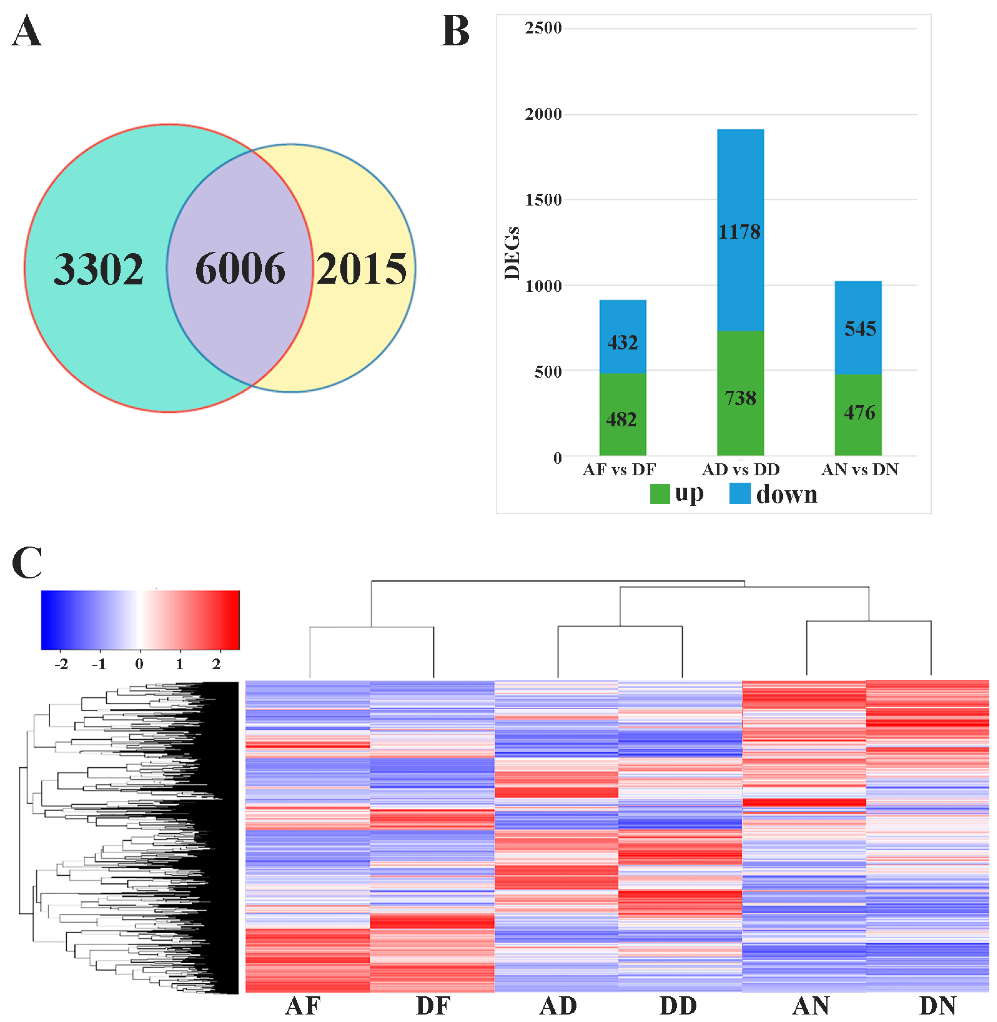
3.1. Transcriptome Sequencing and Data Analysis

The average number of total reads were 30,232,373, 25,297,715, 27,138,468, 307,643,18, 31,738,567, and 28,529,667 from 18 cDNA libraries obtained for AF, TD, TN, DF, DD, and DN, respectively. In total, 89.2%, 88.0%, 90.2%, 90.7%, 90.4%, and 89.3% of the average reads were matched to genomic locations in the AF, AD, AN, DF, DD, and DN libraries, while uniquely mapped reads accounted for 84.8%, 84.5%, 86.0%, 84.9%, 84.1%, and 84.7%. A total of 41,335 genes were obtained from the sequencing results of triploid and diploid leaves, accounting for 91.9% of the genome of Populus trichocarpa, indicating that the sequencing results were relatively reliable. The gene expression level in each sample is shown in Table S2.

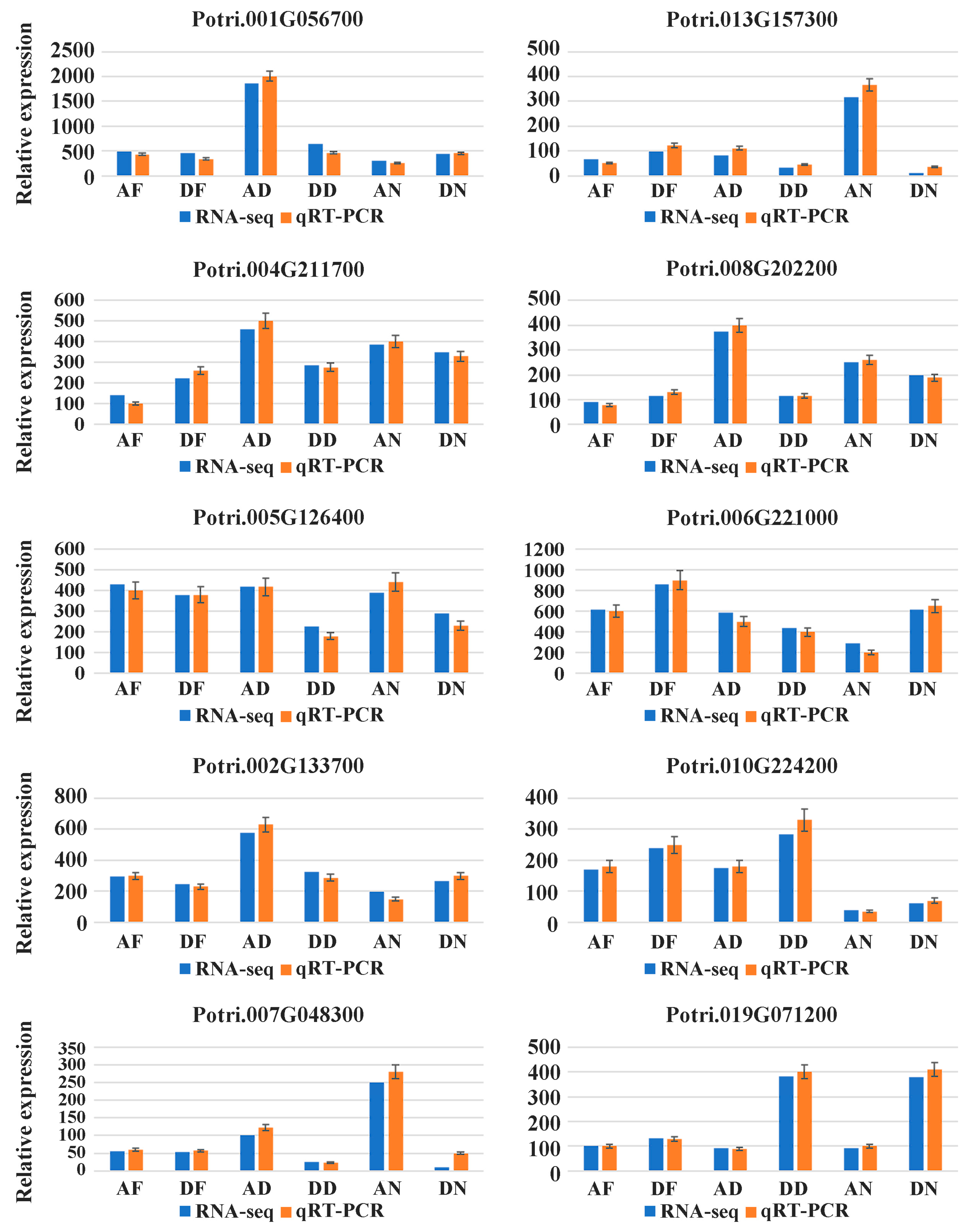
3.2. Differentially Expressed Genes and RT-PCR Validation

A total of 9308 DEGs in triploids were identified at different times of the day, while this number was 8021 in diploid plants (Figure 1A). Triploids have more genes involved in plant circadian regulation than diploids. At the same time, the number of differentially expressed genes between the triploids and diploids is shown in Figure 1B—clearly there were more DEGs at 09:00 than at other times (04:00 and 21:00). We used hierarchical clustering to visualize all genes expressed in samples based on RPKM (Figure 1C). The results showed good consistency among the three biological replicates, the significance of the DEGs among different times was greater than that among different ploidy, and the gene expression patterns were more similar at the same time. Although there was no light in the early morning and midnight, the cluster analysis was still divided into two categories. These results indicate that the gene expression patterns were different between diploid and triploid groups at different times. A total of 10 DEGs at different times were randomly selected for RT-PCR verification, and a comparison of the RT-PCR results and the RNA-seq data suggested that trends in the changes in expression of most DEGs were in agreement (Figure 2).

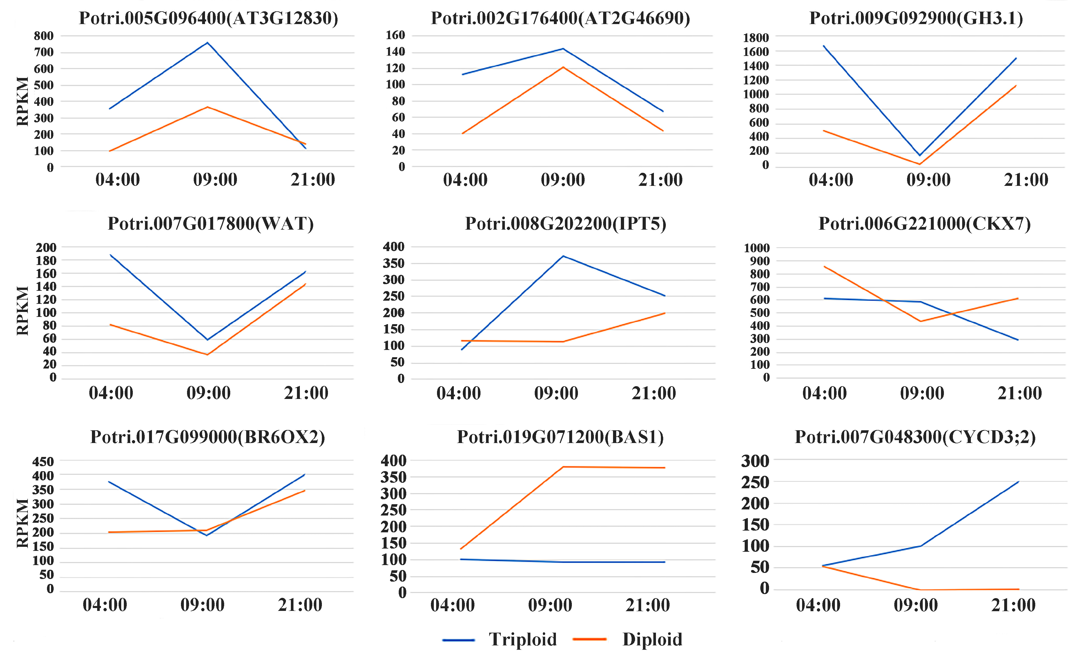
3.3. Differentially Expressed Genes Related to Plant Hormones

Plant hormones are a category of important regulatory factors that affect leaf growth and development. In this study, a series of genes related to the biosynthesis and hormone signal transduction was identified at AFs relative to DFs including the auxin biosynthetic process, auxin polar transport, and positive regulation of the auxin metabolic process (Figure 3A). Among these, five highly expressed genes were identified as auxin-related genes (Walls Are Thin 1, two SAUR-like auxin-responsive proteins, pinoid-binding protein 1, and auxin-responsive GH3); three highly expressed genes were responsible for cytokinin regulation (PSAT, ENO1, FER1, and EBP4) (Table S3). Two genes encoding the WRKY transcription factors WRKY70 and WRKY46 at AF displayed over two-fold higher expression than those at DF (Table 1).
At 09:00, genes related to the synthesis of the indoleacetic acid biosynthetic process and auxin polar transport in triploid leaves were upregulated (Figure 3B). The gene encoding auxin response factor 10 showed a 1.6 fold increased expression. Genes participating in the indoleacetic acid biosynthetic process, which encode the leucine-rich repeat protein kinase family protein, allene oxide synthase and oxophytodienoate-reductase 3, exhibited an over 2 fold increased expression in triploid relative to diploid at 09:00 (Table S4). In addition, a similarly high expression was detected in AF samples for GH3. A group of genes responsible for the cytokinin biosynthetic process, positive regulation of cytokinesis, response to cytokinin, and cytokinin-activated signaling pathway also showed increased expression at AD compared with DD. The genes encoding the isopentenyltransferase 5 and Transducin/WD40 repeat-like superfamily protein showed a 3.5 fold and 3.2 fold increased expression, respectively. A TCP transcription factor, TCP 14, displayed a 2.3 fold higher expression in triploids at 09:00 (Table 2).
At 21:00, genes related to the indoleacetic acid biosynthetic process and which responded to auxin were highly expressed in triploid plants (Figure 3C). Among them, CYP79B2 had a maximum expression of 26.0 fold in triploids relative to diploids. An auxin-responsive factor, MP, was significantly enriched at AN. Other genes coding the auxin-responsive GH3 family proteins, oxophytodienoate-reductase 3, and subtilisin-like serine endopeptidase family proteins were found to be highly expressed at AD. A WRKY transcription factor displayed over 2.3 fold higher expression at AD when compared with DD (Table 3).
This study also identified the growth hormone-related core genes and diurnal expression pattern (Figure 4). Those genes showed differential expression patterns at least once point within a day. Most genes related to auxin exhibited similar expression patterns in both ploidies within a day, and the transcript levels in triploids showed higher expression than diploids. However, transcript levels of BR- and CTK-related genes showed distinct expression patterns between triploids and diploids. The transcript levels of AT3G12830 and AT2G46690 increased rapidly from 04:00 to 09:00 and decreased from 09:00 to 21:00. Both GH3.1 and WAT1 remarkably declined at 09:00, then dropped to a low level at night. The expressions of IPT5 and CKX7 in the triploid and diploid leaves changed in the opposite directions. The transcript level of BAS1 drastically increased at 09:00 in triploids, whereas a gentle trend in diploids. The transcript level of CYCD3;2 was continuously enhanced triploids, whereas it gradually decreased in diploids.

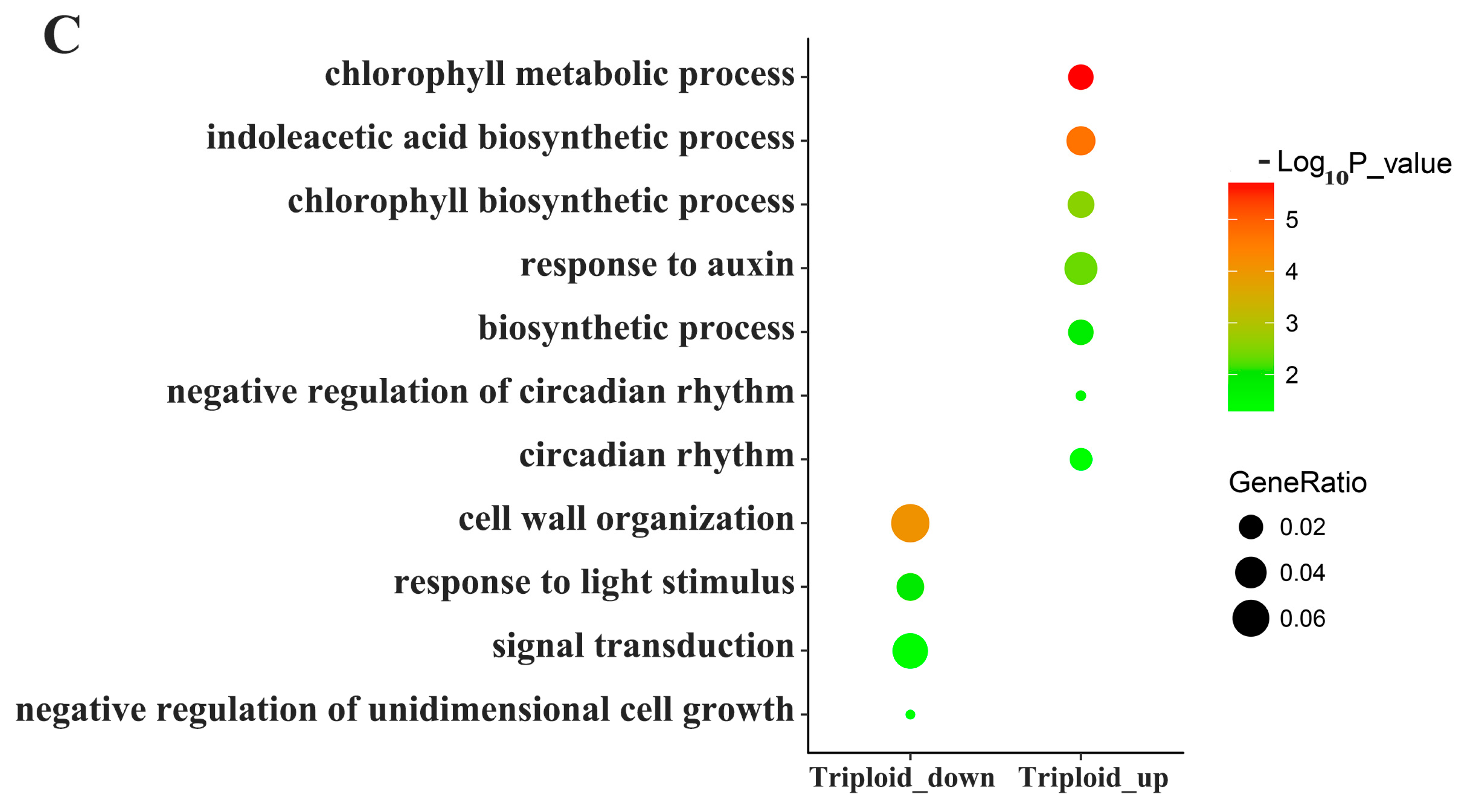
3.4. Differentially Expressed Genes Related to Photosynthesis and Energy Metabolism

Many of the genes related to photosynthesis and energy metabolism showed significant differential expression between the triploid and diploid plants. At 4 a.m., genes associated with the regulation of the anthocyanin and starch biosynthetic processes were upregulated in the triploids. Three transcripts of TT4 exhibited >2 fold higher transcript levels in triploids relative to diploids. Genes involved in the starch biosynthetic process coding glycosyl transferase, family 35, alpha-galactosidase 2, pyruvate kinase family protein, and the NAD(P)-binding Rossmann-fold superfamily protein showed changes of 2.0–3.8 fold in triploids. At 09:00, many of the upregulated genes were involved in the anthocyanin-containing compound biosynthetic process, the biosynthetic process, regulation of the anthocyanin biosynthetic process, the chlorophyll biosynthetic process, photosynthesis, light harvesting, the sugar mediated signaling pathway, the starch catabolic process, and gluconeogenesis and the starch biosynthetic process. Among them, PORA, LHB1B1, and LHCA2 coding protochlorophyllide oxidoreductase A, the light-harvesting chlorophyll–protein complex II subunit B1 and the photosystem I light harvesting complex gene 2 were upregulated in triploids. At 21:00, genes associated with the chlorophyll metabolic process, chlorophyll biosynthetic process, negative regulation of circadian rhythm, and circadian rhythm were highly expressed by more than two-fold in triploids (Figure 3C). Two transcripts of CLA1-encoding deoxyxylulose-5-phosphate synthase were upregulated by 2.0–2.1 fold.

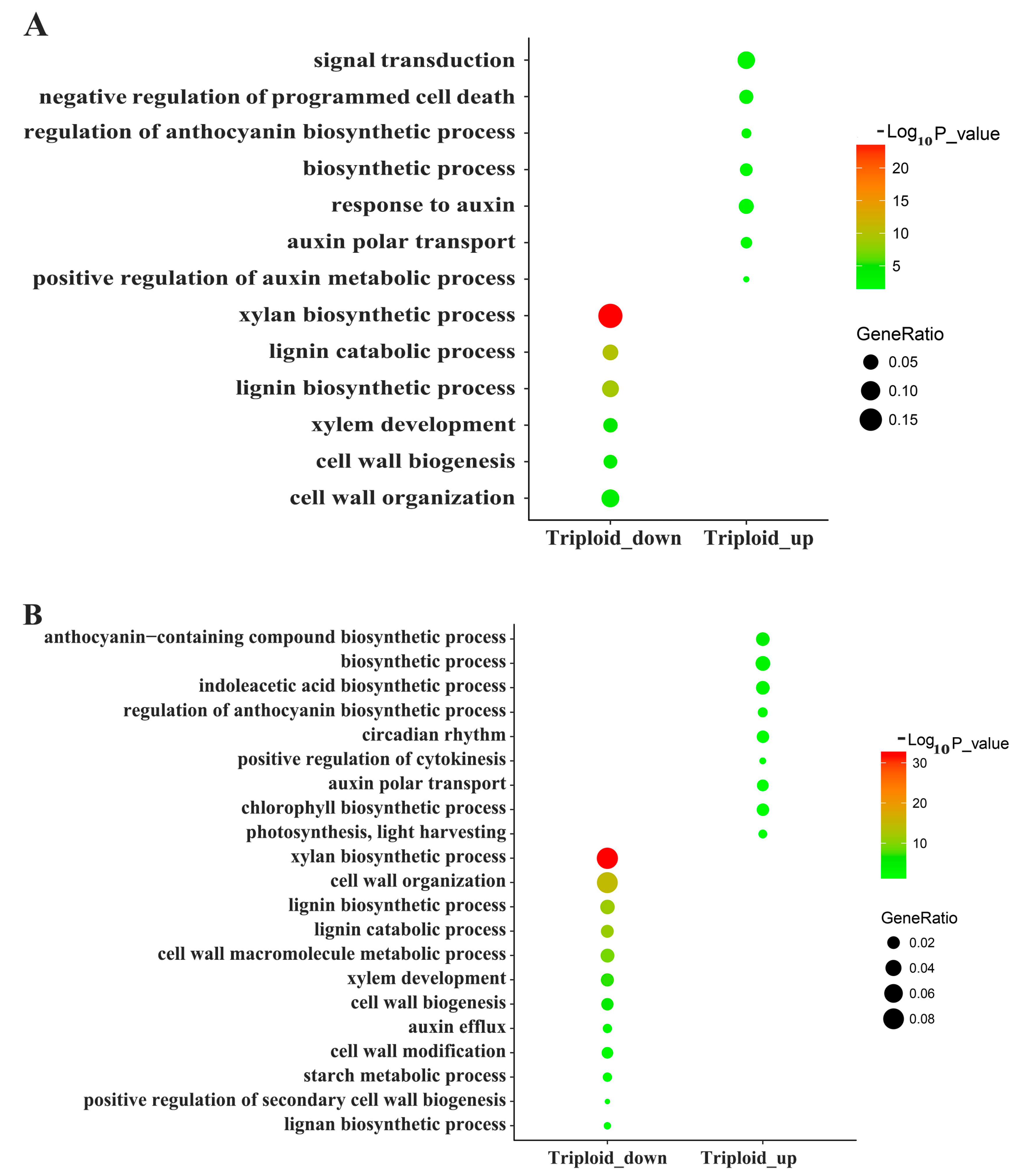
3.5. Differentially Expressed Genes Related to Lignin and Cellulose

Our study also showed that a large number of the DEGs were downregulated in the triploid library when compared with the diploid library. These DEGs were found to encode proteins and enzymes involved in lignin and cellulose synthesis. At 0:400, the gene encoding transcription factor NAC domain-containing protein 12 exhibited significantly lower expression in triploids (Table S3). Multiple plant lignin biosynthetic/catabolic and xylan biosynthetic genes were less enriched in the triploids at 04:00 and 09:00 (Table S4). These included genes encoding the cellulose synthase family proteins, such as CEV1, IRX3, CESA4, and LAC12. The analyses further identified several downregulated genes involved in cell wall biogenesis in triploid. The gene coding the SAUR-like auxin-responsive protein was observed to be highly expressed throughout the day (Table S5).

4. Discussion

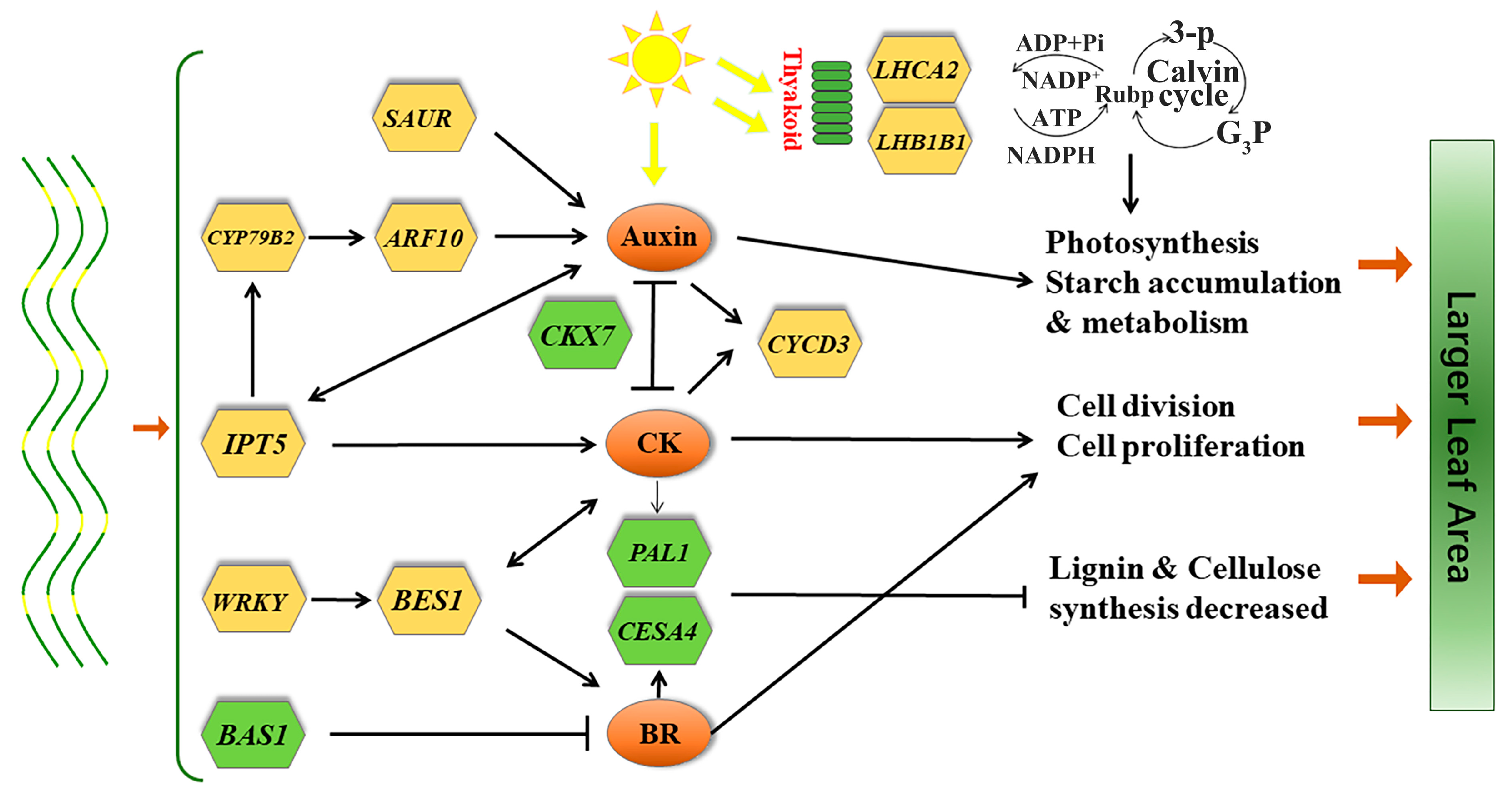
4.1. Endogenous Hormone-Related DEGs

Auxin, cytokinin, and brassinosteroids—all plant hormones that promote plant leaf growth and development—are significantly affected in triploid poplar when compared with the diploid poplar [30,33,34,35]. In this study, eight DEGs related to auxin biosynthesis and signal transduction have been found. Among them, CYP79B2 belongs to cytochrome P450 and converts Trp to indo-3-acetaldoxime (IAOx) as a precursor for IAA and indole glucosinolates [36]. Both AOS and OPR3, which encode allene oxide synthase and oxophytodienoate-reductase 3, are involved in IAA biosynthesis [37,38]. A vacuolar auxin transport facilitator, WAT1, required for auxin homoeostasis31. Auxin regulation factor 10(ARF10) and GH3.1 regulate cell proliferation and increase in response to high concentrations of auxin, and MP encodes a transcription factor (IAA24) that mediates vascular development in leaves [39]. Both AT3G12830 and AT2G46690 belong to the group of SAUR10-clade genes that generally induce cell-elongation [40]. Cytochrome P450 family members were recognized as regulating organ size and development; at 21:00, CYP79B2-induced genes related to indoleacetic acid biosynthesis were highly expressed, promoting auxin biosynthesis. At the same time, the MP response of the transcription factors to a high concentration of auxin also showed a tendency towards high expression in triploids. In the early morning, the accumulation of auxin was transported by WAT1 to activate the auxin responsive protein GH3.1. During the day, allene oxide synthase and oxophytodienoate-reductase 3 were highly expressed, and the auxin response transcription factor ARF10 was also significantly upregulated in the triploid. Compared with the diploid, the triploid had the characteristics of rapid and efficient synthesis of auxin and enhanced transport capacity. Through the regulation of related key genes by the rapid accumulation of auxin, the triploid promoted cell division and elongation leading to an increase in the leaf area.
Cytokinin is a key regulator of plant cell division [41]. In this study, genes positively correlated with the biosynthesis of cytokines were significantly upregulated in triploid, while other genes playing a negative regulation role were downregulated in triploid. A gene IPT5 encodes adenosine phosphate-isopentenyltransferase, which is a rate-limiting enzyme participating in the first step of the biosynthesis of cytokines [42]. When the expression of the IPT5 gene is activated in leaves, the content of cytokinin is increased and the senescence of leaf cells is delayed [43]. The NEDD1 homolog gene plays a critical role in microtubule organization during mitosis, and its function is similar to the gamma–tubulin complex [44]. A transcription factor TCP14 that responds to cytokines and regulates cell proliferation in developing leaves [45]. Overexpression of MYB2 would enhance expression of cytokinin-synthesizing isopentenyltransferase genes, which contain higher levels of cytokinin [46]. A gene CKX7 encodes a protein whose sequence is similar to cytokinin oxidase/dehydrogenase, which catalyzes the degradation of cytokinin [47]. In this study, high expression of isopentenyltransferase genes, activated by transcription factor MYB2, accelerated the biosynthesis process of cytokines. Transcription factor TCP14 responds to high cytokines and promotes cell proliferation. The overall expression abundance of the CKX7 gene was lower in diploids, indicating that the cytokinin degradation of the triploid was sluggish. In the early morning hours, PAST and ENO1, which encode phosphoserine methyltransferase and enolase 1, responded to the cytokinin to regulate the cell cycle. It was proven that the triploid accumulated cytokinin more efficiently and degraded slower than the diploid, and its genes were regulated to accelerate cell division.
Brassin is a steroid that regulates plant leaf growth and development [48]. Brassin increased relative water content, nitrate reductase activity, chlorophyll content, and photosynthesis. These beneficial effects resulted in a higher leaf area, biomass production, grain yield, and yield-related parameters in plants [49]. The transcription factors WRKY70 and WRKY46 promoted the synthesis of BR by mediating the gene expression of the BES1 transcription factor family [50]. The transcription factor BAS1 represents one of the control points between multiple photoreceptor systems and brassinosteroid signal transduction, reducing the levels of brassinosteroids [51]. The BR6OX2-encoded P450 enzyme is the last step in the biosynthesis of brassinolide [52], its overexpression could promote the vegetative growth of plants [53]. Moreover, there is a feedback regulation mechanism between BR6OX2 and BAS1, which reduces the expression level of BR6OX1 and should improve the transcription level of BAS1 [54]. It was proven that BR hormone levels and related genes play important roles in promoting the division and expansion of leaf cells in Arabidopsis thaliana [55]. Before dawn, the WRKY transcription factors were highly expressed, but there was no significant difference in BES1 in the triploid. During the day, the transcription factors WRKY and BES1 showed high expression in the triploid, suggesting that there was lag between the upstream and downstream genes. The transcription factor BES1 was activated by WRKY70 and WRKY46, which accelerated the biosynthesis of BR in the triploid, and had a lower expression of BAS1 from day to night, indicating that the degradation of BR was slower in the triploid.
Different hormones may control the same physiological processes by regulating the expression of the same downstream genes. For example, endogenous cytokinin can induce the expression of CYCD3, participate in the regulation of the cell cycle, regulate cell division, affect the development of leaves, and has an important influence on the number of cells in transverse organs [56]. Stagnation of the cell cycle was observed in plants with cytokine deletion [57]. Treatment with BR can also induce the expression of CYCD3 where the cytokinin can be replaced by BR to maintain cell division in callus culture experiments [58]. CYCD3;1, as a key gene, is also involved in the giant formation of poplar polyploid leaves [59]. Therefore, the more efficient synthetic and low degradation of cytokines and BR maintain high expression of the CYCD3 gene in the triploid within a day. High expression of the CYCD3;2 encoded cell-cycle protein accelerated the division of triploid leaf cells.

4.2. Lignin and Cellulose-Related DEGs

Lignin and cellulose are important components of vascular plants and have an important influence on plant growth and development [60]. Lignin biosynthesis is the process of converting phenylalanine or tyrosine into lignin monomers, which finally polymerize to lignin through a series of enzymatic catalysis steps [61]. Recent studies have shown that transcription factors such as MYB and NAC play important regulatory roles in lignin biosynthesis [62]. A member of the plant-specific NAC transcription factor family, NAC012, negatively regulates xylary fiber development in Arabidopsis thaliana [63]. The 4CL2 encodes an isoform of 4-coumarate: CoA ligase (4CL), which is involved in the last step of the general phenylpropanoid pathway [64]. Ester is an important substrate for lignin synthesis [65,66]. The PAL1 encodes a phenylalanine ammonia-lyase which is the first enzyme of the phenylpropanoid pathway. The disruption of PAL led to a significantly reduced lignin content [67,68]. Gene CCoAOMT is involved in lignin biosynthesis; transgenic poplars with 10% residual CCoAOMT protein levels had a 12% reduced Klason lignin content [69,70]. Cellulose synthase family protein also plays an important role in lignin synthesis. These results demonstrated that inhibition of lignin and cellulose synthesis was caused by abnormal gene expression in the diploid. It is generally considered that the cells of triploid trees are huge and that the decrease in the number of cells per unit volume is caused by the cell enlargement. A decrease in the surface area of the cell wall results in a relative reduction in the components, including lignin and cellulase. Previous studies have shown that the content of lignin in triploid wood per unit volume is generally lower than that in diploids [71]. The downregulated expression of lignin biosynthetic-related genes in triploid indicated that not all metabolic pathway genes exhibit a high expression dose effect in polyploids. Plants regulate gene expression according to their actual needs using the most economical material and energy metabolism to feed the individual growth and development demand.

4.3. Photosynthesis and Energy Metabolism Related DEGs

Photosynthesis is a complex physiological process in green plants, and plant leaf growth is closely related to the accumulation of photosynthates. In this study, several of the upregulated genes were involved in photosynthesis, the chlorophyll biosynthetic process, photosynthesis, and light harvesting at 09:00. Protochlorophyllide oxidoreductase A is not only involved in initiating chlorophyll synthesis during illumination but is also essential for normal growth and plant development [72]. The photosystem I light harvesting complex protein and light-harvesting chlorophyll-protein complex II subunit B1 are involved in the photosynthesis process [73,74]. This means that the triploid has more efficient photosynthesis and increased photosynthetic product accumulation. In previous experiments, a higher photosynthetic rate of triploid leaves was also observed21. A transcription factor, TCP 20, can inhibit the expression of bHLH transcription factor family and facilitate cell differentiation and chloroplast formation [75]. Upregulated expression genes of the triploid were significantly enriched in the anthocyanin synthesis process at 04:00. Those genes could promote the synthesis of chlorophyll in the early morning, increase the content of chlorophyll, and prolong the duration of the leaf photosynthetic activity. At 21:00, the protein 1-deoxyxylulose 5-phosphate synthase was shown to be essential for chloroplast development in Arabidopsis [76]. Highly expressed genes were enriched in the chlorophyll metabolic process and chlorophyll biosynthetic process. Triploid poplars have a higher growth rate and photosynthetic efficiency than diploids. High-performing photosynthesis not only depends on the high expression of related genes in light, but also on efficient synthesis of chlorophyll, which provide a basis for efficient light reaction during the day. Genes associated with starch and carbohydrate metabolism showed high expression in triploids at least at one time point during the day, indicating that the triploid obtained higher material energy reserves through a higher photosynthetic rate capacity, which established a material energy foundation for the formation of giant leaves.

5. Conclusions

Leaf development is a complex process involving various regulatory pathways. In this study, we found that the differentially expressed genes between triploid and diploid leaves of poplar were concentrated in the transcription factors, hormone signal transduction, photosynthetic energy metabolism, and lignin and cellulose metabolism. Significant differences in the expression of the transcription factor family members, such as WRKY, MYB, and ARF, promoted triploid expression of CYP79B2, IPT5, and BES1. Auxin and cytokinin synthesis were related to high expression of BR-related genes, inhibited the expression of BAS1, CKX7 and other plant hormone degradation genes, and modified the formation of the triploid leaf. The increase in the content of BR promoted cell division and elongation, delaying the apoptosis of cells. The synthesis of chlorophyll in the triploid under non-light that improved the photosynthesis ability. The high expression of genes involved in the energy synthesis of carbohydrates enabled the triploid to accumulate more biomass, which is the material energy basis of the giant formation of triploid leaves. The low expression of triploid lignin metabolism-related genes is a potential molecular mechanism for the lower lignin content in triploids compared with diploids. In conclusion, the formation mechanisms of the giant triploid leaves are the acceleration of cell division and giant cell formation. Economically efficient material and energy metabolism complete individual development through polyploid autonomic regulation. This study provides a new basis for establishing the molecular mechanism of the formation of giant young poplar polyploid giant leaves (Figure 5).

Supplementary Materials

The following are available online at https://www.mdpi.com/1999-4907/10/10/920/s1, Figure S1: Sampling schematic, Table S1: The primer sequences for real time-PCR; Table S2: The gene expression level in each sample; Table S3: List of differentially expressed genes and function categories between Triploid and Diploid at 04:00; Table S4: List of differentially expressed genes and function categories between Triploid and Diploid at 09:00; Table S5: List of differentially expressed genes and function categories between Triploid and Diploid at 21:00.

Author Contributions

Conceptualization, K.D. and X.K.; Formal analysis, K.D., Q.H., Y.Z., and X.K.; Investigation, K.D., Y.Z., Q.H., and X.K.; Methodology, K.D. and X.K.; Supervision, X.K.; Writing, K.D. and X.K.

Funding

This work was supported by the National Natural Science Foundation of China (31530012).

Conflicts of Interest

The authors declare that they have no conflict of interest. The experiments were performed in accordance with all relevant Chinese laws.

References

  1. Adams, K.L.; Wendel, J.F. Polyploidy and genome evolution in plants. Curr. Opin. Plant Biol. 2005, 8, 135–141. [Google Scholar] [CrossRef] [PubMed]
  2. Comai, L. The advantages and disadvantages of being polyploid. Nat. Rev. Genet. 2005, 6, 836–846. [Google Scholar] [CrossRef] [PubMed]
  3. Chen, Z.J. Genetic and epigenetic mechanisms for gene expression and phenotypic variation in plant polyploids. Ann. Rev. Plant Biol. 2007, 58, 377–406. [Google Scholar] [CrossRef] [PubMed]
  4. Ni, Z.; Kim, E.D.; Ha, M.; Lackey, E.; Liu, J.; Zhang, Y.; Sun, Q.; Chen, Z.J. Altered circadian rhythms regulate growth vigour in hybrids and allopolyploids. Nature 2009, 457, 327–331. [Google Scholar] [CrossRef]
  5. Miller, M.; Zhang, C.; Chen, Z.J. Ploidy and Hybridity Effects on Growth Vigor and Gene Expression in Arabidopsis thaliana Hybrids and Their Parents. G3 Genes Genomes Genet. 2012, 2, 505–513. [Google Scholar] [CrossRef]
  6. Wang, Z.; Fan, G.; Dong, Y.; Zhai, X.; Deng, M.; Zhao, Z.; Liu, W.; Cao, Y. Implications of polyploidy events on the phenotype, microstructure, and proteome of Paulownia australis. PLoS ONE 2017, 12, e0172633. [Google Scholar] [CrossRef]
  7. Sugiyama, S. Polyploidy and cellular mechanisms changing leaf size: Comparison of diploid and autotetraploid populations in two species of Lolium. Ann. Bot. 2005, 96, 931–938. [Google Scholar] [CrossRef]
  8. Einspahr, D.W. Production and utilization of triploid hybrid aspen [Populus tremuloides, Populus tremula]. Iowa State J. Res. 1984, 58, 401–409. [Google Scholar]
  9. Zhu, Z.; Kang, X.; Zhang, Z. Studies on selection of natural triploids of Populus tomentosa. Sci. Silvae Sin. 1998, 34, 22–31. [Google Scholar]
  10. Mu, H.Z.; Liu, Z.J.; Lin, L.; Li, H.Y.; Jiang, J.; Liu, G.F. Transcriptomic Analysis of Phenotypic Changes in Birch (Betula platyphylla) Autotetraploids. Int. J. Mol. Sci. 2012, 13, 13012–13029. [Google Scholar] [CrossRef]
  11. Zhang, X.; Deng, M.; Fan, G. Differential transcriptome analysis between Paulownia fortunei and its synthesized autopolyploid. Int. J. Mol. Sci. 2014, 15, 5079–5093. [Google Scholar] [CrossRef] [PubMed]
  12. Chen, Z.J.; Birchler, J.A. Polyploid and Hybrid Genomics; Wiley-Blackwell: Hoboken, NJ, USA, 2013; pp. 323–333. [Google Scholar]
  13. Thirulogachandar, V.; Alqudah, A.M.; Koppolu, R.; Rutten, T.; Graner, A.; Hensel, G.; Kumlehn, J.; Brautigam, A.; Sreenivasulu, N.; Schnurbusch, T.; et al. Leaf primordium size specifies leaf width and vein number among row-type classes in barley. Plant J. 2017, 91, 601–612. [Google Scholar] [CrossRef] [PubMed]
  14. Collins, C.; Dewitte, W.; Murray, J.A. D-type cyclins control cell division and developmental rate during Arabidopsis seed development. J. Exp. Bot. 2012, 63, 3571–3586. [Google Scholar] [CrossRef] [PubMed] [Green Version]
  15. Yan, C.; Ling, C.; Sheng, W.; Yongpeng, L.; Xianzong, S.; Han, L.; Lixia, L.; Zhengli, Z.; Fowke, L.C.; Hong, W.J. Downregulation of multiple CDK inhibitor ICK/KRP genes upregulates the E2F pathway and increases cell proliferation, and organ and seed sizes in Arabidopsis. Plant J. 2013, 75, 642–655. [Google Scholar]
  16. Zhou, Y.M.; Wang, H.S.; Whitwill, S.; Fowke, L.C. Effects of co-expressing the plant CDK inhibitor ICK1 and D-type cyclin genes on plant growth, cell size and ploidy in Arabidopsis thaliana. Planta 2003, 216, 604–613. [Google Scholar]
  17. Hu, Y.; Xie, Q.; Chua, N.H. The Arabidopsis auxin-inducible gene ARGOS controls lateral organ size. Plant Cell 2003, 15, 1951–1961. [Google Scholar] [CrossRef]
  18. Kim, G.T.; Tsukaya, H.; Uchimiya, H.J. The ROTUNDIFOLIA3 gene of Arabidopsis thaliana encodes a new member of the cytochrome P-450 family that is required for the regulated polar elongation of leaf cells. Genes Dev. 1998, 12, 2381–2391. [Google Scholar] [CrossRef]
  19. Li, X.; Wang, L.; Wang, S.; Yang, Q.; Zhou, Q.; Huang, X. A preliminary analysis of the effects of bisphenol A on the plant root growth via changes in endogenous plant hormones. Ecotoxicol. Environ. Saf. 2017, 150, 152. [Google Scholar] [CrossRef]
  20. Wang, S.; Chen, W.; Yang, C.; Yao, J.; Xiao, W.; Xin, Y.; Qiu, J.; Hu, W.; Yao, H.; Ying, W. Comparative proteomic analysis reveals alterations in development and photosynthesis-related proteins in diploid and triploid rice. BMC Plant Biol. 2016, 16, 199. [Google Scholar] [CrossRef]
  21. Liao, T.; Cheng, S.; Zhu, X.; Min, Y.; Kang, X. Effects of triploid status on growth, photosynthesis, and leaf area in Populus. Trees 2016, 30, 1137–1147. [Google Scholar] [CrossRef]
  22. Wang, W.; Meng, M.; Zhang, Y.; Wei, C.; Xie, Y.; Jiang, L.; Wang, C.; Yang, F.; Tang, W.; Jin, X. Global transcriptome-wide analysis of CIK cells identify distinct roles of IL-2 and IL-15 in acquisition of cytotoxic capacity against tumor. BMC Med. Genom. 2014, 7, 49. [Google Scholar] [CrossRef]
  23. Langmead, B.; Trapnell, C.; Pop, M.; Salzberg, S.L. Ultrafast and memory-efficient alignment of short DNA sequences to the human genome. Genome Biol. 2009, 10, R25. [Google Scholar] [CrossRef]
  24. Love, M.I.; Huber, W.; Anders, S. Moderated estimation of fold change and dispersion for RNA-seq data with DESeq2. Genome Biol. 2014, 15, 550. [Google Scholar] [CrossRef] [Green Version]
  25. Buchfink, B.; Xie, C.; Huson, D.H. Fast and sensitive protein alignment using DIAMOND. Nat. Methods 2015, 12, 59–60. [Google Scholar] [CrossRef]
  26. Goodstein, D.M.; Shu, S.; Howson, R.; Neupane, R.; Hayes, R.D.; Fazo, J.; Mitros, T.; Dirks, W.; Hellsten, U.; Putnam, N.; et al. Phytozome: a comparative platform for green plant genomics. Nucleic Acids Res. 2012, 40, D1178–D1186. [Google Scholar] [CrossRef]
  27. Sjödin, A.; Street, N.R.; Sandberg, G.; Gustafsson, P.; Jansson, S. The Populus Genome Integrative Explorer (PopGenIE): a new resource for exploring the Populus genome. New Phytol. 2009, 182, 1013–1025. [Google Scholar] [CrossRef]
  28. Lamesch, P.; Berardini, T.Z.; Li, D.; Swarbreck, D.; Wilks, C.; Sasidharan, R.; Muller, R.; Dreher, K.; Alexander, D.L.; Garcia-Hernandez, M.; et al. The Arabidopsis Information Resource (TAIR): improved gene annotation and new tools. Nucleic Acids Res. 2012, 40, D1202–D1210. [Google Scholar] [CrossRef]
  29. Mao, X.; Cai, T.; Olyarchuk, J.G.; Wei, L. Automated genome annotation and pathway identification using the KEGG Orthology (KO) as a controlled vocabulary. Bioinformatics 2005, 21, 3787–3793. [Google Scholar] [CrossRef]
  30. Alexa, A.; Rahnenfuhrer, J.; Lengauer, T. Improved scoring of functional groups from gene expression data by decorrelating GO graph structure. Bioinformatics 2006, 22, 1600–1607. [Google Scholar] [CrossRef] [Green Version]
  31. Draghici, S.; Khatri, P.; Tarca, A.L.; Amin, K.; Done, A.; Voichita, C.; Georgescu, C.; Romero, R. A systems biology approach for pathway level analysis. Genome Res. 2007, 17, 1537–1545. [Google Scholar] [CrossRef] [Green Version]
  32. Jin, J.; Zhang, H.; Kong, L.; Gao, G.; Luo, J. PlantTFDB 3.0: a portal for the functional and evolutionary study of plant transcription factors. Nucleic Acids Res. 2014, 42, D1182–D1187. [Google Scholar] [CrossRef]
  33. Untergasser, A.; Cutcutache, I.; Koressaar, T.; Ye, J.; Faircloth, B.C.; Remm, M.; Rozen, S.G. Primer3—new capabilities and interfaces. Nucleic Acids Res. 2012, 40, e115. [Google Scholar] [CrossRef]
  34. Santner, A.; Calderonvillalobos, L.I.; Estelle, M. Plant hormones are versatile chemical regulators of plant growth. Nat. Chem. Biol. 2009, 5, 301–307. [Google Scholar] [CrossRef]
  35. Rademacher, W. Plant Growth Regulators: Backgrounds and Uses in Plant Production. J. Plant Growth Regul. 2015, 34, 845–872. [Google Scholar] [CrossRef]
  36. Mroue, S.; Simeunovic, A.; Robert, H.S. Auxin production as an integrator of environmental cues for developmental growth regulation. J. Exp. Bot. 2018, 69, 201. [Google Scholar] [CrossRef]
  37. Saini, S.; Sharma, I.; Kaur, N.; Pati, P.K. Auxin: A master regulator in plant root development. Plant Cell Rep. 2013, 32, 741–757. [Google Scholar] [CrossRef]
  38. Hull, A.K.; Vij, R.; Celenza, J.L. Arabidopsis cytochrome P450s that catalyze the first step of tryptophan-dependent indole-3-acetic acid biosynthesis. Proc. Natl. Acad. Sci. USA 2000, 97, 2379–2384. [Google Scholar] [CrossRef]
  39. Zheng, H.; Pan, X.; Deng, Y.; Wu, H.; Liu, P.; Li, X.J. AtOPR3 specifically inhibits primary root growth in Arabidopsis under phosphate deficiency. Sci. Rep. 2016, 6, 24778. [Google Scholar] [CrossRef]
  40. Ranocha, P.; Dima, O.; Nagy, R.; Felten, J.; Corratge-Faillie, C.; Novak, O.; Morreel, K.; Lacombe, B.; Martinez, Y.; Pfrunder, S.; et al. Arabidopsis WAT1 is a vacuolar auxin transport facilitator required for auxin homoeostasis. Nat. Commun. 2013, 4, 2625. [Google Scholar] [CrossRef]
  41. Qiao, L.; Zhang, W.; Li, X.; Zhang, L.; Zhang, X.; Li, X.; Guo, H.; Ren, Y.; Zheng, J.; Chang, Z. Characterization and Expression Patterns of Auxin Response Factors in Wheat. Front. Plant Sci. 2018, 9, 1395. [Google Scholar] [CrossRef]
  42. Van, H.M.; Van, A.D.; Stortenbeker, N.; Angenent, G.C.; Bemer, M. Divergent regulation of ArabidopsisSAURgenes: A focus on theSAUR10-clade. BMC Plant Biol. 2017, 17, 245. [Google Scholar]
  43. Werner, T.; Motyka, V.; Strnad, M.; Schmülling, T. Regulation of plant growth by cytokinin. Proc. Natl. Acad. Sci. USA 2001, 98, 10487–10492. [Google Scholar] [CrossRef] [Green Version]
  44. Akiyoshi, D.E.; Klee, H.; Amasino, R.M.; Nester, E.W.; Gordon, M.P. T-DNA of Agrobacterium tumefaciens encodes an enzyme of cytokinin biosynthesis. Proc. Natl. Acad. Sci. USA 1984, 81, 5994–5998. [Google Scholar] [CrossRef]
  45. Gan, S.; Amasino, R.M. Inhibition of Leaf Senescence by Autoregulated Production of Cytokinin. Science 1995, 270, 1986–1988. [Google Scholar] [CrossRef]
  46. Zeng, C.J.; Lee, Y.R.; Liu, B. The WD40 repeat protein NEDD1 functions in microtubule organization during cell division in Arabidopsis thaliana. Plant Cell 2009, 21, 1129–1140. [Google Scholar] [CrossRef]
  47. Kieffer, M.; Master, V.; Waites, R.; Davies, B. TCP14 and TCP15 affect internode length and leaf shape in Arabidopsis. Plant J. 2011, 68, 147–158. [Google Scholar] [CrossRef] [Green Version]
  48. Guo, Y.; Gan, S. AtMYB2 regulates whole plant senescence by inhibiting cytokinin-mediated branching at late stages of development in Arabidopsis. Plant. Physiol. 2011, 156, 1612–1619. [Google Scholar] [CrossRef]
  49. Köllmer, I.; Novák, O.; Strnad, M.; Schmülling, T.; Werner, T. Overexpression of the cytosolic cytokinin oxidase/dehydrogenase (CKX7) from Arabidopsis causes specific changes in root growth and xylem differentiation. Plant J. 2014, 78, 359. [Google Scholar] [CrossRef]
  50. Khripach, V.; Zhabinskii, V.; Groot, A.D. Twenty Years of Brassinosteroids: Steroidal Plant Hormones Warrant Better Crops for the XXI Century. Ann. Bot. 2000, 86, 441–447. [Google Scholar] [CrossRef] [Green Version]
  51. Sairam, R.K. Effects of homobrassinolide application on plant metabolism and grain yield under irrigated and moisture-stress conditions of two wheat varieties. Plant Growth Regul. 1994, 14, 173–181. [Google Scholar] [CrossRef]
  52. Chen, J.; Nolan, T.; Ye, H.; Zhang, M.; Tong, H.; Xin, P.; Chu, J.; Chu, C.; Li, Z.; Yin, Y. Arabidopsis WRKY46, WRKY54 and WRKY70 Transcription Factors Are Involved in Brassinosteroid-Regulated Plant Growth and Drought Response. Plant Cell 2017, 29, 1425. [Google Scholar] [CrossRef]
  53. Neff, M.M.; Nguyen, S.M.; Malancharuvil, E.J.; Fujioka, S.; Noguchi, T.; Seto, H.; Tsubuki, M.; Honda, T.; Takatsuto, S.; Yoshida, S. BAS1: A gene regulating brassinosteroid levels and light responsiveness in Arabidopsis. Proc. Natl. Acad. Sci. USA 1999, 96, 15316–15323. [Google Scholar] [CrossRef]
  54. Katsumata, T.; Hasegawa, A.; Fujiwara, T.; Komatsu, T.; Notomi, M.; Abe, H.; Natsume, M.; Kawaide, H. Arabidopsis CYP85A2 catalyzes lactonization reactions in the biosynthesis of 2-deoxy-7-oxalactone brassinosteroids. J. Agric. Chem. Soc. Jpn. 2008, 72, 2110–2117. [Google Scholar] [CrossRef]
  55. Chang, S.C.; Takatsuto, S. Arabidopsis CYP85A2, a Cytochrome P450, Mediates the Baeyer-Villiger Oxidation of Castasterone to Brassinolide in Brassinosteroid Biosynthesis. Plant Cell 2005, 17, 2397–2412. [Google Scholar]
  56. Tanaka, K.; Okamoto, S. Brassinosteroid homeostasis in Arabidopsis is ensured by feedback expressions of multiple genes involved in its metabolism. Plant Phys. 2005, 138, 1117. [Google Scholar] [CrossRef]
  57. Zhiponova, M.K.; Vanhoutte, I.; Boudolf, V.; Betti, C.; Dhondt, S.; Coppens, F.; Mylle, E.; Maes, S.; Gonzalez-Garcia, M.P.; Cano-Delgado, A.I.; et al. Brassinosteroid production and signaling differentially control cell division and expansion in the leaf. New Phytol. 2013, 197, 490–502. [Google Scholar] [CrossRef]
  58. Dewitte, W.; Scofield, S.; Alcasabas, A.A.; Maughan, S.C.; Menges, M.; Braun, N.; Collins, C.; Nieuwland, J.; Prinsen, E.; Sundaresan, V.; et al. Arabidopsis CYCD3 D-type cyclins link cell proliferation and endocycles and are rate-limiting for cytokinin responses. Proc. Natl. Acad. Sci. USA 2007, 104, 14537–14542. [Google Scholar] [CrossRef]
  59. Machakova, I.; Zazimalova, E.; George, E.F.; George, E.F.; Hall, M.A.; Klerk, G.J.D. Plant growth regulators I: Introduction; auxins, their analogues and inhibitors. In Plant Propagation by Tissue Culture; Springer: Medford, MA, USA, 2008; pp. 175–204. [Google Scholar]
  60. Hu, Y.; Bao, F.; Li, J. Promotive effect of brassinosteroids on cell division involves a distinct CycD3-induction pathway in Arabidopsis. Plant J. 2000, 24, 693–701. [Google Scholar] [CrossRef]
  61. Zhang, Y.; Wang, B.; Qi, S.; Dong, M.; Wang, Z.; Li, Y.; Chen, S.; Li, B.; Zhang, J. Ploidy and hybridity effects on leaf size, cell size and related genes expression in triploids, diploids and their parents in Populus. Planta 2018, 249, 635–646. [Google Scholar] [CrossRef]
  62. Hancock, J.E.; Loya, W.M.; Giardina, C.P.; Li, L.; Chiang, V.L.; Pregitzer, K.S. Plant growth, biomass partitioning and soil carbon formation in response to altered lignin biosynthesis in Populus tremuloides. New Phytol. 2007, 173, 732–742. [Google Scholar] [CrossRef]
  63. Humphreys, J.M.; Chapple, C. Rewriting the lignin roadmap. Curr. Opin. Plant Biol. 2002, 5, 224–229. [Google Scholar] [CrossRef]
  64. Yang, C.; Xu, Z.; Song, J.; Conner, K.; Barrena, G.V.; Wilson, Z.A. Arabidopsis MYB26/MALE STERILE35 Regulates Secondary Thickening in the Endothecium and Is Essential for Anther Dehiscence. Plant Cell 2007, 19, 534–548. [Google Scholar] [CrossRef]
  65. Ko, J.H.; Yang, S.H.; Park, A.H.; Lerouxel, O.; Han, K.H. ANAC012, a member of the plant-specific NAC transcription factor family, negatively regulates xylary fiber development in Arabidopsis thaliana. Plant J. 2010, 50, 1035–1048. [Google Scholar] [CrossRef]
  66. Hahlbrock, K.; Scheel, D. Physiology and Molecular Biology of Phenylpropanoid Metabolism. Ann. Rev. Plant Physiol. Plant Mol. Biol 1989, 40, 347–369. [Google Scholar] [CrossRef]
  67. Hu, W.J.; Kawaoka, A.; Tsai, C.J.; Lung, J.; Osakabe, K.; Ebinuma, H.; Chiang, V.L. Compartmentalized expression of two structurally and functionally distinct 4-coumarate:CoA ligase genes in aspen (Populus tremuloides). Proc. Natl. Acad. Sci. USA 1998, 95, 5407–5412. [Google Scholar] [CrossRef]
  68. Hu, W.J.; Harding, S.A.; Jrhau, L.; Popko, J.L.; Ralph, J.; Stokke, D.D.; Chungjui, T.; Chiang, V.L. Repression of lignin biosynthesis promotes cellulose accumulation and growth in transgenic trees. Nat. Biotechnol. 1999, 17, 808–812. [Google Scholar] [CrossRef] [Green Version]
  69. Rohde, A.; Morreel, K.; Ralph, J.; Goeminne, G.; Hostyn, V.; Rycke, R.D.; Kushnir, S.; Doorsselaere, J.V.; Joseleau, J.P.; Vuylsteke, M. Molecular Phenotyping of the pal1 and pal2 Mutants of Arabidopsis thaliana Reveals Far-Reaching Consequences on Phenylpropanoid, Amino Acid, and Carbohydrate Metabolism. Plant Cell 2004, 16, 2749–2771. [Google Scholar] [CrossRef]
  70. Olsen, K.M.; Lea, U.S.; Slimestad, R.; Verheul, M.; Lillo, C. Differential expression of four Arabidopsis PAL genes; PAL1 and PAL2 have functional specialization in abiotic environmental-triggered flavonoid synthesis. J. Plant Physiol. 2008, 165, 1491–1499. [Google Scholar] [CrossRef]
  71. Meyermans, H.; Morreel, K.; Lapierre, C.; Pollet, B.; Bruyn, A.D.; Busson, R.; Herdewijn, P.; Devreese, B.; Beeumen, J.V.; Marita, J.M. Modifications in lignin and accumulation of phenolic glucosides in poplar xylem upon down-regulation of caffeoyl-coenzyme A O-methyltransferase, an enzyme involved in lignin biosynthesis. J. Biol. Chem. 2000, 275, 36899. [Google Scholar] [CrossRef]
  72. Zhong, R.; Iii, W.H.; Negrel, J.; Ye, Z.H. Dual methylation pathways in lignin biosynthesis. Plant Cell 1998, 10, 2033–2045. [Google Scholar] [CrossRef]
  73. Feng, W.; Zhang, P.; Pei, J.; Kang, X. Genotypic parameters of wood density and fiber traits in triploid hybrid clones of Populus tomentosa at five clonal trials. Ann. For. Sci. 2013, 70, 751–759. [Google Scholar]
  74. Kim, C.; Apel, K. Arabidopsis light-dependent NADPH: Protochlorophyllide oxidoreductase A (PORA) is essential for normal plant growth and development: An addendum. Plant Mol. Biol. 2012, 80, 237–240. [Google Scholar] [CrossRef]
  75. Wientjes, E.; Croce, R. The light-harvesting complexes of higher-plant Photosystem I: Lhca1/4 and Lhca2/3 form two red-emitting heterodimers. Biochem. J. 2011, 433, 477–485. [Google Scholar] [CrossRef] [Green Version]
  76. Ytterberg, A.J.; Peltier, J.B.; van Wijk, K.J. Protein profiling of plastoglobules in chloroplasts and chromoplasts. A surprising site for differential accumulation of metabolic enzymes. Plant Physiol. 2006, 140, 984–997. [Google Scholar] [CrossRef]
Figure 1. Comparative mRNA transcriptome between triploid and diploid poplar leaf during the day. (A) Venn diagram of the number of differentially expressed genes in triploid (cyan) and diploid (yellow) seed germination. (B) The number of differentially expressed genes in triploid and diploid leaf. Aquamarine color bars refer to downregulated genes; green color bars refer to upregulated genes. (C) Expression of the differentially expressed genes identified in one day. The AF, AD, and AN abbreviations represent the triploids at 04:00, 09:00, and 21:00, respectively; DF, DD, and DN represent the diploids at 04:00, 09:00, and 21:00, respectively.
Forests 10 00920 g001
Figure 2. Analysis of differential expression genes by qPCR and RNA-seq. These 10 genes were randomly selected from the data.
Forests 10 00920 g002
Figure 3. The GO(Gene Ontology databases annotations) terms identified by enrichment analysis for differentially expressed genes. (A) Differentially expressed genes at 04:00. (B) Differentially expressed genes at 09:00. (C) Differentially expressed genes at 21:00. The colors ranging from green to red represents the base 10 logarithm of the enrichment p-value, and the y-axis represents the term of enriched GO terms. The size of the circle indicates the numbers of differentially expressed genes in each GO terms ratio. Up means upregulated genes in triploid; down means downregulated genes in triploid.
Forests 10 00920 g003aForests 10 00920 g003b
Figure 4. Distinct expression patterns of transcription factors and hormone-related genes between triploid and diploid leaves. Red lines represent the transcript levels in diploids; blue lines represent the transcript levels in triploids. Reads per kilobase of transcript per million (RPKM) were used to normalize the transcripts’ expression.
Forests 10 00920 g004
Figure 5. Mechanism model of triploids in forming giant leaves. The yellow hexagons and red ovals represent upregulated in triploids; green hexagons represents downregulated in triploids. Arrows are for facilitation and transversals are for inhibition.
Forests 10 00920 g005
Table 1. Differentially expressed transcription factors at 04:00.
Gene NameAFDFTranscription FactorsDescription
up
Potri.005G09820027.2343.46619NAC1NAC(transcription factor NAC) domain containing protein 1
Potri.004G181900545.664218.295NAC036NAC domain containing protein 36
Potri.009G141600736.5296.549NAC036NAC domain containing protein 36
Potri.016G1379003589.251505.05WRKY70WRKY(transcription factor WRKY) DNA-binding protein 70
Potri.006G1091003168.181614.5WRKY70WRKY DNA-binding protein 70
Potri.002G168700107.43658.9119WRKY46WRKY DNA-binding protein 46
down
Potri.T044100101.378207.353TCP20TEOSINTE BRANCHED 1, TCP (transcription factor TCP)-domain family protein 20
Potri.011G153300100.003219.598NAC012NAC domain containing protein 12
Table 2. Differentially expressed transcription factors at 09:00.
Gene NameADDDTranscription FactorsDescription
up
Potri.004G211700462.101284.943ARF10auxin response factor 10
Potri.005G126400418.002226.958BZR1Brassinosteroid signaling positive regulator (BZR1) family protein
Potri.002G133700576.321326.778BZR1Brassinosteroid signaling positive regulator (BZR1) family protein
Potri.002G14280075.763942.1725GATA2GATA(transcription factor GATA) transcription factor 2
Potri.009G123400588.683357.448GATA5GATA transcription factor 5
Potri.005G11680030.01814.70138VND1Vascular related NAC-domain protein 1
Potri.002G168700379.12294.068WRKY46WRKY DNA-binding protein 46
Potri.006G1091003542.131256.65WRKY70WRKY DNA-binding protein 70
down
Potri.003G05010048.6193101.898ATHB-15Homeobox-leucine zipper family protein
Potri.005G18290076.5924143.312AT1G21580Zinc finger C-x8-C-x5-C-x3-H type family protein
Potri.004G0951002.8945820.2711AT1G66810Zinc finger C-x8-C-x5-C-x3-H type family protein
Potri.010G1187000.334322.2819AT1G68200Zinc finger C-x8-C-x5-C-x3-H type family protein
Potri.001G07980041.333586.3043AT5G51190Integrase-type DNA-binding superfamily protein
Table 3. Differentially expressed transcription factors at 21:00.
Gene NameANDNTranscription FactorsDescription
up
Potri.005G236700121.67150.9552MPTranscriptional factor B3 family protein/auxin-responsive factor AUX/IAA-related
Potri.018G029500182.423103.379HY5Basic-leucine zipper (bZIP) transcription factor family proteisn
Potri.014G11700025.08311.97366MYB2Myb domain protein 2
Potri.002G180800397.419111.409LHYHomeodomain-like superfamily protein
Potri.005G11680027.76854.70297VND1Vascular related NAC-domain protein 1
Potri.014G108100334.082153.051TIPTCV-interacting protein
Potri.002G168700294.105125.42WRKY46WRKY DNA-binding protein 46
down
Potri.019G112000207.012368.607AT1G05710Basic helix-loop-helix (bHLH) DNA-binding superfamily protein
Potri.008G161800234.024436.951BHLH92Basic helix-loop-helix (bHLH) DNA-binding superfamily protein
Potri.003G139300180.925325.849AT1G64380Integrase-type DNA-binding superfamily protein
Potri.001G09240086.1233159.747AT1G64380Integrase-type DNA-binding superfamily protein
Potri.007G07680081.6476160.502ABR1Integrase-type DNA-binding superfamily protein
Potri.002G142800134.963291.083GATA2GATA transcription factor 2
Potri.004G078800472.198830.782SCL8SCARECROW-like 8

