1. Introduction
The treatment of bone defects remains a challenging problem. In a high number of orthopedic surgical procedures a bone substitute is necessary. Autologous bone grafting is currently the most frequently used method for bone replacement, although key disadvantages as donor site morbidity with prolonged hospitalization, graft resorption or limited shaping of these grafts have not been solved [
1,
2].
Autologous free bone grafting serves as gold standard and shows good osteoinduction and osteoconduction in the management of smaller bone defects. In larger reconstructions however, it results in poor osseointegration and graft resorption caused by deficient blood supply. Other techniques such as microvascular grafts or distraction osteogenesis appear better suited, but are technically more difficult and sometimes are associated with even more complications [
3,
4].
In recent years, alternative therapeutic approaches, such as alloplastic bone replacement materials or growth factors have been developed. Among biomaterials currently under investigation one can find ceramics as well as polymers. Available alloplastic bone replacement materials feature unsatisfying biological and mechanical properties. They are inferior to autologous bone and fail to prove in clinical routine [
5,
6].
Poly(lactic acid) (PLA), show hydrolytic degradation, under which PLA forms acidic groups that induce autocatalytic bulk erosion, leading to uncontrolled loss of mechanical properties. Local decrease of pH value and the release of lactic acid can cause inflammation reactions or even tissue necrosis [
7].
(Meth)acrylate-based polymers and their (meth)acrylic groups always show some irritancy and sometimes cytotoxicity. During degradation these residual groups form harmful (meth)acrylic acid. Degradation leads to high molecular (70 kD) poly(meth)acrylic acid that cannot be excreted from the human body and therefore might cause inflammation reactions [
8].
In the group of biodegradable polymers, polyesters such as poly(lactic acid) (PLA), poly(glycolic acid) (PGA) or poly(e-caprolactone) (PCL) comprise the earliest and most extensively investigated class of materials and are used in several clinical applications.
Classical polymer processing methods such as extrusion or injection molding are frequently used for processing of these materials to produce sutures, bone fixation materials and other medical devices. Unfortunately, these techniques have very restricted capability for the manufacturing of 3D cellular structures [
7,
9].
One of the milestones in regenerative medicine has been the development of 3D scaffolds that guide cells to form functional tissue. Recently, layered manufacturing techniques, known as Additive Manufacturing Technology (AMT) or 3D Printing, have been successfully used to fabricate complex scaffolds. Besides Selective Laser Sintering, Stereolithography (SL), inkjet-based techniques that use photopolymerizable formulations are of significant importance [
10,
11].
Photopolymerization is a widely explored technology that has recently been recognized to also have great potentialities in the biomedical field. Well-established applications are contact lenses or dental filling materials [
12,
13,
14].
The two main advantages of using photopolymer-based AMT, compared to other methods, are the excellent achievable feature resolution and the possibility to tune the mechanical and functional properties over a wide range by modifying the resin. Reactivity, processing viscosity, biocompatibility and mechanical properties and degradation behavior can be set according to the specific requirements [
15].
To compensate the given disadvantages of available alloplastic materials, we aimed to develop and test vinyl ester and vinyl carbonate based monomers that are polymerizable by AMT [
16,
17].
The polymer backbone and final degradation product of these poly(vinyl esters) and poly(vinyl carbonates) upon hydrolysis is poly(vinyl alcohol). This is nontoxic, FDA-approved and well known as a pharmaceutical additive and its use in medical implants and in food industry.
Based on own previous studies investigating poly(vinyl esters) and poly(vinyl carbonates), we selected the most promising monomers according to polymerization and mechanical properties for comprehensive
in vitro and
in vivo testing. Standard acrylate-based mono- and polymers served as controls, both
in vitro and
in vivo [
18,
19].
Osteoblast-like murine MC3T3-E1 cells were incubated in vitro with selected monomers in different concentrations and cell growth was determined by the Alamar-Blue assay. Additionally, alkaline phosphatase activity was tested in vitro as a surrogate for the synthetic activity of the MC3T3-E1 cells.
For in vivo studies 3D cellular structures of 5 mm × 5 mm × 8 mm were built by AMT and implanted into surgically created bone defects of 16 New Zealand White Rabbits and observed for 4 to 12 weeks. At end of observation, the animals were sacrificed and the specimens were retrieved for histological and histomorphometric evaluation of the biological behavior of the cellular structures.
2. Experimental Section
2.1. Monomer Synthesis
2.1.1. Acrylates
For this experimental setting, the acrylate monomers were not synthesized, as they are commercially available agents.
Ethoxylated trimethylolpropane triacrylate (
ETA) and trimethylolpropane triacrylate (
TTA) (both Sigma Life Science, St. Louis, MO, USA) were selected to serve as controls within
in vitro and
in vivo experiments (see
Figure 1).
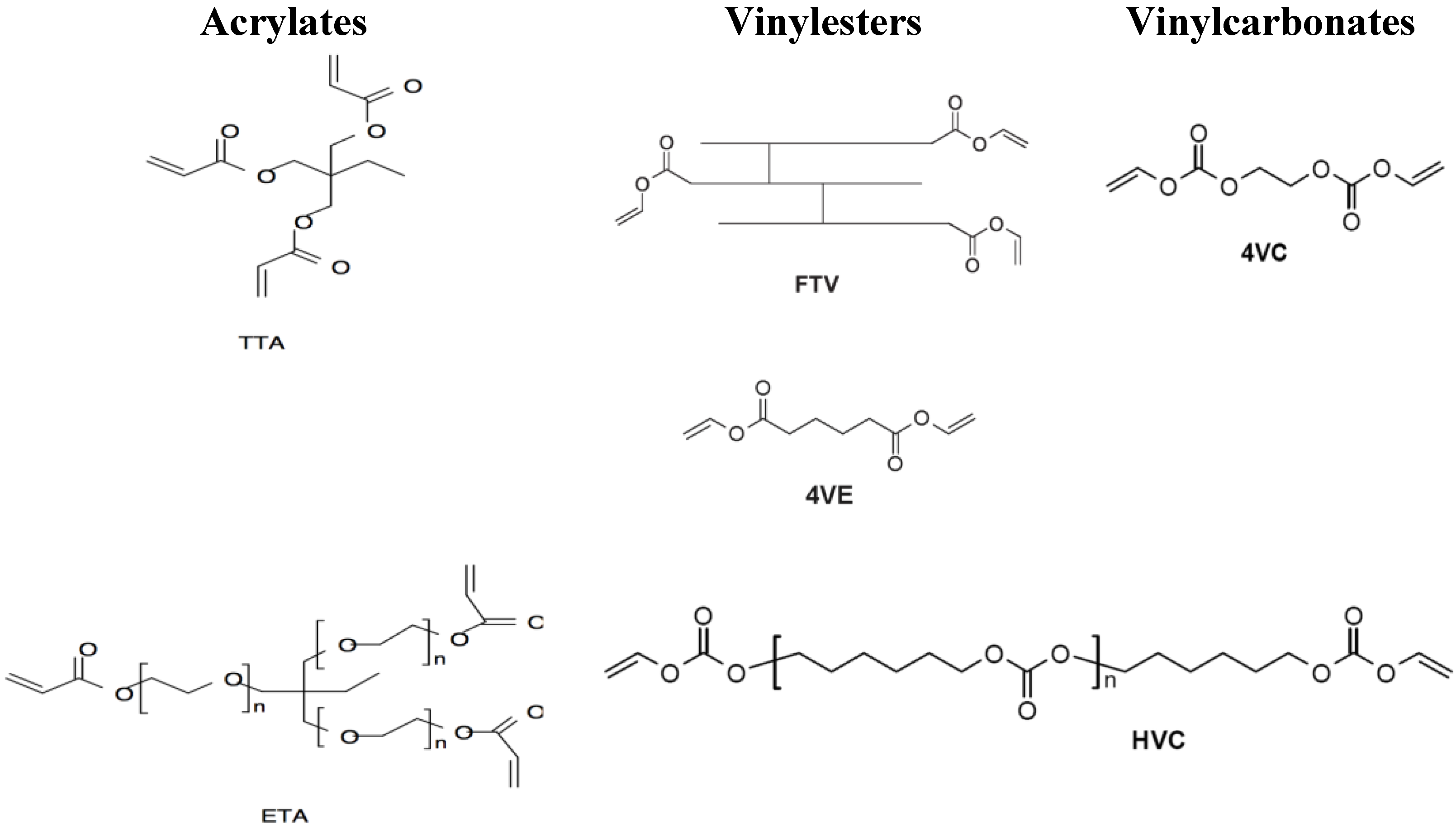
Figure 1.
Investigated vinylester and vinylcarbonate monomers and acrylate references. Acrylates: Ethoxylated trimethylolpropane triacrylate (ETA) and trimethylolpropane triacrylate (TTA); Vinylesters: Adipic acid divinyl ester (4VE) and trimer fatty acid vinyl ester (FTV); Vinylcarbonates: Ethylene glycol divinyl carbonate (4VC) and poly(hexamethylene carbonate) divinyl carbonate (HVC).
2.1.2. Vinylesters
Mercury(II) and hydroquinone as inhibitor were added to a suspension of the appropriate acid in a large excess of vinyl acetate. After stirring under argon atmosphere, p-toluene sulfonic acid was added and the reaction mixture was refluxed for 4–24 h. After cooling the resulting solution was diluted with ethyl acetate and extracted with NaOH.
The organic layer was dried over sodium sulfate and concentrated. The crude product was purified by flash chromatography on silica gel petroleum ether/ethyl acetate.
Based on previous experiments, the monomers adipic acid divinyl ester (
4VE) and trimer fatty acid vinyl ester (
FTV) were selected for the presented trial (see
Figure 1) for the “vinylester” series [
18].
2.1.3. Vinylcarbonates
The most frequently used method for preparation of vinyl carbonates is the conversion of alcohols with vinyl chloroformate. As vinyl chloroformate is commercially available, we chose this route that can convert a large variety of alcohols and primary and secondary amines, to vinyl carbonates and vinyl carbamates in the presence of pyridine or sodium carbonate as acid scavenger.
As representative monomers for vinylcarbonates, ethylene glycol divinyl carbonate (
4VC) and poly (hexamethylene carbonate) divinyl carbonate (
HVC) were used for
in vitro and
in vivo experiments (see
Figure 1) [
19].
2.2. In Vitro Experiments
The highly differentiated pre-osteoblastic cell line MC3T3-E1 (Subclone 4, ATCC®, Catalog No. CRL-2593™) was cultured in DMEM (Sigma Life Science, high glucose, St. Louis, MO, USA) supplemented with 10% FBS (Gibco® by Life Technologies, Carlsbad, NM, USA), 100 IU/mL Penicillin and 100 µg/mL Streptomycin (Gibco® by Life Technologies) at 37 °C with 95% humidity and 5% CO2.
Cells were passaged using 0.05% Trypsin-EDTA (Gibco® by Life Technologies) and cultured on 25 cm2 Flasks (Corning® Costar®).
To determine the effect of monomers on cell viability and alkaline phosphatase activity, cells were seeded onto a 96-well plate (Corning® Costar®) with a density of 6400 cells per well and allowed to attach overnight. The next day supernatants were discarded and cells were treated with different concentrations of monomers (10, 5, 2.5, 1.25, 0.63, 0.31 and 0.16 mM).
For better solubility, monomers were diluted in the previously described medium supplemented with 1% (v/v) Dimethylsulphoxide (DMSO Hybri-Max®, Sigma Life Science). An untreated control with culture medium and a control with culture medium containing 1% (v/v) DMSO were kept.
After 5 days of cultivation, supernatants were collected and cell viability was determined using the viability reagent AlamarBlue® (Invitrogen® by Life Technologies) following the manufacturer’s instructions. The assay measures the irreversible reaction of resazurin to resorufin, which is proportional to aerobic respiration.
The alkaline phosphatase (ALP) activity was determined from supernatants by using a p-Nitrophenylphosphate assay (NPP, Sigma Life Science) according to the technical bulletin (Procedure No.104, Sigma Life Science).
All in vitro experiments were performed in triplicates.
2.3. Fabrication of 3D Cellular Structures by AMT
Based on previous results of reactivity measurements and mechanical testing, we selected the following monomer formulations and ratios for production of the 3D cellular structures [
18,
19]:
Light penetration tests were performed to determine the amount of initiator and absorber required completely to cure one layer of 50–100 μm thickness. Formulations with 3 wt % of IrgacureVR 819 (BASF Schweiz AG, Basel, Switzerland) as photoinitiator and 0.15 wt % of CGL 097 (BASF Schweiz AG) as UV-absorber showed best results [
18].
For the fabrication of 3D cellular structures a lithography-based AMT system was used, which uses dynamic masks based on DLP-technology (digital light processing) for exposing individual layers of a photopolymer [
20].
The layers were structured with a light intensity of 800 mW/dm2, a layer thickness of 50 μm (acrylates and vinylesters) or 100 μm (vinylcarbonates), respectively, and an exposure time of 2 min per layer (3 min for the first three layers).
After completion of the structuring process the part was rinsed with ethanol and the final parts were obtained after post-curing under a UV lamp to lower the amount of residual monomers.
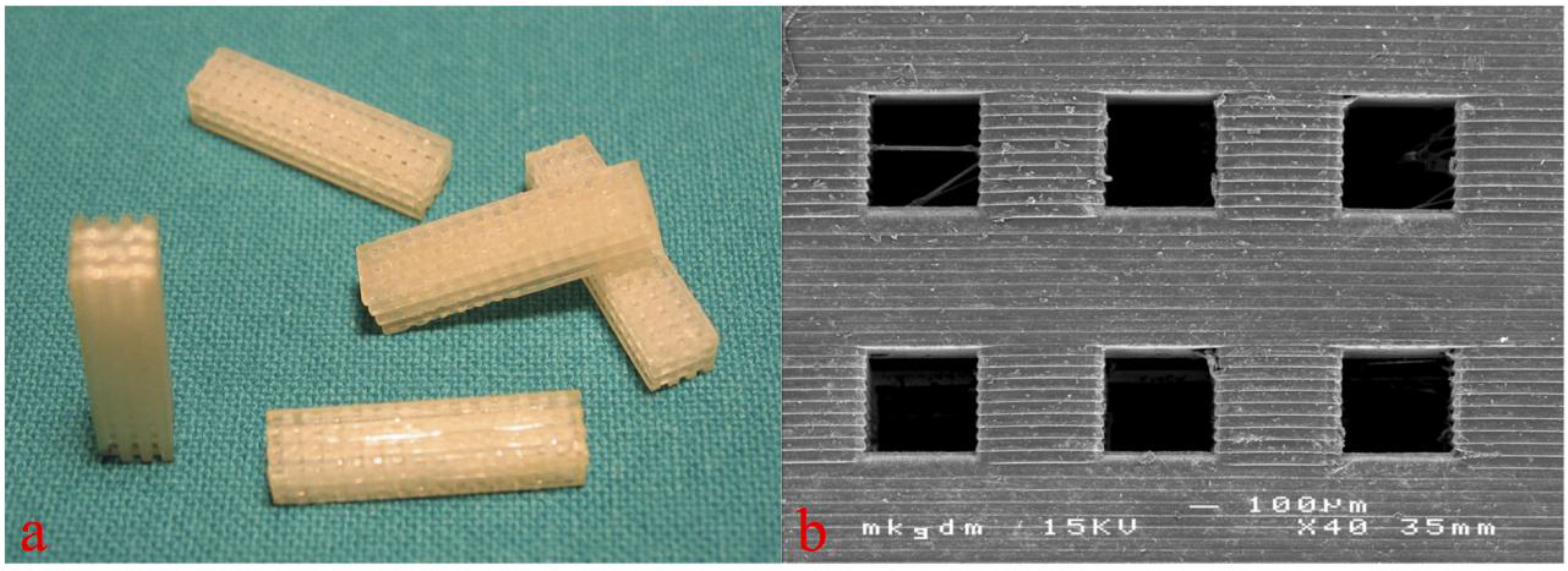
3D cellular structures of 5 mm × 5 mm × 8 mm dimension with pore sizes of 300–500 μm were built by AMT (see
Figure 2). Before implantation, the 3D cellular structures built by AMT were gamma sterilized.
Figure 2.
Macroscopic (
a, figure from [
18]) and scanning electron microscopic view (
b) of the 3D cellular structures for
in vivo testing. Copyright John Wiley and Sons 2015.
Figure 2.
Macroscopic (
a, figure from [
18]) and scanning electron microscopic view (
b) of the 3D cellular structures for
in vivo testing. Copyright John Wiley and Sons 2015.
2.4. In Vivo Experiments and Histologic Sample Preparation
All surgical procedures were performed after approval by the local ethics committee at the animal research facilities of the Department of Biomedical Research, Medical University of Vienna.
Representing a standard animal model for biomedical research, adult female New Zealand White Rabbits (Charles River, Germany), weighing approximately 3 kg were used for in vivo testing. The animals were premedicated with an intramuscular injection of 25 mg/kg ketamine and 2 mg/kg xylazine. For anesthetic induction, additional ketamine and xylazine were administered.
After the onset of muscle relaxation, the respective animal underwent orotracheal intubation followed by the insertion of an orotracheal tube. The anesthesia was maintained with volume-controlled ventilation with an oxygen-air mixture plus 1% to 2% isoflurane and the administration of a fentanyl bolus of 0.025 mg. Postoperative analgesia was managed by subcutaneous administration of 0.05 mg/kg buprenorphine every 8 h for 2 days.
After anesthetization of the animals the rear right leg was washed and shaved. Then the layerwise preparation to the lateral side of the distal femoral condyle was carried out followed by splitting the periosteum above the bone. A drilling hole of 5 mm diameter and 10 mm depth was then placed with carbide drills of increasing size until reaching the final diameter avoiding any perforation into the nearby joint (
Figure 3) [
18,
19].
To reach high primary stability of the 3D cellular structures, they were placed into the drilling holes using press-fit. The wound closure was performed again layerwise using absorbable polyglactin and silk sutures. For the time of observation the animals were kept in single stables at the local animal testing facility.
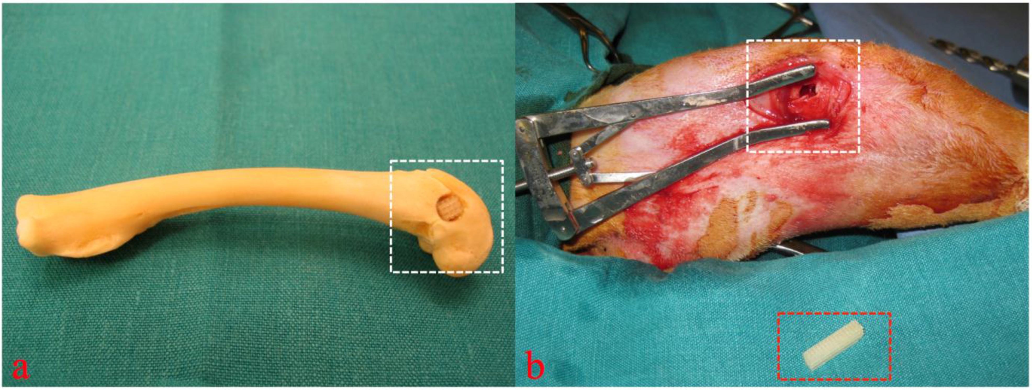
Figure 3.
Cadaver simulation (
a) and intra operative view (
b, figure from [
19]) of implantation site (white rectangular) and 3D cellular structure (red rectangular) before insertion into the drilling defect. Copyright John Wiley and Sons 2015.
Figure 3.
Cadaver simulation (
a) and intra operative view (
b, figure from [
19]) of implantation site (white rectangular) and 3D cellular structure (red rectangular) before insertion into the drilling defect. Copyright John Wiley and Sons 2015.
At the end of the particular observation period the animals were euthanized by injection of 25 mg/kg ketamine and 2 mg/kg xylazine, followed by an overdose of phenobarbital (120 mg/kg).
After exitus of the respective animals, a total number of 16 bone samples containing the whole distal femoral bone were explanted and immediately placed in 4.5% buffered formaldehyde solution (pH 7.4) and initially fixed. The specimens were processed undecalcified with a modification of the Donath technique [
21].
After fixation, samples were dehydrated in a graded ethanol series (70%–100% ethanol). To ensure good fixation and dewatering of the specimen, all the work steps were carried out with agitating equipment and regular changing of the media. Afterwards samples were transferred to synthetic resin monomer (MMA, methylmetacrylate, Sigma Life Science) for 1 day and then placed into plastic embedding mixture of MMA, nonylphenol polyglycol ether (Sigma Life Science) and benzoyl peroxide (Merck, Darmstadt, Germany) as initiator. Controlled constant polymerization then occurred at room temperature over a period of 7 days followed by a curing phase of 8 days at 37 °C in the incubator.
After hardening of the resin, specimens were trimmed to the appropriate size on a manual grinder and prepared according to the grinding technique for undecalcified hard tissue.
The surfaces to be examined were glued to a plastic microscope slide with a fixation adhesive, divided with a precision disk saw and then ground to the desired thickness of 20 μm using a micro-grinding system, with changing abrasive paper grades (1200, 2400, 4000). To remove any remaining grinding marks the ground surfaces were subsequently polished with velvet disks. The thin sections were then subjected to standardized surface staining for ground specimens (1% thionine stain).
2.5. Histologic and Histomorphometric Evaluation
All specimens underwent histomorphometric and histomorphological analysis. After blinding, two cross-sectional slides of each surgical defect, containing the implanted 3D cellular structures, were examined by transmission light microscopy (Eclipse 800, Nikon Corp., Tokyo, Japan) connected to a digital camera (Sony 950 Power HAD, Sony Corp., Tokyo, Japan).
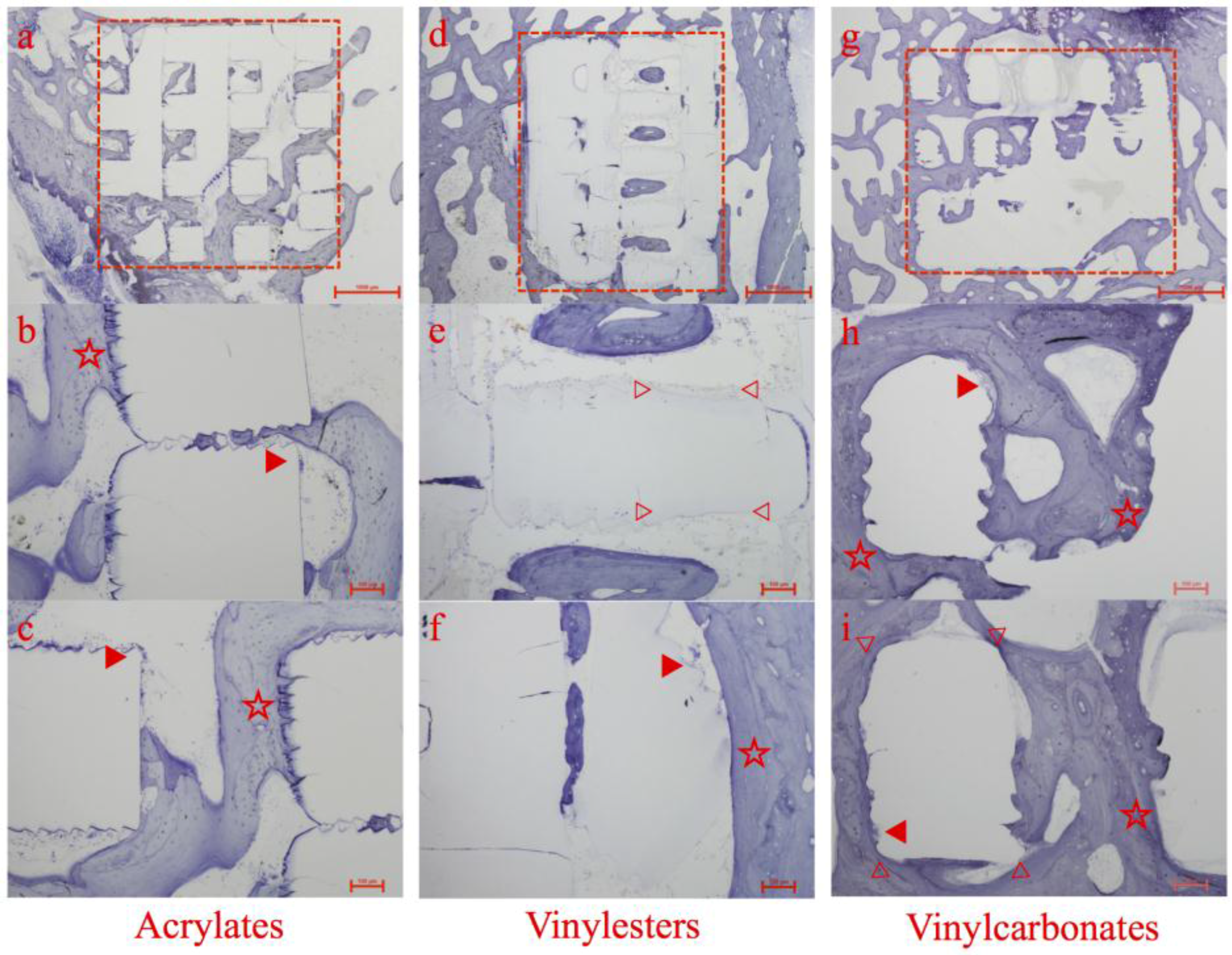
The following were examined: Newly formed bone, sprouting of blood vessels, the turnover of newly formed bone tissue, the processes occurring on the surface of the 3D cellular structures, e.g., bone deposition and potential degradation or surface erosion of the polymer structures.
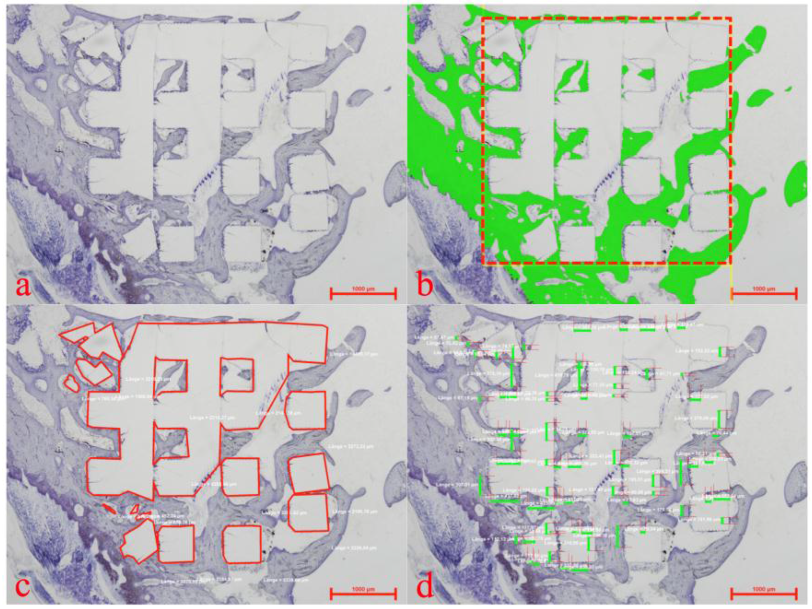
Within the whole 3D cellular structure, percentages of newly formed bone and bone to implant contact (BIC) were assessed by histomorphometry (see
Figure 4) with a semi-automated image analysis system (NIS- Elements AR 2.3.0, Nikon Corp.).
Figure 4.
Histomorphometric evaluation showing a plain thionine stain (a), the region of interest (b) (red rectangular) for assessment of newly formed bone (b) (green), the marked extent of the implanted 3D cellular structure (c) (red lines) and allocation of bone to implant contact (d) (green bars).
2.6. Statistical Methods
As the Poisson distribution is considered to be best for modeling areas or volumes, Poisson regression models were used to model the new bone formation and the bone to implant contact (BIC) within the scaffold area of the implantation site.
The BIC and the newly formed bone were estimated by the resin as dummy factors (baseline was ETA/TTA) and by 1/time, since the regressands were assumed to asymptotically reach a maximum value over time. Each effect was tested using a Wald’s test.
A p-value of <0.05 was defined as significant. All calculations were performed using the statistical programming environment “R” (version 2.15.1, Vienna, Austria).
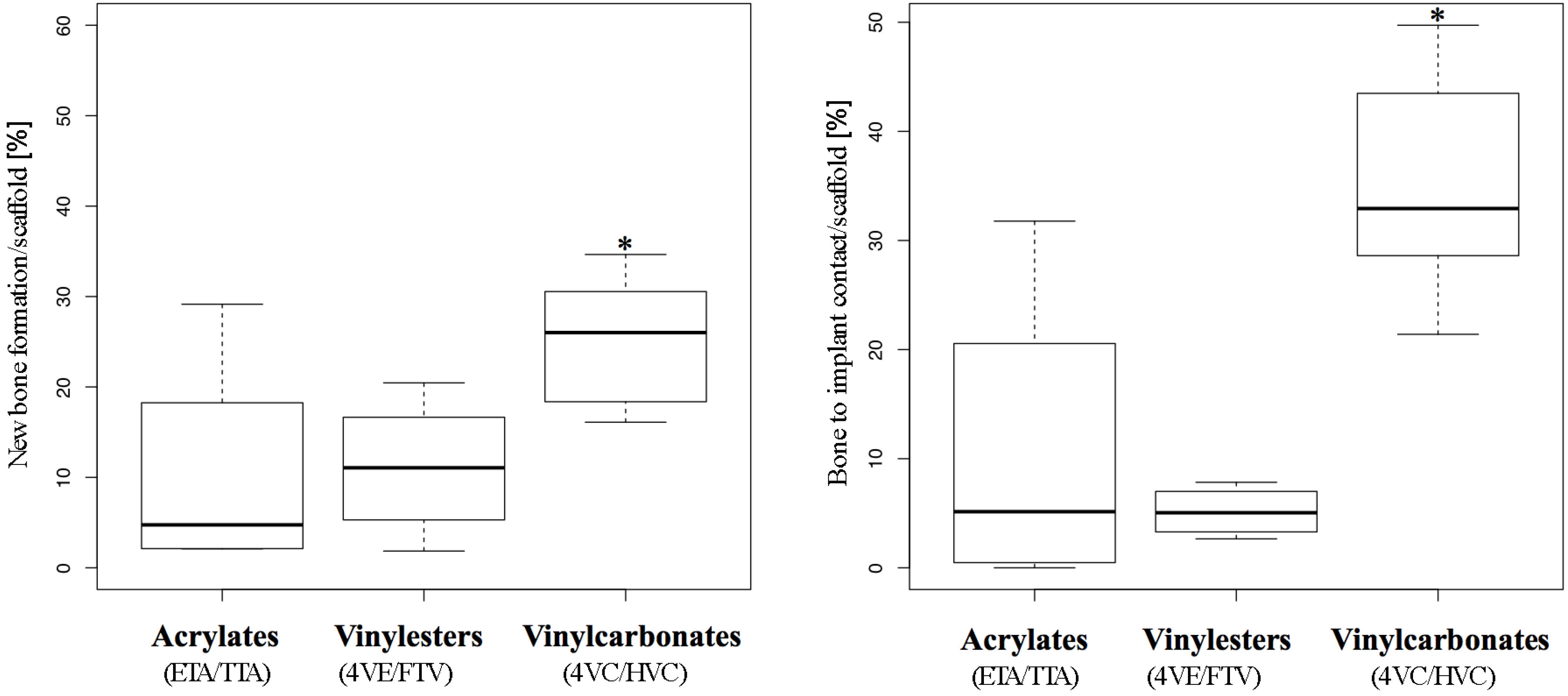
4. Conclusions
To circumvent shortcomings of widely used polymers based whether on poly(lactic acid) or (meth)acrylates, we investigated two previously developed biophotopolymer classes, that are chemically based on non-toxic poly(vinyl alcohol). We successfully compared these vinylesters and vinylcarbonates in vitro and in vivo to standard acrylates.
In vitro, both vinylester and vinylcarbonates showed superior results to acrylates by testing monomer toxicity and alkaline activity on MC3T3-E1 cells and therefore proved the suitability of the new monomers for biomedical application.
In vivo, polymerized 3D cellular structures containing vinylesters showed similar rates of new bone formation but significantly less bone to implant contact, when compared to acrylates. This lack of bone to implant contact may be associated with a high amount of hydrophobic monomers within the resin. As a consequence, the monomer formulation of these vinylesters should be adapted and improved [
22].
The investigated vinylcarbonates showed the best overall results in this trial setting. Besides excellent
in vitro behavior of vinylcarbonate monomers,
in vivo application revealed superior rates of new bone formation, bone to implant contact, and signs of polymer degradation. These findings underline the promising role of vinylcarbonate based biophotopolymers for future biomedical applications [
8,
17].


 ,
, 


