Adhesion and Growth of Vascular Smooth Muscle Cells on Nanostructured and Biofunctionalized Polyethylene

Abstract
:1. Introduction
2. Results and Discussion
2.1. Physical and Chemical Properties of the Polyethylene Samples
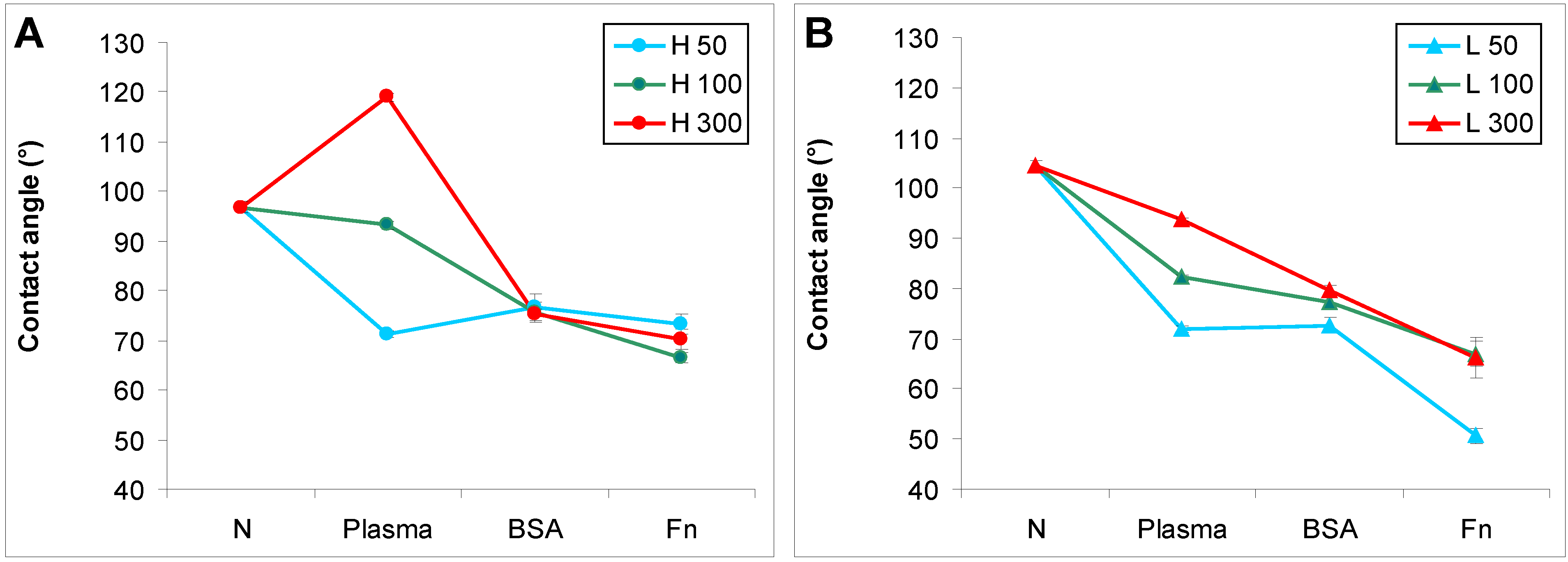
2.1.1. Surface Wettability

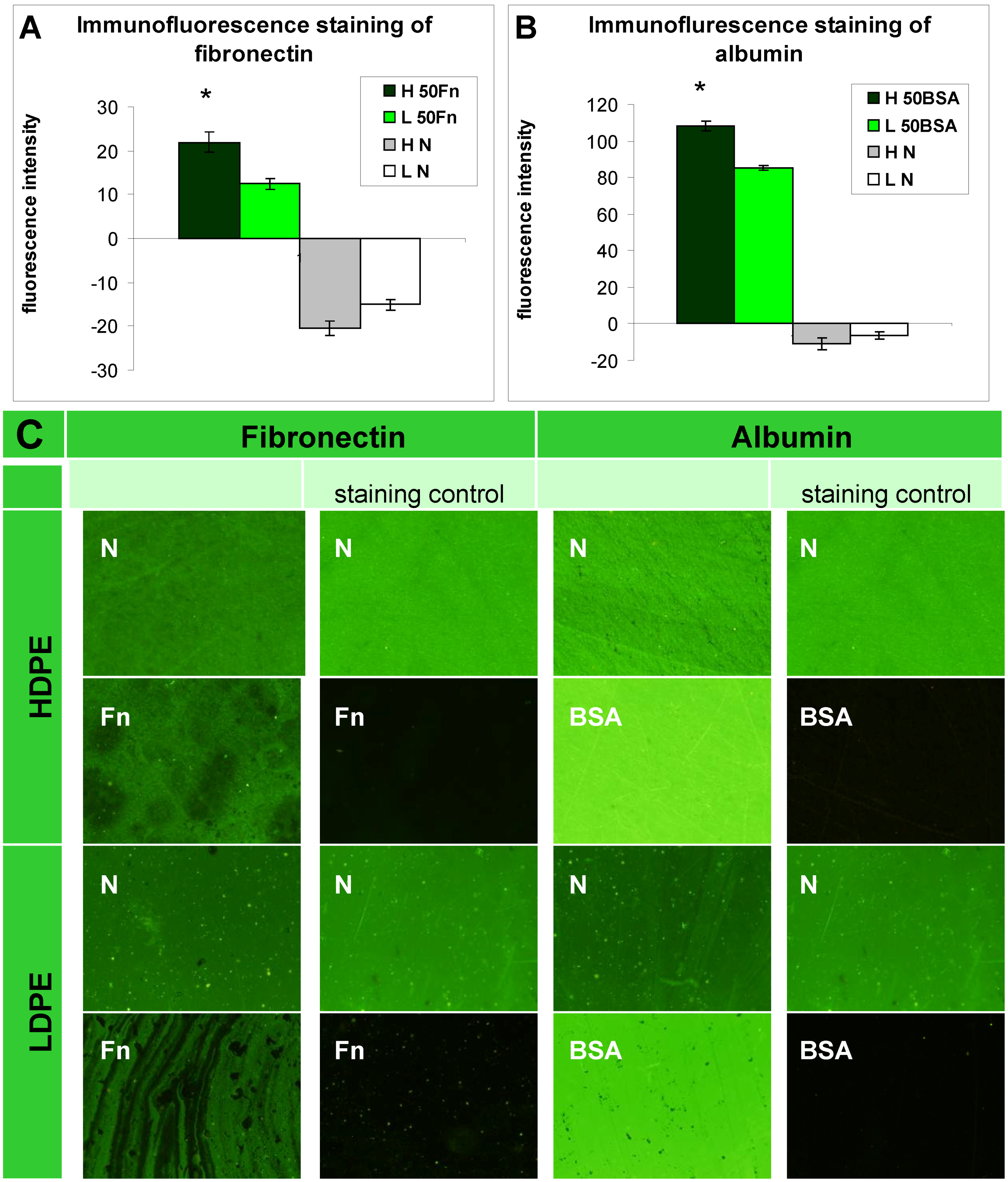
2.1.2. XPS Analysis and Immunofluorescence Staining of Grafted Biomolecules

| Material/Modification | HDPE | LDPE | ||||||
| C (at. %) | O (at. %) | N (at. %) | S (at. %) | C (at. %) | O (at. %) | N (at. %) | S (at. %) | |
| 50 | 82.2 | 14.2 | 2.5 | 0 | 83.0 | 14.8 | 1.5 | 0 |
| 50 Fn | 72.9 | 20.2 | 6.7 | 0.2 | 82.2 | 13.6 | 2.8 | at limit of detection |
| 50 BSA | 74.4 | 15.3 | 9.9 | 0.4 | 72.7 | 15.3 | 10.9 | 0.4 |

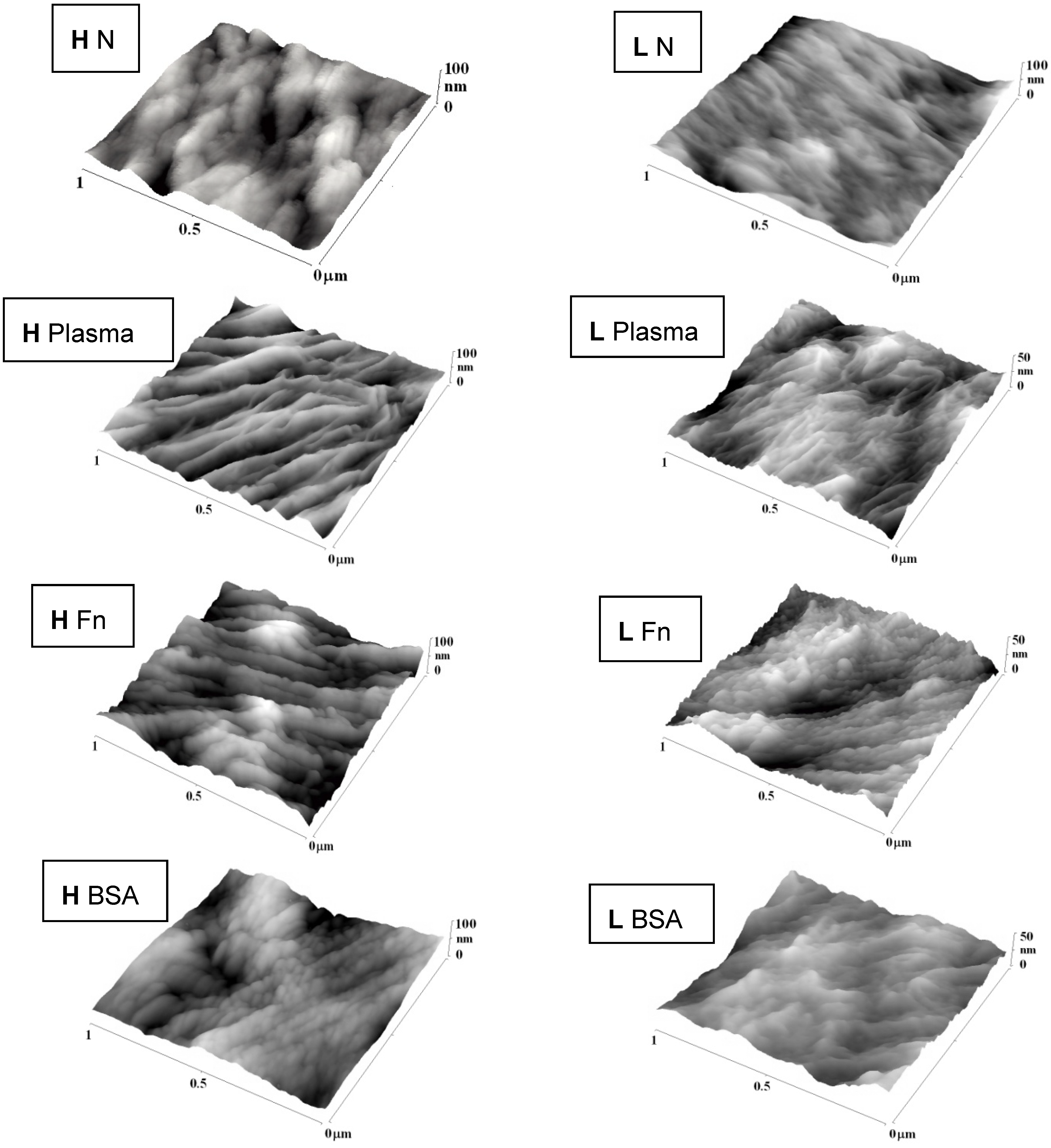
2.1.3. Surface Roughness and Morphology
| Sample | HDPE | LDPE |
|---|---|---|
| N | 6.1 | 5.0 |
| 50 | 7.4 | 4.7 |
| 50 Fn | 10.4 | 5.5 |
| 50 BSA | 6.9 | 5.4 |
| 100 | 8.4 | 6.7 |
| 100 Fn | 11.7 | 4.6 |
| 100 BSA | 9.4 | 4.7 |
| 300 | 11.5 | 6.6 |
| 300 Fn | 12.0 | 5.6 |
| 300 BSA | 7.8 | 4.6 |
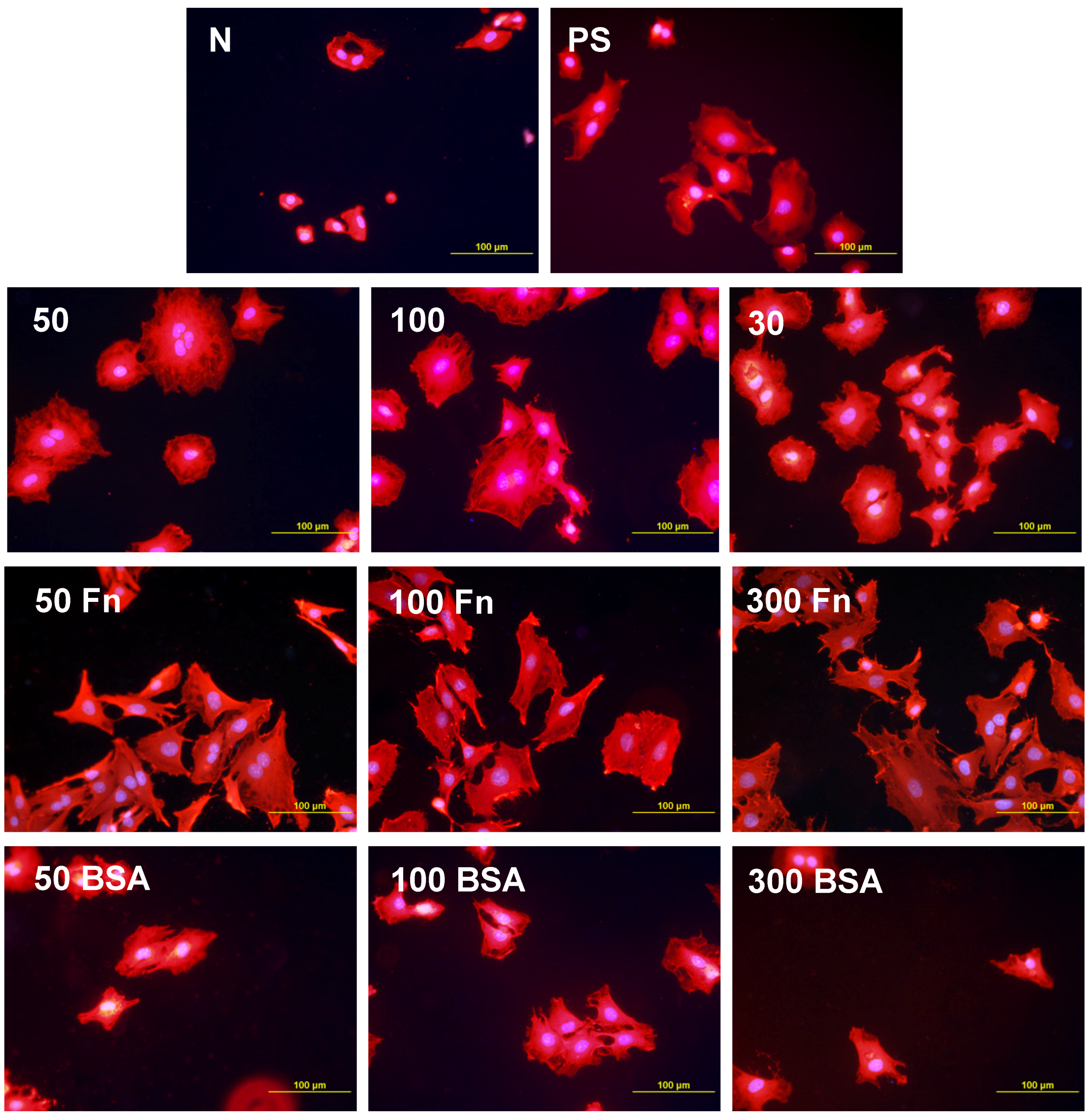
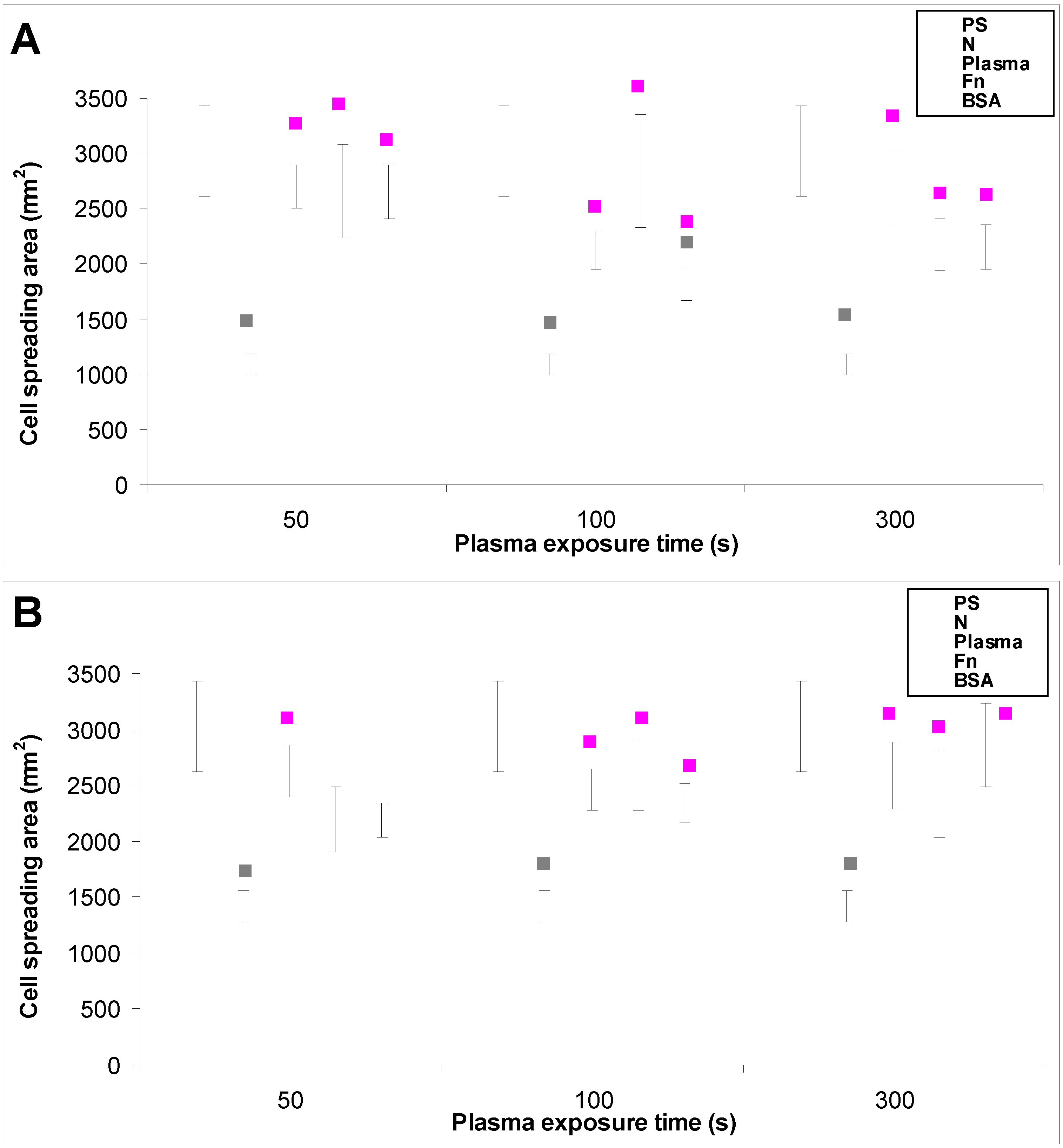
2.2. Cell Adhesion and Growth
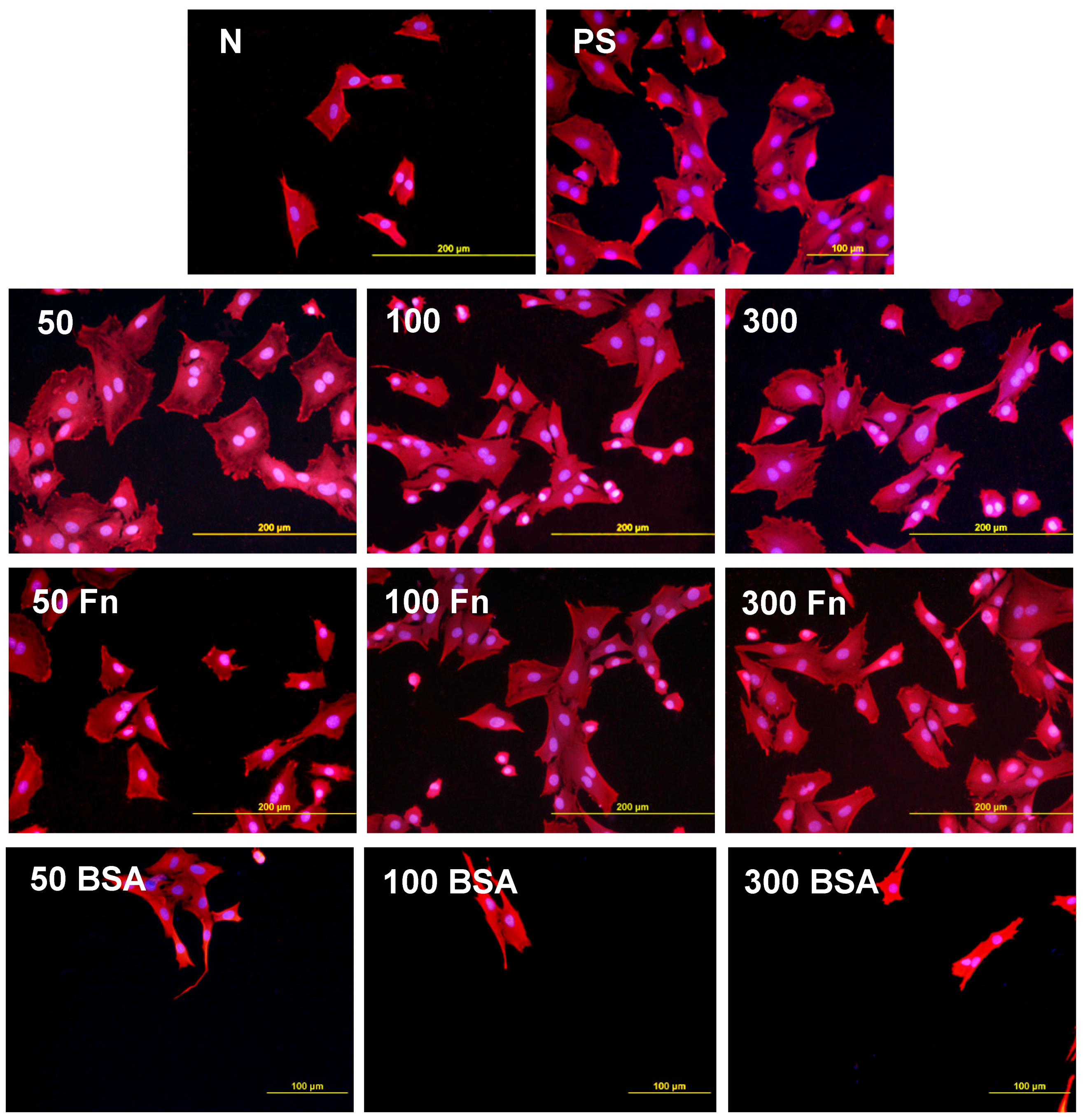
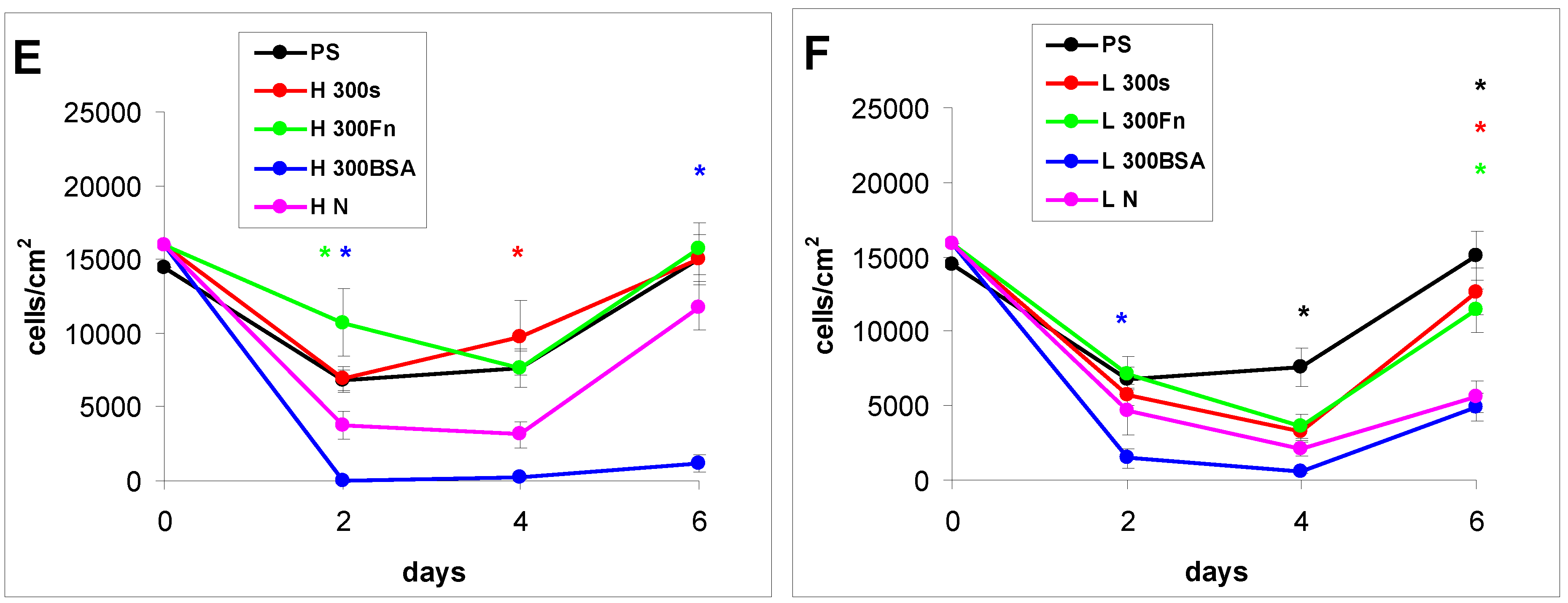
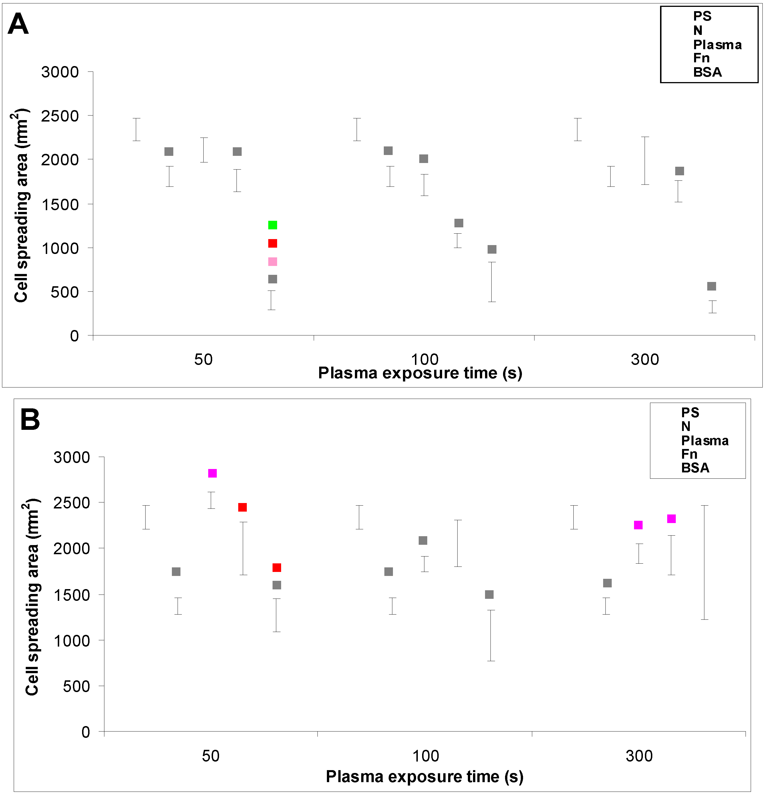
2.2.1. Cell Adhesion and Growth on LDPE and HDPE Samples in a Standard Serum-supplemented Medium
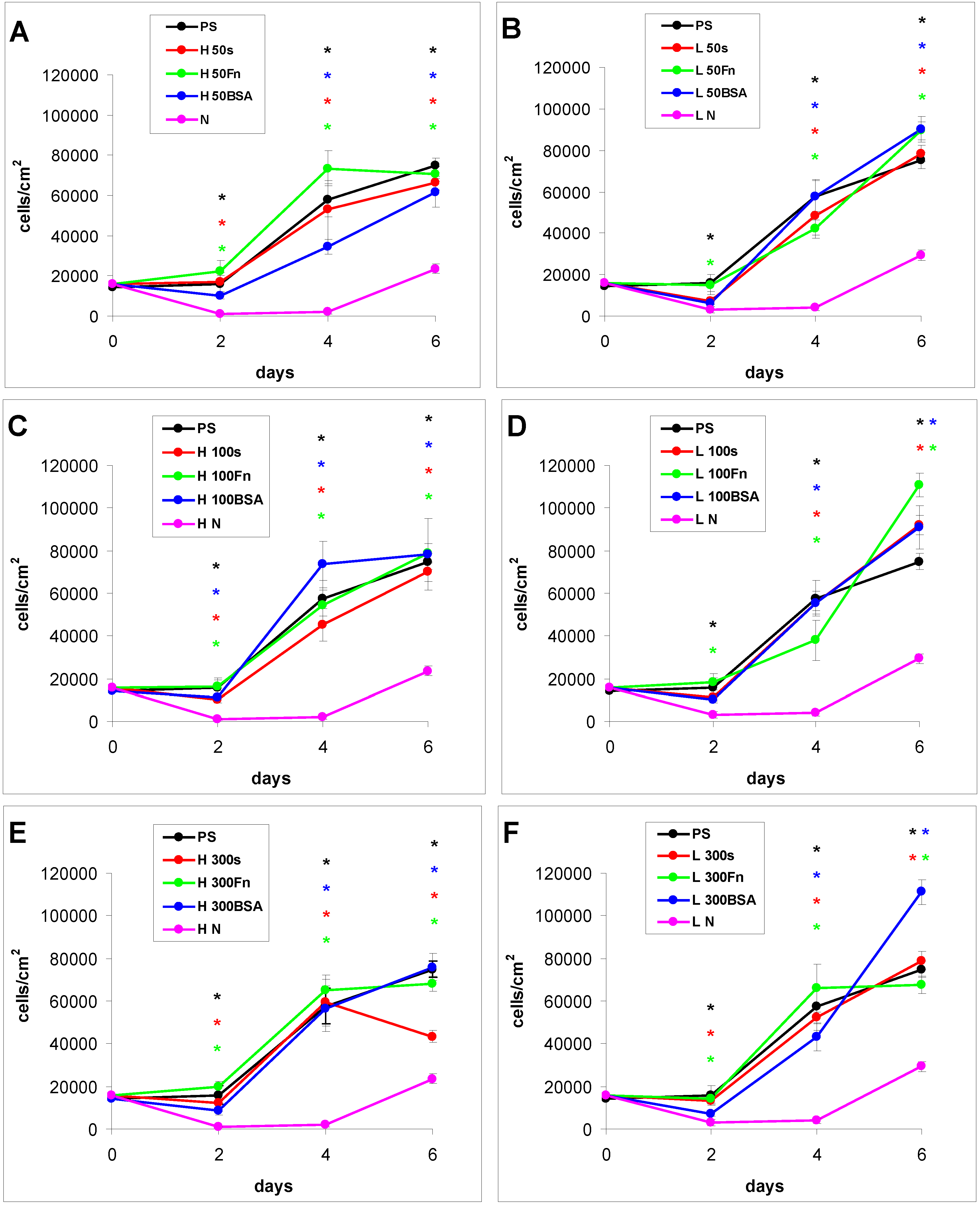
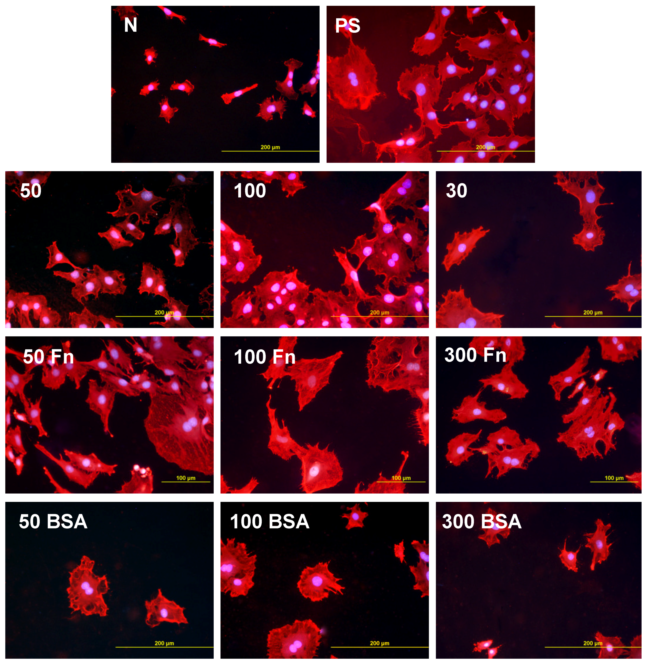


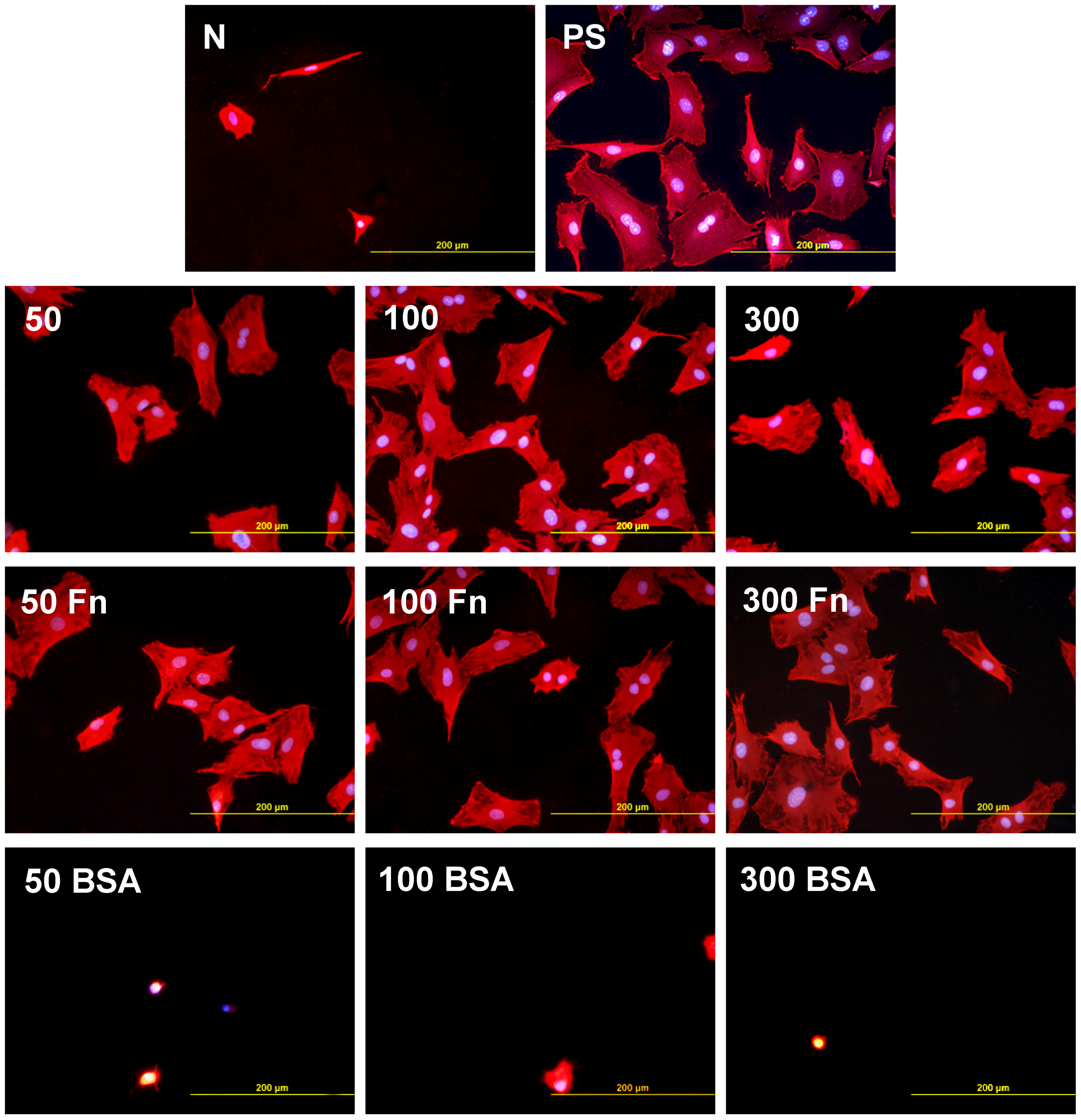
2.2.2. Cell Adhesion and Growth on LDPE and HDPE in a Serum-Free Medium


3. Experimental Section
3.1. Preparation and Modification of Polymer Samples
3.2. Characterization of the Physical and Chemical Properties of the Polymer Surface
3.2.1. Surface Wettability
3.2.2. Chemical Composition of the Polymer Surface
3.2.3. Surface Morphology and Roughness
3.3. Cells and Culture Conditions
3.4. Evaluation of the Cell Number and Cell Spreading Area
4. Conclusions
Acknowledgments
References
- Zhang, W.J.; Liu, W.; Cui, L.; Cao, Y. Tissue engineering of blood vessel. J. Cell. Mol. Med. 2007, 11, 945–957. [Google Scholar] [CrossRef] [PubMed]
- Yu, H.; Wagner, E. Bioresponsive polymers for nonviral gene delivery. Curr. Opin. Mol. Ther. 2009, 2, 165–178. [Google Scholar]
- Bacakova, L.; Filova, E.; Rypacek, F.; Svorcik, V.; Stary, V. Cell adhesion on artificial materials for tissue engineering. Physiol. Res. 2004, 53, S35–S45. [Google Scholar] [PubMed]
- Bacakova, L.; Svorcik, V. Cell colonization control by physical and chemical modification of materials. In Cell Growth Processes: New Research; Kimura, D., Ed.; Nova Science Publishers, Inc.: Hauppauge, NY, USA, 2008; pp. 5–56. [Google Scholar]
- Bozukova, D.; Pagnoulle, C.; de Pauw-Gillet, M.C.; Desbief, S.; Lazzaroni, R.; Ruth, N.; Jerome, R.; Jerome, C. Improved performances of intraocular lenses by poly(ethylene glycol) chemical coatings. Biomacromolecules 2007, 8, 2379–2387. [Google Scholar] [CrossRef] [PubMed]
- Poulsson, A.H.; Mitchell, S.A.; Davidson, M.R.; Johnstone, A.J.; Emmison, N.; Bradley, R.H. Attachment of human primary osteoblast cells to modified polyethylene surfaces. Langmuir 2009, 25, 3718–3727. [Google Scholar] [CrossRef] [PubMed]
- Granke, K.; Ochsner, J.L.; McClugage, S.G.; Zdrahal, P. Analysis of graft healing in a new elastomer-coated vascular prosthesis. Cardiovasc. Surg. 1993, 1, 254–261. [Google Scholar] [PubMed]
- Bacakova, L.; Filova, E.; Kubies, D.; Machova, L.; Proks, V.; Malinova, V.; Lisa, V.; Rypacek, F. Adhesion and growth of vascular smooth muscle cells in cultures on bioactive RGD peptide-carrying polylactides. J. Mater. Sci. Mater. Med. 2007, 18, 1317–1323. [Google Scholar] [CrossRef] [PubMed]
- Bacakova, L.; Filova, E.; Parizek, M.; Ruml, T.; Svorcik, V. Modulation of cell adhesion, proliferation and differentiation on materials designed for body implants. Biotechnol. Adv. 2011, 29, 739–767. [Google Scholar] [CrossRef] [PubMed]
- Heitz, J.; Svorcik, V.; Bacakova, L.; Rockova, K.; Ratajova, E.; Gumpenberger, T.; Bäuerle1, D.; Dvorankova, B.; Kahr, H.; Graz, I.; et al. Cell adhesion on polytetrafluoroethylene modified by UV-irradiation in an ammonia atmosphere. J. Biomed. Mater. Res. 2003, 67, 130–137. [Google Scholar] [CrossRef]
- Svorcik, V.; Rybka, V.; Hnatowicz, V.; Smetana, K., Jr. Structure and biocompatibility of ion beam modified polyethylene. J. Mater. Sci. Mater. Med. 1997, 8, 435–440. [Google Scholar] [CrossRef] [PubMed]
- Bacakova, L.; Mares, V.; Lisa, V.; Svorcik, V. Molecular mechanisms of improved adhesion and growth of an endothelial cell line cultured on polystyrene implanted with fluorine ions. Biomaterials 2000, 21, 1173–1179. [Google Scholar] [CrossRef] [PubMed]
- Walachova, K.; Svorcik, V.; Bacakova, L.; Hnatowicz, V. Colonization of ion-modified polyethylene with vascular smooth muscle cells in vitro. Biomaterials 2002, 23, 2989–2996. [Google Scholar] [CrossRef] [PubMed]
- Wang, Y.; Lu, L.; Zheng, Y.; Chen, X. Improvement in hydrophilicity of PHBV films by plasma treatment. J. Biomed. Mater. Res. A 2006, 76, 589–595. [Google Scholar] [CrossRef] [PubMed]
- Tajima, S.; Chu, J.S.; Li, S.; Komvopoulos, K. Differential regulation of endothelial cell adhesion, spreading, and cytoskeleton on low-density polyethylene by nanotopography and surface chemistry modification induced by argon plasma treatment. J. Biomed. Mater. Res. A 2008, 84, 828–836. [Google Scholar] [CrossRef] [PubMed]
- Pareta, R.A.; Reising, A.B.; Miller, T.; Storey, D.; Webster, T.J. Increased endothelial cell adhesion on plasma modified nanostructured polymeric and metallic surfaces for vascular stent applications. Biotechnol. Bioeng. 2009, 103, 459–471. [Google Scholar] [CrossRef] [PubMed]
- Zhang, Y.; Tanner, K.E.; Gurav, N.; di Silvio, L. In vitro osteoblastic response to 30 vol% hydroxyapatite-polyethylene composite. J. Biomed. Mater. Res. A 2007, 81, 409–417. [Google Scholar] [CrossRef] [PubMed]
- Homaeigohar, S.S.; Shokrgozar, M.A.; Javadpour, J.; Khavandi, A.; Sadi, A.Y. Effect of reinforcement particle size on in vitro behavior of beta-tricalcium phosphate-reinforced high-density polyethylene: A novel orthopedic composite. J. Biomed. Mater. Res. A 2006, 78, 129–138. [Google Scholar] [CrossRef] [PubMed]
- Fouad, H.; Elleithy, R. High density polyethylene/graphite nano-composites for total hip joint replacements: processing and in vitro characterization. J. Mech. Behav. Biomed. Mater. 2011, 4, 1376–1383. [Google Scholar] [CrossRef] [PubMed]
- Oldinski, R.A.; Ruckh, T.T.; Staiger, M.P.; Popat, K.C.; James, S.P. Dynamic mechanical analysis and biomineralization of hyaluronan-polyethylene copolymers for potential use in osteochondral defect repair. Acta Biomater. 2011, 7, 1184–1191. [Google Scholar] [CrossRef] [PubMed]
- Mokal, N.J.; Desai, M.F. Calvarial reconstruction using high-density porous polyethylene cranial hemispheres. Indian J. Plast. Surg. 2011, 44, 422–431. [Google Scholar]
- Caldwell, R.A.; Woodell, J.E.; Ho, S.P.; Shalaby, S.W.; Boland, T.; Langan, E.M.; LaBerge, M. In vitro evaluation of phosphonylated low-density polyethylene for vascular applications. J. Biomed. Mater. Res. 2002, 62, 514–524. [Google Scholar] [CrossRef] [PubMed]
- Svorcik, V.; Kolarova, K.; Slepicka, P.; Mackova, A.; Novotna, M.; Hnatowicz, V. Modification of surface properties of high and low density polyethylene by Ar plasma discharge. Polym. Degrad. Stabil. 2006, 91, 1219–1225. [Google Scholar] [CrossRef]
- Svorcik, V.; Kasalkova, N.; Slepicka, P.; Zaruba, K.; Kral, V.; Bacakova, L. Cytocompatibility of Ar + plasma treated and Au nanoparticle-grafted PE. Nucl. Instrum. Meth. B 2009, 267, 1904–1910. [Google Scholar] [CrossRef]
- Parizek, M.; Kasalkova, N.; Bacakova, L.; Slepicka, P.; Lisa, V.; Blazkova, M.; Svorcik, V. Improved adhesion, growth and maturation of vascular smooth muscle cells on polyethylene grafted with bioactive molecules and carbon particles. Int. J. Mol. Sci. 2009, 4352–4374. [Google Scholar] [CrossRef]
- Parizek, M.; Kasalkova, N.S.; Bacakova, L.; Lisa, V.; Svindrych, Z.; Slepicka, P.; Svorcik, V. Adhesion, growth and maturation of vascular smooth muscle cells on low-density polyethylene grafted with bioactive substance. J. Biomed. Biotechnol. 2013, in press. [Google Scholar]
- Kella, N.K.; Kang, Y.J.; Kinsella, J.E. Effect of oxidative sulfitolysis of disulfide bonds of bovine serum albumin on its structural properties: A physiochemical study. J. Protein Chem. 1988, 7, 535–548. [Google Scholar] [CrossRef] [PubMed]
- Pankov, R.; Yamada, K.M. Fibronectin at a glance. J. Cell Sci. 2002, 15, 3861–3863. [Google Scholar] [CrossRef]
- Xiao, Y.; Isaacs, S.N. Enzyme-linked immunosorbent assay (ELISA) and blocking with bovine serum albumin (BSA)—Not all BSAs are alike. J. Immunol. Methods 2012, 384, 148–151. [Google Scholar] [CrossRef] [PubMed]
- Bacakova, L.; Lisa, V.; Kubinova, L.; Wilhelm, J.; Novotna, J.; Eckhart, A.; Herget, J. UV light—Irradiated collagen III modulates expression of cytoskeletal and surface adhesion molecules in rat aortic smooth muscle cells in vitro. Virchows Arch. 2002, 440, 50–62. [Google Scholar] [CrossRef] [PubMed]
- Kim, K.S.; Ryu, C.M.; Park, C.S.; Sur, G.S.; Park, C.E. Investigation of crystallinity effects on the surface of oxygen plasma treated low density polyethylene using X-ray photoelectron spectroscopy. Polymer 2003, 44, 6287–6295. [Google Scholar] [CrossRef]
- Kowalczynska, H.M.; Nowak-Wyrzykowska, M.; Szczepankiewicz, A.A.; Dobkowski, J.; Dyda, M.; Kaminski, J.; Kołos, R. Albumin adsorption on unmodified and sulfonated polystyrene surfaces, in relation to cell-substratum adhesion. Colloids Surf. B Biointerfaces 2011, 84, 536–544. [Google Scholar] [CrossRef] [PubMed]
- Brynda, E.; Houska, M.; Jirouskova, M.; Dyr, J.E. Albumin and heparin multilayer coatings for blood-contacting medical devices. J. Biomed. Mater. Res. 2000, 51, 249–257. [Google Scholar] [CrossRef] [PubMed]
- Yamazoe, H.; Tanabe, T. Drug-carrying albumin film for blood-contacting biomaterials. J. Biomater. Sci. Polym. Ed. 2010, 21, 647–657. [Google Scholar] [CrossRef] [PubMed]
- Glukhova, M.A.; Koteliansky, V.E. Integrins, cytoskeletal and extracellular matrix proteins in developing smooth muscle cells of human aorta. In The Vascular Smooth Muscle Cell: Molecular and Biological Responses to the Extracellular Matrix; Schwartz, S.M., Mecham, R.P., Eds.; Academic Press Inc.: Waltham, MA, USA, 1995; pp. 37–79. [Google Scholar]
- Shipley, G.D.; Ham, R.G. Multiplication of Swiss 3T3 cells in a serum-free medium. Exp. Cell Res. 1983, 146, 249–260. [Google Scholar] [CrossRef] [PubMed]
- Maroudas, N.G. Sulphonated polystyrene as an optimal substratum for the adhesion and spreading of mesenchymal cells in monovalent and divalent saline solutions. J. Cell. Physiol. 1976, 90, 511–520. [Google Scholar] [CrossRef]
- Curtis, A.S.G.; Forrester, J.V.; McInnes, C.; Lawrie, F. Adhesion of cells to polystyrene surfaces. J. Cell. Biol. 1983, 97, 1500–1506. [Google Scholar] [CrossRef] [PubMed]
- Burmeister, J.S.; Vrany, J.D.; Reichert, W.M.; Truskey, G.A. Effect of fibronectin amount and conformation on the strength of endothelial cell adhesion to HEMA/EMA copolymers. J. Biomed. Mater. Res. 1996, 30, 13–22. [Google Scholar] [CrossRef] [PubMed]
- Bacakova, L.; Mares, V.; Lisa, V.; Bottone, M.G.; Pellicciari, C.; Kocourek, F. Sex-related differences in the migration and proliferation of rat aortic smooth muscle cells in short and long term culture. In Vitro Cell. Develop. Biol. Anim. 1997, 33, 410–413. [Google Scholar] [CrossRef]
© 2013 by the authors; licensee MDPI, Basel, Switzerland. This article is an open access article distributed under the terms and conditions of the Creative Commons Attribution license (http://creativecommons.org/licenses/by/3.0/).
Share and Cite
Novotna, K.; Bacakova, M.; Kasalkova, N.S.; Slepicka, P.; Lisa, V.; Svorcik, V.; Bacakova, L. Adhesion and Growth of Vascular Smooth Muscle Cells on Nanostructured and Biofunctionalized Polyethylene. Materials 2013, 6, 1632-1655. https://doi.org/10.3390/ma6051632
Novotna K, Bacakova M, Kasalkova NS, Slepicka P, Lisa V, Svorcik V, Bacakova L. Adhesion and Growth of Vascular Smooth Muscle Cells on Nanostructured and Biofunctionalized Polyethylene. Materials. 2013; 6(5):1632-1655. https://doi.org/10.3390/ma6051632
Chicago/Turabian StyleNovotna, Katarina, Marketa Bacakova, Nikola Slepickova Kasalkova, Petr Slepicka, Vera Lisa, Vaclav Svorcik, and Lucie Bacakova. 2013. "Adhesion and Growth of Vascular Smooth Muscle Cells on Nanostructured and Biofunctionalized Polyethylene" Materials 6, no. 5: 1632-1655. https://doi.org/10.3390/ma6051632
APA StyleNovotna, K., Bacakova, M., Kasalkova, N. S., Slepicka, P., Lisa, V., Svorcik, V., & Bacakova, L. (2013). Adhesion and Growth of Vascular Smooth Muscle Cells on Nanostructured and Biofunctionalized Polyethylene. Materials, 6(5), 1632-1655. https://doi.org/10.3390/ma6051632