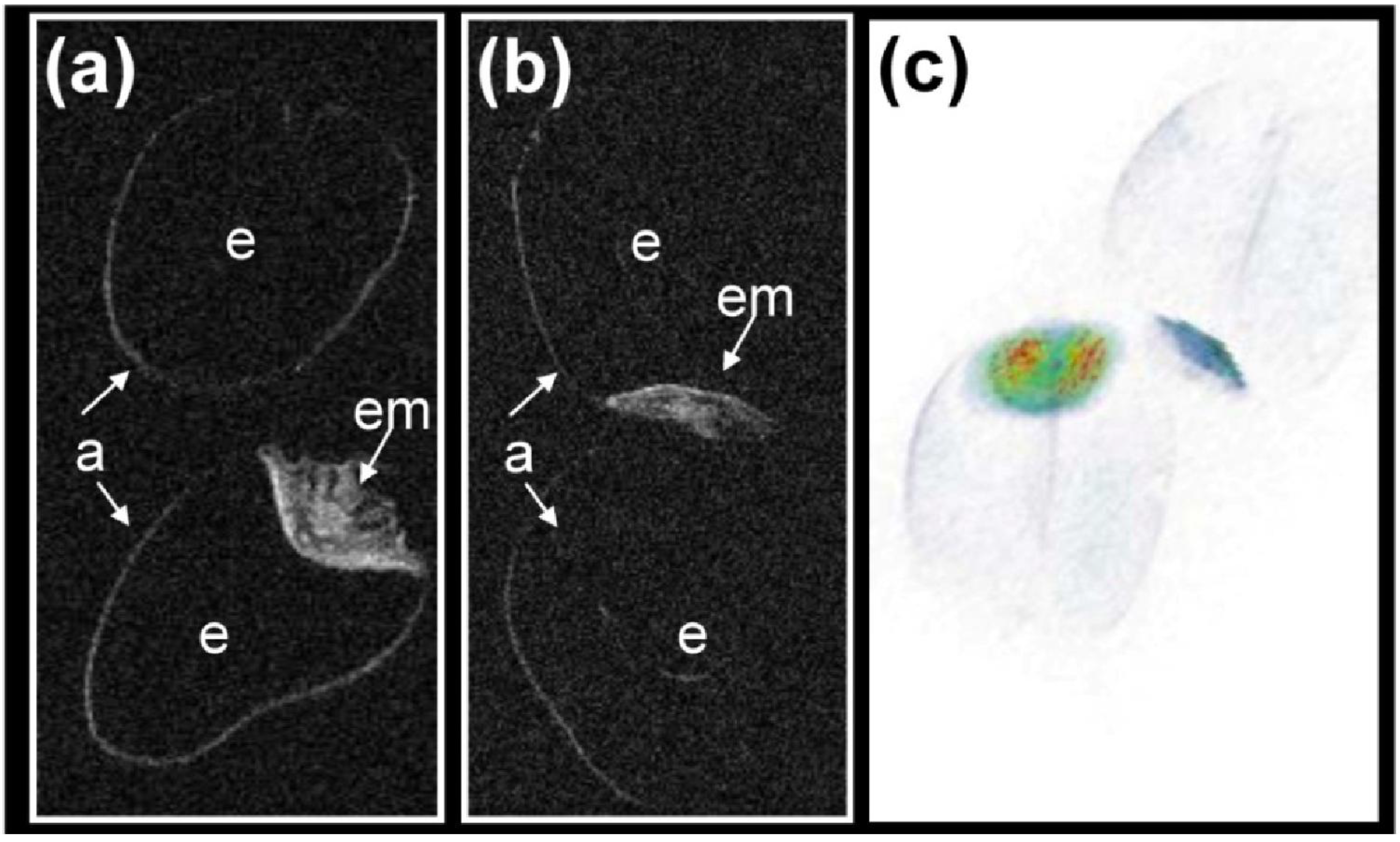
Low and High Field Magnetic Resonance for in Vivo Analysis of Seeds

Abstract
1. Introduction
2. Methods
2.1. Hardware
2.2. Plant Seed Material
2.3. Application of MQ60
2.4. Lipid Mapping of Mature Seeds Using 1H MRS-Imaging
2.5. Localized 1H NMR and 1H Spectroscopic Imaging on Fresh Harvested Seed
2.6. Double Resonant 13C / 1H NMR for Tracing 13C Metabolites in Developing Seeds
3. Results and Discussion
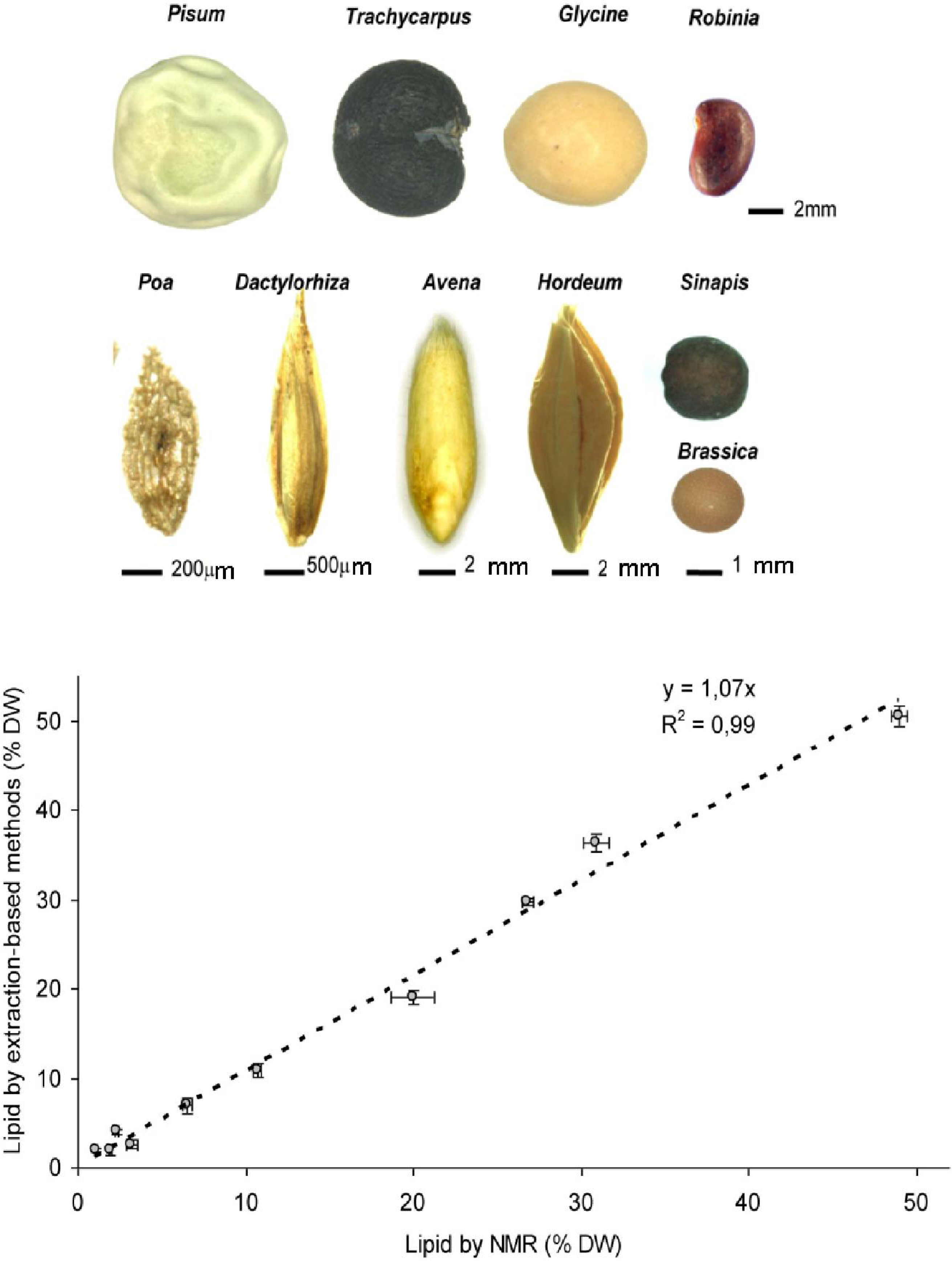
3.1. Low Field NMR Enables Accurate and Fast Quantification of Seed Lipids in a Wide Range of Species
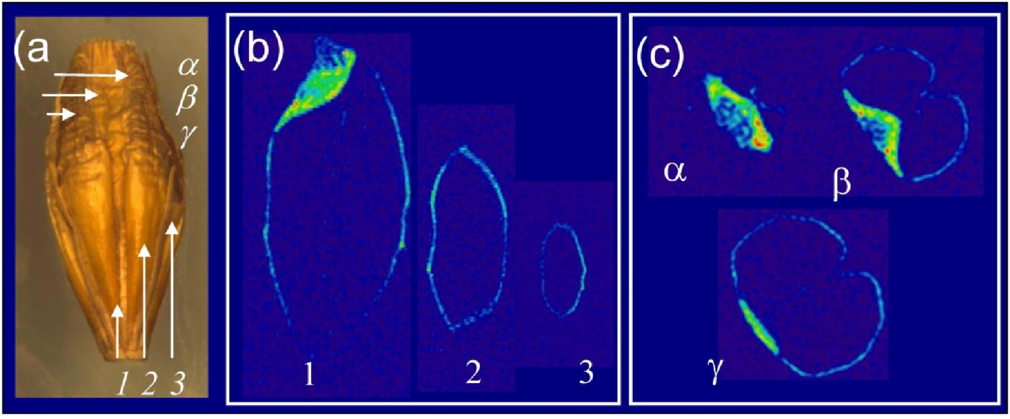
3.2. High Field MRI is a Versatile Tool for Exploring Grain Lipid Composition

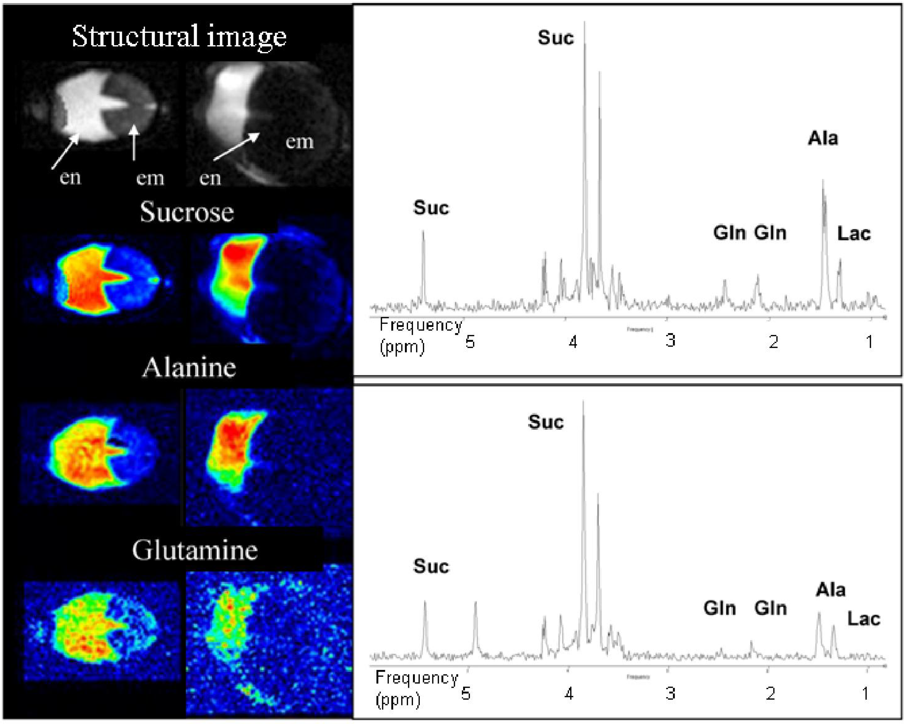
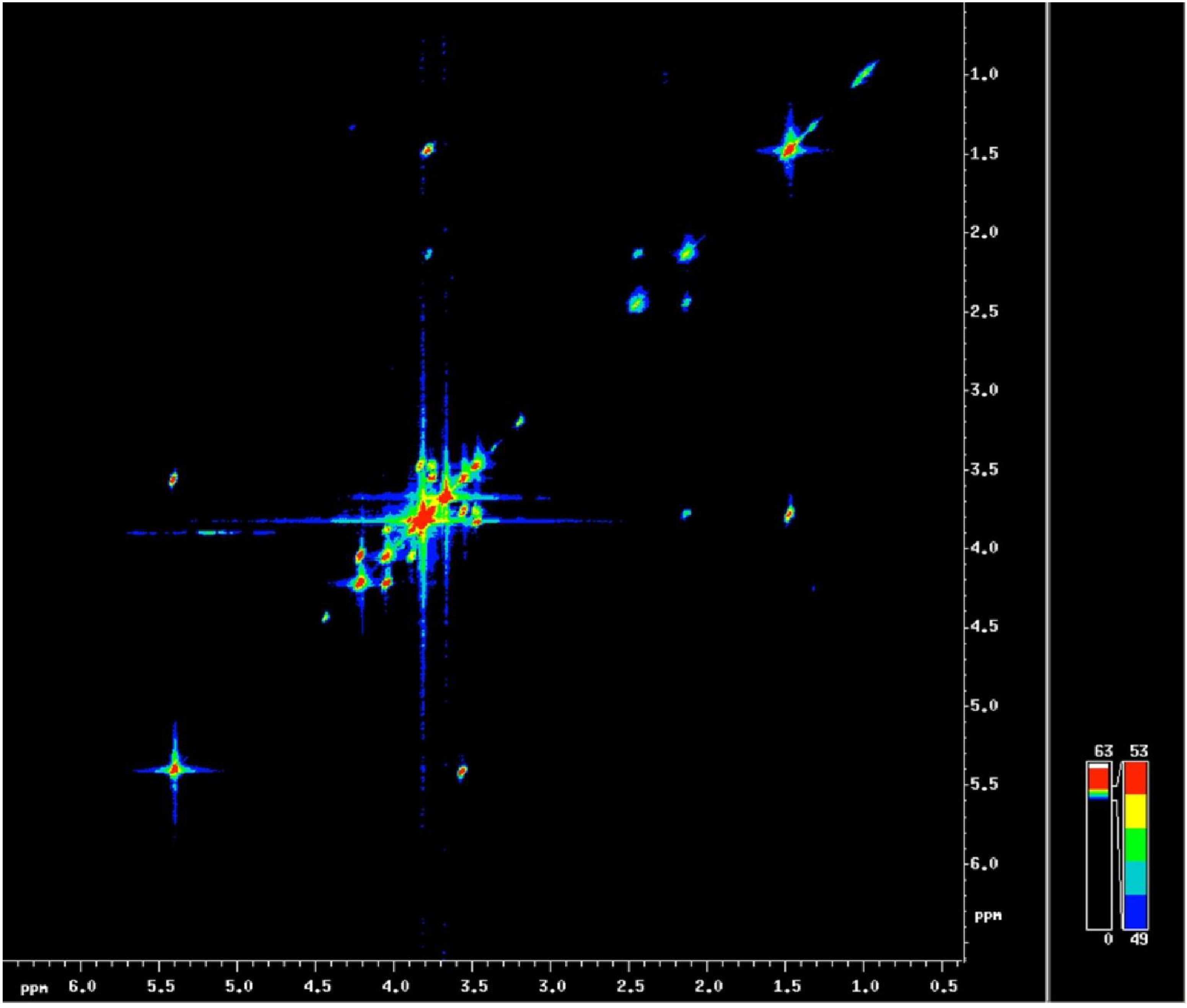
3.3. Localized 1H NMR and 1H Spectroscopic Imaging Allows Assessment of Metabolite Distribution within the Living Seed
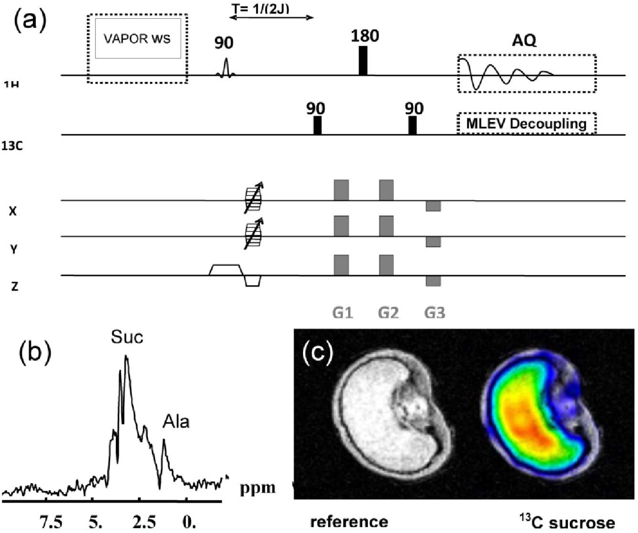
3.4. Double Resonant 1H /13C NMR Allows the Tracing of 13C Labelled Metabolites within the Seed
4. Conclusions
Acknowledgments
References
- Krygsman, P.H.; Barrett, A.E. Simple methods for measuring total oil content by benchtop NMR. Oil Extraction and Analisis 2004, 152–165. [Google Scholar]
- Todt, H.; Guthausen, G.; Burk, W.; Schmalbein, D.; Kamlowski, A. Water/moisture and fat analysis by time-domain NMR. Food Chem. 2005, 96, 436–440. [Google Scholar] [CrossRef]
- Ghosh, S.; Tombokan, X. TD-NMR technology: A noninvasive tool for high-throughput QC and rapid cereal product improvement. Cereal Foods World 2009, 54, 152–157. [Google Scholar]
- Van Duynhoven, J.; Voda, A.; Witek, M.; van As, H. Time-domain Nmr applied to food products. Ann. Rep. NMR Spectrsp. 2010, 69, 145–197. [Google Scholar]
- U.S. Department of Agriculture GIPSA. GIPSA Procedure for Determining the Oil Content of Pre-Dried Sunflower Seed Using the Bruker Minispec 7.5 Pulsed NMR Instrument; Certificate No. FGIS 00-101; U.S. Department of Agriculture: Washington, DC, USA, 1999. [Google Scholar]
- International Standard Organization (ISO) 10565. Oilseeds-Simultaneous Determination of Oil and Moisture Contents-Method Using Pulsed Nuclear Magnetic Resonance Spectrometry; International Organization for Standardization: Geneva, Switzerland, 1999. [Google Scholar]
- Vaughan, J.T.; Hetherington, H.P.; Out, J.O.; Pan, J.W.; Pohost, G.M. High frequency volume coils for clinical NMR imaging and spectroscopy. Magn. Reson. Med. 1994, 32, 206–218. [Google Scholar] [CrossRef] [PubMed]
- Köckenberger, W.; DePanfilis, C.; Santoro, D.; Dahiva, P.; Rawsthorne, S. High resolution NMR microscopy of plants and fungi. J. Microsc. 2004, 214, 182–189. [Google Scholar] [CrossRef]
- Judith, R.; Rathjen, E.; Strounina, V.; Mares, D.J. Water movement into dormant and non-dormant wheat (Triticum aestivum L.) grains. J. Exp. Bot. 2009, 60, 1619–1631. [Google Scholar] [CrossRef] [PubMed]
- Koizumi, M.; Kikuchi, K.; Isobe, S.; Ishida, N.; Naito, S.; Kano, H. Role of seed coat in imbibing soybean seeds observed by micro-magnetic resonance imaging. Ann Bot. 2008, 102, 343–352. [Google Scholar] [CrossRef] [PubMed]
- Lakshinarayana, M.R.; Syamasudar, J.; Gowda, G.; Khetrapal, C.L. Spatial distribution of oil in groundnut and sunflower seeds by nuclear magnetic resonance imaging. J. Biosci. 1992, 17, 87–93. [Google Scholar]
- Ischida, N.; Koizumi, M.; Kano, H. The NMR microscope: A unique and promising tool for plant science. Ann. Bot. 2000, 86, 259–278. [Google Scholar] [CrossRef]
- Baranska, M.; Schulz, H.; Reitzenstein, S.; Uhlemann, U.; Strehle, M.A.; Kruger, H.; Quilitzsch, R.; Foley, W.; Popp, J. Vibrational spectroscopic studies to acquire a quality control method of Eucalyptus essential oils. Biopolymers 2005, 78, 237–248. [Google Scholar] [CrossRef] [PubMed]
- Knothe, G.; Kenar, J.A. Determination of the fatty acid profile by 1H-NMR spectroscopy. Eur. J. Lipid Sci. Technol. 2004, 106, 88–96. [Google Scholar] [CrossRef]
- Borisjuk, L.; Nguyen, T.H.; Neuberger, T.; Rutten, T.; Tschiersch, H.; Claus, B.; Feussner, I.; Webb, A.G.; Jakob, P.; Weber, H.; Wobus, U.; Rolletschek, H. Gradients of lipid storage, photosynthesis and plastid differentiation in developing soybean seeds. New Phytol. 2005, 167, 761–776. [Google Scholar] [CrossRef] [PubMed]
- Tse, T.Y.; Spanswick, R.M.; Jelinski, L.W. Quantitative evaluation of NMR and MRI methods to measure sucrose concentrations in plants. Protoplasma 1996, 194, 54–62. [Google Scholar] [CrossRef]
- Melkus, G.; Rolletschek, H.; Radchuk, R.; Fuchs, J.; Rutten, T.; Wobus, U.; Altmann, T.; Jakob, P.; Borisjuk, L. The metabolic role of legume endosperm: A non-invasive imaging study. Plant Physiol. 2009, 151, 1139–1154. [Google Scholar] [CrossRef] [PubMed]
- Kalusche, B.; Verscht, J.; Gebauer, G.; Komor, E.; Haase, A. Sucrose unloading in the hypocotyl of the Ricinus communis L. seedling measured by 13C-nuclear magnetic resonance spectroscopy in vivo. Planta 1999, 208, 358–364. [Google Scholar] [CrossRef]
- Ratcliffe, R.G.; Roscher, A.; Shachar-Hill, Y. Plant NMR spectroscopy. Prog. Nucl. Magn. Reson. Sp. 2001, 39, 267–300. [Google Scholar] [CrossRef]
- Köckenberger, W. Functional imaging of plants by magnetic resonance experiments. Trends Plant Sci. 2001, 6, 286–292. [Google Scholar] [CrossRef] [PubMed]
- Bardet, M.; Foray, M.F.; Guillermo, A. High-resolution solid-state NMR as an analytical tool to study plant seeds. Modern Magn. Reson. 2006, 7, 1777–1781. [Google Scholar]
- Terskikh, V.V.; Feurtado, J.A.; Ren, C.; Abrams, S.R.; Kermode, A.R. Water uptake and oil distribution during imbibition of seeds of western white pine (Pinus monticola Dougl. ex D. Don) monitored in vivo using magnetic resonance imaging. Planta 2005, 221, 17–27. [Google Scholar] [CrossRef] [PubMed]
- Annarao, S.; Sidhu, O.P.; Roy, R.; Tuli, R.; Khetrapal, C.L. Lipid profiling of developing Jatropha curcas L. seeds using 1H NMR spectroscopy. Bioresource Technol. 2008, 99, 9032–9035. [Google Scholar] [CrossRef]
- Neuberger, T.; Sreenivasulu, N.; Rokitta, M.; Rolletschek, H.; Göbel, C.; Rutten, T.; Radchuk, V.; Feussner, I.; Wobus, U.; Jakob, P.; Borisjuk, L. Quantitative imaging of oil storage in developing crop seeds. Plant Biotechnol. J. 2008, 6, 31–45. [Google Scholar] [PubMed]
- Minchin, P.E.H.; Thorpe, M.R. Using the short-lived isotope 11C in mechanistic studies of photosynthate transport. Funct. Plant Biol. 2003, 30, 831–841. [Google Scholar] [CrossRef]
- Jahnke, S.; Menzel, M.I.; van Dusschoten, D.; Roeb, G.W.; Bühler, J.; Minwuyelet, S.; Blümler, P.; Temperton, V.M.; Hombach, T.; Streun, M.; et al. Combined MRI-PET dissects dynamic changes in plant structures and functions. Plant J. 2009, 59, 634–644. [Google Scholar] [CrossRef]
- Melkus, G.; Rolletschek, H.; Fuchs, J.; Radchuk, V.; Grafahrend-Belau, E.; Sreenivasulu, N.; Rutten, T.; Weier, D.; Heinzel, N.; Schreiber, F.; et al. Dynamic 13C/1H NMR imaging uncovers sugar allocation in the living seed. Plant Biotechnol. J. 2011. [Google Scholar] [CrossRef]
- Nagel, M.; Vogel, H.; Landjeva, S.; Buck-Sorlin, G.; Lohwasser, U.; Scholz, U.; Börner, A. Seed conservation in ex-situ genebanks-genetic studies on longevity in barley. Euphytica 2009, 170, 1–10. [Google Scholar] [CrossRef]
- Neuberger, T.; Rolletschek, H.; Webb, A.; Borisjuk, L. Non-invasive mapping of lipids in plant tissue using magnetic resonance imaging. Methods Mol. Biol. 2009, 579, 485–496. [Google Scholar]
- Seal, C.E.; Kranner, I.; Pritchard, H.W. Quantification of seed oil from species with varying oil content using supercritical fluid extraction. Phytochem. Anal. 2008, 19, 493–498. [Google Scholar] [CrossRef] [PubMed]
- Brown, T.R.; Kincaid, B.M.; Ugurbil, K. NMR chemical shift imaging in three dimensions. Proc. Natl. Acad. Sci. USA 1982, 79, 3523–3526. [Google Scholar] [CrossRef] [PubMed]
- Tkác, I.; Starcuk, Z.; Choi, I.Y.; Gruetter, R. In vivo 1H NMR spectroscopy of rat brain at 1 ms echo time. Magn. Reson. Med. 1999, 41, 649–656. [Google Scholar] [CrossRef] [PubMed]
- Soher, B.J.; van Zijl, P.C.; Duyn, J.H.; Barker, P.B. Quantitative proton MR spectroscopic imaging of the human brain. Magn. Reson. Med. 1996, 35, 356–363. [Google Scholar] [CrossRef] [PubMed]
- Thomas, M.A.; Yue, K.; Binesh, N. Localized two-dimensional shift correlated MR spectroscopy of human brain. Magn. Reson. Med. 2001, 46, 58–67. [Google Scholar] [CrossRef] [PubMed]
- Hurd, R.E.; John, B.K. Gradient-enhanced proton-detected heteronuclear multiple-quantum coherence spectroscopy. J. Magn. Resonance 1991, 91, 648–653. [Google Scholar]
- Novotny, E.J., Jr.; Ogino, T.; Rothman, D.L.; Petroff, O.A.; Prichard, J.W.; Shulman, R.G. Direct carbon versus proton heteronuclear editing of 2-13C ethanol in rabbit brain in vivo: A sensitivity comparison. Magn. Reson. Med. 1990, 16, 431–43. [Google Scholar] [CrossRef] [PubMed]
- Rance, M.; Byrd, R.A. Obtaining high-fidelity spin-1/2 powder spectra in anisotropic media: Phase-cycled Hahn Echo spectroscopy. J. Magn. Resonance 1983, 52, 221–240. [Google Scholar]
- Davis, D.G. Elimination of baseline distortions and minimization of artefacts from phased 2D NMR spectra. J. Magn. Resonance 1989, 81, 603–607. [Google Scholar]
- McDonald, M.B. Seed deterioration: Physiology, repair and assessment. Seed Sci. Technol. 1999, 27, 177–237. [Google Scholar]
- Terskikh, V.V.; Zeng, Y.; Feurtado, J.A.; Giblin, M.; Abrams, S.R.; Kermode, A.R. Deterioration of western redcedar (Thuja plicata Donn ex D. Don) seeds: Protein oxidation and in vivo NMR monitoring of storage oils. J. Exp. Bot. 2008, 59, 765–777. [Google Scholar] [CrossRef] [PubMed]
- Walters, C.; Wheeler, L.M.; Grotenhuis, J.M. Longevity of seeds stored in a genebank: Species characteristics. Seed Sci. Res. 2005, 15, 1–20. [Google Scholar] [CrossRef]
- Chen-Miller, J.; Schopf, W.J.; Harbottle, G.; Cao, R.; Ouyang, S.; Zhou, K.; Southon, J.R.; Guo-hai, L. Long-living lotus: Germination and soil γ-irradiation of centuries-old fruits, and cultivation, growth, and phenotypic abnormalities of offspring. Am. J. Bot. 2002, 89, 236–247. [Google Scholar] [CrossRef] [PubMed]
- Alonso-Blanco, C.; Blankestijn-de Vries, H.; Hanhart, C.J.; Koornneef, M. Natural allelic variation at seed size loci in relation to other life history traits of Arabidopsis thaliana. Proc. Natl. Acad. Sci. USA 1999, 96, 4710–4717. [Google Scholar] [CrossRef] [PubMed]
- Melkus, G.; Mörchel, P.; Behr, V.C.; Kotas, M.; Flentje, M.; Jakob, P.M. Short echo spectroscopic imaging combined with lactate editing in a single scan. NMR Biomed. 2008, 21, 1076–1086. [Google Scholar] [CrossRef] [PubMed]
- Weiss, K.; Melkus, G.; Jakob, P.M.; Faber, C. Quantitative in vivo 1H spectroscopic imaging of metabolites in the early postnatal mouse brain at 17.6 T. Magma 2009, 22, 62–72. [Google Scholar] [CrossRef]
- Fisher, D.B.; Gifford, R.M. Accumulation and conversion of sugars by developing wheat grains: VI. Gradients along the transport pathway from the peduncle to the endosperm cavity during grain filling. Plant Physiol. 1986, 82, 1024–1030. [Google Scholar] [CrossRef] [PubMed]
© 2011 by the authors; licensee MDPI, Basel, Switzerland. This article is an open access article distributed under the terms and conditions of the Creative Commons Attribution license (http://creativecommons.org/licenses/by/3.0/).
Share and Cite
Borisjuk, L.; Rolletschek, H.; Fuchs, J.; Melkus, G.; Neuberger, T. Low and High Field Magnetic Resonance for in Vivo Analysis of Seeds. Materials 2011, 4, 1426-1439. https://doi.org/10.3390/ma4081426
Borisjuk L, Rolletschek H, Fuchs J, Melkus G, Neuberger T. Low and High Field Magnetic Resonance for in Vivo Analysis of Seeds. Materials. 2011; 4(8):1426-1439. https://doi.org/10.3390/ma4081426
Chicago/Turabian StyleBorisjuk, Ljudmilla, Hardy Rolletschek, Johannes Fuchs, Gerd Melkus, and Thomas Neuberger. 2011. "Low and High Field Magnetic Resonance for in Vivo Analysis of Seeds" Materials 4, no. 8: 1426-1439. https://doi.org/10.3390/ma4081426
APA StyleBorisjuk, L., Rolletschek, H., Fuchs, J., Melkus, G., & Neuberger, T. (2011). Low and High Field Magnetic Resonance for in Vivo Analysis of Seeds. Materials, 4(8), 1426-1439. https://doi.org/10.3390/ma4081426