Review

Mechanical Properties and Corrosion Behavior of Biodegradable Metals: Current Challenges

by
Fatemeh Nazeran
1,
Seyed Mahmood Fatemi
2,*,
Nafiseh Mollaei
3,4,
Mohammad H. Pishbin
1,
Jessica Calvo
2 and
Jose Maria Cabrera
2,5
1
School of Metallurgy and Materials Engineering, College of Engineering, University of Tehran, Tehran P.O. Box 11155-4563, Iran
2
Department of Materials Science and Engineering, Campus Diagonal Besòs-EEBE, Universitat Politècnica de Catalunya-BarcelonaTech, 08019 Barcelona, Spain
3
IMDEA Materials Institute, 28906 Getafe, Madrid, Spain
4
Department of Materials Science, Universidad Politécnica de Madrid, 28040 Madrid, Spain
5
Fundació Centre CIM, Llorens i Artigas 12, 08028 Barcelona, Spain
*
Author to whom correspondence should be addressed.
Materials 2026, 19(17), 3646; https://doi.org/10.3390/ma19173646
Submission received: 13 July 2026 / Revised: 14 August 2026 / Accepted: 25 August 2026 / Published: 27 August 2026

Abstract

Biodegradable metals have emerged as promising alternatives to permanent implants because they provide temporary mechanical support while gradually degrading after tissue healing, eliminating the need for removal surgery. Among the main biodegradable systems, Fe-, Mg-, and Zn-based alloys each offer unique advantages but also face important challenges related to the balance between mechanical performance, corrosion behavior, and biocompatibility. This review summarizes recent advances in the development of these materials, focusing on how alloy design, thermomechanical processing, porous architectures, surface engineering, and additive manufacturing influence their mechanical properties and degradation mechanisms. Special attention is given to the interplay between corrosion and mechanical integrity throughout the implant lifetime, as well as to current strategies for tailoring degradation rates to match tissue healing. Finally, the review discusses the remaining challenges, including the need for standardized testing, improved long-term in vivo validation, and the development of next-generation biodegradable metallic implants with optimized structural, biological, and functional performance.

Graphical Abstract

1. Introduction

The aging global population has increased the demand for biodegradable metallic materials for temporary biomedical devices, particularly orthopedic implants and cardiovascular stents [1,2]. Unlike conventional permanent implants such as stainless steels, titanium alloys, and Co-Cr alloys, biodegradable metals provide temporary mechanical support and gradually degrade after tissue healing, eliminating the need for secondary removal surgeries and reducing long-term complications such as stress shielding, chronic inflammation, implant failure, and impaired tissue remodeling [1,3]. Biodegradable metals are mainly classified into Fe-, Mg-, and Zn-based systems, each exhibiting distinct advantages and limitations. Fe-based alloys offer excellent mechanical strength but degrade too slowly in physiological environments, potentially leading to long-term persistence of corrosion products [3,4]. Mg-based alloys possess an elastic modulus closer to bone, reducing stress shielding, but their highly negative electrochemical potential causes excessively rapid degradation and hydrogen evolution, which may compromise mechanical integrity and induce gas-related complications [1,5,6,7]. Zn-based alloys have emerged as an attractive intermediate solution, exhibiting degradation rates between those of Fe and Mg while avoiding hydrogen generation. Zinc also plays important physiological roles in tissue regeneration and cellular activity. However, pure Zn generally exhibits insufficient strength and limited ductility in the as-cast state, restricting its direct use in load-bearing applications. Nevertheless, its ductility and mechanical performance are strongly processing-dependent and can be significantly improved by extrusion, rolling, drawing, and other thermomechanical routes [8,9,10].
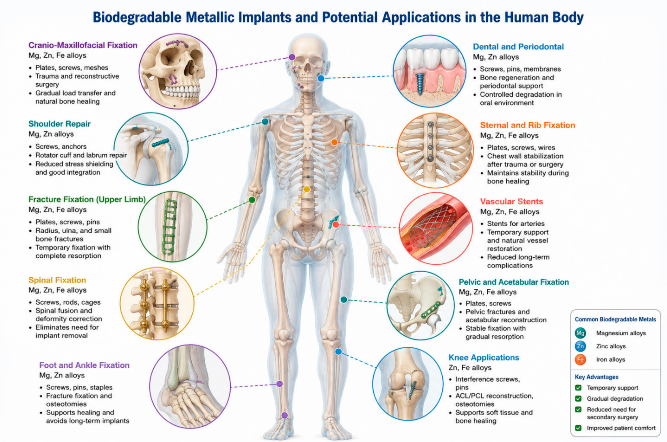
A major challenge in biodegradable metallic systems is achieving a balance between mechanical integrity, corrosion behavior, and biocompatibility, as these factors are strongly interconnected. During degradation, microstructural changes, pore formation, cracking, and corrosion products progressively reduce mechanical performance, making it essential to maintain structural integrity throughout the healing period [3,11]. To address these limitations, significant efforts have focused on alloy design, thermomechanical processing, microstructural engineering, surface modification, and additive manufacturing. These approaches aim to tailor degradation behavior, improve mechanical properties, and enable the fabrication of patient-specific implants with controlled architectures and porosity [1,2,11]. Bioresorbable metallic alloys are commonly produced using casting, powder metallurgy, thermomechanical processing, surface modification, and additive manufacturing routes [1,2,3,11]. Casting is widely used for initial alloy development but often produces coarse grains, segregation, and brittle secondary phases [1,3,11]. Thermomechanical processing, including rolling, extrusion, forging, drawing, ECAP, and HPT, is applied to refine grains, redistribute intermetallic phases, modify texture, and improve the strength–ductility–corrosion balance [12,13,14,15]. Powder metallurgy and space-holder methods enable porous architectures with controlled pore size and porosity [16,17,18,19], while additive manufacturing techniques such as LPBF, SLM, binder jetting, and extrusion-based 3D printing provide greater control over patient-specific geometry and scaffold topology [20,21,22,23,24]. Surface treatments, including plasma electrolytic oxidation, hydroxyapatite-based coatings, and other protective conversion layers, are frequently combined with bulk processing to regulate early-stage degradation and biological response [25,26,27]. More recently, hybrid and multi-material systems combining different biodegradable metals or metal-polymer combinations have been proposed to achieve optimized mechanical performance, degradation kinetics, and biological functionality [3]. Despite recent progress, the relationships among processing methods, microstructure evolution, corrosion behavior, mechanical degradation, and biological performance are still not fully understood. In addition, rapid advances in alloy development, thermomechanical processing, porous structures, and additive manufacturing have generated a large and scattered body of knowledge across different biodegradable metallic systems. Therefore, a comprehensive assessment of current progress and remaining challenges is still needed. Unlike previous reviews, which have primarily examined alloy composition, degradation behavior, or additive manufacturing as separate topics [1,2,3,20,21], this article focuses on the interrelationship among processing routes, microstructural evolution, corrosion kinetics, and the preservation of mechanical integrity in iron-, magnesium-, and zinc-based biodegradable metals. Attention is also given to thermomechanical processing regimes, heat treatments, porous architectures, and additive manufacturing strategies to clarify how processing-induced grain refinement, crystallographic texture, secondary phases, and scaffold topology can simultaneously govern degradation behavior and load-bearing performance [12,13,14,15,28]. By integrating processing–structure–property relationships, this review provides a more analytical perspective for the design of the next generation of biodegradable metallic implants. This review focuses on the corrosion behavior and mechanical performance of Fe-, Mg-, and Zn-based biodegradable metals, highlighting the effects of alloying, processing routes, porous architectures, and additive manufacturing on their overall performance. Finally, it discusses the main challenges and future opportunities for developing next-generation biodegradable metallic biomaterials [2,3,11]. The potential biomedical applications of biodegradable metallic implants in different regions of the human body are schematically illustrated in Figure 1.

2. Standard Requirements and Design Criteria for Biodegradable Metals

Unlike conventional biomaterials such as titanium alloys and stainless steels, biodegradable metals must be evaluated not only for their initial mechanical properties and biocompatibility but also for the changes that occur during in vivo degradation [2,3,11]. These changes include degradation kinetics, corrosion product formation, ion release, hydrogen evolution, and the gradual loss of mechanical integrity. To address these challenges, standards, including ISO/TS 20721 and ASTM F3268, were specifically established for absorbable metallic implants. These standards consider key aspects such as degradation kinetics, corrosion–mechanical interactions, ion release, hydrogen evolution, and time-dependent property changes. Their goal is to assess the safety and functionality of biodegradable metals under physiologically relevant conditions [31,32].
However, many studies continue to employ conventional standards originally developed for permanent implants or engineering materials, such as ASTM E8 [33] for tensile testing, ASTM G31 [34] for immersion corrosion testing, and ISO 10993 [35] for biological evaluation. While these methods provide reliable and reproducible and well-established procedures, they do not fully represent the dynamic degradation processes of biodegradable metals. For example, tensile tests measure only the initial mechanical properties, whereas conventional corrosion tests often fail to reproduce the complexity of the physiological environment or the simultaneous interaction between corrosion and mechanical degradation [32,33,34,35].
As a result, current assessment of biodegradable metals generally relies on a combination of dedicated biodegradable-metal standards and adapted conventional methodologies. This hybrid approach remains necessary because no single standard currently addresses all aspects of biodegradable implant performance. Future standardization efforts should focus on integrating mechanical, corrosion, and biological evaluations to enable more reliable prediction of long-term clinical performance [3,31,32,35].

2.1. Mechanical Performance Criteria

The mechanical requirements of biodegradable metallic implants depend strongly on their intended application. In orthopedic devices, materials must provide sufficient load-bearing support during bone healing while minimizing stress shielding through an elastic modulus closer to that of cortical bone [1,2]. For vascular stents, materials must combine adequate radial strength, high ductility for expansion, low elastic recoil, and fatigue resistance under cyclic loading [4,9].
For biodegradable stents, typical benchmark requirements include yield strength exceeding ~200 MPa, ultimate tensile strength above ~300 MPa, and elongation to failure greater than 15–20% to withstand crimping and balloon expansion without fracture. Elastic recoil should generally remain below 4% to ensure effective vessel support. Young’s modulus must also be optimized to balance radial strength and mechanical compatibility with the surrounding tissues [4,9,36].
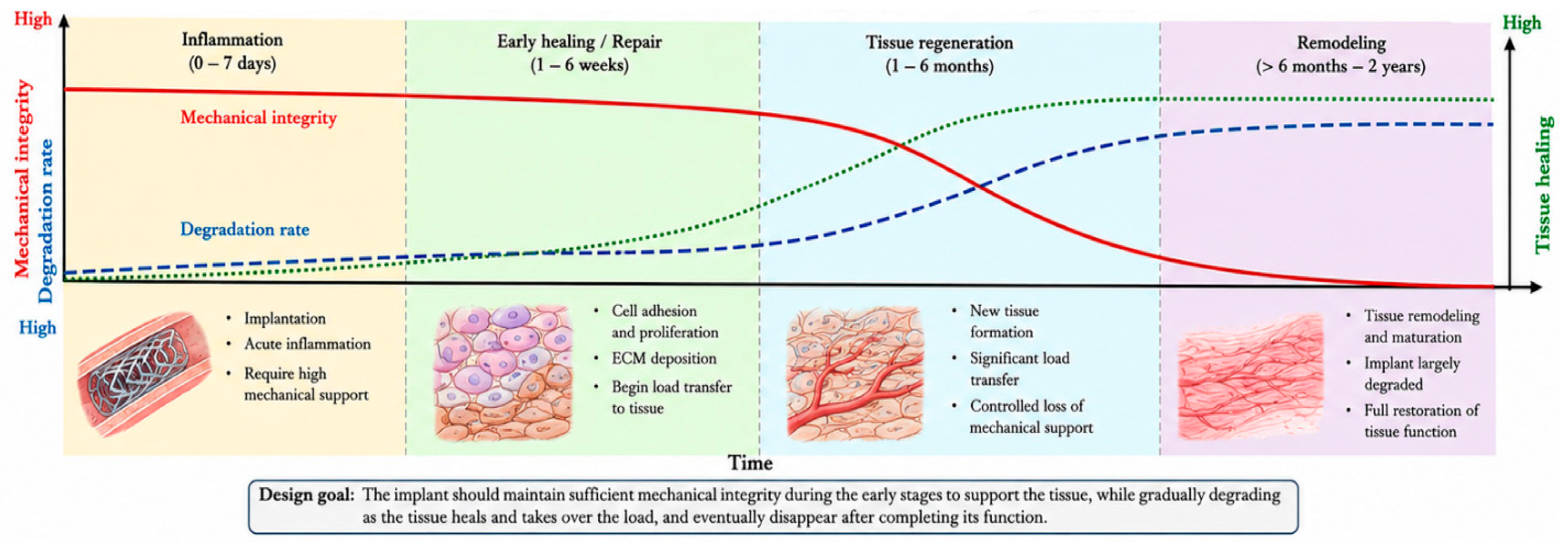
In orthopaedic applications, implants must maintain mechanical integrity throughout healing while progressively transferring load to the regenerating tissue. Therefore, degradation behavior and mechanical integrity should evolve in a synchronized manner, as illustrated in Figure 2. Premature degradation may result in the loss of mechanical support and implant failure, whereas excessively slow degradation reduces the clinical benefits of biodegradable systems [1,3,11]. Table 1 summarizes the mechanical benchmark criteria for biodegradable orthopedic implants and vascular stents.
Table 2 compares the typical mechanical properties of Mg-, Zn-, and Fe-based biodegradable metals with cortical bone, highlighting the challenge of simultaneously achieving suitable strength, ductility, stiffness, and degradation behavior. Fe-based alloys provide the highest strength but are limited by their high elastic modulus and slow degradation rate. Mg alloys exhibit a bone-like modulus but often degrade too rapidly, leading to premature loss of mechanical support. Zn alloys offer intermediate degradation behavior and good biocompatibility without hydrogen evolution; however, pure Zn still shows limited strength and ductility, motivating extensive research on alloying strategies and thermomechanical processing routes to enhance its mechanical performance while maintaining favorable biodegradation characteristics [1,8,9,10]. Overall, these benchmark values should be regarded as application-dependent guidelines rather than fixed acceptance limits. Several Fe-, Mg-, and Zn-based biodegradable alloys have approached the required mechanical ranges after appropriate alloying and processing; however, meeting the initial strength requirement alone is insufficient. For biodegradable implants, mechanical performance must be evaluated together with degradation rate, corrosion mode, retention of mechanical integrity, and biological response over time. Therefore, materials that satisfy tensile or compressive benchmarks in the undegraded state may still be unsuitable if rapid localized corrosion, excessive gas evolution, or long-term accumulation of corrosion products compromises their in vivo functionality.
Although Table 2 summarizes the baseline mechanical properties of pure Fe, Mg, and Zn, alloying and processing can substantially modify these properties and bring selected systems closer to application-specific mechanical benchmarks. Therefore, representative Fe-, Mg-, and Zn-based biodegradable systems that approach these requirements after alloying and processing, together with their main advantages and remaining limitations, are summarized in Table 3.
This comparison indicates that meeting the initial mechanical benchmark is necessary but not sufficient, because each alloy family still faces specific degradation-related limitations that must be considered together with mechanical performance.

2.2. Corrosion Requirements: Pure Fe, Mg, and Zn

The corrosion behavior of biodegradable metals plays a critical role in determining both their mechanical integrity and biological performance. Unlike permanent implant materials, biodegradable metals are designed to undergo controlled degradation in physiological environments, progressively transferring load to the healing tissue before complete resorption. Consequently, the degradation rate must be carefully tailored to the tissue healing process to prevent either premature loss of mechanical support or excessive implant retention [3,11].
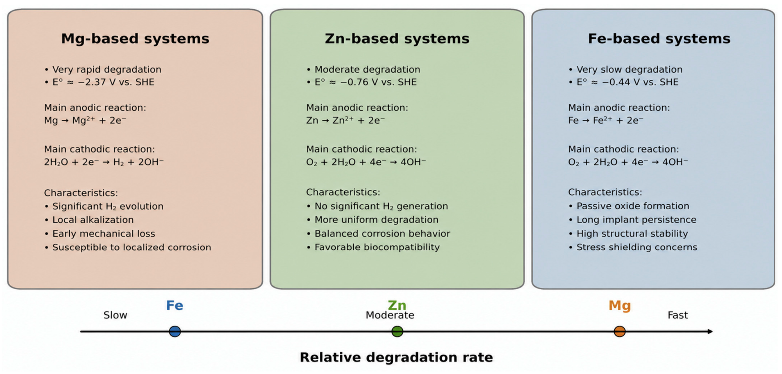
A comparison of the electrochemical and corrosion characteristics of Fe-, Mg-, and Zn-based biodegradable metals is provided in Table 4, while Figure 3 schematically summarizes the corresponding degradation mechanisms, relative degradation rates, hydrogen-gas evolution, and mechanical-integrity retention. As shown in Table 4 and Figure 3, Fe-based systems generally corrode slowly because of passive oxide/hydroxide film formation, Mg-based systems exhibit rapid dissolution accompanied by hydrogen evolution and early mechanical loss, and Zn-based systems show an intermediate degradation tendency without significant gas generation. However, Zn corrosion should not be considered intrinsically uniform, because alloy composition, secondary-phase distribution, crystallographic texture, processing route, and local galvanic coupling can produce heterogeneous or localized corrosion. Therefore, Zn degradation is better described as moderate but microstructure-dependent rather than uniformly homogeneous [1,5,6,42,43].
To address these limitations, considerable research has focused on alloy design, microstructural engineering, surface modification, and thermomechanical processing. More recently, additive manufacturing and functionally graded biodegradable materials have attracted increasing interest, enabling patient-specific degradation profiles through spatial control of porosity, microstructure, and corrosion behavior within a single implant [2,11].
The measured corrosion behavior in biodegradable Mg-, Zn-, and Fe-based metals strongly depends on the selected physiological test medium (Table 5). Commonly used solutions such as SBF, HBSS, PBS, DMEM, Ringer’s solution, and Tyrode’s solution differ in buffering capacity, chloride concentration, bicarbonate content, phosphate level, calcium ions, proteins, amino acids, and organic components. These differences can directly affect pH evolution, corrosion-product formation and stability, surface passivation, localized corrosion tendency, and the apparent degradation rate [44,45,46,47].
Therefore, corrosion rates reported for biodegradable metals should not be compared without considering the test medium, pH control, solution renewal, immersion time, temperature, surface-area/solution-volume ratio, and the presence or absence of organic components. This issue is particularly important for Mg alloys because hydrogen evolution and alkalization are highly sensitive to buffering capacity, but it is also relevant for Zn and Fe alloys because phosphate, carbonate, chloride, and protein-containing species can alter corrosion-product stability and surface passivation [8,12,44,45,46,47].

2.3. Biocompatibility

Biocompatibility is a fundamental requirement for biodegradable metals because both the implant and its degradation products interact continuously with surrounding tissues. Unlike permanent implants, biodegradable materials release ions and corrosion products during degradation, making their biological response dynamic and time-dependent [3,11,35].
Magnesium and zinc offer clear advantages as they are essential elements involved in bone metabolism, enzymatic activity, cell proliferation, immune response, and tissue regeneration. Consequently, their degradation products are generally well-tolerated and may even promote healing when released in controlled amounts. Iron is also an essential element; however, its slow degradation may lead to the accumulation of corrosion products, potentially affecting tissue remodeling and causing localized inflammatory responses [5,48,49].
Recent developments increasingly focus on biofunctionalization, where biodegradable metals are designed not only to be biocompatible but also to actively stimulate biological processes. This includes alloying strategies to enhance antibacterial activity, promote cell growth, and improve tissue integration, as well as surface treatments that regulate ion release and biological interactions. Another emerging trend is the integration of mechanical and biological design, where degradation is tailored to release beneficial ions at specific stages of healing. These developments reflect the transition from passive biomaterials toward multifunctional implants. The strong interrelationship between mechanical properties, corrosion behavior, and biocompatibility highlights the need for a holistic design approach, combining alloy development, processing, and surface engineering to achieve optimal performance [1,2,3,11].

3. Fe-Based Alloys

3.1. Overview and Main Limitations

Fe based biodegradable alloys have attracted considerable attention owing to their excellent mechanical properties, including high strength, good ductility, and superior fatigue resistance. These characteristics make them attractive candidates for load-bearing orthopedic implants and cardiovascular stents. In addition, iron is an essential trace element involved in numerous physiological processes, providing an inherent advantage from a biological perspective [12,40,42,50].
Despite these benefits, the clinical application of Fe-based biodegradable implants remains limited by their extremely slow degradation rate in physiological environments. The slow corrosion behavior of Fe is primarily associated with the formation of stable passive oxide layers that protect the surface from further degradation. While such passivation is beneficial for permanent implants, it is undesirable for biodegradable systems, where controlled resorption is required. As a result, Fe implants may persist for several years after implantation, exceeding the intended healing period [49,51,52]. Furthermore, the high elastic modulus of Fe (~200 GPa) is substantially higher than that of cortical bone (10–30 GPa), increasing the risk of stress shielding and subsequent bone resorption. The accumulation of iron oxides and hydroxides may also interfere with tissue remodeling and induce localized biological responses. Consequently, extensive research has focused on accelerating the degradation of Fe-based materials while preserving their favorable mechanical properties and biocompatibility [42,49,50]. From a clinical perspective, the main difficulty in Fe-based biodegradable alloys is not the lack of initial mechanical strength, but the mismatch between their slow degradation kinetics and the required healing time of temporary implants. Although alloying and processing can improve strength, ductility, and corrosion activity, Fe-based systems often remain protected by relatively stable oxide/hydroxide layers, which limit ion release and delay complete resorption. This slow degradation may reduce the biological advantage of a bioresorbable implant and increase the risk of long-term corrosion-product retention, stress shielding, and incomplete tissue remodeling. Therefore, future Fe-based implant design should not focus only on increasing corrosion rate, but should also consider corrosion uniformity, corrosion-product transport, local tissue response, magnetic compatibility, and the retention of mechanical integrity during the intended healing period [4,12,40].

3.2. Development: Fe Alloying, Bulk Processing, and Porous Architectures

The development of Fe-based biodegradable alloys has followed two complementary strategies: The first strategy focuses on dense bulk components, in which alloying and thermomechanical processing are used to modify phase stability, refine the microstructure, improve mechanical properties, and accelerated degradation. The second strategy involves the design of porous Fe-based scaffolds, where controlled porosity and interconnected pore networks increase the exposed surface area, facilitate electrolyte penetration, reduce stiffness and promote tissue ingrowth. Together, these approaches address the intrinsic limitation of Fe systems, namely their excessively slow degradation compared with clinical healing times [12,20,50,53].
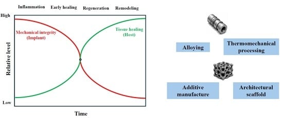
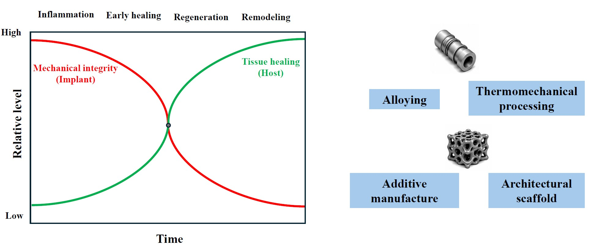
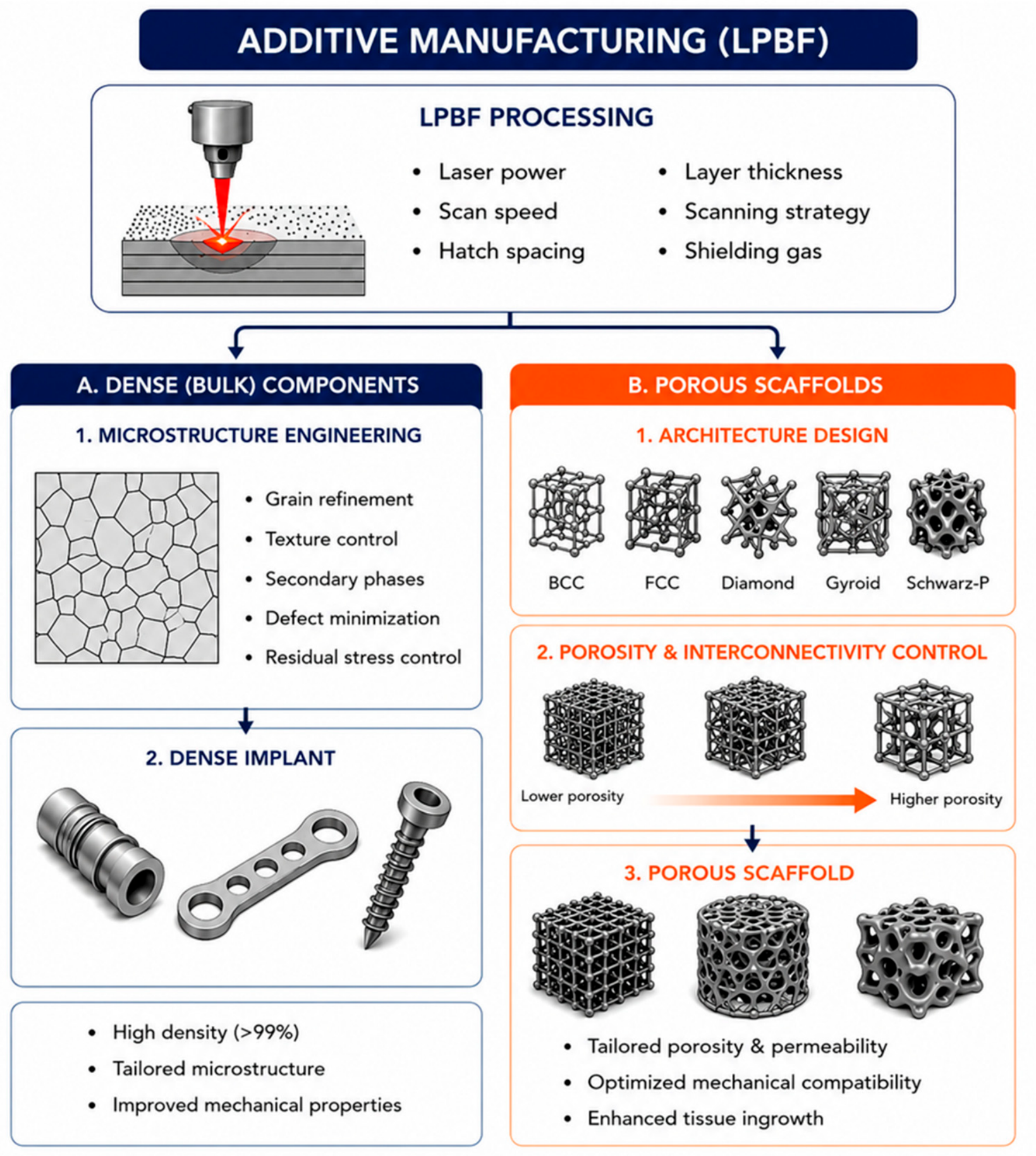
As schematically illustrated in Figure 4, biodegradable metallic systems can generally be developed through two complementary routes: dense bulk components produced via microstructural engineering and porous scaffolds designed through architectural control.

3.2.1. Alloying Strategies in Fe-Based Bioresorbable Alloys

Alloying remains one of the most effective approaches for improving the performance of biodegradable Fe-based alloys. Because the primary limitation of pure Fe is its extremely slow degradation rate, alloy design has largely focused on accelerating corrosion while preserving the excellent mechanical properties that make Fe attractive for biomedical applications. Depending on their primary function, alloying elements can generally be categorized as austenite stabilizers, degradation accelerators, and biofunctional elements [40,52,57].
Mn, C, and Si are among the most widely investigated alloying additions in Fe-based bioresorbable alloys because they simultaneously influence phase stability, mechanical behavior, magnetic response, and corrosion activity. Mn is particularly important because it stabilizes the γ-austenite phase, reduces ferromagnetism, and improves ductility, making Fe–Mn alloys one of the benchmark systems for biodegradable implant applications [40,51,58]. Carbon further enhances strength and work hardening in Fe–Mn–C alloys through solid-solution strengthening and TWIP/TRIP-assisted deformation mechanisms; however, carbide formation must be carefully controlled because it may introduce local micro-galvanic effects and promote localized corrosion [57,59,60,61,62,63], whereas Si promotes ε-martensite formation and shape-memory behavior in Fe–Mn–Si systems, while also affecting Young’s modulus, corrosion response, and phase transformation behavior [64,65,66,67,68,69]. Therefore, Mn, C, and Si do not simply act as strengthening additions; rather, they control the balance between phase constitution, deformation mechanism, magnetic compatibility, and degradation behavior in Fe-based biodegradable alloys [40,51,58,59,60,61,64,66].
A second group of alloying elements is primarily introduced to accelerate degradation. Noble elements such as Pd, Ag, Au, Pt, and Cu promote micro-galvanic corrosion through the formation of cathodic second phases [52,70,71,72,73,74,75,76,77,78,79,80,81,82]. Among these, Pd is particularly effective, increasing degradation rates by nearly an order of magnitude while retaining high mechanical strength [70,71,72,73]. Ag and Cu can also accelerate degradation and provide antibacterial activity, although their effectiveness depends on particle distribution, ion release, and processing route [41,74,75,76,77,78,83,84,85,86,87]. Ca and Mg are less commonly used but are attractive for bone applications because of their osteogenic potential; however, their limited solubility in Fe creates challenges related to segregation, secondary phases, and localized corrosion [88,89,90,91]. Overall, future Fe alloy design is moving toward multi-element systems that combine degradation control, mechanical integrity, antibacterial response, magnetic compatibility, and radiopacity [12,52,79,80,81,82,92,93,94]. Representative Fe-based bioresorbable alloy systems and their main alloying strategies, processing routes, reported effects, and remaining limitations are summarized in Table 6.
This comparison highlights that no single Fe-based alloying strategy simultaneously resolves all key requirements, including controlled and sufficiently uniform degradation, adequate mechanical integrity, magnetic compatibility, and acceptable biological response [12,49,50,53].

3.2.2. Fe Thermomechanical Processing

Thermomechanical processing is used not only to improve strength and ductility but also to modify corrosion behavior, phase stability, and microstructural evolution. Hot rolling, cold rolling, rolling plus heat treatment, forging, swaging, wire/tube drawing, SPD, and hot extrusion refine grains, increase defect density, modify texture, and redistribute phases [12,64,114,115]. Hot rolling has been widely applied to Fe–Mn, Fe–Mn–Si, Fe–Mn–C, and Fe–Mn–C–Ag alloys at 600–1100 °C, improving microstructural homogeneity and changing the γ-austenite/ε-martensite balance [63,65,92,97,98,116]. Cold rolling increases strength through work hardening but may reduce ductility; its effect on corrosion depends on alloy chemistry, strain level, and annealing [57,59,64,92].
Rolling combined with annealing, recrystallization, solution treatment, or quenching is particularly important in Fe–Mn–Si systems because it controls grain size, residual stress, γ ↔ ε martensitic transformation, Young’s modulus, and electrochemical response [65,66,96,98,114]. Bulk compressive processes such as forging, rotary swaging, radial-shear rolling, and hot compression improve homogeneity and can generate deformation bands, twins, and phase changes that affect both mechanical properties and degradation [41,51,67,114]. Wire and tube drawing are essential for Fe–Mn-based stents and neurovascular devices, as they produce fine geometries with high radial strength, although intermediate annealing is often required to prevent excessive loss of ductility [58,62,93,94,95]. SPD methods such as ECAP and HPT can generate ultrafine or nanostructured Fe alloys, but corrosion behavior does not always increase with grain refinement because phase transformations may reduce micro-galvanic activity [68,69,99]. Hot extrusion remains less explored, mainly in Fe–Mn–Ca alloys, and systematic studies on extrusion temperature, ratio, and speed are still lacking [100]. The main thermomechanical and manufacturing routes applied to Fe-based biodegradable alloys are summarized in Table 7. These processing routes not only improve mechanical properties, but also modify phase stability, defect density, texture, secondary-phase distribution, and corrosion response [12,64,88,114,115].
Overall, thermomechanical processing provides an important route for improving the mechanical performance of Fe-based biodegradable alloys; however, its effect on degradation is not straightforward. Grain refinement, increased defect density, and phase redistribution may enhance corrosion activity in some systems, whereas processing-induced phase transformations may reduce micro-galvanic effects or change the stability of the surface film. Therefore, processing parameters must be optimized together with alloy composition rather than considered as an independent variable [12,64,66,68,99].

3.2.3. Fe Porous Architectures and Manufacturing Approaches

Porous Fe-based structures provide an extrinsic route to accelerate degradation by increasing exposed surface area, improving electrolyte penetration, and reducing stiffness toward values closer to bone. This approach is especially relevant because bulk Fe alloys often remain too corrosion-resistant for biodegradable applications. Porous Fe-based scaffolds have been fabricated using several processing routes, including powder metallurgy with space holders, sponge replication, LPBF/SLM, binder jetting, extrusion-based 3D printing/direct ink writing (DIW), and hybrid 3D printing followed by sintering. Among these methods, powder metallurgy and space-holder techniques provide relatively simple control over porosity and pore size, whereas LPBF/SLM enables more precise control of ordered lattice architectures. Binder jetting is also relevant because it can fabricate complex porous Fe-based scaffolds without direct laser melting; however, its final properties are strongly affected by debinding, sintering shrinkage, residual porosity, and post-sintering microstructure. In contrast, extrusion-based 3D printing/DIW has recently attracted attention because it enables the fabrication of highly porous Fe, Fe–Mn, and Fe-based bioactive composite scaffolds with controlled architecture and improved biological functionality. Therefore, this section emphasizes the routes for which more complete relationships among processing route, scaffold architecture, degradation behavior, mechanical response, and biological performance have been reported [12,24,50,53,110,111,112,117]. However, increasing porosity reduces load-bearing capacity and fatigue resistance, making the balance between degradation rate, mechanical integrity, and biological performance critical [20,50,53,101,102,112,118]. Porous architectures also promote tissue integration by enabling cell migration, vascularization, and nutrient transport and new tissue ingrowth [20,50,53], as schematically shown in Figure 4.
Powder metallurgy and space-holder techniques are widely used conventional routes because they allow control of pore size and porosity through powder characteristics, space-holder content, compaction pressure, and sintering parameters [16,53,102,104]. Porosity levels around 30–50% can maintain properties suitable for selected orthopedic applications, and bioactive phases such as hydroxyapatite can be incorporated to improve biological response [16,102,105]. Sponge replica methods produce highly interconnected open-cell structures; Fe–Mn scaffolds with ~85% porosity and 375–500 μm pores have shown properties comparable to cancellous bone, although load-bearing capacity remains limited [106,119,120,121] LPBF/SLM enables digital control of pore architecture, strut thickness, and porosity, and Fe–35Mn scaffolds have shown promising biodegradation, biocompatibility, and mechanical performance for porous load-bearing bone scaffold applications [53,107,108,109]. However, defects, residual stress, surface roughness, and elemental evaporation must be controlled [107,108,109]. Extrusion-based 3D printing/DIW offers a lower-cost alternative and has enabled Fe–Mn and FeMn–akermanite scaffolds with improved biodegradability, osteogenic response, and MRI compatibility [24,110,111,112,113]. Hybrid approaches combining AM, powder metallurgy, and sintering can create hierarchical porous structures with CAD-designed macropores and sintering-induced micropores [103,122,123,124]. Overall, the most promising route for Fe-based biodegradable implants is the integration of alloy design, thermomechanical processing, and controlled porous architecture to achieve degradation synchronized with tissue healing while maintaining temporary mechanical support [12,20,24,50,53,107].

4. Mg-Based Alloys

4.1. Comparison to Fe and Clinical Motivation

Magnesium-based alloys have attracted considerable attention as biodegradable implants because they address several limitations of permanent metallic biomaterials and Fe-based biodegradable systems. Unlike stainless steels and titanium alloys, Mg exhibits an elastic modulus of approximately 40–45 GPa, much closer to cortical bone, reducing stress shielding and promoting more natural load transfer during healing. Compared with Fe-based alloys, Mg offers superior biomechanical compatibility and a more suitable degradation timeframe. However, Mg alloys generally possess lower mechanical strength and significantly higher corrosion rates, making degradation control the primary challenge for clinical application [5,6,13,125].

4.2. Challenges

The main limitation of Mg alloys is their high chemical reactivity in physiological environments. Corrosion leads to the formation of Mg(OH)2 and the evolution of hydrogen gas, often resulting in degradation rates that exceed tissue healing requirements. Rapid corrosion can generate gas cavities around the implant, affecting tissue regeneration, implant stability, and, in severe cases, causing local tissue damage. Simultaneously, the accelerated loss of material may lead to premature reduction in mechanical integrity and implant failure before healing is complete. In addition, Mg degradation causes local alkalization through Mg(OH)2 formation and OH accumulation; however, the magnitude of pH increase strongly depends on the test solution, buffering capacity, ion composition, and solution volume/surface area ratio. For example, Lamaka et al. measured the local pH 10–50 μm above Mg alloy surfaces in Hank’s solution and reported near-surface pH values of approximately 9.9–10.5 in simple Hank’s solution, whereas Ca2+-modified Hank’s solution maintained lower values of about 7.8–8.5 owing to the formation of calcium-phosphate/carbonate surface layers. Yang and Zhang further showed that Mg alloy corrosion in Hank’s solution and simulated blood plasma is affected by chloride concentration, Ca2+/PO43− content, and solution volume/surface area ratio, confirming that pH evolution and corrosion kinetics must be interpreted together with the selected immersion medium and testing geometry. Such localized alkalization may impair cellular activity and tissue compatibility if buffering and corrosion-product formation are insufficient [46,47,126].
Beyond corrosion, Mg alloys exhibit relatively low fatigue resistance and are highly sensitive to microstructural heterogeneities, which can further promote localized degradation. Consequently, controlling corrosion kinetics while maintaining adequate mechanical performance remains the key challenge limiting the broader clinical adoption of Mg-based biodegradable implants [5,13,14,127]. It should also be noted that the corrosion kinetics of Mg-based biodegradable alloys cannot be fully described by a single corrosion-rate value, because the degradation process is strongly time-dependent and affected by the selected testing method. Electrochemical techniques such as OCP, potentiodynamic polarization, and EIS mainly reflect early-stage surface reactions, passive-film stability, and charge-transfer resistance, whereas hydrogen-evolution, immersion, and weight-loss measurements provide longer-term information on dissolution, corrosion-product accumulation, and degradation progression. Therefore, corrosion-rate data for Mg alloys should be interpreted together with test duration, solution composition, pH evolution, solution renewal, and the surface-area/solution-volume ratio [44,45,47].
From an electrochemical-kinetics perspective, Mg corrosion is mainly governed by rapid anodic dissolution of Mg and cathodic hydrogen evolution, whereas the temporary formation and breakdown of Mg(OH)2-based corrosion layers control the transition from early-stage surface activation to longer-term degradation. Potentiodynamic polarization can provide useful parameters such as corrosion potential, corrosion current density, and apparent corrosion rate; however, for Mg alloys these values should be interpreted cautiously because negative difference effects, film rupture, hydrogen evolution, and localized attack may distort Tafel extrapolation. EIS is therefore particularly useful for evaluating the evolution of surface-film resistance, charge-transfer resistance, and diffusion-related processes during immersion. A larger capacitive loop and higher polarization resistance generally indicate improved short-term corrosion resistance, whereas the appearance of inductive loops or decreasing impedance at low frequency often reflects film instability, adsorption/desorption of intermediate species, or localized corrosion activity. Thus, polarization and EIS results should be correlated with immersion observations, hydrogen evolution, pH variation, and corrosion-product characterization rather than used as isolated indicators of Mg degradation behavior [44,45,128,129,130].

4.3. Mg Development: Alloying, Bulk Processing, and Porous Architectures

Following the general development strategy illustrated in Figure 4, research on Mg-based biodegradable materials has focused on two parallel directions: optimization of dense bulk alloys through alloy design and thermomechanical processing and the development of porous architectures to improve mechanical compatibility and regulate degradation behavior. While bulk alloy development primarily aims to reduce the intrinsically high corrosion rate of magnesium, porous structures exploit geometric design to simultaneously promote biological integration and control degradation kinetics. These two approaches are increasingly combined to achieve a balanced relationship between mechanical integrity, corrosion resistance, and tissue regeneration [13,14,21,125].

4.3.1. Development of Bulk Mg-Based Alloys

In bulk form, Mg alloys are primarily optimized through alloying and thermomechanical processing to improve mechanical performance while reducing corrosion rates [13,14,125,127]. Alloying additions such as Zn, Ca, Mn, Zr, Sr, and rare earth (RE) elements play a central role in tailoring microstructure and electrochemical behavior. Ca and Zn are particularly attractive because they are naturally present in the human body and contribute to bone metabolism. Controlled additions of Ca improve biocompatibility and osteogenic potential; however, excessive Ca promotes the formation of Mg2Ca intermetallic phases that can accelerate localized galvanic corrosion and reduce ductility [131,132,133,134,135]. Similarly, Zn contributes to solid-solution strengthening, grain refinement, and stabilization of corrosion products, although excessive Zn may increase micro-galvanic activity through the formation of Zn-rich secondary phases [125,136,137,138,139,140].
Rare earth elements such as Y, Nd, and Gd are among the most effective alloying additions for biodegradable Mg alloys. These elements refine grains, modify crystallographic texture, improve mechanical strength, and contribute to the formation of more stable surface films, thereby reducing corrosion rates [141,142,143,144,145,146]. Additional alloying elements including Sr, Zr, Ag, Cu, Li, Si, and Sn have also been investigated to improve degradation behavior, antibacterial performance, and mechanical properties, although their use requires careful control to avoid excessive galvanic corrosion or potential biocompatibility concerns [125,147,148,149,150,151,152,153,154,155,156,157].
Beyond alloy composition, thermomechanical processing is one of the most effective strategies for microstructural optimization. Hot rolling, hot extrusion, cold rolling followed by annealing, forging, multidirectional forging (MDF), equal-channel angular pressing (ECAP), and high-pressure torsion (HPT) have all been employed to refine grain size, redistribute secondary phases, and reduce casting defects. Dynamic recrystallization during rolling and extrusion produces finer and more homogeneous microstructures, improving both mechanical strength and corrosion resistance through grain refinement, reduced segregation, and a more uniform distribution of secondary phases. In Mg-based biodegradable alloys, hot extrusion is particularly important because it converts coarse cast structures into refined recrystallized microstructures, breaks up brittle intermetallic networks, and redistributes cathodic second phases that otherwise act as local galvanic sites. Shiri et al. reviewed the role of extrusion parameters in biodegradable Mg alloys and showed that extrusion temperature, extrusion ratio, and extrusion speed strongly influence degradation behavior through their effects on dynamic recrystallization, basal texture, grain size, and secondary-phase distribution. For example, in extruded Mg–Y–Zn–Zr alloys containing W and I phases, increasing the extrusion temperature from 250 °C to 300 °C improved corrosion resistance because of more complete dynamic recrystallization, grain refinement, and a more homogeneous distribution of broken second phases; however, further increasing the temperature to 350 °C reduced the benefit because grain coarsening and second-phase effects became more pronounced. More specifically, Wu et al. reported that Mg–2Y–1Zn–0.6Zr alloy extruded at 300 °C exhibited the best corrosion resistance in SBF, with a weight-loss corrosion rate of 0.4601 mm/year, whereas the corrosion-rate sequence followed E250 °C > E350 °C > E300 °C. This behavior was attributed to sufficient dynamic recrystallization and finer, more uniform grains at 300 °C, while undissolved W and I phases or unrecrystallized regions at lower extrusion temperature, and grain coarsening at higher extrusion temperature, promoted stronger micro-galvanic corrosion [158]. Similar observations have been reported in Mg–Zn–Ca–Zr systems, where optimized extrusion temperatures produced lower corrosion rates and more uniform degradation, whereas non-optimized extrusion conditions or excessive secondary-phase precipitation could increase localized corrosion. Therefore, the beneficial effect of extrusion on Mg corrosion is not universal; it depends on the balance between grain refinement, texture evolution, residual strain, and the size, chemistry, and distribution of intermetallic phases [14,158,159,160,161,162,163,164].
The effect of heat treatment on Mg corrosion should be interpreted according to alloy family rather than treated as a universal response. In Mg–Al systems, solution treatment can dissolve or redistribute β-Mg17Al12 phases and reduce micro-galvanic coupling when the secondary phase network is discontinuous; however, inappropriate aging may reintroduce cathodic precipitates and increase localized corrosion. In Mg–Zn and Mg–Zn–Zr alloys, T4 treatment may improve corrosion resistance by dissolving Zn-rich secondary phases and homogenizing solute distribution, whereas T6 aging can either improve strength through precipitation hardening or reduce corrosion resistance if precipitates become coarse, continuous, or strongly cathodic relative to the Mg matrix. In Mg–Y–Nd–Zr and other Mg–RE systems, heat treatment modifies RE-rich β′ and β1 nanophases, grain-boundary precipitates, and surface-film stability; therefore, T6 treatment may improve passivation and mechanical strength in some WE43-type alloys but may also promote localized attack when precipitates or eutectic remnants are non-uniformly distributed. Thus, heat treatment should be optimized separately for each Mg alloy family because corrosion resistance depends on the combined effects of solute homogenization, precipitate morphology, grain size, and cathodic second-phase distribution [165,166,167]. The alloy-family-dependent effects of heat treatment on the microstructure and corrosion behavior of representative biodegradable Mg alloys are summarized in Table 8. Representative studies further show that the corrosion response after heat treatment is mainly governed by the morphology, continuity, and electrochemical role of secondary phases rather than by the heat-treatment condition alone. In Mg–Al alloys, solution treatment can reduce micro-galvanic corrosion by dissolving or disrupting continuous β-Mg17Al12 networks; however, subsequent aging may reintroduce cathodic β-Mg17Al12 precipitates and increase localized corrosion if the precipitates become coarse or continuous. In Mg–Zn and Mg–Zn–Zr alloys, T4 treatment can improve corrosion resistance by dissolving Zn-rich phases and homogenizing solute distribution, whereas T6 aging may either improve mechanical strength through precipitation hardening or reduce corrosion resistance when Zn-rich precipitates form strong local cathodic sites. In Mg–Y–Nd–Zr and WE43-type alloys, heat treatment affects RE-rich precipitates, especially β′ and β1 nanophases, as well as grain-boundary particles and surface-film stability. Therefore, the beneficial or detrimental effect of heat treatment depends on whether secondary phases become fine, discontinuous, and uniformly distributed or coarse, continuous, and electrochemically active relative to the Mg matrix [165,166,167].
This comparison shows that heat treatment cannot be evaluated independently of alloy chemistry. In Mg alloys, the same treatment condition may improve corrosion resistance in one alloy family by dissolving harmful secondary phases but decrease corrosion resistance in another system by producing coarse or continuous cathodic precipitates. Therefore, the corrosion response of heat-treated Mg alloys should be interpreted together with phase constitution, precipitate distribution, electrochemical potential differences, and the stability of the corrosion-product layer [165,166,167]. Overall, bulk processing strategies for Mg-based biodegradable alloys should be evaluated through the combined effects of grain refinement, crystallographic texture, dynamic recrystallization, residual strain, and secondary-phase redistribution. Although extrusion, rolling, and heat treatment can substantially improve strength and ductility, their influence on corrosion is alloy-dependent and sometimes contradictory. Grain refinement and solute homogenization may reduce micro-galvanic heterogeneity and promote more uniform degradation, whereas strong basal texture, residual deformation defects, or continuous cathodic intermetallic phases may accelerate localized attack. Therefore, future development of bulk Mg-based implants should move from single-parameter optimization toward integrated control of processing route, alloy chemistry, microstructure, corrosion kinetics, and mechanical-integrity retention during degradation [14,158,164,165,167].
Processing-induced crystallographic texture also plays a critical role. Magnesium’s hexagonal close-packed (HCP) structure makes corrosion behavior strongly orientation-dependent. Consequently, texture control has become as important as grain refinement, since strong basal textures may induce mechanical and corrosion anisotropy that can compromise implant reliability [168,169,170,171]. Similarly, forging and MDF processing routes improve strength and corrosion resistance by refining grains and modifying texture, although excessive deformation can increase residual stresses and promote localized corrosion [172,173,174,175,176].
Severe plastic deformation techniques such as ECAP and HPT represent advanced processing routes capable of producing ultrafine-grained Mg alloys. These methods significantly enhance strength through grain refinement and may improve corrosion resistance when microstructural homogeneity is maintained. However, excessive residual stresses and heterogeneous distributions of secondary phases may offset some of these benefits [127,177,178,179,180,181,182,183].
Surface modification strategies are commonly employed alongside alloy and processing optimization. Calcium-phosphate coatings, hydroxyapatite layers, polymeric coatings, plasma electrolytic oxidation (PEO), fluoride treatments, and oxide films act as temporary barriers that reduce the initial corrosion rate and preserve mechanical integrity during early healing stages. Nevertheless, their long-term effectiveness depends on coating adhesion, degradation kinetics, and interactions with the underlying substrate [25,26,27,184,185].
Despite these advances, bulk Mg alloys continue to face challenges associated with rapid degradation, hydrogen evolution, localized alkalization, and premature mechanical failure in aggressive physiological environments. Consequently, structural approaches based on engineered porosity have emerged as a complementary strategy [5,6,21,186].

4.3.2. Development of Porous Mg-Based Structures

Porous Mg-based structures represent an alternative approach to overcoming the limitations of bulk materials by exploiting architectural design to tailor mechanical properties, degradation behavior, and biological performance. The introduction of interconnected porosity facilitates cell migration, vascularization, nutrient transport, and bone ingrowth while simultaneously reducing the elastic modulus toward values closer to those of cancellous bone, thereby mitigating stress shielding effects [186,187].
Unlike Fe-based systems, where porosity is primarily introduced to accelerate degradation, Mg scaffolds require careful architectural design because increased surface area generally accelerates corrosion and hydrogen evolution. Therefore, scaffold design must balance porosity, pore size, interconnectivity, wall thickness, mechanical strength, and degradation rate [21,186,187].
Powder metallurgy combined with space-holder techniques remains one of the most widely used fabrication routes. In these methods, temporary pore-forming agents such as ammonium bicarbonate, urea, or salt particles are removed after compaction and sintering, producing interconnected pore networks with controllable pore morphology [17,18,186,188,189,190]. These techniques enable tuning of pore size and porosity, although challenges remain related to powder oxidation, incomplete sintering, residual MgO formation, and insufficient control of local corrosion processes [17,18,186,190].
Additive manufacturing has emerged as one of the most promising technologies for biodegradable Mg scaffolds because it enables precise control of pore geometry, unit-cell architecture, strut thickness, and spatial porosity distribution [21,185,191,192,193,194,195]. Technologies such as laser powder bed fusion (LPBF) and selective laser melting (SLM) have demonstrated the ability to fabricate highly ordered porous structures with mechanical properties approaching those of trabecular bone. However, magnesium processing remains challenging due to its high vapor pressure, oxidation tendency, flammability, and susceptibility to powder spattering and evaporation during laser processing [21,185,194].
Recent studies have demonstrated that scaffold architecture can significantly influence degradation behavior. Parameters such as pore size, pore morphology, strut thickness, and surface roughness directly affect corrosion kinetics, mechanical stability, and osteogenic response [185,192,195]. Larger interconnected pores generally improve bone ingrowth and vascularization, whereas excessively high porosity may lead to rapid loss of mechanical integrity and accelerated degradation [189,192].
Alternative fabrication routes include infiltration casting, sacrificial-template methods, replica techniques, and metallic foaming approaches [27,196,197,198,199]. These methods can generate highly interconnected open-cell structures and often provide better metallurgical continuity than powder-based routes. However, they require careful control of melt infiltration, template removal, and oxidation during processing. Residual contaminants from templates may significantly influence corrosion behavior and biocompatibility [196,197,198]. The main fabrication routes for porous Mg-based biodegradable scaffolds and their architecture-related advantages and limitations are summarized in Table 9.
This comparison emphasizes that porous Mg scaffold design is more complex than simply increasing [17,18,27,185,186,191,192,193,196,197,198,199] porosity. Although interconnected pores improve cell migration, vascularization, nutrient transport, and bone ingrowth, they also increase the exposed surface area and may accelerate Mg dissolution, hydrogen evolution, and early mechanical loss. Therefore, the design of porous Mg-based implants requires simultaneous optimization of pore size, porosity, interconnectivity, strut thickness, surface condition, coating stability, and alloy composition. In this regard, graded and hierarchical architectures are particularly attractive because dense regions can preserve load-bearing capacity, whereas porous regions can promote tissue integration and controlled degradation [17,18,27,185,186,191,192,193,196,197,198,199].
To address the conflicting requirements of mechanical support and biological performance, recent developments increasingly focus on graded and hierarchical architectures. Dense regions provide load-bearing capacity, whereas porous regions promote tissue integration and controlled degradation. Furthermore, combining porous architectures with surface coatings such as hydroxyapatite, calcium-phosphate, fluoride, or oxide layers has shown considerable potential for mitigating the accelerated corrosion typically associated with highly porous Mg structures [27,185,192,199].
Overall, the integration of alloy design, thermomechanical processing, advanced manufacturing technologies, and surface engineering is driving the development of next-generation biodegradable Mg implants. Future research is expected to focus on hierarchical scaffold architectures, patient-specific additive manufacturing, and multiscale strategies that simultaneously optimize mechanical performance, degradation kinetics, and biological functionality [5,6,21,53,185,192].

5. Zn-Based Alloys

5.1. Comparison to Fe and Mg

Zinc-based alloys have emerged as a promising alternative to both Mg- and Fe-based biodegradable metals because they exhibit an intermediate degradation rate that is closer to the clinically desired window. Unlike Mg, Zn does not generate hydrogen gas during corrosion, while compared with Fe it degrades more rapidly, reducing the risk of long-term implant persistence. Mechanically, Zn alloys occupy an intermediate position between Mg and Fe, combining moderate strength, ductility, and stiffness, making them attractive for orthopedic and cardiovascular applications [7,9,200,201].

5.2. Challenges

Despite these advantages, pure Zn generally suffers from relatively low mechanical strength in the as-cast condition, often remaining below the levels required for demanding load-bearing applications. Its ductility may also be restricted in the as-cast state because of coarse grains, segregation, and limited activation of deformation mechanisms. However, this limitation should not be considered intrinsic to Zn, because extrusion, rolling, drawing, and severe plastic deformation can substantially improve ductility, strength, and mechanical reliability by refining grains, modifying texture, and redistributing second phases. In addition, Zn corrosion should not be regarded as intrinsically uniform. Alloying-induced second phases, eutectic constituents, crystallographic texture, additively manufactured defects, and local micro-galvanic couples can promote heterogeneous or localized corrosion. Therefore, the performance of Zn alloys is highly processing-sensitive and depends on the combined control of alloy chemistry, grain size, texture, secondary-phase morphology, and manufacturing route [9,15,109,202,203].

5.3. Development of Zn-Based Biodegradable Metals: From Bulk Alloys to Porous Architectures

5.3.1. Alloy Design of Bulk Zn-Based Biodegradable Alloys

Alloying is one of the main strategies used to improve the limited mechanical properties of pure Zn while preserving its favorable degradation behavior. Different alloying elements are added to enhance strength, ductility, corrosion resistance, biocompatibility, antibacterial activity, and bone-regeneration potential [9,200,201,204,205].
Zn–Mg alloys are among the most successful and widely studied Zn-based systems. Mg is biocompatible, promotes grain refinement, improves cell adhesion, and can bring the mechanical response of Zn alloys closer to that of natural bone [204,206,207]. However, Mg forms brittle Mg2Zn11 and MgZn2 phases, which increase strength through precipitation hardening but may reduce ductility and promote localized galvanic corrosion [206,207,208]. Therefore, Mg content and intermetallic morphology must be carefully controlled. Processing routes such as extrusion and ECAP can fragment and homogenize these phases, improving strength, ductility, and corrosion uniformity [204,207,209].
Li is one of the strongest alloying additions for Zn. It increases strength and hardness while reducing elastic modulus, which is beneficial for bone-related applications [10,204,210]. Its strengthening effect is mainly associated with the β-LiZn4 phase, which restricts dislocation motion. However, excessive Li may cause brittleness [10,210]. Thermomechanical processing, such as extrusion and rolling, refines the microstructure and secondary phases, while additions such as Mn can further improve the strength–ductility balance [211,212,213]. Li-containing corrosion products such as LiOH and Li2CO3 may form a relatively protective layer in SBF, although corrosion behavior strongly depends on the testing medium [184,214].
Mn is attractive due to its biocompatibility, low toxicity, and role in bone metabolism [203,204,215]. It contributes to grain refinement, solid-solution strengthening, twinning, texture modification, and improved plastic deformation [203,204,215]. At low contents, MnZn13 intermetallics may be beneficial, while excessive Mn can increase localized corrosion and stress concentration [200,215]. In some systems, Mn produces exceptional ductility; for example, Zn–0.8Li–0.8Mn showed elongation of about 103% [10]. In extruded Zn–0.2Mg, Mn promoted grain rotation and increased the Schmid factor for non-basal slip, improving plasticity [216].
Cu improves strength, antibacterial activity, bone growth, and angiogenesis [204,215,217]. Its strengthening effect is mainly related to solid-solution strengthening, grain refinement, and hard intermetallic phases such as ε-CuZn5/CuZn4 [10]. However, coarse Cu-rich phases can reduce ductility and intensify localized corrosion due to galvanic coupling [204,217]. Thermomechanical processing can refine these phases and improve corrosion uniformity. In Zn–Cu alloys, refined precipitates may activate phase-boundary sliding at Zn/CuZn4 interfaces, enabling room-temperature superplasticity [217,218,219].
Ca is especially relevant for bone implants because of its high bioactivity, low toxicity, and positive role in bone regeneration [200,206]. However, its very low solubility in Zn leads to the formation of coarse and brittle CaZn13 intermetallics, which reduce ductility and promote localized galvanic corrosion [200,203]. Therefore, controlling Ca-containing precipitates is essential. Recent studies show that ultrasonic treatment, hot rolling, and optimized casting can refine CaZn13 particles and significantly improve ductility; for example, Zn–0.3Ca achieved 49% elongation after strong refinement of CaZn13 particles and Zn grains [220,221].
Sr stimulates osteogenesis, mineralization, and bioactivity [200,222]. Like Ca, it forms SrZn13 intermetallics due to limited solubility in Zn but generally causes less brittleness [200,204]. Thermomechanical processing can refine SrZn13 phases and improve the balance between strength, ductility, and degradation. Low Sr contents may promote more uniform corrosion, while excessive Sr can increase intermetallic content and microgalvanic corrosion [200,222]. A Zn–1Sr alloy showed compressive superplasticity and a yield strength of about 340 MPa [223].
Ag provides solid-solution strengthening and antibacterial functionality [200,204,224]. It forms ε-AgZn3 intermetallics, improving creep resistance, hardness, and mechanical performance at body temperature [224]. Ag may also increase strain hardening, which is important for stents because insufficient strain hardening can cause recoil or deformation instability [225]. However, Ag-rich phases may promote localized or pitting corrosion because the Zn matrix corrodes preferentially relative to Ag-rich regions [200,224]. Under cold working, Zn/AgZn3 interfaces may activate phase-boundary sliding, leading to strain softening and even room-temperature superplasticity [15,224,226].
Rare-earth elements such as Y, Nd, Er, Ho, and Dy have recently gained attention in biodegradable Zn alloys. They usually have low solubility in Zn and act through intermetallic formation, grain refinement, grain-boundary pinning, dynamic recrystallization stimulation, texture modification, and corrosion control [227,228,229,230]. Among various rare-earth elements, Y, Ho, and Lu showed the strongest improvements in mechanical properties and biocompatibility, while Eu, Gd, and Dy showed weaker effects [228]. However, their influence on corrosion is dual: they may improve corrosion resistance by refining grains and forming denser corrosion layers, but Zn–REE intermetallics may also act as microgalvanic corrosion sites. Therefore, the morphology, size, and distribution of rare-earth phases must be carefully controlled [228,231,232].

5.3.2. Thermomechanical Processing of Bulk Zn-Based Alloys

Hot extrusion, hot rolling, cold rolling, ECAP, hydrostatic extrusion, and HPT are the main thermomechanical routes applied to biodegradable Zn-based alloys. These processes aim to promote dynamic recrystallization and grain refinement, fragment intermetallic phases, homogenize precipitate distribution, modify crystallographic texture, and improve corrosion behavior [15,200,204,233]. Their effect on corrosion can be both positive and negative. Beneficial effects include the formation of a more uniform corrosion layer and fewer galvanic cells due to grain refinement, homogeneous phase distribution, and elimination of coarse eutectic structures [200,204,207]. However, increased dislocation density, grain-boundary area, and fragmented phases may also increase electrochemical activity and initial corrosion rate [15,204]. Overall, thermomechanical processing generally makes corrosion more uniform, even if the early corrosion rate slightly increases [200,204].
Hot extrusion is one of the most effective processes for simultaneously improving strength and ductility in Zn-based alloys. Through dynamic recrystallization, it reduces grain size, breaks brittle phases, and distributes them more uniformly, increasing yield strength, ultimate tensile strength, and elongation [10,204,206]. In Zn–Mg and Zn–Li systems, extrusion and rolling can increase tensile strength above 400 MPa while maintaining useful ductility [10,213]. In Zn–Mg alloys, typical post-extrusion tensile strength is 220–280 MPa with elongation of 10–20%, depending on composition, extrusion ratio, temperature, and final microstructure [206,207,209]. Reported extrusion temperatures range from 100 to 320 °C, with 200–300 °C being the most common and optimal range because it activates dynamic recrystallization, reduces flow stress, prevents hot cracking, limits grain growth, and improves both strength and ductility [15,228,234,235,236,237,238,239,240,241,242,243,244,245]. Extrusion ratios from 1:9 to 1:100 have been reported [15,227,228,234,235,236,237,238,239,241,242,243,244,245]. Extrusion speed has been less systematically studied, but typical values range from 0.1 to 20 mm/s. Nienaber et al. showed that increasing extrusion speed can increase flow heterogeneity during Zn-based wire processing [246].
Hot rolling improves ductility, reduces porosity, orients the microstructure, and enhances plastic deformation in Zn alloys [203,247]. In Zn–Mn alloys, elongation can increase from below 1% in the as-cast state to 35–40% after hot rolling [201,203]. Similar ductility improvements have been reported for Zn–Cu and Zn–Li alloys [201,213,247]. The reported temperature range is 210–350 °C, while 220–350 °C is considered optimal due to dynamic recrystallization, texture improvement, reduced edge cracking, and enhanced ductility [218,230,232,240,247,248,249,250]. Because Zn alloys have an HCP crystal structure, reduction per pass must be carefully controlled; excessive reduction may cause cracking, strain localization, texture intensification, and fracture of intermetallic particles. Reported reductions range from 0.5 mm/pass to 25–33% per pass [251], while total reductions are commonly 50–90%, with 50–75% considered optimal. Small reductions per pass promote gradual recrystallization, weaker texture, fewer edge cracks, and more uniform corrosion [218,227,230,232,247,248,250,252].
ECAP is one of the most effective severe plastic deformation methods for producing ultrafine-grained Zn alloys, leading to strong strengthening but sometimes reduced ductility [15,253,254]. In Zn–3Mg, two ECAP passes increased tensile strength from about 84 MPa in the as-cast state to about 220 MPa [253]. In multicomponent alloys, hybrid processing can produce tensile strengths of 350–420 MPa depending on composition, number of passes, and temperature [212,255]. ECAP temperatures between 50 and 200 °C have been reported, while 100–200 °C is most common because it prevents cracking, activates dynamic recrystallization, promotes grain refinement, and limits grain growth [15,253,254]. Up to eight ECAP passes have been reported for Zn alloys [15,253]. Recent work on Zn–Li alloys shows that controlled ECAP processing can increase tensile strength above 400 MPa [255]. Hybrid processing, especially hot extrusion followed by ECAP, has become increasingly important because it combines strong grain refinement with eutectic fragmentation, texture modification, and improved mechanical and corrosion behavior [212].
Cold rolling increases dislocation density, promotes nanoscale precipitation, produces ultrafine grains, and enhances strength in Zn alloys [213,217,247]. In Zn–Cu alloys, cold rolling can form nanoscale ε-CuZn4 precipitates and, under certain conditions, induce superplastic behavior [217]. Due to the low processing temperature and HCP structure of Zn, reduction per pass and total reduction must be lower than in hot processing. Typical reductions are 3–10% per pass and 10–50% total reduction, with 20–40% considered optimal [201,247].
HPT is among the most severe SPD processes. By imposing extremely high strain, it produces ultrafine-grained or nanostructured microstructures, homogenizes intermetallic phase distribution, and strongly increases strength [15,254]. In some multicomponent Zn alloys, tensile strength close to 500 MPa has been reported after HPT, together with improved microstructural uniformity and corrosion behavior [245,254].

5.3.3. Additive Manufacturing of Dense Zn-Based Materials

As illustrated in Figure 4, the development of additively manufactured biodegradable metals also follows the two complementary pathways of dense bulk components and porous scaffolds. In Zn-based systems, however, particular attention has been devoted to optimizing LPBF processing conditions because of the low melting and boiling temperatures of Zn, followed by alloy design, microstructure engineering, and scaffold architecture optimization [22,247,256,257]. Early studies on LPBF-processed Zn primarily focused on improving densification and establishing stable processing windows. Montani et al. [256] were the first to demonstrate the feasibility of fabricating biodegradable Zn using LPBF; however, the relative density of the fabricated samples reached only 88%, mainly due to severe Zn evaporation during processing. To alleviate vapor accumulation and improve process stability, Demir et al. [257] and Lietaert et al. [258] optimized the processing environment and gas-flow conditions, enabling the fabrication of pure Zn components with relative densities exceeding 98%, although within a relatively narrow processing window. Further improvements were achieved by Wen et al. [259], who developed a dedicated gas-circulation system to suppress smoke accumulation and spatter formation, resulting in a relative density of 99.9%. The optimized pure Zn exhibited an ultimate tensile strength of approximately 122 MPa and an elongation of ~8%, demonstrating the feasibility of producing near fully dense Zn components by LPBF. Nevertheless, these mechanical properties remained insufficient for many load-bearing biomedical applications [258].
Beyond densification, several studies investigated the influence of LPBF processing conditions on the microstructure and performance of pure Zn. Qin et al. [260] showed that grain refinement induced by higher scanning speeds improved both strength and ductility, while build orientation introduced pronounced mechanical anisotropy. Similarly, Dong et al. [261] reported tensile strengths exceeding 120 MPa together with elongations approaching 14%, highlighting the beneficial effects of the refined non-equilibrium microstructures generated during rapid solidification. Although LPBF significantly improved the mechanical properties of pure Zn compared with its casting, the achieved strength levels still fell considerably below the requirements for biodegradable fixation devices and vascular implants [260,261].
Consequently, research efforts increasingly shifted toward alloy design and microstructural engineering to overcome the intrinsic strength limitations of pure Zn while maintaining its favorable biodegradation characteristics. Among the various alloying strategies explored, Mg was one of the earliest and most extensively investigated alloying elements because of its strong strengthening effect and good biocompatibility. The development of LPBF-fabricated Zn alloys has since expanded to include Ag, Cu, Li, Mn, and multicomponent alloy systems, aiming to achieve an improved balance between strength, ductility, degradation behavior, and biological performance [226,262,263,264,265,266,267].
Among the various alloy systems investigated, Mg was one of the earliest and most extensively studied alloying elements for LPBF-processed Zn. Owing to its high solid-solution strengthening capability and the formation of intermetallic phases such as Mg2Zn11 and MgZn2, Mg additions substantially increased the strength of Zn alloys. Qin et al. [263] fabricated LPBF Zn–Mg alloys with relative densities above 99.5% and demonstrated that moderate Mg additions improved both hardness and strength through grain refinement and precipitation strengthening. However, increasing Mg content also promoted the formation of brittle intermetallic phases, resulting in a progressive reduction in ductility. Similar trends were reported by Voshage [262], highlighting the persistent challenge of balancing strength and plasticity in Zn–Mg systems. Recent work has further demonstrated that the mechanical behavior of LPBF Zn–Mg alloys is strongly governed by their unique additively manufactured microstructures.
To overcome the ductility limitations associated with binary Zn–Mg alloys, researchers subsequently explored more complex alloy systems. Shuai et al. [264] introduced Ag into Zn–Mg alloys fabricated by selective laser melting and reported the formation of unique spiral eutectic structures consisting of α-Zn and MgZn2 phases. These eutectic structures altered crack propagation paths and promoted energy dissipation during deformation, leading to simultaneous improvements in strength and ductility while maintaining good biocompatibility.
Silver has also been investigated as an independent alloying element in LPBF Zn alloys. Shuai et al. [264] reported that Ag additions promoted constitutional undercooling and stimulated heterogeneous nucleation during solidification, leading to significant grain refinement. At higher Ag contents, AgZn3 precipitates further contributed to strengthening through precipitation hardening. As a result, both compressive strength and hardness were substantially enhanced compared with pure Zn.
Another promising strategy involved Cu alloying. Lan et al. [265] fabricated LPBF Zn–2Cu alloys and demonstrated that the rapid solidification conditions of LPBF generated ultrafine equiaxed grains with an average size of only ~0.2 μm. The combined effects of grain refinement supersaturated solid solution strengthening, and precipitation of ε-CuZn5 and γ-Cu5Zn8 phases increased the yield and tensile strengths to approximately 145 MPa and 188 MPa, respectively. In addition to mechanical strengthening, Cu alloying offers potential antibacterial functionality, which is highly attractive for orthopedic applications.
More recently, multi-component alloy systems have been developed to achieve a more favorable balance between strength and ductility. Waqas et al. [266] reported a LPBF Zn–0.4Li–0.4Cu alloy exhibiting a tensile strength of 296 MPa and a yield strength of 260 MPa, representing one of the highest strength levels reported for additively manufactured biodegradable Zn alloys. The remarkable strengthening was attributed to the synergistic effects of grain refinement, solid-solution strengthening, and the precipitation of CuZn4 and ZnLi4 phases. Importantly, the alloy maintained a uniform degradation behavior and a relatively low corrosion rate, making it attractive for biodegradable stent applications. In parallel, Mn-containing alloys have attracted increasing attention because Mn can promote both grain refinement and biological functionality. Huang et al. [267] recently developed LPBF Zn-1Mn and Zn–1Mn–0.4Mg alloys with unique heterogeneous microstructures containing dispersed MnZn13 and Mg2Zn11 phases. The ternary Zn–Mn–Mg alloy achieved an unprecedented combination of strength and ductility among additively manufactured biodegradable Zn alloys, exhibiting a yield strength of 213.5 MPa, ultimate tensile strength of 289 MPa, and elongation exceeding 20%. Furthermore, the alloy demonstrated a moderate biodegradation rate (~0.15 mm/year) together with enhanced osteogenic activity, indicating that carefully designed multi-component alloy systems can simultaneously optimize mechanical, corrosion, and biological performance [267].

5.3.4. Processing and Design of Porous Zn-Based Structures

Porous Zn-based structures are mainly developed for orthopedic and bone-regeneration applications, where controlled porosity is used to reduce stiffness, improve tissue ingrowth, enhance nutrient transport, and minimize stress shielding while preserving the moderate degradation behavior of Zn-based systems. Unlike dense Zn components, porous scaffolds are governed not only by alloy composition and microstructure but also by pore size, porosity, interconnectivity, strut thickness, surface condition, and scaffold topology. Therefore, this section focuses on the main fabrication routes for porous Zn-based materials, including powder metallurgy, replication methods, and additive manufacturing, with emphasis on how each route controls scaffold architecture, degradation behavior, and mechanical compatibility [19,20,55,56].
Powder Metallurgy
In powder metallurgy, alloy powders are mixed with space-holder particles, compacted, sintered, and subsequently processed to remove the space-holder phase [19,268,269]. Key processing parameters include the size and morphology of Zn powders, the type and size of space-holder particles (which determine pore size), compaction conditions (temperature, pressure, and holding time), sintering temperature and duration, total porosity, and pore interconnectivity [19,270]. The main advantages of this method are its relatively good control over pore size and porosity level, the ability to produce interconnected pore networks, and the formation of relatively large pores suitable for tissue ingrowth. However, improper process control may result in interparticle defects and incomplete densification [19,268,269]. For porous Zn scaffolds fabricated by powder metallurgy, porosity levels between 40 and 60% are generally considered optimal for balancing mechanical strength, permeability, and degradation behavior, while porosities of 40–50% are regarded as particularly promising for bone scaffold applications [19,269,271]. Most studies have focused on pure Zn and, to a lesser extent, Zn–Mg systems [19,270].
Replication Method
In the replication method, a polymer foam template is coated with a metallic slurry or suspension, after which the polymer is removed by thermal burnout, leaving behind a metallic skeleton that is subsequently sintered [272]. The most important parameters include the pore size of the initial foam, foam removal temperature, sintering conditions, slurry viscosity, and the number of coating cycles [272]. This method can produce highly interconnected porous structures that closely resemble the architecture of cancellous bone, making it particularly attractive for scaffold applications [19,268,272]. However, limitations include significant shrinkage during processing, limited control over precise pore geometry, potential cracking during foam burnout, and lower mechanical strength compared with LPBF-produced scaffolds [262,272].
Additive Manufacturing of Porous Zn-Based Scaffolds
In recent years, Selective Laser Melting (SLM) and Laser Powder Bed Fusion (LPBF) have become highly attractive technologies for manufacturing porous Zn-based scaffolds because they enable precise control of pore architecture, pore size, porosity gradients, and biomimetic structures [56,262,273]. The most important processing parameters include laser power, scanning speed, hatch spacing, and atmosphere/oxygen control [257,259,273]. Process conditions must provide sufficient energy for complete melting while avoiding excessive Zn evaporation, compositional changes, keyhole porosity, and poor surface quality. An energy density range of approximately 60–135 J/mm3 has been reported to achieve high relative densities (~99.5%) and acceptable forming quality, indicating a relatively narrow processing window [257,259,273]. Therefore, careful optimization of laser power, scanning speed, hatch spacing, layer thickness, and shielding gas flow is required to obtain high density, suitable microstructures, desirable mechanical properties, and controlled degradation behavior [256,257,273].
While dense Zn components are mainly developed for temporary fixation devices and load-bearing applications, porous Zn scaffolds are specifically designed for bone regeneration, where implant architecture becomes as important as the material itself. In these systems, pore size, porosity, interconnectivity, and topology directly influence mechanical compatibility, degradation kinetics, mass transport, and biological performance [19,56,258,263]. Consequently, the emergence of additive manufacturing has shifted scaffold development from simple material optimization toward the integrated design of both composition and architecture [56,263,274].
As illustrated in Figure 4, LPBF enables precise control over scaffold geometry, allowing the fabrication of periodic lattice structures, triply periodic minimal surface (TPMS) architectures, and functionally graded porous implants. This design freedom provides an additional degree of control that is not available in conventional processing routes, enabling the simultaneous tailoring of mechanical properties, degradation behavior, and biological response [55,56,258,274].
Initial studies on additively manufactured porous Zn scaffolds primarily focused on demonstrating manufacturing feasibility and achieving mechanical properties compatible with cancellous bone. These investigations showed that increasing porosity reduces elastic modulus and compressive strength while improving permeability and tissue accessibility, thereby reducing the risk of stress shielding. At the same time, the larger specific surface area of porous structures accelerates ion release and degradation relative to dense materials, highlighting the strong coupling between architecture and biodegradation behavior [19,56,258].
More recently, research has increasingly focused on the influence of scaffold topology. Shi et al. [55] systematically compared LPBF-fabricated Zn-Mn-Mg scaffolds with Diamond, FCC, Gyroid, and Schwarz-P architectures and demonstrated that topology alone can significantly influence both degradation behavior and mechanical performance. Beam-based structures such as Diamond and FCC exhibited relatively uniform degradation and higher permeability owing to their open-channel morphology. In contrast, TPMS architectures such as Gyroid and Schwarz-P generated more complex mass-transport pathways and promoted localized accumulation of degradation products, resulting in distinct degradation kinetics. These findings demonstrated that scaffold topology can serve as an effective design parameter for controlling biodegradation independently of alloy composition [55].
Architecture also plays a critical role in determining mechanical behavior. TPMS-based structures generally exhibit smoother stress distributions and higher energy-absorption capability than conventional lattice structures, whereas beam-based architectures often provide superior permeability and easier control of porosity. Consequently, scaffold optimization requires balancing stiffness, strength, permeability, and degradation rate according to the targeted clinical application [55,56]. Recent studies on LPBF-fabricated Zn-Mg-Sr porous scaffolds demonstrated that appropriately designed architectures can achieve compressive properties approaching those of trabecular bone while maintaining favorable degradation behavior and excellent cytocompatibility [274].
Beyond mechanical and corrosion considerations, porous Zn scaffolds provide significant biological advantages through their interconnected pore networks, which facilitate cell migration, nutrient transport, vascularization, and new tissue ingrowth. Furthermore, controlled Zn-ion release has been associated with enhanced osteogenesis, angiogenesis, and antibacterial activity [19,56,263]. Several in vitro and in vivo studies have shown that optimized scaffold architectures can significantly improve bone regeneration even when identical alloy compositions are employed, emphasizing the importance of structural design in determining biological performance [19,56,222,271].
Overall, the development of additively manufactured porous Zn scaffolds has evolved from the fabrication of simple porous structures toward architecture-driven design strategies [55,56,258]. Current research increasingly recognizes topology, porosity, and interconnectivity as engineering variables with an influence comparable to alloy composition itself. Future developments are therefore expected to combine alloy design, LPBF process optimization, topology engineering, functionally graded architectures, and patient-specific manufacturing to achieve multifunctional biodegradable implants with simultaneously optimized mechanical, degradation, and biological performance [55,56,263,274].
Compared with dense additively manufactured Zn components, porous Zn-based scaffolds should be evaluated primarily through topology-dependent degradation and mechanical-integrity retention rather than processing feasibility alone. Increasing porosity and pore interconnectivity can enhance tissue ingrowth, nutrient transport, and surface-area-driven degradation, but excessive porosity may reduce compressive strength and accelerate premature mechanical loss. In addition, additively manufactured defects, unmelted particles, rough strut surfaces, and microstructural heterogeneity can create local electrochemical differences and promote non-uniform corrosion. Therefore, future porous Zn scaffold design should integrate alloy composition, AM processing parameters, pore architecture, surface condition, and post-processing treatments to achieve a balanced combination of degradation rate, load-bearing capacity, and biological performance [19,20,55,56,202].

6. Conclusions and Future Perspectives

Biodegradable metals are promising alternatives to permanent implants because they can support tissue during healing and then gradually degrade, avoiding the need for removal surgery. Fe, Mg, and Zn-based alloys each have different strengths and limitations. Fe alloys have high strength but degrade too slowly. Mg alloys have a bone-like modulus and good biocompatibility but corrode too fast and produce hydrogen gas. Zn alloys show a more balanced degradation rate and do not generate significant hydrogen, but their strength, ductility, and fatigue resistance still need improvement. Recent progress in alloy design, thermomechanical processing, porous structures, and additive manufacturing has shown that the properties of biodegradable metals can be tailored by controlling composition, microstructure, secondary phases, and implant architecture. Additive manufacturing is especially promising because it enables patient-specific implants and porous scaffolds with controlled geometry and mechanical behavior. However, several challenges remain. Fe-based alloys require faster degradation without losing mechanical strength. Mg-based alloys require better control of rapid corrosion and hydrogen evolution. Zn-based alloys require further improvements in strength, ductility, fatigue resistance, and long-term degradation predictability. For all three systems, more attention is needed on long-term in vivo studies, corrosion–fatigue behavior, standardized testing methods, and the biological effects of degradation products.
The future development of biodegradable Fe-, Mg-, and Zn-based metals should focus on the combined control of processing, microstructure, mechanical properties, and degradation behavior. The evidence reviewed in this article indicates that processing route, microstructural evolution, secondary-phase distribution, crystallographic texture, scaffold architecture, and testing environment collectively determine degradation behavior and mechanical-integrity retention. Therefore, the next generation of biodegradable metallic implants should be designed using an integrated processing–structure–property–degradation framework. Therefore, thermomechanical processing and additive manufacturing should be used not only as fabrication routes, but also as tools to tailor degradation, mechanical stability, and biological response throughout the healing period [12,14,20,165,275].

Author Contributions

Conceptualization, S.M.F.; methodology, S.M.F., N.M. and J.C.; validation, J.M.C., M.H.P. and J.C.; investigation, F.N. and N.M.; data curation, F.N. and N.M.; writing—original draft preparation, F.N., N.M. and M.H.P.; writing—review and editing, S.M.F. and J.M.C. All authors have read and agreed to the published version of the manuscript.

Funding

This research received no external funding.

Data Availability Statement

No new data were created or analyzed in this study. Data sharing is not applicable to this article.

Conflicts of Interest

The authors declare no conflict of interest.

References

  1. Li, H.; Zheng, Y.; Qin, L. Progress of biodegradable metals. Prog. Nat. Sci. Mater. Int. 2014, 24, 414–422. [Google Scholar] [CrossRef] [Scilit]
  2. Han, H.S.; Loffredo, S.; Jun, I.; Edwards, J.; Kim, Y.C.; Seok, H.K.; Witte, F.; Mantovani, D.; Glyn-Jones, S. Current status and outlook on the clinical translation of biodegradable metals. Mater. Today 2019, 23, 57–71. [Google Scholar] [CrossRef] [Scilit]
  3. Liu, Y.; Zheng, Y.; Chen, X.H.; Yang, J.A.; Pan, H.; Chen, D.; Wang, L.; Zhang, J.; Zhu, D.; Wu, S.; et al. Fundamental Theory of Biodegradable Metals—Definition, Criteria, and Design. Adv. Funct. Mater. 2019, 29, 1805402. [Google Scholar] [CrossRef] [Scilit]
  4. Moravej, M.; Mantovani, D. Biodegradable metals for cardiovascular stent application: Interests and new opportunities. Int. J. Mol. Sci. 2011, 12, 4250–4270. [Google Scholar] [CrossRef] [Scilit]
  5. Virtanen, S. Biodegradable Mg and Mg alloys: Corrosion and biocompatibility. Mater. Sci. Eng. B 2011, 176, 1600–1608. [Google Scholar] [CrossRef] [Scilit]
  6. Banerjee, P.C.; Al-Saadi, S.; Choudhary, L.; Harandi, S.E.; Singh, R. Magnesium implants: Prospects and challenges. Materials 2019, 12, 136. [Google Scholar] [CrossRef] [Scilit] [PubMed]
  7. Levy, G.K.; Goldman, J.; Aghion, E. The prospects of zinc as a structural material for biodegradable implants—A review paper. Metals 2017, 7, 402. [Google Scholar] [CrossRef] [Scilit]
  8. Venezuela, J.; Dargusch, M.S. The influence of alloying and fabrication techniques on the mechanical properties, biodegradability and biocompatibility of zinc: A comprehensive review. Acta Biomater. 2019, 87, 1–40. [Google Scholar] [CrossRef] [Scilit] [PubMed]
  9. Mostaed, E.; Sikora-Jasinska, M.; Drelich, J.W.; Vedani, M. Zinc-based alloys for degradable vascular stent applications. Acta Biomater. 2018, 71, 1–23. [Google Scholar] [CrossRef] [Scilit] [PubMed]
  10. Yang, H.; Jia, B.; Zhang, Z.; Qu, X.; Li, G.; Lin, W.; Zhu, D.; Dai, K.; Zheng, Y. Alloying design of biodegradable zinc as promising bone implants for load-bearing applications. Nat. Commun. 2020, 11, 401. [Google Scholar] [CrossRef] [Scilit] [PubMed]
  11. Cherqaoui, A.; Paternoster, C.; Mantovani, D. Biodegradable Metals and Corrosion Control: Challenges, Limits and New Opportunities for Innovating in Orthopedic Fixations. Materials 2026, 19, 1789. [Google Scholar] [CrossRef] [Scilit] [PubMed]
  12. Rabeeh, V.P.M.; Hanas, T. Progress in manufacturing and processing of degradable Fe-based implants: A review. Prog. Biomater. 2022, 11, 163–191. [Google Scholar] [CrossRef] [Scilit] [PubMed]
  13. Zhao, H.; Cheng, J.; Zhao, C.; Wen, M.; Wang, R.; Wu, D.; Wu, Z.; Yang, F.; Sheng, L. The Recent Developments of Thermomechanical Processing for Biomedical Mg Alloys and Their Clinical Applications. Materials 2025, 18, 1718. [Google Scholar] [CrossRef] [Scilit] [PubMed]
  14. Shiri, M.; Jafari, H.; Singh, R. Effect of extrusion parameters on degradation of magnesium alloys for bioimplant applications: A review. Trans. Nonferrous Met. Soc. China 2022, 32, 2787–2813. [Google Scholar] [CrossRef] [Scilit]
  15. Mollaei, N.; Fatemi, S.M.; Abutalebi, M.R.; Razavi, S.H. Zinc based bioalloys processed by severe plastic deformation: A review. J. Ultrafine Grained Nanostruct. Mater. 2020, 53, 39–47. [Google Scholar] [CrossRef] [Scilit]
  16. Čapek, J.; Vojtěch, D.; Jablonská, E.; Lipov, J.; Ruml, T. Microstructural and mechanical characteristics of porous iron prepared by powder metallurgy. Mater. Sci. Eng. C 2014, 43, 494–501. [Google Scholar] [CrossRef] [Scilit] [PubMed]
  17. Cifuentes, S.C.; Alvarez, L.; Arias, L.; Fey, T.; Tsipas, S.A. Strategies to Control In Vitro Degradation of Mg Scaffolds Processed by Powder Metallurgy. Metals 2022, 12, 566. [Google Scholar] [CrossRef] [Scilit]
  18. Jia, G.; Hou, Y.; Chen, C.; Niu, J.; Zhang, H.; Huang, H.; Xiong, M.; Yuan, G. Precise fabrication of open porous Mg scaffolds using NaCl templates: Relationship between space holder particles, pore characteristics and mechanical behavior. Mater. Des. 2018, 140, 106–113. [Google Scholar] [CrossRef] [Scilit]
  19. Cockerill, I.; Su, Y.; Sinha, S.; Qin, Y.; Zheng, Y.; Young, M.L.; Zhu, D. Porous zinc scaffolds for bone tissue engineering applications: A novel additive manufacturing and casting approach. Mater. Sci. Eng. C 2020, 110, 110738. [Google Scholar] [CrossRef] [Scilit] [PubMed]
  20. Li, Y.; Jahr, H.; Zhou, J.; Zadpoor, A.A. Additively manufactured biodegradable porous metals. Acta Biomater. 2020, 115, 29–50. [Google Scholar] [CrossRef] [Scilit] [PubMed]
  21. Sezer, N.; Evis, Z.; Koc, M. Additive manufacturing of biodegradable magnesium implants and scaffolds: Review of the recent advances and research trends. J. Magnes. Alloy. 2021, 9, 392–415. [Google Scholar] [CrossRef] [Scilit]
  22. Lowther, M.; Louth, S.; Davey, A.; Hussain, A.; Ginestra, P.; Carter, L.; Eisenstein, N.; Grover, L.; Cox, S. Clinical, industrial, and research perspectives on powder bed fusion additively manufactured metal implants. Addit. Manuf. 2019, 28, 565–584. [Google Scholar] [CrossRef] [Scilit]
  23. Zhou, Y.; Wang, J.; Yang, Y.; Yang, M.; Zheng, H.; Xie, D.; Wang, D.; Shen, L. Laser Additive Manufacturing of Zinc Targeting for Biomedical Application. Int. J. Bioprinting 2022, 8, 501. [Google Scholar] [CrossRef] [Scilit] [PubMed]
  24. Putra, N.E.; Leeflang, M.A.; Taheri, P.; Fratila-Apachitei, L.E.; Mol, J.M.C.; Zhou, J.; Zadpoor, A.A. Extrusion-based 3D printing of ex situ-alloyed highly biodegradable MRI-friendly porous iron–manganese scaffolds. Acta Biomater. 2021, 134, 774–790. [Google Scholar] [CrossRef] [Scilit] [PubMed]
  25. Kaseem, M.; Fatimah, S.; Nashrah, N.; Ko, Y.G. Recent progress in surface modification of metals coated by plasma electrolytic oxidation: Principle, structure, and performance. Prog. Mater. Sci. 2021, 117, 100735. [Google Scholar] [CrossRef] [Scilit]
  26. Ali, W.; Li, M.; Tillmann, L.; Mayer, T.; González, C.; LLorca, J.; Kopp, A. Bioabsorbable WE43 Mg alloy wires modified by continuous plasma-electrolytic oxidation for implant applications. Part I: Processing, microstructure and mechanical properties. Biomater. Adv. 2023, 146, 213314. [Google Scholar] [CrossRef] [Scilit] [PubMed]
  27. Kang, M.H.; Jung, H.D.; Kim, S.W.; Lee, S.M.; Kim, H.E.; Estrin, Y.; Koh, Y.H.; Kim, H.J. Production and bio-corrosion resistance of porous magnesium with hydroxyapatite coating for biomedical applications. Mater. Lett. 2013, 108, 122–124. [Google Scholar] [CrossRef] [Scilit]
  28. Yu, Z.; Hu, W.; Chen, Z.; Shi, L.; Yang, L.; Jin, J.; Zhang, E. Effect of Extrusion Process on Microstructure, Corrosion Properties, and Mechanical Properties of Micro-Alloyed Mg–Zn–Ca–Zr Alloy. Materials 2024, 17, 4263. [Google Scholar] [CrossRef] [Scilit] [PubMed]
  29. Manivasagam, G.; Dhinasekaran, D.; Rajamanickam, A. Biomedical Implants: Corrosion and its Prevention—A Review. Recent Pat. Corros. Sci. 2010, 2, 40–54. [Google Scholar] [CrossRef] [Scilit]
  30. Yang, K.; Zhou, C.; Fan, H.; Fan, Y.; Jiang, Q.; Song, P.; Fan, H.; Chen, Y.; Zhang, X. Bio-Functional Design, Application and Trends in Metallic Biomaterials. Int. J. Mol. Sci. 2018, 19, 24. [Google Scholar] [CrossRef] [Scilit] [PubMed]
  31. ISO/TS 20721:2025; Implants for Surgery—Absorbable Implants—General Guidelines and Requirements for Assessment of Absorbable Metallic Implants. International Organization for Standardization: Geneva, Switzerland, 2025. Available online: https://www.iso.org/standard/89265.html (accessed on 11 June 2026).
  32. ASTM F3268-26; Standard Guide for In Vitro Degradation Testing of Absorbable Metals. ASTM International: West Conshohocken, PA, USA, 2026. Available online: https://store.astm.org/f3268-26.html (accessed on 11 June 2026).
  33. ASTM E8/E8M-25; Standard Test Methods for Tension Testing of Metallic Materials. ASTM International: West Conshohocken, PA, USA, 2025. Available online: https://store.astm.org/e0008_e0008m-25.html (accessed on 11 June 2026).
  34. ASTM G31-21; Standard Guide for Laboratory Immersion Corrosion Testing of Metals. ASTM International: West Conshohocken, PA, USA, 2021. Available online: https://store.astm.org/g0031-21.html (accessed on 11 June 2026).
  35. ISO 10993-1:2025; Biological Evaluation of Medical Devices—Part 1: Requirements and General Principles for the Evaluation of Biological Safety Within a Risk Management Process. International Organization for Standardization: Geneva, Switzerland, 2025. Available online: https://www.iso.org/standard/10993-1 (accessed on 11 June 2026).
  36. Bowen, P.K.; Guillory, R.J.; Shearier, E.R.; Seitz, J.M.; Drelich, J.; Bocks, M.; Zhao, F.; Goldman, J. Metallic zinc exhibits optimal biocompatibility for bioabsorbable endovascular stents. Mater. Sci. Eng. C 2015, 56, 467–472. [Google Scholar] [CrossRef] [Scilit] [PubMed]
  37. Hermawan, H. Updates on the Research and Development of Absorbable Metals for Biomedical Applications. Prog. Biomater. 2018, 7, 93–110. [Google Scholar] [CrossRef] [Scilit] [PubMed]
  38. Zheng, Y.F.; Gu, X.N.; Witte, F. Biodegradable Metals. Mater. Sci. Eng. R Rep. 2014, 77, 1–34. [Google Scholar] [CrossRef] [Scilit]
  39. Chen, K.; Gu, X.; Zheng, Y. Feasibility, challenges and future prospects of biodegradable zinc alloys as orthopedic internal fixation implants. Smart Mater. Manuf. 2024, 2, 100042. [Google Scholar] [CrossRef] [Scilit]
  40. Hermawan, H.; Dubé, D.; Mantovani, D. Degradable metallic biomaterials: Design and development of Fe–Mn alloys for stents. J. Biomed. Mater. Res. Part A 2009, 93, 1–11. [Google Scholar] [CrossRef] [Scilit] [PubMed]
  41. Mandal, S.; Kishore, V.; Bose, M.; Nandi, S.K.; Roy, M. In vitro and in vivo degradability, biocompatibility and antimicrobial characteristics of Cu added iron–manganese alloy. J. Mater. Sci. Technol. 2021, 84, 159–172. [Google Scholar] [CrossRef] [Scilit]
  42. Gorejová, R.; Haverová, L.; Oriňaková, R.; Oriňak, A.; Oriňak, M. Recent advancements in Fe-based biodegradable materials for bone repair. J. Mater. Sci. 2019, 54, 1913–1947. [Google Scholar] [CrossRef] [Scilit]
  43. Dong, H.; Lin, F.; Boccaccini, A.R.; Virtanen, S. Corrosion behavior of biodegradable metals in two different simulated physiological solutions: Comparison of Mg, Zn and Fe. Corros. Sci. 2021, 182, 109278. [Google Scholar] [CrossRef] [Scilit]
  44. Kirkland, N.T.; Birbilis, N.; Staiger, M.P. Assessing the corrosion of biodegradable magnesium implants: A critical review of current methodologies and their limitations. Acta Biomater. 2012, 8, 925–936. [Google Scholar] [CrossRef] [Scilit] [PubMed]
  45. Yamamoto, A.; Hiromoto, S. Effect of inorganic salts, amino acids and proteins on the degradation of pure magnesium in vitro. Mater. Sci. Eng. C 2009, 29, 1559–1568. [Google Scholar] [CrossRef] [Scilit]
  46. Kieke, M.; Feyerabend, F.; Lemaitre, J.; Behrens, P.; Willumeit-Römer, R. Degradation rates and products of pure magnesium exposed to different aqueous media under physiological conditions. BioNanoMaterials 2016, 17, 131–143. [Google Scholar] [CrossRef] [Scilit]
  47. Yang, L.; Zhang, E. Biocorrosion behavior of magnesium alloy in different simulated fluids for biomedical application. Mater. Sci. Eng. C 2009, 29, 1691–1696. [Google Scholar] [CrossRef] [Scilit]
  48. Liu, Q.; Li, A.; Liu, S.; Fu, Q.; Xu, Y.; Dai, J.; Li, P.; Xu, S. Cytotoxicity of Biodegradable Zinc and Its Alloys: A Systematic Review. J. Funct. Biomater. 2023, 14, 206. [Google Scholar] [CrossRef] [Scilit] [PubMed]
  49. Scarcello, E.; Lison, D. Are Fe-based stenting materials biocompatible? A critical review of in vitro and in vivo studies. J. Funct. Biomater. 2020, 11, 2. [Google Scholar] [CrossRef] [Scilit] [PubMed]
  50. Salama, M.; Vaz, M.F.; Colaco, R.; Santos, C.; Carmezim, M. Biodegradable Iron and Porous Iron: Mechanical Properties, Degradation Behaviour, Manufacturing Routes and Biomedical Applications. J. Funct. Biomater. 2022, 13, 72. [Google Scholar] [CrossRef] [Scilit] [PubMed]
  51. Hermawan, H.; Purnama, A.; Dube, D.; Couet, J.; Mantovani, D. Fe–Mn alloys for metallic biodegradable stents: Degradation and cell viability studies. Acta Biomater. 2009, 6, 1852–1860. [Google Scholar] [CrossRef] [Scilit] [PubMed]
  52. Schinhammer, M.; Hänzi, A.C.; Loffler, J.F.; Uggowitzer, P.J. Design strategy for biodegradable Fe-based alloys for medical applications. Acta Biomater. 2010, 6, 1705–1713. [Google Scholar] [CrossRef] [Scilit] [PubMed]
  53. Li, Y.; Jahr, H.; Lietaert, K.; Pavanram, P.; Yilmaz, A.; Fockaert, L.I.; Leeflang, M.A.; Pouran, B.; Gonzalez-Garcia, Y.; Weinans, H.; et al. Additively manufactured biodegradable porous iron. Acta Biomater. 2018, 77, 380–393. [Google Scholar] [CrossRef] [Scilit] [PubMed]
  54. Yang, X.; Peng, Z.; Fang, Y.; Tang, Y.; Shen, H.-H.; Zhu, Y. Review of Additively Manufactured Zinc Alloys by Laser Powder Bed Fusion for Biomedical Applications. Rare Met. 2025, 44, 8262–8291. [Google Scholar] [CrossRef] [Scilit]
  55. Shi, Y.; Xu, W.; Che, H.; Zhao, S.; Chang, W.; Li, X.; Lu, Y.; Xue, C.; Zhang, D.; Wang, L.-N.; et al. The effect of topological design on the degradation behavior of additively manufactured porous zinc alloy. npj Mater. Degrad. 2024, 8, 42. [Google Scholar] [CrossRef] [Scilit]
  56. Li, Y.; Pavanram, P.; Zhou, J.; Lietaert, K.; Taheri, P.; Li, W.; San, H.; Leaflang, M.A.; Mol, J.M.C.; Jahr, H.; et al. Additive Manufactured biodegradable porous zinc. Acta Biomater. 2020, 101, 609–623. [Google Scholar] [CrossRef] [Scilit] [PubMed]
  57. Liu, B.; Zheng, Y.F. Effects of alloying elements (Mn, Co, Al, W, Sn, B, C and S) on biodegradability and in vitro biocompatibility of pure iron. Acta Biomater. 2011, 7, 1407–1420. [Google Scholar] [CrossRef] [Scilit] [PubMed]
  58. Hermawan, H.; Alamdari, H.; Mantovani, D.; Dube, D. Iron–manganese: New class of metallic degradable biomaterials prepared by powder metallurgy. Powder Metall. 2008, 51, 38–45. [Google Scholar] [CrossRef] [Scilit]
  59. Conti, M.C.; Aquilina, D.; Paternoster, C.; Vella, D.A.; Sinagra, E.; Mantovani, D.; Cassar, G.; Schembri-Wismayer, P.; Buhagiar, J.P. Influence of cold rolling on in vitro cytotoxicity and electrochemical behaviour of an Fe–Mn–C biodegradable alloy in physiological solutions. Heliyon 2018, 4, e00926. [Google Scholar] [CrossRef] [Scilit] [PubMed]
  60. Gambaro, S.; Paternoster, C.; Occhionero, B.; Fiocchi, J.; Biffi, C.A.; Tuissi, A.; Mantovani, D. Mechanical and degradation behavior of three Fe–Mn–C alloys for potential biomedical applications. Mater. Today Commun. 2021, 27, 102250. [Google Scholar] [CrossRef] [Scilit]
  61. Mouzou, E.; Paternoster, C.; Tolouei, R.; Purnama, A.; Chevallier, P.; Dubé, D.; Prima, F.; Mantovani, D. In vitro degradation behavior of Fe–20Mn–1.2C alloy in three different pseudo-physiological solutions. Mater. Sci. Eng. C 2016, 61, 564–573. [Google Scholar] [CrossRef] [Scilit] [PubMed]
  62. Rybalchenko, O.V.; Anisimova, N.Y.; Kiselevsky, M.V.; Rybalchenko, G.V.; Martynenko, N.S.; Bochvar, N.R.; Tabachkova, N.Y.; Shchetinin, I.V.; Shibaeva, T.V.; Konushkin, S.V.; et al. Effect of equal-channel angular pressing on structure and properties of Fe–Mn–C alloys for biomedical applications. Mater. Today Commun. 2022, 30, 103048. [Google Scholar] [CrossRef] [Scilit]
  63. Loffredo, S.; Gambaro, S.; Copes, F.; Paternoster, C.; Giguere, N.; Vedani, M.; Mantovani, D. Effect of Silver in Thermal Treatments of Fe–Mn–C Degradable Metals: Implications for Stent Processing. Bioact. Mater. 2021, 12, 30–41. [Google Scholar] [CrossRef] [Scilit] [PubMed]
  64. Fiocchi, J.; Lemke, J.N.; Zilio, S.; Biffi, C.A.; Coda, A.; Tuissi, A. The effect of Si addition and thermomechanical processing in an Fe–Mn alloy for biodegradable implants: Mechanical performance and degradation behavior. Mater. Today Commun. 2021, 27, 102447. [Google Scholar] [CrossRef] [Scilit]
  65. Prokoshkin, S.D.; Pustov, Y.A.; Zhukova, Y.S.; Kadirov, P.; Dubinskiy, S.; Sheremetyev, V.; Karavaeva, M. Effect of thermomechanical treatment on functional properties of biodegradable Fe–30Mn–5Si shape memory alloy. Metall. Mater. Trans. A 2021, 52, 2024–2032. [Google Scholar] [CrossRef] [Scilit]
  66. Drevet, R.; Zhukova, Y.; Malikova, P.; Dubinskiy, S.; Korotitskiy, A.; Pustov, Y.; Prokoshkin, S. Martensitic Transformations and Mechanical and Corrosion Properties of Fe–Mn–Si Alloys for Biodegradable Medical Implants. Metall. Mater. Trans. A 2018, 49, 1006–1013. [Google Scholar] [CrossRef] [Scilit]
  67. Kadirov, P.; Pustov, Y.; Zhukova, Y.; Karavaeva, M.; Sheremetyev, V.; Korotitskiy, A.; Baranova, A.; Prokoshkin, S. Dependence of electrochemical characteristics of a biodegradable Fe–30Mn–5Si wt.% alloy on compressive deformation in a wide temperature range. Metals 2023, 13, 1830. [Google Scholar] [CrossRef] [Scilit]
  68. Kadirov, P.; Zhukova, Y.; Gunderov, D.; Antipina, M.; Teplyakova, T.; Tabachkova, N.; Baranova, A.; Gunderova, S.; Pustov, Y.; Prokoshkin, S. Effect of accumulative high-pressure torsion on structure and electrochemical behavior of biodegradable Fe–30Mn–5Si (wt.%) alloy. Crystals 2025, 15, 351. [Google Scholar] [CrossRef] [Scilit]
  69. Rybalchenko, O.; Anisimova, N.; Martynenko, N.; Rybalchenko, G.; Belyakov, A.; Shchetinin, I.; Lukyanova, E.; Chernogorova, O.; Raab, A.; Pashintseva, N.; et al. Biocompatibility and Degradation of Fe-Mn-5Si Alloy after Equal-Channel Angular Pressing: In Vitro and In Vivo Study. Appl. Sci. 2023, 13, 9628. [Google Scholar] [CrossRef] [Scilit]
  70. Moszner, F.; Sologubenko, A.S.; Schinhammer, M.; Lerchbacher, C.; Hanzi, A.C.; Leitner, H.; Uggowitzer, P.J.; Loffler, J.F. Precipitation hardening of biodegradable Fe–Mn–Pd alloys. Acta Mater. 2011, 59, 981–991. [Google Scholar] [CrossRef] [Scilit]
  71. Schinhammer, M.; Steiger, P.; Moszner, F.; Loffler, J.F.; Uggowitzer, P.J. Degradation performance of biodegradable Fe–Mn–C(–Pd) alloys. Mater. Sci. Eng. C 2013, 33, 1882–1893. [Google Scholar] [CrossRef] [Scilit] [PubMed]
  72. Capek, J.; Stehlíková, K.; Michalcova, A.; Msallamova, S.; Vojtech, D. Microstructure, mechanical and corrosion properties of biodegradable powder metallurgical Fe–2 wt.% X (X = Pd, Ag and C) alloys. Mater. Chem. Phys. 2016, 181, 501–511. [Google Scholar] [CrossRef] [Scilit]
  73. Capek, J.; Msallamova, S.; Jablonska, E.; Lipov, J.; Vojtech, D. A novel high-strength and highly corrosive biodegradable Fe–Pd alloy: Structural, mechanical and in vitro corrosion and cytotoxicity study. Mater. Sci. Eng. C 2017, 79, 550–562. [Google Scholar] [CrossRef] [Scilit] [PubMed]
  74. Liu, R.Y.; He, R.G.; Xu, L.Q.; Guo, S.F. Design of Fe–Mn–Ag Alloys as Potential Candidates for Biodegradable Metals. Acta Metall. Sin. (Engl. Lett.) 2018, 31, 584–590. [Google Scholar] [CrossRef] [Scilit]
  75. Mandal, S.; Ummadi, R.; Bose, M.; Balla, V.K.; Roy, M. Fe–Mn–Cu alloy as biodegradable material with enhanced antimicrobial properties. Mater. Lett. 2019, 237, 323–327. [Google Scholar] [CrossRef] [Scilit]
  76. Ma, Z.; Gao, M.; Na, D.; Li, Y.; Tan, L.; Yang, K. Study on a biodegradable antibacterial Fe–Mn–C–Cu alloy as urinary implant material. Mater. Sci. Eng. C 2019, 103, 109718. [Google Scholar] [CrossRef] [Scilit] [PubMed]
  77. Paul, B.; Kiel, A.; Otto, M.; Gemming, T.; Hoffmann, V.; Giebeler, L.; Kaltschmidt, B.; Hütten, A.; Gebert, A.; Kaltschmidt, C.; et al. Inherent Antibacterial Properties of Biodegradable FeMnC(Cu) Alloys for Implant Application. ACS Appl. Bio Mater. 2024, 7, 839–852. [Google Scholar] [CrossRef] [Scilit] [PubMed]
  78. Deng, B.; Guo, Y.; Zhao, M.-C.; Li, Q.-F.; Ma, B.; Duan, B.; Yin, D.; Atrens, A. Study on a Novel Biodegradable and Antibacterial Fe-Based Alloy Prepared by Microwave Sintering. Materials 2021, 14, 3784. [Google Scholar] [CrossRef] [Scilit] [PubMed]
  79. Lemke, J.N.; Fiocchi, J.; Biffi, C.A.; Coda, A.; Tuissi, A. On the addition of Au and Pt to a Fe–Mn–Si alloy for biodegradable implants. Heliyon 2025, 11, e42663. [Google Scholar] [CrossRef] [Scilit] [PubMed]
  80. Cheng, J.; Huang, T.; Zheng, Y.F. Relatively uniform and accelerated degradation of pure iron coated with micro-patterned Au disc arrays. Mater. Sci. Eng. C 2015, 48, 679–687. [Google Scholar] [CrossRef] [Scilit] [PubMed]
  81. Huang, T.; Zheng, Y. Uniform and accelerated degradation of pure iron patterned by Pt disc arrays. Sci. Rep. 2016, 6, 23627. [Google Scholar] [CrossRef] [Scilit] [PubMed]
  82. Rabeeh, V.P.M.; Rahim, S.A.; Parambath, S.K.; Rajanikant, G.K.; Hanas, T. Iron–Gold Composites for Biodegradable Implants: In Vitro Investigation on Biodegradation and Biomineralization. ACS Biomater. Sci. Eng. 2023, 9, 4255–4268. [Google Scholar] [CrossRef] [Scilit] [PubMed]
  83. Sotoudehbagha, P.; Sheibani, S.; Khakbiz, M.; Ebrahimi-Barough, S.; Hermawan, H. Novel antibacterial biodegradable Fe–Mn–Ag alloys produced by mechanical alloying. Mater. Sci. Eng. C 2018, 88, 88–94. [Google Scholar] [CrossRef] [Scilit] [PubMed]
  84. Babacan, N.; Kochta, F.; Hoffmann, V.; Gemming, T.; Kühn, U.; Giebeler, L.; Gebert, A.; Hufenbach, J. Effect of silver additions on the microstructure, mechanical properties and corrosion behavior of biodegradable Fe–30Mn–6Si. Mater. Today Commun. 2021, 28, 102689. [Google Scholar] [CrossRef] [Scilit]
  85. Loffredo, S.; Gambaro, S.; Marin de Andrade, L.; Paternoster, C.; Casati, R.; Giguere, N.; Vedani, M.; Mantovani, D. Six-Month Long In Vitro Degradation Tests of Biodegradable Twinning-Induced Plasticity Steels Alloyed with Ag for Stent Applications. ACS Biomater. Sci. Eng. 2021, 7, 3669–3682. [Google Scholar] [CrossRef] [Scilit] [PubMed]
  86. Bartkowska, A.; Careta, O.; Turner, A.B.; Blanquer, A.; Ibanez, E.; Trobos, M.; Nogues, C.; Pellicer, E.; Sort, J. Biodegradable porous FeMn(–xAg) alloys: Assessment of cytocompatibility, mechanical, magnetic and antibiofilm properties. Mater. Adv. 2022, 4, 616–630. [Google Scholar] [CrossRef] [Scilit]
  87. Saliba, L.; Sammut, K.; Tonna, C.; Pavli, F.; Valdramidis, V.; Gatt, R.; Giordmaina, R.; Camilleri, L.; Atanasio, W.; Buhagiar, J.; et al. FeMn and FeMnAg biodegradable alloys: An in vitro and in vivo investigation. Heliyon 2023, 9, e15671. [Google Scholar] [CrossRef] [Scilit] [PubMed]
  88. Zhang, Y.J.; Wang, Y.M.; Li, S.P. Preparation and Properties of Biodegradable Fe30Mn-xCa Alloys. J. Northeast. Univ. (Nat. Sci.) 2021, 42, 333–339. [Google Scholar] [CrossRef]
  89. Trincă, L.C.; Burtan, L.; Mareci, D.; Fernández-Pérez, B.M.; Stoleriu, I.; Stanciu, T.; Stanciu, S.; Solcan, C.; Izquierdo, J.; Souto, R.M. Evaluation of in vitro corrosion resistance and in vivo osseointegration properties of a FeMnSiCa alloy as potential degradable implant biomaterial. Mater. Sci. Eng. C 2021, 118, 111436. [Google Scholar] [CrossRef] [Scilit] [PubMed]
  90. Ueki, K.; Hirano, R.; Nakai, M. Development of biodegradable Fe–Mn–Mg alloys by mechanical alloying and spark plasma sintering. Mater. Today Commun. 2023, 34, 105465. [Google Scholar] [CrossRef] [Scilit]
  91. Cimpoeşu, N.; Săndulache, F.; Istrate, B.; Cimpoeşu, R.; Zegan, G. Electrochemical Behavior of Biodegradable FeMnSi–MgCa Alloy. Metals 2018, 8, 541. [Google Scholar] [CrossRef] [Scilit]
  92. Lemke, J.N.; Fiocchi, J.; Biffi, C.A.; Tuissi, A.; Copes, F.; Paternoster, C.; Mantovani, D.; Coda, A. Design, development and performance of a Fe-Mn-Si-Cu alloy for bioabsorbable medical implants. J. Mater. Chem. B 2025, 13, 2737–2752. [Google Scholar] [CrossRef] [Scilit] [PubMed]
  93. Oliver, A.A.; Bilgin, C.; Connon, M.L.; Vercnocke, A.J.; Bayraktar, E.A.; Cortese, J.; Dai, D.; Ding, Y.H.; Erdahl, S.A.; Pederson, J.; et al. Evaluation of FeMnN alloy bioresorbable flow diverting stents in the rabbit abdominal aorta. Bioact. Mater. 2025, 48, 18–28. [Google Scholar] [CrossRef] [Scilit] [PubMed]
  94. Griebel, A.; Maier, P.; Summers, H.; Clausius, B.; Kanasty, I.; He, W.; Peterson, N.; Czerniak, C.; Oliver, A.A.; Kallmes, D.F.; et al. Radiopaque FeMnN–Mo composite drawn filled tubing wires for braided absorbable neurovascular devices. Bioact. Mater. 2024, 40, 74–87. [Google Scholar] [CrossRef] [Scilit] [PubMed]
  95. Escobar, J.; Jimenez, J.L.; Artigas, A.; Perez-Ipina, J.; Monsalve, A. Influence of cold deformation on carbide precipitation kinetics in a Fe–22Mn–0.45C TWIP steel. Materials 2022, 15, 3748. [Google Scholar] [CrossRef] [Scilit] [PubMed]
  96. Zuo, Y.; Sun, L.; Yong, L.; Peng, H.; Yuan, B.; Nie, Y.; Wen, Y. Effect of Grain Size on the Mechanical Properties of Fe–30Mn–6Si Biodegradable Alloy. Mater. Charact. 2022, 193, 112272. [Google Scholar] [CrossRef] [Scilit]
  97. Roman, A.M.; Geantă, V.; Cimpoeșu, R.; Munteanu, C.; Lohan, N.M.; Zegan, G.; Cernei, E.R.; Ionita, I.; Cimpoeșu, N.; Ioanid, N. In-Vitro Analysis of FeMn-Si Smart Biodegradable Alloy. Materials 2022, 15, 568. [Google Scholar] [CrossRef] [Scilit] [PubMed]
  98. Prokoshkin, S.; Pustov, Y.; Zhukova, Y.; Kadirov, P.; Karavaeva, M.; Prosviryakov, A.; Dubinskiy, S. Effect of thermomechanical treatment on structure and functional fatigue characteristics of biodegradable Fe–30Mn–5Si wt.% shape memory alloy. Materials 2021, 14, 3327. [Google Scholar] [CrossRef] [Scilit] [PubMed]
  99. Rybalchenko, O.; Anisimova, N.; Martynenko, N.; Rybalchenko, G.; Kiselevskiy, M.; Tabachkova, N.; Shchetinin, I.; Raab, A.; Dobatkin, S. Structure optimization of a Fe–Mn–Pd alloy by equal-channel angular pressing for biomedical use. Materials 2023, 16, 45. [Google Scholar] [CrossRef] [Scilit] [PubMed]
  100. Wei, S.; Ma, J.X.; Xu, L.; Gu, X.S.; Ma, X.L. Biodegradable materials for bone defect repair. Mil. Med. Res. 2020, 7, 54. [Google Scholar] [CrossRef] [Scilit] [PubMed]
  101. Wegener, B.; Sichler, A.; Milz, S.; Sprecher, C.; Pieper, K.; Hermanns, W.; Jansson, V.; Nies, B.; Kieback, B.; Muller, P.E.; et al. Development of a novel biodegradable porous iron-based implant for bone replacement. Sci. Rep. 2020, 10, 9141. [Google Scholar] [CrossRef] [Scilit] [PubMed]
  102. Capek, J.; Vojtech, D.; Oborna, A. Microstructural and mechanical properties of biodegradable iron foam prepared by powder metallurgy. Mater. Des. 2015, 83, 468–482. [Google Scholar] [CrossRef] [Scilit]
  103. Sharma, P.; Pandey, P.M. Rapid manufacturing of biodegradable pure iron scaffold using amalgamation of three-dimensional printing and pressureless microwave sintering. Proc. Inst. Mech. Eng. Part C J. Mech. Eng. Sci. 2019, 233, 1876–1895. [Google Scholar] [CrossRef] [Scilit]
  104. Cakyova, V.; Gorejova, R.; Macko, R.; Petrus, O.; Sopcak, T.; Kupkova, M.; Kalavsky, F.; Orinakova, R. Biodegradable iron-based foams prepared by the space holder technique using urea. J. Appl. Electrochem. 2024, 54, 625–634. [Google Scholar] [CrossRef] [Scilit]
  105. Heiden, M.; Nauman, E.; Stanciu, L. Bioresorbable Fe–Mn and Fe–Mn–HA Materials for Orthopedic Implantation: Enhancing Degradation through Porosity Control. Adv. Healthc. Mater. 2017, 6, 1700120. [Google Scholar] [CrossRef] [Scilit] [PubMed]
  106. Liu, P.; Zhang, D.; Dai, Y.; Lin, J.; Li, Y.; Wen, C. Microstructure, mechanical properties, degradation behavior, and biocompatibility of porous Fe–Mn alloys fabricated by sponge impregnation and sintering techniques. Acta Biomater. 2020, 114, 485–496. [Google Scholar] [CrossRef] [Scilit] [PubMed]
  107. Carluccio, D.; Xu, C.; Venezuela, J.; Cao, Y.; Kent, D.; Bermingham, M.; Demir, A.G.; Previtali, B.; Ye, Q.; Dargusch, M.S. Additively manufactured iron–manganese for biodegradable porous load-bearing bone scaffold applications. Acta Biomater. 2020, 103, 346–360. [Google Scholar] [CrossRef] [Scilit] [PubMed]
  108. Carluccio, D.; Demir, A.G.; Caprio, L.; Previtali, B.; Bermingham, M.J.; Dargusch, M.S. The influence of laser processing parameters on the densification and surface morphology of pure Fe and Fe–35Mn scaffolds produced by selective laser melting. J. Manuf. Process. 2019, 40, 113–121. [Google Scholar] [CrossRef] [Scilit]
  109. Dargusch, M.S.; Dehghan-Manshadi, A.; Shahbazi, M.; Venezuela, J.; Tran, X.; Song, J.; Liu, N.; Xu, C.; Ye, Q.; Wen, C. Exploring the Role of Manganese on the Microstructure, Mechanical Properties, Biodegradability, and Biocompatibility of Porous Iron-Based Scaffolds. ACS Biomater. Sci. Eng. 2019, 5, 1686–1702. [Google Scholar] [CrossRef] [Scilit] [PubMed]
  110. Putra, N.E.; Borg, K.G.N.; Díaz-Payno, P.J.; Leeflang, M.A.; Klimopoulou, M.; Taheri, P.; Mol, J.M.C.; Fratila-Apachitei, L.E.; Huan, Z.; Chang, J.; et al. Additive manufacturing of bioactive and biodegradable porous iron-akermanite composites for bone regeneration. Acta Biomater. 2022, 148, 355–373. [Google Scholar] [CrossRef] [Scilit] [PubMed]
  111. Putra, N.E.; Leeflang, M.A.; Klimopoulou, M.; Dong, J.; Taheri, P.; Huan, Z.; Fratila-Apachitei, L.E.; Mol, J.M.C.; Chang, J.; Zhou, J.; et al. Extrusion-based 3D printing of biodegradable, osteogenic, paramagnetic, and porous FeMn-akermanite bone substitutes. Acta Biomater. 2023, 162, 182–198. [Google Scholar] [CrossRef] [Scilit] [PubMed]
  112. Putra, N.E.; Leeflang, M.A.; Minneboo, M.; Taheri, P.; Fratila-Apachitei, L.E.; Mol, J.M.C.; Zhou, J.; Zadpoor, A.A. Extrusion-based 3D printed biodegradable porous iron. Acta Biomater. 2021, 121, 741–756. [Google Scholar] [CrossRef] [Scilit] [PubMed]
  113. Putra, N.E.; Moosabeiki, V.; Leeflang, M.A.; Zhou, J.; Zadpoor, A.A. Biodegradation-affected fatigue behavior of extrusion-based additively manufactured porous iron–manganese scaffolds. Acta Biomater. 2024, 178, 340–351. [Google Scholar] [CrossRef] [Scilit] [PubMed]
  114. Kadirov, P.; Sheremetyev, V.; Pustov, Y.; Karavaeva, M.; Zhukova, Y.; Cheverikin, V.; Galkin, S.; Prokoshkin, S. Effect of combined thermomechanical treatment on structure, mechanical properties, electrochemical behavior and functional corrosion fatigue of biodegradable Fe–30Mn–5Si alloy. J. Alloys Compd. 2024, 1008, 176635. [Google Scholar] [CrossRef] [Scilit]
  115. da Silva, K.B.; Carobolante, J.P.A.; Nakazato, R.Z.; Filho, A.C.; Alves, A.P.R. Influence of a Novel Thermomechanical Processing Route on the Structural, Mechanical, and Corrosion Properties of a Biodegradable Fe–35Mn Alloy. Metals 2025, 15, 462. [Google Scholar] [CrossRef] [Scilit]
  116. Donik, C.; Kocijan, A.; Paulin, I.; Hocevar, M.; Gregorcic, P.; Godec, M. Improved biodegradability of Fe–Mn alloy after modification of surface chemistry and topography by a laser ablation. Appl. Surf. Sci. 2018, 453, 383–393. [Google Scholar] [CrossRef] [Scilit]
  117. Hong, D.; Chou, D.-T.; Velikokhatnyi, O.I.; Roy, A.; Lee, B.; Swink, I.; Issaev, I.; Kuhn, H.A.; Kumta, P.N. Binder-jetting 3D printing and alloy development of new biodegradable Fe-Mn-Ca/Mg alloys. Acta Biomater. 2016, 45, 375–386. [Google Scholar] [CrossRef] [Scilit] [PubMed]
  118. Sharma, P.; Pandey, P.M. Corrosion behaviour of the porous iron scaffold in simulated body fluid for biodegradable implant application. Mater. Sci. Eng. C 2019, 99, 838–852. [Google Scholar] [CrossRef] [Scilit] [PubMed]
  119. Fey, T.; Betke, U.; Rannabauer, S.; Scheffler, M. Reticulated Replica Ceramic Foams: Processing, Functionalization, and Characterization. Adv. Eng. Mater. 2017, 19, 1700369. [Google Scholar] [CrossRef] [Scilit]
  120. Wang, C.; Chen, H.; Zhu, X.; Xiao, Z.; Zhang, K.; Zhang, X. An improved polymeric sponge replication method for biomedical porous titanium scaffolds. Mater. Sci. Eng. C 2017, 70, 1192–1199. [Google Scholar] [CrossRef] [Scilit] [PubMed]
  121. Fiume, E.; Ciavattini, S.; Verné, E.; Baino, F. Foam Replica Method in the Manufacturing of Bioactive Glass Scaffolds: Out-of-Date Technology or Still Underexploited Potential? Materials 2021, 14, 2795. [Google Scholar] [CrossRef] [Scilit] [PubMed]
  122. Sharma, P.; Jain, K.G.; Pandey, P.M.; Mohanty, S. In vitro degradation behaviour, cytocompatibility and hemocompatibility of topologically ordered porous iron scaffold prepared using 3D printing and pressureless microwave sintering. Mater. Sci. Eng. C 2020, 106, 110247. [Google Scholar] [CrossRef] [Scilit] [PubMed]
  123. Sharma, P.; Kumar, A.; Kumar, A. An economically viable hybrid additive manufacturing process for the fabrication of hierarchically controlled porous biodegradable Fe–Mn scaffolds. Mater. Lett. 2025, 380, 137701. [Google Scholar] [CrossRef] [Scilit]
  124. Sharma, P.; Pandey, P.M. Morphological and mechanical characterization of topologically ordered open-cell porous iron foam fabricated using 3D printing and pressureless microwave sintering. Mater. Des. 2018, 160, 442–454. [Google Scholar] [CrossRef] [Scilit]
  125. Ding, Y.; Wen, C.; Hodgson, P.; Li, Y. Effects of alloying elements on the corrosion behavior and biocompatibility of biodegradable magnesium alloys: A review. J. Mater. Chem. B 2014, 2, 1912–1933. [Google Scholar] [CrossRef] [Scilit] [PubMed]
  126. Lamaka, S.V.; Gonzalez, J.; Mei, D.; Feyerabend, F.; Willumeit-Römer, R.; Zheludkevich, M.L. Local pH and Its Evolution Near Mg Alloy Surfaces Exposed to Simulated Body Fluids. Adv. Mater. Interfaces 2018, 5, 1800169. [Google Scholar] [CrossRef] [Scilit]
  127. Hashemi, M.; Alizadeh, R.; Langdon, T.G. Recent advances using equal-channel angular pressing to improve the properties of biodegradable Mg–Zn alloys. J. Magnes. Alloy. 2023, 11, 2260–2284. [Google Scholar] [CrossRef] [Scilit]
  128. Song, G.L.; Atrens, A. Corrosion Mechanisms of Magnesium Alloys. Adv. Eng. Mater. 1999, 1, 11–33. [Google Scholar] [CrossRef]
  129. Xin, Y.; Huo, K.; Tao, H.; Tang, G.; Chu, P.K. Influence of aggressive ions on the degradation behavior of biomedical magnesium alloy in physiological environment. Acta Biomater. 2008, 4, 2008–2015. [Google Scholar] [CrossRef] [Scilit] [PubMed]
  130. Atrens, A.; Song, G.-L.; Liu, M.; Shi, Z.; Cao, F.; Dargusch, M.S. Review of Recent Developments in the Field of Magnesium Corrosion. Adv. Eng. Mater. 2015, 17, 400–453. [Google Scholar] [CrossRef] [Scilit]
  131. Bakhsheshi-Rad, H.R.; Idris, M.H.; Abdul-Kadir, M.R.; Ourdjini, A.; Medraj, M.; Daroonparvar, M.; Hamzah, E. Mechanical and bio-corrosion properties of quaternary Mg–Ca–Mn–Zn alloys compared with binary Mg–Ca alloys. Mater. Des. 2014, 53, 283–292. [Google Scholar] [CrossRef] [Scilit]
  132. Wan, Y.; Xiong, G.; Luo, H.; He, F.; Huang, Y.; Zhou, X. Preparation and characterization of a new biomedical magnesium–calcium alloy. Mater. Des. 2008, 29, 2034–2037. [Google Scholar] [CrossRef] [Scilit]
  133. Kim, W.C.; Kim, J.G.; Lee, J.Y.; Seok, H.K. Influence of Ca on the corrosion properties of magnesium for biomaterials. Mater. Lett. 2008, 62, 4146–4148. [Google Scholar] [CrossRef] [Scilit]
  134. Li, Z.; Gu, X.; Lou, S.; Zheng, Y. The development of binary Mg–Ca alloys for use as biodegradable materials within bone. Biomaterials 2008, 29, 1329–1344. [Google Scholar] [CrossRef] [Scilit] [PubMed]
  135. Huo, L.; Li, Q.; Jiang, L.; Jiang, H.; Zhao, J.; Yang, K.; Dong, Q.; Shao, Y.; Chu, C.; Xue, F.; et al. Porous Mg–Zn–Ca scaffolds for bone repair: A study on microstructure, mechanical properties and in vitro degradation behavior. J. Mater. Sci. Mater. Med. 2024, 35, 22. [Google Scholar] [CrossRef] [Scilit] [PubMed]
  136. Zhang, S.; Zhang, X.; Zhao, C.; Li, J.; Song, Y.; Xie, C.; Tao, H.; Zhang, Y.; He, Y.; Jiang, Y.; et al. Research on an Mg–Zn alloy as a degradable biomaterial. Acta Biomater. 2010, 6, 626–640. [Google Scholar] [CrossRef] [Scilit] [PubMed]
  137. Zhang, S.; Li, J.; Song, Y.; Zhao, C.; Zhang, X.; Xie, C.; Zhang, Y.; Tao, H.; He, Y.; Jiang, Y.; et al. In vitro degradation, hemolysis and MC3T3-E1 cell adhesion of biodegradable Mg–Zn alloy. Mater. Sci. Eng. C 2009, 29, 1907–1912. [Google Scholar] [CrossRef] [Scilit]
  138. Zberg, B.; Uggowitzer, P.J.; Loffler, J.F. MgZnCa glasses without clinically observable hydrogen evolution for biodegradable implants. Nat. Mater. 2009, 8, 887–891. [Google Scholar] [CrossRef] [Scilit] [PubMed]
  139. Lu, Y.; Bradshaw, A.R.; Chiu, Y.L.; Jones, I.P. Effects of secondary phase and grain size on the corrosion of biodegradable Mg–Zn–Ca alloys. Mater. Sci. Eng. C 2015, 48, 480–486. [Google Scholar] [CrossRef] [Scilit] [PubMed]
  140. Zhang, E.; Yin, D.; Xu, L.; Yang, L.; Yang, K. Microstructure, mechanical and corrosion properties and biocompatibility of Mg–Zn–Mn alloys for biomedical application. Mater. Sci. Eng. C 2009, 29, 987–993. [Google Scholar] [CrossRef] [Scilit]
  141. Dargusch, M.S.; Balasubramani, N.; Venezuela, J.; Johnston, S.; Wang, G.; Lau, C.; Bermingham, M.J.; Kent, D.; StJohn, D.H. Improved biodegradable magnesium alloys through advanced solidification processing. Scr. Mater. 2020, 177, 234–240. [Google Scholar] [CrossRef] [Scilit]
  142. Jin, S.; Zhang, D.; Lu, X.; Zhang, Y.; Tan, L.; Liu, Y.; Wang, Q. Mechanical properties, biodegradability and cytocompatibility of biodegradable Mg–Zn–Zr–Nd/Y alloys. J. Mater. Sci. Technol. 2020, 47, 190–201. [Google Scholar] [CrossRef] [Scilit]
  143. Chou, D.T.; Hong, D.; Saha, P.; Ferrero, J.; Lee, B.; Tan, Z.; Dong, Z.; Kumta, P.N. In vitro and in vivo corrosion, cytocompatibility and mechanical properties of biodegradable Mg–Y–Ca–Zr alloys as implant materials. Acta Biomater. 2013, 9, 8518–8533. [Google Scholar] [CrossRef] [Scilit] [PubMed]
  144. Zhang, Y.; Huang, Y.; Feyerabend, F.; Blawert, C.; Gan, W.; Maawad, E.; You, S.; Gavras, S.; Scharnagl, N.; Bode, J.; et al. Influence of the amount of intermetallics on the degradation of Mg–Nd alloys under physiological conditions. Acta Biomater. 2021, 121, 695–712. [Google Scholar] [CrossRef] [Scilit] [PubMed]
  145. Elkaiam, L.; Hakimi, O.; Goldman, J.; Aghion, E. The Effect of Nd on Mechanical Properties and Corrosion Performance of Biodegradable Mg–5%Zn Alloy. Metals 2018, 8, 438. [Google Scholar] [CrossRef] [Scilit]
  146. Peruzzi, N.; Galli, S.; Helmholz, H.; Kardjilov, N.; Krüger, D.; Markötter, H.; Moosmann, J.; Orlov, D.; Prgomet, Z.; Willumeit-Römer, R.; et al. Multimodal ex vivo methods reveal that Gd-rich corrosion byproducts remain at the implant site of biodegradable Mg-Gd screws. Acta Biomater. 2021, 136, 582–591. [Google Scholar] [CrossRef] [Scilit] [PubMed]
  147. Gu, X.N.; Xie, X.H.; Li, N.; Zheng, Y.F.; Qin, L. In vitro and in vivo studies on a Mg–Sr binary alloy system developed as a new kind of biodegradable metal. Acta Biomater. 2012, 8, 2360–2374. [Google Scholar] [CrossRef] [Scilit] [PubMed]
  148. Sun, M.; Yang, D.; Zhang, Y.; Mao, L.; Li, X.; Pang, S. Recent Advances in the Grain Refinement Effects of Zr on Mg Alloys: A Review. Metals 2022, 12, 1388. [Google Scholar] [CrossRef] [Scilit]
  149. Ding, Y.; Li, Y.; Lin, J.; Wen, C. Effects of zirconium and strontium on the biocorrosion of Mg–Zr–Sr alloys for biodegradable implant applications. J. Mater. Chem. B 2015, 3, 3714–3729. [Google Scholar] [CrossRef] [Scilit] [PubMed]
  150. Tie, D.; Feyerabend, F.; Müller, W.D.; Schade, R.; Liefeith, K.; Kainer, K.U.; Willumeit, R. Antibacterial biodegradable Mg–Ag alloys. Eur. Cells Mater. 2013, 25, 284–298. [Google Scholar] [CrossRef] [Scilit] [PubMed]
  151. Mohammadi-Zerankeshi, M.; Alizadeh, R.; Labbaf, S. Improving mechanical, degradation and biological behavior of biodegradable Mg–2Ag alloy: Effects of Y addition and heat treatment. J. Mater. Res. Technol. 2023, 22, 1677–1694. [Google Scholar] [CrossRef] [Scilit]
  152. Li, Y.; Liu, L.; Wan, P.; Zhai, Z.; Mao, Z.; Ouyang, Z.; Yu, D.; Sun, Q.; Tan, L.; Ren, L.; et al. Biodegradable Mg–Cu alloy implants with antibacterial activity for the treatment of osteomyelitis: In vitro and in vivo evaluations. Biomaterials 2016, 106, 250–263. [Google Scholar] [CrossRef] [Scilit] [PubMed]
  153. Liu, C.; Fu, X.; Pan, H.; Wan, P.; Wang, L.; Tan, L.; Wang, K.; Zhao, Y.; Yang, K.; Chu, P.K. Biodegradable Mg–Cu alloys with enhanced osteogenesis, angiogenesis, and long-lasting antibacterial effects. Sci. Rep. 2016, 6, 27374. [Google Scholar] [CrossRef] [Scilit] [PubMed]
  154. Xia, D.; Liu, Y.; Wang, S.; Zeng, R.C.; Liu, Y.; Zheng, Y.; Zhou, Y. In vitro and in vivo investigation on biodegradable Mg–Li–Ca alloys for bone implant application. Sci. China Mater. 2019, 62, 256–272. [Google Scholar] [CrossRef] [Scilit]
  155. Rosalbino, F.; De Negri, S.; Saccone, A.; Angelini, E.; Delfino, S. Bio-corrosion characterization of Mg–Zn–X (X = Ca, Mn, Si) alloys for biomedical applications. J. Mater. Sci. Mater. Med. 2010, 21, 1091–1098. [Google Scholar] [CrossRef] [Scilit] [PubMed]
  156. Kubásek, J.; Vojtěch, D.; Lipov, J.; Ruml, T. Structure, mechanical properties, corrosion behavior and cytotoxicity of biodegradable Mg–X (X = Sn, Ga, In) alloys. Mater. Sci. Eng. C 2013, 33, 2421–2432. [Google Scholar] [CrossRef] [Scilit] [PubMed]
  157. Jiang, W.; Wang, J.; Liu, Q.; Zhao, W.; Jiang, D.; Guo, S. Low hydrogen release behavior and antibacterial property of Mg–4Zn–xSn alloys. Mater. Lett. 2019, 241, 88–91. [Google Scholar] [CrossRef] [Scilit]
  158. Wu, X.; Xu, C.; Kuan, J.; Zhang, Z.; Zhang, J.; Yang, W. Effects of Hot Extrusion Temperature on Mechanical and Corrosion Properties of Mg-Y-Zn-Zr Biological Magnesium Alloy Containing W Phase and I Phase. Materials 2020, 13, 1147. [Google Scholar] [CrossRef] [Scilit] [PubMed]
  159. Liu, D.; Guo, C.; Chai, L.; Sherman, V.R.; Qin, X.; Ding, Y.; Meyers, M.A. Mechanical properties and corrosion resistance of hot extruded Mg–2.5Zn–1Ca alloy. Mater. Sci. Eng. B 2015, 195, 50–58. [Google Scholar] [CrossRef] [Scilit]
  160. Bazhenov, V.E.; Li, A.V.; Komissarov, A.A.; Koltygin, A.V.; Tavolzhanskii, S.A.; Bautin, V.A.; Voropaeva, O.O.; Mukhametshina, A.M.; Tokar, A.A. Microstructure and mechanical and corrosion properties of hot-extruded Mg–Zn–Ca–(Mn) biodegradable alloys. J. Magnes. Alloy. 2021, 9, 1428–1442. [Google Scholar] [CrossRef] [Scilit]
  161. Jafari, H.; Tehrani, A.H.M.; Heydari, M. Effect of extrusion process on microstructure and mechanical and corrosion properties of biodegradable Mg–5Zn–1.5Y magnesium alloy. Int. J. Miner. Metall. Mater. 2022, 29, 490–502, Erratum in Int. J. Miner. Metall. Mater. 2022, 29, 1305. [Google Scholar] [CrossRef] [Scilit]
  162. Gui, Z.Z.; Kang, Z.X.; Li, Y.Y. Mechanical and corrosion properties of Mg–Gd–Zn–Zr–Mn biodegradable alloy by hot extrusion. J. Alloys Compd. 2016, 685, 222–230. [Google Scholar] [CrossRef] [Scilit]
  163. Xu, Y.Z.; Li, J.Y.; Qi, M.F.; Guo, W.H.; Deng, Y. A newly developed Mg–Zn–Gd–Mn–Sr alloy for degradable implant applications: Influence of extrusion temperature on microstructure, mechanical properties and in vitro corrosion behavior. Mater. Charact. 2022, 188, 111867. [Google Scholar] [CrossRef] [Scilit]
  164. Ghorbani, F.; Mirzadeh, H.; Dehghanian, C.; Emamy, M. Mechanical Properties and Corrosion Behavior of Biodegradable Mg-0.5Zr-0.5Ca-xZn Magnesium Alloys. Adv. Eng. Mater. 2023, 25, 2201778. [Google Scholar] [CrossRef] [Scilit]
  165. Mohammadi Zerankeshi, M.; Alizadeh, R.; Gerashi, E.; Asadollahi, M.; Langdon, T.G. Effects of heat treatment on the corrosion behavior and mechanical properties of biodegradable Mg alloys. J. Magnes. Alloy. 2022, 10, 1737–1785. [Google Scholar] [CrossRef] [Scilit]
  166. Szklarz, Z.; Rogal, Ł. Influence of Heat Treatment on the Microstructure and Corrosion Behavior of Thixo-cast Mg-Y-Nd-Zr. J. Mater. Eng. Perform. 2020, 29, 6181–6195. [Google Scholar] [CrossRef] [Scilit]
  167. Dargusch, M.; Wang, Y.; Sha, C.; Yang, N.; Chen, X.; Venezuela, J.; Otte, J.; Johnston, S.; Lau, C.; Allavena, R.; et al. Insights into heat treatments of biodegradable Mg-Y-Nd-Zr alloys in clinical settings: Unveiling roles of β′ and β1 nanophases and latent in vivo hydrogen evolution. Acta Biomater. 2024, 190, 605–622. [Google Scholar] [CrossRef] [Scilit] [PubMed]
  168. Zeng, Z.R.; Zhu, Y.M.; Xu, S.W.; Bian, M.Z.; Davies, C.H.J.; Birbilis, N.; Nie, J.F. Texture evolution during static recrystallization of cold-rolled magnesium alloys. Acta Mater. 2016, 105, 479–494. [Google Scholar] [CrossRef] [Scilit]
  169. Kim, D.W.; Suh, B.C.; Shim, M.S.; Bae, J.H.; Kim, D.H.; Kim, N.J. Texture Evolution in Mg–Zn–Ca Alloy Sheets. Metall. Mater. Trans. A 2013, 44, 2950–2961. [Google Scholar] [CrossRef] [Scilit]
  170. Nakata, T.; Xu, C.; Kamado, S. Formation of anomalous twinning and its effect on texture development in a cold-rolled Mg–Zn–Ca alloy sheet. Mater. Charact. 2021, 181, 111507. [Google Scholar] [CrossRef] [Scilit]
  171. Seong, J.W.; Kim, W.J. Development of biodegradable Mg–Ca alloy sheets with enhanced strength and corrosion properties through the refinement and uniform dispersion of the Mg2Ca phase by high-ratio differential speed rolling. Acta Biomater. 2015, 11, 531–542. [Google Scholar] [CrossRef] [Scilit] [PubMed]
  172. Harandi, S.E.; Idris, M.H.; Jafari, H. Effect of forging process on microstructure, mechanical and corrosion properties of biodegradable Mg–1Ca alloy. Mater. Des. 2011, 32, 2596–2603. [Google Scholar] [CrossRef] [Scilit]
  173. Gerashi, E.; Alizadeh, R.; Mahmudi, R. Improved corrosion resistance and mechanical properties of biodegradable Mg–4Zn–xSr alloys: Effects of heat treatment, Sr additions, and multi-directional forging. J. Mater. Res. Technol. 2022, 20, 3363–3380. [Google Scholar] [CrossRef] [Scilit]
  174. Kudva, S.A.; Anne, G.; Kumar, P.; Prabhu, A.; Ramesh, S. Development of biodegradable Mg–Zn–Zr-based temporary implant materials through multidirectional forging and laser shock peening. Sci. Rep. 2025, 15, 41613. [Google Scholar] [CrossRef] [Scilit] [PubMed]
  175. Yurchenko, N.Y.; Stepanov, N.D.; Salishchev, G.A.; Serebryany, V.N.; Martynenko, N.S.; Lukyanova, E.A.; Rokhlin, L.L.; Birbilis, N.; Dobatkin, S.V.; Estrin, Y.Z. Effect of multiaxial deformation on structure, mechanical properties, and corrosion resistance of a Mg–Ca alloy. J. Magnes. Alloy. 2022, 10, 266–280. [Google Scholar] [CrossRef] [Scilit]
  176. Mohammad, F.K.; Rokkala, U.; Mohammed, S.M.A.K.; Altammar, H.; Moinuddin, S.Q.; Mohammed, R. Strength–Ductility Synergy in Biodegradable Mg-Rare Earth Alloy Processed via Multi-Directional Forging. J. Funct. Biomater. 2025, 16, 391. [Google Scholar] [CrossRef] [Scilit] [PubMed]
  177. Mostaed, E.; Vedani, M.; Hashempour, M.; Bestetti, M. Influence of ECAP process on mechanical and corrosion properties of pure Mg and ZK60 magnesium alloy for biodegradable stent applications. Biomatter 2014, 4, e28283. [Google Scholar] [CrossRef] [Scilit] [PubMed]
  178. Mostaed, E.; Hashempour, M.; Fabrizi, A.; Dellasega, D.; Bestetti, M.; Bonollo, F.; Vedani, M. Microstructure, texture evolution, mechanical properties and corrosion behavior of ECAP processed ZK60 magnesium alloy for biodegradable applications. J. Mech. Behav. Biomed. Mater. 2014, 37, 307–322. [Google Scholar] [CrossRef] [Scilit] [PubMed]
  179. Vinogradov, A.; Vasilev, E.; Kopylov, V.I.; Linderov, M.; Brilevesky, A.; Merson, D. High Performance Fine-Grained Biodegradable Mg–Zn–Ca Alloys Processed by Severe Plastic Deformation. Metals 2019, 9, 186. [Google Scholar] [CrossRef] [Scilit]
  180. Alawad, M.O.; Alateyah, A.I.; El-Garaihy, W.H.; BaQais, A.; Elkatatny, S.; Kouta, H.; Kamel, M.; El-Sanabary, D. Optimizing the ECAP Parameters of Biodegradable Mg–Zn–Ca–Mn Alloy Based on Experimental, Mathematical Empirical, and Response Surface Methodology. Materials 2022, 15, 7719. [Google Scholar] [CrossRef] [Scilit] [PubMed]
  181. Kumar, P.; Anne, G.; Ramesh, S.; Kudva, S.A.; Ramesh, M.R.; Doddamani, M.; Prabhu, A.; Sahu, S. High-pressure torsion of biodegradable Mg–Zn–Mn alloy and investigate mechanical and corrosion behaviour. Sci. Rep. 2025, 15, 36168. [Google Scholar] [CrossRef] [Scilit] [PubMed]
  182. Horky, J.; Ghaffar, A.; Werbach, K.; Mingler, B.; Pogatscher, S.; Schaublin, R.; Setman, D.; Uggowitzer, P.J.; Loffler, J.F.; Zehetbauer, M.J. Exceptional Strengthening of Biodegradable Mg–Zn–Ca Alloys through High Pressure Torsion and Subsequent Heat Treatment. Materials 2019, 12, 2460. [Google Scholar] [CrossRef] [Scilit] [PubMed]
  183. Prithivirajan, S.; Nyahale, M.B.; Naik, G.M.; Narendranath, S.; Prabhu, A.; Rekha, P.D. Bio-corrosion impacts on mechanical integrity of ZM21 Mg for orthopaedic implant application processed by equal channel angular pressing. J. Mater. Sci. Mater. Med. 2021, 32, 65. [Google Scholar] [CrossRef] [Scilit] [PubMed]
  184. Wang, M.; Yang, L.; Zhu, X.; Yang, L.; Shen, J.; Lu, T.; Liu, H.; Song, Z. Corrosion mechanisms of a biodegradable Zn-0.4Li alloy in simulated gastrointestinal environment. Coatings 2023, 13, 529. [Google Scholar] [CrossRef] [Scilit]
  185. Li, M.; Benn, F.; Derra, T.; Kröger, N.; Zinser, M.; Smeets, R.; Molina-Aldareguía, J.M.; Kopp, A.; LLorca, J. Microstructure, mechanical properties, corrosion resistance and cytocompatibility of WE43 Mg alloy scaffolds fabricated by laser powder bed fusion for biomedical applications. Mater. Sci. Eng. C 2021, 119, 111623. [Google Scholar] [CrossRef] [Scilit] [PubMed]
  186. Yazdimamaghani, M.; Razavi, M.; Vashaee, D.; Moharamzadeh, K.; Boccaccini, A.R.; Tayebi, L. Porous magnesium-based scaffolds for tissue engineering. Mater. Sci. Eng. C 2017, 71, 1253–1266. [Google Scholar] [CrossRef] [Scilit] [PubMed]
  187. Witte, F.; Ulrich, H.; Palm, C.; Willbold, E. Biodegradable magnesium scaffolds: Part II: Peri-implant bone remodeling. J. Biomed. Mater. Res. Part A 2007, 81, 757–765. [Google Scholar] [CrossRef] [Scilit] [PubMed]
  188. Mutlu, I. Production and characterization of highly porous biodegradable Mg alloy scaffolds containing Ca, Zn and Co. Bio-Med. Mater. Eng. 2018, 29, 119–135. [Google Scholar] [CrossRef] [Scilit] [PubMed]
  189. Cheng, M.Q.; Wahafu, T.; Jiang, G.F.; Liu, W.; Qiao, Y.Q.; Peng, X.C.; Cheng, T.; Zhang, X.L.; He, G.; Liu, X.Y. A novel open-porous magnesium scaffold with controllable microstructures and properties for bone regeneration. Sci. Rep. 2016, 6, 24134. [Google Scholar] [CrossRef] [Scilit] [PubMed]
  190. Ghabour, O.; Taha, N.; Gheit, S.A.; El Din, M.M. Evaluation of magnesium-based scaffolds fabricated using a modified sintering technique and two types of space holding agents (in vitro study). BDJ Open 2025, 11, 20. [Google Scholar] [CrossRef] [Scilit] [PubMed]
  191. Antoniac, I.; Manescu, V.; Paltanea, G.; Antoniac, A.; Nemoianu, I.V.; Petrescu, M.I.; Dura, H.; Bodog, A.D. Additive Manufactured Magnesium-Based Scaffolds for Tissue Engineering. Materials 2022, 15, 8693. [Google Scholar] [CrossRef] [Scilit] [PubMed]
  192. Wang, C.; Liu, J.; Min, S.; Liu, Y.; Liu, B.; Hu, Y.; Wang, Z.; Mao, F.; Wang, C.; Ma, X.; et al. The effect of pore size on the mechanical properties, biodegradation and osteogenic effects of additively manufactured magnesium scaffolds after high temperature oxidation: An in vitro and in vivo study. Bioact. Mater. 2023, 28, 537–548. [Google Scholar] [CrossRef] [Scilit] [PubMed]
  193. Dong, J.; Li, Y.; Lin, P.; Leeflang, M.A.; van Asperen, S.; Yu, K.; Tümer, N.; Norder, B.; Zadpoor, A.A.; Zhou, J. Solvent-cast 3D printing of magnesium scaffolds. Acta Biomater. 2020, 114, 497–514. [Google Scholar] [CrossRef] [Scilit] [PubMed]
  194. Wang, Y.; Fu, P.; Wang, N.; Peng, L.; Kang, B.; Zeng, H.; Yuan, G.; Ding, W. Challenges and Solutions for the Additive Manufacturing of Biodegradable Magnesium Implants. Engineering 2020, 6, 1267–1275. [Google Scholar] [CrossRef] [Scilit]
  195. Li, Y.; Zhou, J.; Pavanram, P.; Leeflang, M.A.; Fockaert, L.I.; Pouran, B.; Tumer, N.; Schröder, K.-U.; Mol, J.M.C.; Weinans, H.; et al. Additively manufactured biodegradable porous magnesium. Acta Biomater. 2018, 67, 378–392. [Google Scholar] [CrossRef] [Scilit] [PubMed]
  196. Nguyen, T.L.; Staiger, M.P.; Dias, G.J.; Woodfield, T.B.F. A Novel Manufacturing Route for Fabrication of Topologically-Ordered Porous Magnesium Scaffolds. Adv. Eng. Mater. 2011, 13, 872–881. [Google Scholar] [CrossRef] [Scilit]
  197. Trinidad, J.; Marco, I.; Arruebarrena, G.; Wendt, J.; Letzig, D.; de Argandoña, E.S.; Goodall, R. Processing of Magnesium Porous Structures by Infiltration Casting for Biomedical Applications. Adv. Eng. Mater. 2014, 16, 241–247. [Google Scholar] [CrossRef] [Scilit]
  198. Temiz, A.; Yasar, M.; Koc, E. Fabrication of Open-Pore Biodegradable Magnesium Alloy Scaffold via Infiltration Technique. Int. J. Met. 2022, 16, 317–328. [Google Scholar] [CrossRef] [Scilit]
  199. Jiang, G.F.; He, G. A new approach to the fabrication of porous magnesium with well-controlled 3D pore structure for orthopedic applications. Mater. Sci. Eng. C 2014, 43, 317–320. [Google Scholar] [CrossRef] [Scilit] [PubMed]
  200. Kong, L.; Heydari, Z.; Lami, G.H.; Saberi, A.; Baltatu, M.S.; Vizureanu, P. A Comprehensive Review of the Current Research Status of Biodegradable Zinc Alloys and Composites for Biomedical Applications. Materials 2023, 16, 4797. [Google Scholar] [CrossRef] [Scilit] [PubMed]
  201. Kabir, H.; Munir, K.; Wen, C.; Li, Y. Recent research and progress of biodegradable zinc alloys and composites for biomedical applications: Biomechanical and biocorrosion perspectives. Bioact. Mater. 2021, 6, 836–879. [Google Scholar] [CrossRef] [Scilit] [PubMed]
  202. Pinc, J.; Školáková, A.; Hybášek, V.; Msallamová, Š.; Veřtát, P.; Ashcheulov, P.; Vondráček, M.; Duchoň, J.; McCarroll, I.; Hývl, M.; et al. A detailed mechanism of degradation behaviour of biodegradable as-ECAPed Zn-0.8Mg-0.2Sr with emphasis on localized corrosion attack. Bioact. Mater. 2023, 27, 447–460. [Google Scholar] [CrossRef] [Scilit] [PubMed]
  203. Shi, Z.Z.; Li, H.Y.; Xu, J.Y.; Gao, X.X.; Liu, X.F. Microstructure evolution of a high-strength low-alloy Zn-Mn-Ca alloy through casting, hot extrusion and warm caliber rolling. Mater. Sci. Eng. A 2020, 771, 138626. [Google Scholar] [CrossRef] [Scilit]
  204. Zhuo, X.; Wu, Y.; Ju, J.; Liu, H.; Jiang, J.; Hu, Z.; Bai, J.; Xue, F. Recent progress of novel biodegradable zinc alloys: From the perspective of strengthening and toughening. J. Mater. Res. Technol. 2022, 17, 244–269. [Google Scholar] [CrossRef] [Scilit]
  205. Chen, C.; Fan, S.; Niu, J.; Huang, H.; Jin, Z.; Kong, L.; Zhu, D.; Yuan, G. Alloying design strategy for biodegradable zinc alloys based on first-principles study of solid solution strengthening. Mater. Des. 2021, 204, 109676. [Google Scholar] [CrossRef] [Scilit]
  206. Vojtěch, D.; Kubásek, J.; Šerák, J.; Novák, P. Mechanical and corrosion properties of newly developed biodegradable Zn-based alloys for bone fixation. Acta Biomater. 2011, 7, 3515–3522. [Google Scholar] [CrossRef] [Scilit] [PubMed]
  207. Kubásek, J.; Vojtěch, D.; Jablonská, E.; Pospíšilová, I.; Lipov, J.; Ruml, T. Structure, mechanical characteristics and in vitro degradation, cytotoxicity, genotoxicity and mutagenicity of novel biodegradable Zn–Mg alloys. Mater. Sci. Eng. C 2016, 58, 24–35. [Google Scholar] [CrossRef] [Scilit] [PubMed]
  208. Mostaed, E.; Sikora-Jasinska, M.; Mostaed, A.; Loffredo, S.; Demir, A.G.; Previtali, B.; Mantovani, D.; Beanland, R.; Vedani, M. Novel Zn-based alloys for biodegradable stent applications: Design, development and in vitro degradation. J. Mech. Behav. Biomed. Mater. 2016, 60, 581–602. [Google Scholar] [CrossRef] [Scilit] [PubMed]
  209. Jarzębska, A.; Bieda, M.; Kawałko, J.; Rogal, Ł.; Koprowski, P.; Sztwiertnia, K.; Pachla, W.; Kulczyk, M. A new approach to plastic deformation of biodegradable zinc alloy with magnesium and its effect on microstructure and mechanical properties. Mater. Lett. 2018, 211, 58–61. [Google Scholar] [CrossRef] [Scilit]
  210. Li, G.; Yang, H.; Zheng, Y.; Chen, X.H.; Yang, J.A.; Zhu, D.; Ruan, L.; Takashima, K. Challenges in the use of zinc and its alloys as biodegradable metals: Perspective from biomechanical compatibility. Acta Biomater. 2019, 97, 23–45. [Google Scholar] [CrossRef] [Scilit] [PubMed]
  211. Yang, X.; Bao, W.; Xiang, T.; Cai, Z.; Liu, X.; Xie, G. Biodegradable Zn–Mn–Li alloy with a promising balance of mechanical property and corrosion resistance. J. Mater. Res. Technol. 2024, 30, 1852–1863. [Google Scholar] [CrossRef] [Scilit]
  212. Cao, F.; Li, L.; Suo, Y.; Wang, X.; Yang, Y.; Duan, J. High-strength and high-ductility low alloy biodegradable Zn-Mg-Li alloy via hybrid extrusion-ECAP processing: In vitro and in vivo study. J. Alloys Compd. 2025, 1038, 182884. [Google Scholar] [CrossRef] [Scilit]
  213. Li, Z.; Shi, Z.Z.; Hao, Y.; Li, H.F.; Liu, X.F.; Volinsky, A.A.; Zhang, H.J.; Wang, L.N. High-performance hot-warm rolled Zn–0.8Li alloy with nano-sized metastable precipitates and sub-micron grains for biodegradable stents. J. Mater. Sci. Technol. 2019, 35, 2618–2624. [Google Scholar] [CrossRef] [Scilit]
  214. Li, Z.; Shi, Z.Z.; Hao, Y.; Li, H.F.; Zhang, H.J.; Liu, X.F.; Wang, L.N. Insight into role and mechanism of Li on the key aspects of biodegradable Zn-Li alloys: Microstructure evolution, mechanical properties, corrosion behavior and cytotoxicity. Mater. Sci. Eng. C 2020, 114, 111049. [Google Scholar] [CrossRef] [Scilit] [PubMed]
  215. Shi, Z.Z.; Yu, J.; Liu, X.F.; Wang, L.N. Fabrication and characterization of novel biodegradable Zn-Mn-Cu alloys. J. Mater. Sci. Technol. 2017, 34, 1008–1015. [Google Scholar] [CrossRef] [Scilit]
  216. Mollaei, N.; Razavi, S.H.; Abutalebi, M.R.; Fatemi, S.M. Effect of Mn addition on the microstructure, mechanical, and corrosion properties of an extruded biodegradable Zn-0.2Mg alloy. J. Mater. Res. Technol. 2023, 22, 1983–1998. [Google Scholar] [CrossRef] [Scilit]
  217. Jarzębska, A.; Maj, L.; Bieda, M.; Chulist, R.; Wojtas, D.; Watroba, M.; Janus, K.; Rogal, L.; Sztwiertnia, K. Dynamic recrystallization and its effect on superior plasticity of cold-rolled Zn-Cu alloys. Materials 2021, 14, 3483. [Google Scholar] [CrossRef] [Scilit] [PubMed]
  218. Huang, S.; Wang, L.; Zheng, Y.; Qiao, L.; Yan, Y. In vitro degradation behavior of novel Zn–Cu–Li alloys: Roles of alloy composition and rolling processing. Mater. Des. 2021, 212, 110288. [Google Scholar] [CrossRef] [Scilit]
  219. Mostaed, E.; Ardakani, M.S.; Sikora-Jasinska, M.; Drelich, J.W. Precipitation induced room temperature superplasticity in Zn-Cu alloys. Mater. Lett. 2019, 244, 203–206. [Google Scholar] [CrossRef] [Scilit] [PubMed]
  220. Balasubramani, N.; Yang, N.; Venezuela, J.; Dargusch, M. Ultrasonic treatment for the refinement of brittle CaZn13 phases in a biomedical Zn-Cu-Ca alloy. Mater. Lett. 2021, 305, 130754. [Google Scholar] [CrossRef] [Scilit]
  221. Li, X.M.; Shi, Z.Z.; Tuoliken, A.; Gou, W.; Li, C.H.; Wang, L.N. Highly plastic Zn-0.3Ca alloy for guided bone regeneration membrane: Breaking the trade-off between antibacterial ability and biocompatibility. Bioact. Mater. 2024, 42, 550–572. [Google Scholar] [CrossRef] [Scilit] [PubMed]
  222. Jia, B.; Yang, H.; Zhang, Z.; Qu, X.; Jia, X.; Wu, Q.; Han, Y.; Zheng, Y.; Dai, K. Biodegradable Zn-Sr alloy for bone regeneration in rat femoral condyle defect model: In vitro and in vivo studies. Bioact. Mater. 2021, 6, 1588–1604. [Google Scholar] [CrossRef] [Scilit] [PubMed]
  223. Li, H.F.; Xie, X.H.; Zheng, Y.F.; Cong, Y.; Zhou, F.Y.; Qiu, K.J.; Wang, X.; Chen, S.H.; Huang, L.; Tian, L.; et al. Development of biodegradable Zn-1X binary alloys with nutrient alloying elements Mg, Ca and Sr. Sci. Rep. 2015, 5, 10719. [Google Scholar] [CrossRef] [Scilit] [PubMed]
  224. Sikora-Jasinska, M.; Mostaed, E.; Mostaed, A.; Beanland, R.; Mantovani, D.; Vedani, M. Fabrication, mechanical properties and in vitro degradation behavior of newly developed ZnAg alloys for degradable implant applications. Mater. Sci. Eng. C 2017, 77, 1170–1181. [Google Scholar] [CrossRef] [Scilit] [PubMed]
  225. Heiss, A.; Thatikonda, V.S.; Richter, A.; Schmit, L.Y.; Park, D.; Klotz, U.E. Development, processing and aging of novel Zn-Ag-Cu based biodegradable alloys. Materials 2023, 16, 3198. [Google Scholar] [CrossRef] [Scilit] [PubMed]
  226. Gao, C.; Li, C.; Peng, S.; Shuai, C. Spiral-eutectic-reinforced Biodegradable Zn–Mg–Ag Alloy Prepared via Selective Laser Melting. Chin. J. Mech. Eng. Addit. Manuf. Front. 2022, 1, 100022. [Google Scholar] [CrossRef] [Scilit]
  227. Tong, X.; Han, Y.; Zhou, R.; Zeng, J.; Wang, C.; Yuan, Y.; Zhu, L.; Huang, S.; Ma, J.; Li, Y.; et al. Mechanical properties, corrosion and degradation behaviors, and in vitro cytocompatibility of a biodegradable Zn–5La alloy for bone-implant applications. Acta Biomater. 2023, 169, 641–660. [Google Scholar] [CrossRef] [Scilit] [PubMed]
  228. Du, S.; Shen, Y.; Zheng, Y.; Cheng, Y.; Xu, X.; Chen, D.; Xia, D. Systematic in vitro and in vivo study on biodegradable binary Zn-0.2 at% Rare Earth alloys (Zn-RE: Sc, Y, La–Nd, Sm–Lu). Bioact. Mater. 2023, 24, 507–523. [Google Scholar] [CrossRef] [Scilit] [PubMed]
  229. Zhang, Y.; Li, Q.; Guo, T.; Li, S. Effect of Y Content on Properties of Extruded Zn-1.5Mg-xY Alloys for Medical Applications. Mater. Res. 2019, 22, e20190004. [Google Scholar] [CrossRef] [Scilit]
  230. Tong, X.; Zhang, D.; Lin, J.; Dai, Y.; Luan, Y.; Sun, Q.; Shi, Z.; Wang, K.; Gao, Y.; Lin, J.; et al. Development of biodegradable Zn–1Mg–0.1RE (RE = Er, Dy, and Ho) alloys for biomedical applications. Acta Biomater. 2020, 117, 384–399. [Google Scholar] [CrossRef] [Scilit] [PubMed]
  231. Li, M.; Zhu, Z.; Zhang, L.; Hu, S.; Li, J.; Li, S.; Jiang, X.; Peng, B.; Hao, S. Enhancement of mechanical, corrosion, and biocompatibility properties of zinc alloys through magnesium and neodymium alloying for bone implant applications. RSC Adv. 2025, 15, 45438–45452. [Google Scholar] [CrossRef] [Scilit] [PubMed]
  232. Tong, X.; Dong, Y.; Han, Y.; Zhou, R.; Zhu, L.; Zhang, D.; Dai, Y.; Shen, X.; Li, Y.; Wen, C.; et al. A Biodegradable Zn-5Gd Alloy with Biomechanical Compatibility, Cytocompatibility, Antibacterial Ability, and In Vitro and In Vivo Osteogenesis for Orthopedic Applications. Acta Biomater. 2024, 177, 538–559. [Google Scholar] [CrossRef] [Scilit] [PubMed]
  233. Bowen, P.K.; Drelich, J.; Goldman, J. Zinc exhibits ideal physiological corrosion behavior for bioabsorbable stents. Adv. Mater. 2013, 25, 2577–2582. [Google Scholar] [CrossRef] [Scilit] [PubMed]
  234. Gao, Z.; Zhang, X.; Huang, H.; Chen, C.; Jiang, J.; Niu, J.; Dargusch, M.; Yuan, G. Microstructure evolution, mechanical properties and corrosion behavior of biodegradable Zn-2Cu-0.8Li alloy during room temperature drawing. Mater. Charact. 2022, 185, 111722. [Google Scholar] [CrossRef] [Scilit]
  235. Jiang, J.; Qian, Y.; Huang, H.; Niu, J.; Yuan, G. Biodegradable Zn-Cu-Mn alloy with suitable mechanical performance and in vitro degradation behavior as a promising candidate for vascular stents. Biomater. Adv. 2022, 133, 112652. [Google Scholar] [CrossRef] [Scilit] [PubMed]
  236. Wang, L.; Lou, D.; Ren, Y.; Qin, G.; Bai, P.; Zhao, Z. Effect of Mn content on the microstructure and mechanical properties of as-extruded Zn-0.2Mg-Mn (wt%) alloys. Mater. Charact. 2022, 194, 112400. [Google Scholar] [CrossRef] [Scilit]
  237. Li, H.; Huang, Y.; Ji, X.; Wen, C.; Wang, L. Fatigue and corrosion fatigue behaviors of biodegradable Zn-Li and Zn-Cu-Li under physiological conditions. J. Mater. Sci. Technol. 2022, 131, 48–59. [Google Scholar] [CrossRef] [Scilit]
  238. Ding, F.; Zhu, X.; Guo, P.; Yang, L.; Zhang, Q.; Xu, C.; Zhang, Y.; Sun, W.; Song, Z. Softening and structural instability mechanism of biodegradable Zn-0.45Mn alloy at different heat treatment temperatures. Mater. Today Commun. 2022, 33, 104768. [Google Scholar] [CrossRef] [Scilit]
  239. Guo, P.; Ren, T.; Liu, Y.; Zhu, X.; Yang, L.; Li, B.Q.; Cho, K.; Song, Z.; Zhang, Y. Bimodal coarse-grained and unimodal ultrafine-grained biodegradable Zn-0.5Mn alloy: Superplastic mechanism and short-term biocompatibility in vivo. Mater. Today Commun. 2022, 31, 103660. [Google Scholar] [CrossRef] [Scilit]
  240. Tong, X.; Shen, T.; Zhou, X.; Zeng, J.; Tao, J.; Munir, K.; Li, Y.; Huang, S.; Wu, X.; Ma, J.; et al. Biodegradable Zn–Cu–Li alloys with ultrahigh strength, ductility, antibacterial ability, cytocompatibility, and suitable degradation rate for potential bone-implant applications. Smart Mater. Manuf. 2023, 1, 100012. [Google Scholar] [CrossRef] [Scilit]
  241. Farabi, E.; Sharp, J.; Vahid, A.; Wang, J.; Fabijanic, D.M.; Barnett, M.R.; Gallo, S.C. Novel biodegradable Zn alloy with exceptional mechanical and in vitro corrosion properties for biomedical applications. ACS Biomater. Sci. Eng. 2021, 7, 5555–5572. [Google Scholar] [CrossRef] [Scilit] [PubMed]
  242. Gong, H.; Wang, K.; Strich, R.; Zhou, J.G. In vitro biodegradation behavior, mechanical properties, and cytotoxicity of biodegradable Zn-Mg alloy. J. Biomed. Mater. Res. Part B 2015, 103, 1632–1640. [Google Scholar] [CrossRef] [Scilit] [PubMed]
  243. Mostaed, E.; Sikora-Jasinska, M.; Ardakani, M.S.; Mostaed, A.; Reaney, I.M.; Goldman, J.; Drelich, J.W. Towards revealing key factors in mechanical instability of bioabsorbable Zn-based alloys for intended vascular stenting. Acta Biomater. 2020, 105, 319–335. [Google Scholar] [CrossRef] [Scilit] [PubMed]
  244. Li, H.; Wang, P.; Liu, X. Effect of rare earth elements Ce and Yb on the in vitro properties of biodegradable Zn alloys. J. Mater. Res. Technol. 2024, 30, 8228–8239. [Google Scholar] [CrossRef] [Scilit]
  245. Watroba, M.; Bednarczyk, W.; Kawałko, J.; Lech, S.; Wieczerzak, K.; Longdon, T.G.; Bala, P. A novel high-strength Zn-3Ag-0.5Mg alloy processed by hot extrusion, cold rolling, or high-pressure torsion. Metall. Mater. Trans. A 2020, 51, 3335–3348. [Google Scholar] [CrossRef] [Scilit]
  246. Nienaber, M.; Bramkamp, S.; Ben Khalifa, N.; Bohlen, J. How alloying and processing effects can influence the microstructure and mechanical properties of directly extruded thin zinc wires. Mater. Sci. Eng. A 2024, 905, 146720. [Google Scholar] [CrossRef] [Scilit]
  247. Zhuo, X.; Shao, C.; Zhang, P.; Hu, Z.; Liu, H. Effect of Hot Rolling on the Microstructure and Mechanical Performance of a Mg–5Sn Alloy. Materials 2022, 15, 5973. [Google Scholar] [CrossRef] [Scilit] [PubMed]
  248. Yang, X.; Du, P.; Li, K.; Bao, W.; Xiang, T.; Chen, J.; Liu, X.; Xie, G. Optimization of mechanical properties and corrosion resistance of Zn-0.4Mn-0.8Li alloy using the hot rolling process. J. Mater. Sci. Technol. 2023, 145, 136–147. [Google Scholar] [CrossRef] [Scilit]
  249. Huang, X.; Miao, D.; Zhou, R.; Shen, X.; Tong, X.; Lin, J.; Wang, S. Enhancing mechanical strength, tribological properties, cytocompatibility, osteogenic differentiation, and antibacterial capacity of biodegradable Zn-5RE (RE = Nd, Y, and Ho) alloys for potential bone-implant application. J. Mater. Res. Technol. 2024, 30, 2865–2878. [Google Scholar] [CrossRef] [Scilit]
  250. Liu, Y.; Yue, R.; Yang, L.; Song, Z. Effects of various rolling deformations on the microstructure mechanical properties corrosion behavior and cytocompatibility of biodegradable Zn-0.5 Mn alloys. Corros. Commun. 2025, 18, 1–18. [Google Scholar] [CrossRef] [Scilit]
  251. Li, Z.; Shi, Z.Z.; Yan, Y.; Zhang, D.; Yang, K.; Li, H.F.; Zhang, H.; Wang, L.N. Suppression mechanism of initial pitting corrosion of pure Zn by Li alloying. Corros. Sci. 2021, 189, 109564. [Google Scholar] [CrossRef] [Scilit]
  252. Huang, T.; Yang, L.; Xu, C.; Xu, C.; Zhang, Q.; Zhang, J.; Song, Z. Effect of Extrusion on Mechanical Property, Corrosion Behavior, and In Vitro Biocompatibility of the As-Cast Mg–Zn–Y–Sr Alloy. Materials 2024, 17, 1297. [Google Scholar] [CrossRef] [Scilit] [PubMed]
  253. Dambatta, M.S.; Izman, S.; Kurniawan, D.; Hermawan, H. Processing of Zn–3Mg alloy by equal channel angular pressing for biodegradable metal implants. J. King Saud Univ.-Sci. 2018, 29, 455–461. [Google Scholar] [CrossRef] [Scilit]
  254. Bednarczyk, W.; Kawałko, J.; Wróbel, M.; Hadasik, E.; Bała, P. Enhanced mechanical properties and microstructural stability of ultrafine-grained biodegradable Zn-Li-Mn-Cu alloys produced by rapid solidification and high-pressure torsion. Mater. Sci. Eng. A 2024, 892, 146027. [Google Scholar] [CrossRef] [Scilit]
  255. Cai, J.; Li, Q.; Li, S.; Chengyue, Y.; Liu, Z.; Zhang, Z.; Cheng, B. Developing 400 MPa grade biodegradable Zn alloys with superior osteogenic and antibacterial performance. Sci. Rep. 2025, 16, 2537. [Google Scholar] [CrossRef] [Scilit] [PubMed]
  256. Montani, M.; Demir, A.G.; Mostaed, E.; Vedani, M.; Previtali, B. Processability of pure Zn and pure Fe by SLM for biodegradable metallic implant manufacturing. Rapid Prototyp. J. 2017, 23, 514–523. [Google Scholar] [CrossRef] [Scilit]
  257. Demir, A.G.; Monguzzi, L.; Previtali, B. Selective laser melting of pure Zn with high density for biodegradable implant manufacturing. Addit. Manuf. 2017, 15, 20–28. [Google Scholar] [CrossRef] [Scilit]
  258. Lietaert, K.; Zadpoor, A.A.; Sonnaert, M.; Schrooten, J.; Weber, L.; Mortensen, A.; Vleugels, J. Mechanical properties and cytocompatibility of dense and porous Zn produced by laser powder bed fusion for biodegradable implant applications. Acta Biomater. 2020, 110, 289–302. [Google Scholar] [CrossRef] [Scilit] [PubMed]
  259. Wen, P.; Jauer, L.; Voshage, M.; Chen, Y.; Poprawe, R.; Schleifenbaum, J.H. Densification behavior of pure Zn metal parts produced by selective laser melting for manufacturing biodegradable implants. J. Mater. Process. Technol. 2018, 258, 128–137. [Google Scholar] [CrossRef] [Scilit]
  260. Qin, Y.; Wen, P.; Xia, D.; Guo, H.; Voshage, M.; Jauer, L.; Zheng, Y.; Schleifenbaum, J.H.; Tian, Y. Effect of grain structure on the mechanical properties and in vitro corrosion behavior of additively manufactured pure Zn. Addit. Manuf. 2020, 33, 101134. [Google Scholar] [CrossRef] [Scilit]
  261. Dong, Z.; Han, C.; Liu, G.; Zhang, J.; Li, Q.; Zhao, Y.; Wu, H.; Yang, Y.; Wang, J. Revealing anisotropic mechanisms in mechanical and degradation properties of zinc fabricated by laser powder bed fusion additive manufacturing. J. Mater. Sci. Technol. 2024, 214, 87–104. [Google Scholar] [CrossRef] [Scilit]
  262. Voshage, M.; Megahed, S.; Schückler, P.G.; Wen, P.; Qin, Y.; Jauer, L.; Poprawe, R.; Schleifenbaum, J.H. Additive manufacturing of biodegradable Zn-xMg alloys: Effect of Mg content on manufacturability, microstructure and mechanical properties. Mater. Today Commun. 2022, 32, 103805. [Google Scholar] [CrossRef] [Scilit]
  263. Qin, Y.; Liu, A.; Guo, H.; Shen, Y.; Wen, P.; Lin, H.; Xia, D.D.; Voshage, M.; Tian, Y.; Zheng, Y. Additive manufacturing of Zn-Mg alloy porous scaffolds with enhanced osseointegration: In vitro and in vivo studies. Acta Biomater. 2022, 145, 403–415. [Google Scholar] [CrossRef] [Scilit] [PubMed]
  264. Shuai, C.; Xue, L.; Gao, C.; Yang, Y.; Peng, S.; Zhang, Y. Selective laser melting of Zn–Ag alloys for bone repair: Microstructure, mechanical properties and degradation behaviour. Virtual Phys. Prototyp. 2018, 13, 146–154. [Google Scholar] [CrossRef] [Scilit]
  265. Lan, C.; Hu, Y.; Wang, C.; Li, W.; Gao, X.; Lin, X. Phase formation and strengthening mechanism of Zn–2 wt% Cu alloy fabricated by laser powder bed fusion. Addit. Manuf. 2024, 85, 104153. [Google Scholar] [CrossRef] [Scilit]
  266. Waqas, M.; He, D.; He, C.; Tan, Z.; Wu, X.; Ji, G.; Guo, X. A novel biodegradable Zn-0.4Li-0.4Cu alloy with superior strength-ductility synergy and corrosive behavior by laser powder bed fusion. J. Alloys Compd. 2024, 1002, 175482. [Google Scholar] [CrossRef] [Scilit]
  267. Huang, C.; Wang, Y.; Yang, F.; Shi, Y.; Zhao, S.; Li, X.; Lu, Y.; Wu, Y.; Zhou, J.; Zadpoor, A.A.; et al. Additively manufactured biodegradable Zn-Mn-based implants with an unprecedented balance of strength and ductility. Acta Biomater. 2025, 196, 506–522. [Google Scholar] [CrossRef] [Scilit] [PubMed]
  268. Ryan, G.; Pandit, A.; Apatsidis, D.P. Fabrication methods of porous metals for use in orthopaedic applications. Biomaterials 2006, 27, 2651–2670. [Google Scholar] [CrossRef] [Scilit] [PubMed]
  269. Yao, R.; Han, S.; Sun, Y.; Zhao, Y.; Shan, R.; Liu, L.; Yao, X.; Hang, R. Fabrication and characterization of biodegradable Zn scaffold by vacuum heating-press sintering for bone repair. Biomater. Adv. 2022, 138, 212968. [Google Scholar] [CrossRef] [Scilit] [PubMed]
  270. Demir, G.; Mutlu, I. Production of biodegradable highly porous Zn-Mg alloy foam by space holder method. Asian J. Microbiol. Biotechnol. 2021, 6, 30–35. [Google Scholar]
  271. Bose, S.; Roy, M.; Bandyopadhyay, A. Recent advances in bone tissue engineering scaffolds. Trends Biotechnol. 2012, 30, 546–554. [Google Scholar] [CrossRef] [Scilit] [PubMed]
  272. Banhart, J. Manufacture, characterization and application of cellular metals and metal foams. Prog. Mater. Sci. 2001, 46, 559–632. [Google Scholar] [CrossRef] [Scilit]
  273. Wen, P.; Voshage, M.; Jauer, L.; Chen, Y.; Qin, Y.; Poprawe, R.; Schleifenbaum, J.H. Laser Additive Manufacturing of Zn Metal Parts for Biodegradable Applications: Processing, Formation Quality and Mechanical Properties. Mater. Des. 2018, 155, 36–45. [Google Scholar] [CrossRef] [Scilit]
  274. Cao, X.; Wang, X.; Chen, J.; Geng, X.; Tian, H. 3D Printing of a Porous Zn-1Mg-0.1Sr Alloy Scaffold: A Study on Mechanical Properties, Degradability, and Biosafety. J. Funct. Biomater. 2024, 15, 109. [Google Scholar] [CrossRef] [Scilit] [PubMed]
  275. Shi, X.; Lv, Y.; Zhang, Y.; Liu, X.; Dong, Z.; Zhang, X. Role of Mn in improving the corrosion resistance and cytocompatibility of antibacterial Mg–Ag–Mn alloy. J. Alloys Compd. 2024, 1008, 176590. [Google Scholar] [CrossRef] [Scilit]
Figure 1. Schematic illustration of the potential biomedical applications of biodegradable metallic implants in the human body. Based on Refs. [29,30].
Materials 19 03646 g001
Figure 2. Schematic illustration of the relationship between implant degradation, mechanical integrity, and tissue healing during the implantation period. Based on Refs. [37,38].
Materials 19 03646 g002
Figure 3. Comparison of the degradation behavior of Fe, Mg, and Zn-based biodegradable metals in physiological environments, including electrochemical reactions, relative degradation rates, and mechanical integrity retention during implantation. Detailed corrosion products are summarized in Table 4. Based on Refs. [38,43].
Materials 19 03646 g003
Figure 4. Schematic illustration of the main development routes for additively manufactured biodegradable metallic implants. Dense components are optimized through alloy design and microstructure control, while porous scaffolds are designed by tailoring architecture, porosity, and interconnectivity. Based on Refs. [54,55,56].
Materials 19 03646 g004
Table 1. Mechanical benchmark criteria for biodegradable orthopedic implants and vascular stents based on the literature and standards [8,9].
ParameterVascular StentsOrthopedic Fixation DevicesImportance
Yield strength (YS)>200 MPa>230 MPaRadial support/load-bearing capability
Ultimate tensile strength (UTS)>300 MPa>300 MPaStructural integrity
Elongation to failure15–20%>15%Deformation without fracture
Elastic recoil<4%Prevent vessel collapse/restenosis
Elastic modulusHigh enough for radial supportClose to cortical bone (10–20 GPa)Mechanical compatibility
Mechanical integrity retention3–6 months3–6 monthsMaintain support during healing
Full degradation1–2 years1–2 yearsAvoid secondary surgery
Table 2. Comparison of the typical mechanical properties of cortical bone and pure biodegradable metallic systems commonly investigated for biomedical applications [8,39].
PropertyCortical BonePure FePure MgPure Zn
Elastic modulus (GPa)10–3021041–4590–110
Yield strength (MPa)100–200120–18020–6520–50
Ultimate tensile strength (MPa)130–200200–25090–15030–120
Elongation (%)5–2030–503–100.5–5
Table 3. Representative biodegradable metallic systems approaching mechanical benchmark requirements and their remaining limitations.
Material SystemRepresentative Alloy/Processing RouteReported AdvantageRemaining LimitationRef.
Fe-based alloysFe–Mn, Fe–Mn–C, Fe–Mn–Si, Fe–Mn–Ag processed by rolling, drawing, or additive manufacturingHigh strength, good ductility, and suitable load-bearing capacityVery slow degradation, high elastic modulus, and possible long-term corrosion-product retention[12,40,41]
Mg-based alloysWE43, Mg–Zn–Ca, Mg–Zn–Zr, Mg–RE alloys processed by extrusion, rolling, or heat treatmentBone-like modulus, improved strength after processing, and good biological potentialRapid corrosion, hydrogen evolution, local alkalization, and premature mechanical loss[2,13,14]
Zn-based alloysZn–Mg, Zn–Li, Zn–Mn, Zn–Cu, Zn–RE processed by extrusion, rolling, ECAP, or LPBFIntermediate degradation rate, no hydrogen evolution, and improved strength/ductility after processingMicrostructure-dependent and sometimes heterogeneous corrosion; pure Zn is weak in the as-cast state[8,9,10]
Table 4. Comparison of the electrochemical and corrosion characteristics of Fe, Mg, and Zn-based biodegradable metallic systems [6,42,43].
ParameterFe-Based SystemsMg-Based SystemsZn-Based Systems
Standard electrode potential (V vs. SHE)−0.44−2.37−0.76
Relative corrosion rateVery slowVery fastModerate
Main corrosion productsFe oxides/hydroxidesMg(OH)2, MgOZnO, Zn(OH)2, Zn phosphates
Hydrogen gas evolutionVery limitedSignificantNegligible
Typical degradation modePassive oxide-controlledOften localized/pittingMicrostructure-dependent
Table 5. Influence of common physiological test media on the corrosion behavior of biodegradable Mg-, Zn-, and Fe-based metals [8,12,44,45,46,47].
Test MediumMain Chemical FeatureMg-Based MetalsZn-Based MetalsFe-Based MetalsMain Limitation
SBFRich in inorganic ions; promotes apatite-like precipitationMay form Mg(OH)2 and calcium-phosphate layers; pH can increase rapidlyMay form zinc phosphate, carbonate, or oxide/hydroxide products; degradation may appear more stableCan promote phosphate/oxide-containing corrosion products but degradation remains slowDoes not fully reproduce proteins, cells, and dynamic in vivo fluid flow
HBSS/Hank’s solutionContains chloride, calcium, phosphate, and bicarbonate depending on formulationChloride promotes Mg dissolution, while Ca/P species may form partially protective depositsCan reveal microstructure-dependent corrosion and phosphate-containing productsUsually shows slow degradation due to oxide/hydroxide layer formationResults depend strongly on buffering and whether the solution contains Ca2+/Mg2+
PBSHigh phosphate-buffering capacityMay suppress or modify Mg corrosion by phosphate-containing deposits; not always representative for MgCan promote zinc phosphate formation and alter apparent corrosion rateMay enhance phosphate-rich film formation and apparent passivationStrong buffering and high phosphate may underestimate or misrepresent degradation
DMEMContains salts, glucose, amino acids, vitamins, and often proteins/serumOrganic components and proteins can change film formation, pH evolution, and corrosion rateMore biologically relevant for cell-related degradation responseProteins and organic molecules may affect Fe ion release and corrosion-product stabilityComposition changes with serum addition and incubation conditions
Ringer’s solutionSimple chloride-rich salt solutionCan accelerate Mg corrosion because Cl attacks Mg(OH)2 layersUseful for evaluating chloride-driven Zn corrosionFe corrosion remains relatively slow but chloride can destabilize surface filmsToo simple; lacks phosphate, proteins, and organic components
Tyrode’s solutionContains NaCl, KCl, CaCl2, MgCl2, bicarbonate/phosphate/glucose depending on formulationCan better represent ionic physiological conditions; pH and carbonate species affect Mg corrosionMay produce mixed oxide, hydroxide, carbonate, or phosphate productsCan affect oxide/hydroxide film stability and ion releaseDifferent formulations make comparison between studies difficult
Table 6. Representative Fe-based bioresorbable alloy systems, alloying strategies, main effects, and remaining limitations.
Alloy SystemMain Alloying StrategyProcessing/Fabrication RouteMain Reported EffectRemaining LimitationRef.
Fe–MnAustenite stabilization and magnetic response controlCasting, rolling, drawing, swaging, LPBF/SLMImproved ductility, reduced ferromagnetism, good load-bearing potentialDegradation still slower than desired; possible retention of corrosion products[51,58]
Fe–Mn–CSolid-solution strengthening and TWIP/TRIP-assisted deformationHot/cold rolling, annealing, thermomechanical processingIncreased strength and work hardening; improved mechanical integrityCarbide formation may promote micro-galvanic corrosion and reduce corrosion uniformity[59,60,61,62,95]
Fe–Mn–Siε-martensite formation and shape-memory behaviorRolling, annealing, solution treatment, quenchingTunable phase transformation, reduced Young’s modulus, and shape-memory responseComplex phase control; corrosion response depends strongly on heat treatment and martensite fraction[64,65,66,67,68,69,96,97,98]
Fe–PdMicro-galvanic degradation accelerationCasting, thermomechanical processingNearly one-order increase in degradation rate while retaining high strengthHigh cost and possible concerns about noble-metal addition for large-scale implants[70,71,72,73,99]
Fe–Ag/Fe–CuCathodic second-phase formation and antibacterial functionalityCasting, powder metallurgy, rolling, AM-related routesAccelerated corrosion through galvanic coupling; potential antibacterial activityLocalized corrosion risk depends on particle size, distribution, and ion release[63,74,75,76,84,85,86,87]
Fe–Mn–Ca/Fe–Mn–MgBiofunctional alloying for bone-related applicationsCasting, hot extrusion, thermomechanical processingPotential osteogenic contribution and increased corrosion activityLimited solubility in Fe; segregation and brittle secondary phases may promote localized degradation[88,89,90,91,100]
Porous Fe/Fe–MnArchitectural acceleration of degradationPowder metallurgy, space-holder, sponge replica, LPBF/SLM, DIWIncreased surface area, lower stiffness, improved tissue ingrowth, and faster degradation than dense FeReduced fatigue/load-bearing capacity at high porosity; degradation may still be insufficient[16,53,101,102,103,104,105,106,107,108,109]
Fe–Mn–bioactive ceramic compositesBioactive phase incorporationDIW, extrusion-based 3D printing, sinteringImproved osteogenic response, biodegradability, and scaffold functionalityProcessing complexity; need for stronger long-term in vivo validation[24,110,111,112,113]
Table 7. Thermomechanical and manufacturing routes used for Fe-based biodegradable alloys and their main microstructural and corrosion-related effects.
Processing RouteRepresentative Alloy SystemProcessing Condition/FeatureMain Microstructural EffectEffect on Properties/CorrosionRemaining LimitationRef.
Hot rollingFe–Mn, Fe–Mn–Si, Fe–Mn–C, Fe–Mn–C–AgTypically applied at elevated temperatures, about 600–1100 °C depending on alloy systemGrain refinement, improved homogeneity, modification of γ-austenite/ε-martensite balanceImproves strength and ductility; may modify corrosion behavior through phase redistributionCorrosion response remains strongly dependent on phase fraction and alloy chemistry[63,64,65,97,114]
Cold rollingFe–Mn, Fe–Mn–C, Fe–Mn–SiPlastic deformation at room temperature, often followed by annealingIncreased dislocation density, work hardening, texture developmentIncreases strength but may reduce ductility; corrosion behavior depends on strain level and annealingExcessive deformation may reduce plasticity and promote localized corrosion[57,59,63,64]
Rolling + annealing/solution treatmentFe–Mn–Si, Fe–Mn–C, Fe–Mn–Si-based alloysRolling followed by recrystallization, solution treatment, quenching, or annealingControls grain size, residual stress, γ ↔ ε transformation, and carbide/second-phase evolutionCan tune Young’s modulus, shape-memory response, and electrochemical behaviorRequires precise control of temperature and phase transformation[65,66,96,98,114]
Forging/hot compression/swagingFe–Mn, Fe–Mn–Si, Fe–Mn–C-based alloysBulk compressive deformationGenerates deformation bands, twins, refined grains, and phase changesImproves homogeneity and mechanical strength; may affect degradation through defect density and phase redistributionLimited systematic corrosion data compared with rolling and drawing[51,63,67,114]
Wire and tube drawingFe–Mn-based stents and neurovascular devicesSevere shape reduction to produce thin wires or tubesProduces fine geometries, high strength, and deformation textureImportant for radial strength and stent geometryIntermediate annealing is often required to avoid excessive loss of ductility[41,58,62,95]
ECAP/HPTFe–Mn–C and Fe–Mn–Si alloysSevere plastic deformationProduces ultrafine or nanostructured grains and high defect densityStrong strengthening and possible corrosion modificationCorrosion does not always improve because phase transformations may reduce micro-galvanic activity[67,68,99]
Hot extrusionFe–Mn–Ca-based alloysLess explored route; extrusion parameters still limitedCan improve densification, phase distribution, and microstructural alignmentPotentially useful for producing dense biodegradable Fe componentsSystematic studies on extrusion temperature, ratio, and speed are still lacking[88]
Table 8. Alloy-family-dependent effects of heat treatment on the microstructure and corrosion behavior of biodegradable Mg alloys.
Alloy FamilyTypical Heat TreatmentMain Microstructural ChangeEffect on Corrosion BehaviorKey LimitationReferences
Mg–Al alloysT4, T6, solution treatment, agingDissolution, redistribution, or precipitation of β-Mg17Al12Discontinuous β-Mg17Al12 may act as a barrier, but continuous/cathodic β networks can promote galvanic corrosionResponse depends strongly on β-phase morphology and continuity[165]
Mg–Zn/Mg–Zn–Zr alloysT4, T6, extrusion + agingDissolution of Zn-rich phases during T4; precipitation during agingT4 may improve corrosion resistance by homogenizing solute distribution; T6 may improve strength but can increase localized corrosion if precipitates are coarse or continuousBalance between strengthening and corrosion resistance is difficult[164,165]
Mg–Y–Nd–Zr/WE43-type alloysSolution treatment, aging, T6Modification of RE-rich β′, β1, and grain-boundary precipitatesProper aging may improve strength and film stability, but non-uniform RE-rich precipitates can promote localized attack and delayed hydrogen evolutionIn vivo corrosion may differ from short-term in vitro tests[166,167]
Mg–Gd-based alloysSolution treatment and agingFormation and evolution of Gd-rich precipitatesCan improve mechanical strength and sometimes corrosion resistance by modifying precipitate distributionExcessive precipitates may intensify micro-galvanic corrosion[165]
Mg–Nd-based alloysT4, T6, agingRedistribution of Nd-rich intermetallics and precipitatesCorrosion response depends on precipitate size, distribution, and matrix/second-phase potential differenceLimited systematic comparison among heat-treatment states[165]
Mg–Ag/Mg–RE systemsAging and precipitation hardeningFormation of strengthening precipitates and modified surface-film chemistryMay improve mechanical performance, but corrosion behavior remains highly dependent on precipitate chemistry and distributionBiocompatibility and ion-release effects require careful evaluation[165]
Table 9. Fabrication routes for porous Mg-based biodegradable scaffolds and their main architecture-related effects.
Fabrication RouteMain FeatureAdvantageMain LimitationReferences
Powder metallurgy/space-holderTemporary particles such as NaCl, urea, or ammonium bicarbonate are used to create pores after compaction and sinteringRelatively simple control of pore size, porosity, and interconnectivityPowder oxidation, incomplete sintering, residual MgO, and non-uniform local corrosion[17,18,186]
LPBF/SLMLayer-by-layer laser melting enables ordered porous structures and controlled lattice geometriesHigh architectural precision, patient-specific design, and controlled strut/pore geometryMg evaporation, oxidation, flammability, spattering, and narrow processing window[185,191,192]
Solvent-cast 3D printingMg-based scaffolds are produced through printed polymer/particle mixtures followed by post-processingAllows complex porous geometries without direct laser melting of Mg powderBinder removal, shrinkage, residual contamination, and limited densification control[193]
Infiltration castingMolten Mg infiltrates a sacrificial porous templateGood metallurgical continuity and interconnected open-cell structuresDifficult control of melt infiltration, oxidation, template removal, and residual contamination[197,198]
Sacrificial template/replica methodsPolymer or salt templates define the final pore networkHigh interconnectivity and cancellous-bone-like architectureShrinkage, cracking during template removal, and limited precision of pore geometry[196,199]
Surface-coated porous MgPorous Mg is combined with HA, calcium-phosphate, fluoride, oxide, or PEO coatingsReduces initial corrosion rate and improves bioactivityLong-term protection depends on coating adhesion, degradation stability, and coating/substrate interaction[27,185,192]
Disclaimer/Publisher’s Note: The statements, opinions and data contained in all publications are solely those of the individual author(s) and contributor(s) and not of MDPI and/or the editor(s). MDPI and/or the editor(s) disclaim responsibility for any injury to people or property resulting from any ideas, methods, instructions or products referred to in the content.