Antibacterial Interventions for Orthodontic Appliances; Surface Modifications, Coatings and Bulk-Incorporated Antibacterial Agents: Materials, Mechanisms and Clinical Application—A Scoping Review




Highlights
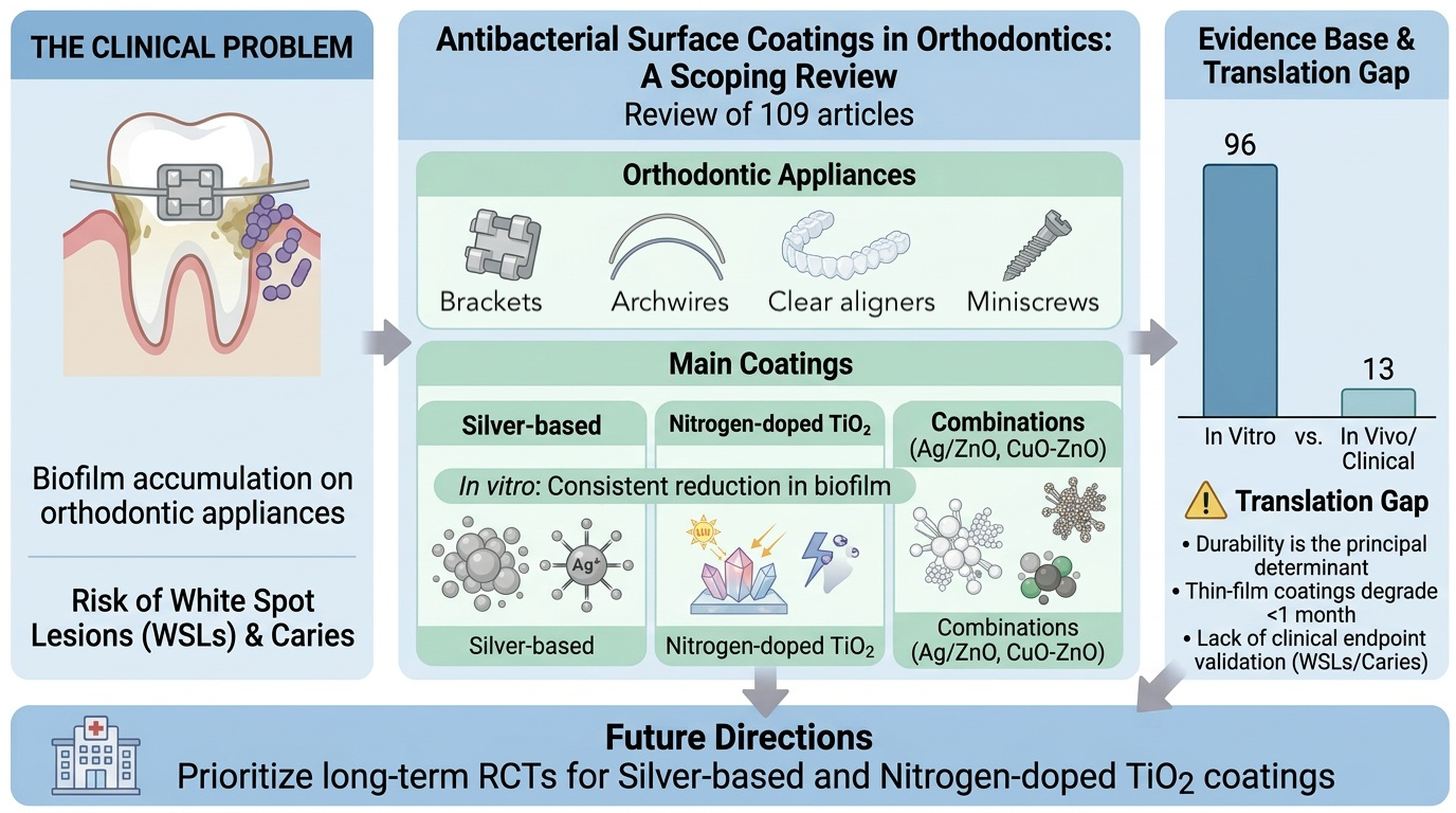
- Across 96 in vitro and 13 in vivo studies, antibacterial coatings on orthodontic appliance surfaces consistently and significantly reduce bacterial growth and adhesion compared to uncoated controls, with most coatings achieving 50–99% reductions in colony counts.
- Durability varies substantially by coating type with elution-dependent systems losing efficacy over months while contact-active and bulk-distributed coatings maintain longer-term protection.
- Under the laboratory conditions evaluated, nitrogen-doped TiO2 demonstrated favorable combinations of antibacterial activity, durability, and reported biocompatibility. However, long-term clinical effectiveness has not been established.
- TiN-Cu and layered polydopamine/PEDOT/AgNP systems seem to balance high antibacterial efficacy, sustained activity over 4–6 weeks, and demonstrated biocompatibility, though all evidence remains preclinical, and these coatings have not yet been validated in clinical use.
- Combination coatings of silver and metal oxides (particularly Ag/ZnO and Ag/TiO2) and nitrogen-doped TiO2 showed a favorable balance of antibacterial activity and reported biocompatibility under controlled laboratory conditions; however, clinical validation remains lacking, while pure TiO2 coatings are limited by UV dependence and the rutile phase of TiO2 show higher levels of cytotoxicity, based on a single in vitro study; this phase-dependent difference is highly subject to particle size, dose, agglomeration state, and preparation conditions.
- Antibacterial coatings, particularly silver-based, nitrogen-doped titanium dioxide, and copper oxide formulations, consistently and significantly reduce bacterial adhesion and biofilm formation on orthodontic device surfaces compared to uncoated controls in vitro, though clinical validation through human trials remains lacking.
- Antibacterial coatings reliably reduce bacterial adhesion and biofilm formation on orthodontic devices compared to uncoated controls, with silver nanoparticles bulk-incorporated into acrylic appliance bases or combined with structural matrices (such as Ag/ZnO, Ag+ TiO2, or CS-Ag composites) showing favorable laboratory profiles with greater durability among the coating types investigated, while surface-deposited photocatalytic coatings like TiO2 show strong in vitro results but frequently fail in vivo due to mechanical delamination and light-dependence, and all major coating types demonstrate acceptable toxicity at orthodontically relevant concentrations with the exception of TiO2 rutile phase (based on a single in vitro study; TiO2 cytotoxicity is highly dependent on particle size, dose, and preparation conditions) and high-concentration silver formulations lacking antifouling modifications.
- Nitrogen-doped TiO2 and silver/zinc oxide composite coatings on metallic brackets demonstrated a favorable balance of antibacterial activity and reported biocompatibility under laboratory conditions; however, clinical validation remains lacking.
- Antibacterial coatings on orthodontic devices consistently reduce bacterial adhesion and biofilm formation in vitro compared to uncoated controls, but clinical in vivo evidence remains limited and shows more modest, time-dependent effects than laboratory data suggest.
Abstract
1. Introduction
2. Materials and Methods
2.1. Protocol and Registration
2.2. Eligibility Criteria
2.3. Information Sources and Search Strategy
2.3.1. Electronic Databases
- -
- PubMed/MEDLINE (National Library of Medicine)
- -
- Scopus (Elsevier)
- -
- Web of Science Core Collection (Clarivate Analytics)
- -
- Cochrane Central Register of Controlled Trials (CENTRAL) (Cochrane Library)
- -
- Google Scholar (first 200 results ranked by relevance)
2.3.2. Search Strategy
2.4. Study Selection Process
Screening Phases
2.5. Data Collection Process
2.5.1. Data Extraction
2.5.2. Data Items
2.6. Methodological Characterization and Evidence Quality Appraisal
2.7. Biological Relevance Hierarchy and Evidence Classification Framework
2.8. Evidence Synthesis
2.9. Data Synthesis and Analysis
2.10. Reporting and Transparency
3. Results
3.1. Selection of Sources of Evidence
3.2. Characteristics of Sources of Evidence
3.3. Methodological Characteristics of Included Studies
3.3.1. Methodological Characteristics of In Vitro Studies (n = 96)
3.3.2. Methodological Characteristics of In Vivo and Clinical Studies (13)
3.3.3. Biological Relevance Tier Distribution
3.3.4. Summary of Methodological Quality Signals
- -
- Studies using single-species planktonic assays reported systematically higher antibacterial efficacy (reported CFU reduction range across studies: 85–99%) than studies using mature multi-species biofilms or salivary inoculum models (reported CFU reduction range across studies: 40–70%), suggesting that published efficacy ranges overestimate real-world performance.
- -
- Only 33% of in vitro studies (n = 32/96) included any form of durability assessment beyond the as-prepared state, limiting conclusions about long-term clinical performance.
- -
- No included clinical study evaluated white spot lesion incidence or caries increment as a primary outcome, precluding direct assessment of clinical effectiveness for caries prevention.
- -
- The median follow-up duration for clinical studies was 6 weeks (range: 2–12 weeks), far shorter than typical orthodontic treatment duration (18–24 months).
3.4. In Vitro Studies
3.4.1. Orthodontic Archwires
3.4.2. Orthodontic Brackets
3.4.3. Orthodontic Bands
3.4.4. Orthodontic Ligatures
3.4.5. Orthodontic Miniscrews
3.4.6. Orthodontic Acrylic and Clear Appliances
3.5. Clinical and In Vivo Evidence
3.6. Synthesis of Results

| Material | Coating Category | Representative Agents | Bacterial Target | Efficacy Range | No. of Studies | Evidence Level | Key References |
|---|---|---|---|---|---|---|---|
| Brackets | Silver nanoparticles | AgNPs (8–25 nm), Ag-W matrix, Ag-Pt alloy | S. mutans | 60–99% CFU reduction; ZOI 4.5–10 mm | 15 | 14 in vitro, 1 in situ | Łyczek 2023 [83], Denis 2022 [44], León 2018 [82] |
| Brackets | TiO2 photocatalytic | N-doped TiO2, anatase/rutile TiO2, TiO2:Ag | S. mutans, L. acidophilus | 79–98% bacterial reduction; 90–95% antimicrobial rate | 12 | 11 in vitro, 1 RCT | Salehi 2018 [12], Monica 2022 [75], Zhang 2018 [14] |
| Brackets | Metal oxide NPs | ZnO, CuO, CuO-ZnO hybrid | S. mutans, L. acidophilus | 45–96% inhibition; zero colonies at 2 h (CuO) | 8 | 8 in vitro | Zeidan 2022 [10], Sharma 2025 [84], Ramazanzadeh 2015 [11] |
| Brackets | Polymer-based | Polydopamine (PDA), PDA-HCDs, chitosan | S. mutans, E. coli | 78–98% CFU reduction; >80% bacterial killing | 6 | 6 in vitro | Singer 2025 [85], Wang 2023 [86], Mayma 2023 [87] |
| Brackets | Hybrid/composite | Ag-CuO, Ag/ZnO, TiO2 + Ag, gold-oxoborate | S. mutans, L. acidophilus | 45–98% reduction; 78% adhesion reduction | 6 | 6 in vitro | Sharma 2025 [84], Fatani 2017 [13], Łyczek 2023 [83] |
| Archwires | Silver nanoparticles | AgNPs (10–50 nm), Ag/PTFE, PDA/PEDOT/AgNP | S. mutans, L. acidophilus, S. sanguinis | 50–90% CFU reduction; >90% bacterial reduction | 12 | 12 in vitro | Mhaske 2015 [88], Gil 2020 [81], Lee 2020 [31] |
| Archwires | TiO2 photocatalytic | N-doped TiO2, TiO2:Ag, anatase TiO2 | S. mutans, P. gingivalis, A. actinomycetemcomitans | 74–98% adhesion/biofilm reduction; 87% reduction under visible light | 8 | 7 in vitro, 1 split-mouth | Chun 2007 [89], Liu 2017 [36], Bacela 2022 [29] |
| Archwires | ZnO nanoparticles | ZnO NPs (CVD, sol-gel, precipitation) | S. mutans, S. pyogenes, S. aureus | 72–98.6% microbial reduction; complete growth inhibition | 7 | 7 in vitro | Gholami 2021 [39], Kachoei 2016 [35], Hammad 2020 [90] |
| Archwires | Graphene/polymer | Graphene oxide (GO), PDA-GO, lysozyme | S. mutans, S. aureus | 23–93% bacterial adhesion reduction (dose-dependent) | 5 | 5 in vitro | Dai 2022 [91], Chen 2023 [34], He 2020 [92] |
| Archwires | TiN-Cu coating | Titanium nitride with copper | S. mutans, S. mitis | Significant CFU reduction (p < 0.05); sustained 28 days | 1 | 1 in vitro | Ilic 2023 [32] |
| Miniscrews | Silver nanoparticles | AgNPs, Ag/HA NPs, Ag/a-C:H nanocomposite | S. mutans, S. aureus, E. coli | 3–6 log reduction; ZOI 10–50 mm2 | 6 | 6 in vitro | Venugopal 2017 [62], Thukkaram 2020 [64], Abo-Elmahasen 2022 [57] |
| Miniscrews | ZnO-based | ZnO NPs, ZnO-doped TiO2 nanotubes + doxycycline | P. gingivalis, S. mutans, S. aureus | ZOI 13–39 mm (Day 5); sustained 30 days with drug | 4 | 4 in vitro | Noorollahian 2022 [58], Mohamed 2025 [63], Othman 2024 [93] |
| Miniscrews | Chitosan-based | Chitosan, chitosan-AgNPs, chitosan + azithromycin | S. mutans, S. sobrinus, P. gingivalis | 31–53% biofilm reduction; ZOI 9–40 mm (dose-dependent) | 4 | 4 in vitro | Nguyen 2019 [94], Sreenivasagan 2020 [59], Anggani 2021 [55] |
| Aligners/Acrylic | Gold nanoparticles | QA-GNCs, AuDAPT | S. mutans, P. gingivalis | 55–95% biofilm/viability reduction; growth stopped at 104 CFU/mL | 2 | 2 in vitro | Xie 2020 [70], Zhang 2020 [73] |
| Aligners/Acrylic | ZnO/MgO NPs | ZnO, MgO, ZnO + MgO | S. mutans, Lactobacillus spp., P. gingivalis | Significant CFU reduction; ZOI > 10–25 mm | 2 | 2 in vitro | Gharibnavaz 2025 [95] |
| Aligners/Acrylic | Curcumin-based | Curcumin NPs, Curcumin-Nisin-PLLA | S. mutans, C. albicans | 68–78% reduction; ZOI 3.8–16 mm (dose-dependent) | 2 | 2 in vitro | Soleymanijadidi 2023 [69], Pourhajibagher 2022 [72] |
| Aligners/Acrylic | TiO2 photocatalytic | TiO2 under UVA | S. mutans, S. sobrinus, S. gordonii | 99.9% CFU reduction; viability 0.2–5.4% | 1 | 1 in vitro | Kuroiwa 2018 [68] |
| Ligatures | Silver-based | Nano-Ag fluoride, AgNPs (green synthesis) | S. mutans, S. aureus, E. coli, L. casei | 57% CFU reduction; 86% biofilm thickness reduction; ZOI present for all species | 4 | 4 in vitro | Choi 2024 [52], Pasala 2024 [96], Schubert 2024 [97], Hernández-Gómora 2017 [98] |
| Bands | Silver/ZnO NPs | Nano-Ag, nano-ZnO | S. mutans, L. acidophilus, C. albicans | 2–3.4 log10 reduction (Ag); 0.6–2.14 log10 (ZnO) | 2 | 2 in vitro | Bahrami 2023 [50], Prabha 2016 [51] |
| Coating Type/Material | Durability Finding | Ion Release/Safety Data | Mechanical Impact | Evidence Level | Key References |
|---|---|---|---|---|---|
| Silver NPs on brackets | Sustained 30 days (electroplated); 2 months (PDA); 3 months (CuO/TiO2/HA-SNPs) | Ag ions within safe limits (~2 ppm); no cytotoxicity at therapeutic concentrations | Nano-Ag: no significant friction increase (0.77–0.82 N vs. 0.55 N control) | 14 in vitro, 1 in situ | Arash 2016 [42], Singer 2025 [85], Ameli 2022 [43], Ghasemi 2017 [99], Ryu 2012 [47] |
| TiO2 on brackets | 90 days (N-doped); 3 months storage (nano-TiO2); 60% loss after 1 month (clinical) | Grade 0 cytotoxicity; no mucosal irritation; anatase 77–89% viability, rutile 21–40% (severe) | Nano-TiO2: significant friction increase (1.52–1.57 N vs. 0.55 N, p < 0.05) | 11 in vitro, 1 RCT | Salehi 2018 [12], Ghasemi 2017 [99], Venkatesan 2020 [80], Baby 2017 [46], Cao 2016 [100], Cao 2013 [45] |
| Ag-W matrix (brackets) | Maintained after simulated 2-year abrasion | Tungsten considered innocuous; Ag release controlled by matrix | Not assessed | 1 in situ | Denis 2022 [44] |
| ZnO/CuO on brackets | 28 days (ZnO/CuO NPs); some decline from baseline to 4 months (Ag-CuO hybrid) | Ion release below toxic thresholds at all time points; peak at day 7, declined thereafter | ZnO: 64% friction reduction; chitosan: 53% reduction vs. uncoated | 8 in vitro | Mobeen 2022 [48], Sharma 2025 [84], Elhelbawy 2021 [101] |
| TiN-Cu on archwires | Effective 28 days; stable in neutral and acidic environments | Highest cell viability in 28-day eluates; Cu within safety limits; lower Ni release vs. uncoated NiTi | Not assessed | 1 in vitro | Ilic 2023 [32] |
| AgNPs on archwires | Stable under orthodontic conditions; up to 42 days (PDA/PEDOT/AgNP system) | Ag+ release within safe limits (ICP-MS); good osteoblast morphology; Ni ion release low | Not assessed | 12 in vitro | Gil 2020 [81], Lee 2019 [31] |
| ZnO on archwires | Durable after bending and friction cycling; no bacterial growth at 48 h | No viability reduction ≤ 5 µg/mL; 20% reduction at 10 µg/mL; >55% reduction at 25 µg/mL (fibroblasts) | 21% friction reduction | 7 in vitro | Kachoei 2016 [35] |
| TiO2:Ag on archwires | Biofilm reduction declined from 98% (24 h) to 40% (96 h) | Not assessed | Not assessed | 7 in vitro, 1 split-mouth | Bacela 2022 [29], Kielan-Grabowska 2021 [102] |
| Graphene oxide on archwires | Stable at 4 weeks under mechanical stress and saliva exposure | Cell viability ~85% after 5 days; meets ISO 10993-5:2009; high GO induces oxidative stress | Not assessed | 5 in vitro | Chen 2023 [34], Dai 2021 [91] |
| ZnO-doped TiO2 nanotubes + doxycycline (Miniscrews) | Sustained antimicrobial action 30 days; progressive decline in ZOI from Day 5 to Day 30 | Not assessed | Not assessed | 4 in vitro | Noorollahian 2022 [58] |
| QA-GNCs on aligners | Maintained effectiveness > 3 months and after > 3 usage cycles | Negligible toxicity in vitro; no inflammatory response or mucosa irritation in vivo; safe up to 4.22 µg/cm2 | Not assessed | 2 in vitro (1 with in vivo biocompatibility) | Xie 2020 [70] |
| Curcumin NPs in acrylic | Sustained release ≥ 30 days (curcumin NPs); significant at 30 d, diminishing by 60 d (Cur-Nis-PLLA) | All tested concentrations (0.5–5%) non-cytotoxic | All groups > 50 MPa flexural strength; 5% acceptable, 10% unacceptable (Cur-Nis-PLLA) | 2 in vitro | Soleymanijadidi 2023 [69], Pourhajibagher 2022 [72] |
4. Discussion
4.1. Scoping Review Methodology and Rationale
- Standardized in vitro testing protocols (bacterial strains, biofilm maturation time, outcome measures, statistical analysis) to enable cross-study comparison and reproducibility.
- Long-term intraoral durability studies (≥12 months) under realistic mechanical and chemical stress to assess coating stability over full treatment duration.
- Adequately powered, multi-center randomized clinical trials with white spot lesion incidence as the primary endpoint and follow-up extending through debonding and retention.
- Post-market surveillance for rare adverse events (e.g., argyria, hypersensitivity reactions, systemic absorption of nanoparticles).
4.2. In Vivo and Clinical Evidence
4.3. Durability as the Principal Determinant of Clinical Relevance
4.4. The In Vitro to In Vivo Translational Gap
4.5. Light Dependence of Photocatalytic Coatings
4.6. Dose-Response Relationships and Biocompatibility
4.7. Comparative Evidence by Coating Type
4.8. Sterilization Compatibility
4.9. Methodological Limitations of the Included Studies
4.10. Future Research Priorities
5. Conclusions
Supplementary Materials
Author Contributions
Funding
Institutional Review Board Statement
Informed Consent Statement
Data Availability Statement
Acknowledgments
Conflicts of Interest
References
- Ren, Y.; Jongsma, M.A.; Mei, L.; van der Mei, H.C.; Busscher, H.J. Orthodontic treatment with fixed appliances and biofilm formation—A potential public health threat? Clin. Oral Investig. 2014, 18, 1711–1718. [Google Scholar] [CrossRef] [Scilit]
- Papaioannou, W.; Gizani, S.; Nassika, M.; Kontou, E.; Nakou, M. Adhesion of Streptococcus mutans to different types of brackets. Angle Orthod. 2007, 77, 1090–1095. [Google Scholar] [CrossRef] [Scilit]
- Sardana, D.; Zhang, J.; Ekambaram, M.; Yang, Y.; McGrath, C.P.; Yiu, C.K.Y. Effectiveness of professional fluorides against enamel white spot lesions during fixed orthodontic treatment: A systematic review and meta-analysis. J. Dent. 2019, 82, 1–10. [Google Scholar] [CrossRef] [Scilit]
- Lupi, L.; Paggetti, H.; Bertrand, M.F.; Charavet, C. Biofilm and Orthodontic Materials: Literature reviews and Scanning Electron Microscopy (SEM) images gallery. Orthod. Fr. 2022, 93, 111–123. [Google Scholar]
- Liu, Y.; Xu, Y.; Song, Q.; Wang, F.; Sun, L.; Liu, L.; Yang, X.; Yi, J.; Bao, Y.; Ma, H.; et al. Anti-biofilm Activities from Bergenia crassifolia Leaves against Streptococcus mutans. Front. Microbiol. 2017, 8, 1738. [Google Scholar] [CrossRef] [Scilit]
- Santonocito, S.; Polizzi, A. Oral Microbiota Changes during Orthodontic Treatment. Front. Biosci. (Elite Ed) 2022, 14, 19. [Google Scholar] [CrossRef] [Scilit]
- Hua, F.; Yan, J.; Zhao, S.; Yang, H.; He, H. In vitro remineralization of enamel white spot lesions with a carrier-based amorphous calcium phosphate delivery system. Clin. Oral Investig. 2020, 24, 2079–2089. [Google Scholar] [CrossRef] [Scilit]
- Hua, F.; Yang, H.; He, H. Current Enamel Remineralization Therapies Have Limited Effects on Postorthodontic White Spot Lesions. J. Evid. Based Dent. Pract. 2018, 18, 339–342. [Google Scholar] [CrossRef] [Scilit]
- Zhang, Y.; Du, Q.; Fei, W.; Li, X.B. Application of antibacterial nanoparticles in orthodontic materials. Nanotechnol. Rev. 2022, 11, 2433–2450. [Google Scholar] [CrossRef] [Scilit]
- Zeidan, N.K.; Enany, N.M.; Mohamed, G.G.; Marzouk, E.S. The antibacterial effect of silver, zinc-oxide and combination of silver/ zinc oxide nanoparticles coating of orthodontic brackets (an in vitro study). BMC Oral Health 2022, 22, 230. [Google Scholar] [CrossRef] [Scilit]
- Ramazanzadeh, B.; Jahanbin, A.; Yaghoubi, M.; Shahtahmassbi, N.; Ghazvini, K.; Shakeri, M.; Shafaee, H. Comparison of Antibacterial Effects of ZnO and CuO Nanoparticles Coated Brackets against Streptococcus mutans. J. Dent. 2015, 16, 200–205. [Google Scholar]
- Salehi, P.; Babanouri, N.; Roein-Peikar, M.; Zare, F. Long-term antimicrobial assessment of orthodontic brackets coated with nitrogen-doped titanium dioxide against Streptococcus mutans. Prog. Orthod. 2018, 19, 35. [Google Scholar] [CrossRef] [Scilit]
- Fatani, E.J.; Almutairi, H.H.; Alharbi, A.O.; Alnakhli, Y.O.; Divakar, D.D.; Muzaheed; Alkheraif, A.A.; Khan, A.A. In vitro assessment of stainless steel orthodontic brackets coated with titanium oxide mixed Ag for anti-adherent and antibacterial properties against Streptococcus mutans and Porphyromonas gingivalis. Microb. Pathog. 2017, 112, 190–194. [Google Scholar] [CrossRef] [Scilit]
- Zhang, J.X.; An, X.L.; Li, X.A.; Liao, X.Z.; Nie, Y.Y.; Fan, Z.J. Enhanced antibacterial properties of the bracket under natural light via decoration with ZnO/carbon quantum dots composite coating. Chem. Phys. Lett. 2018, 706, 702–707. [Google Scholar] [CrossRef] [Scilit]
- Bącela, J.; Łabowska, M.B.; Detyna, J.; Zięty, A.; Michalak, I. Functional Coatings for Orthodontic Archwires-A Review. Materials 2020, 13, 3257. [Google Scholar] [CrossRef] [Scilit]
- Almoudi, M.M.; Hussein, A.S.; Abu Hassan, M.I.; Mohamad Zain, N. A systematic review on antibacterial activity of zinc against Streptococcus mutans. Saudi Dent. J. 2018, 30, 283–291. [Google Scholar] [CrossRef] [Scilit]
- Kalwar, K.; Shan, D. Antimicrobial effect of silver nanoparticles (AgNPs) and their mechanism–a mini review. Micro Nano Lett. 2018, 13, 277–280. [Google Scholar] [CrossRef] [Scilit]
- Tricco, A.C.; Lillie, E.; Zarin, W.; O’Brien, K.K.; Colquhoun, H.; Levac, D.; Moher, D.; Peters, M.D.J.; Horsley, T.; Weeks, L.; et al. PRISMA Extension for Scoping Reviews (PRISMA-ScR): Checklist and Explanation. Ann. Intern. Med. 2018, 169, 467–473. [Google Scholar] [CrossRef] [Scilit] [PubMed]
- Arksey, H.; O’Malley, L. Scoping studies: Towards a methodological framework. Int. J. Soc. Res. Methodol. 2005, 8, 19–32. [Google Scholar] [CrossRef] [Scilit]
- Levac, D.; Colquhoun, H.; O’Brien, K.K. Scoping studies: Advancing the methodology. Implement. Sci. 2010, 5, 69. [Google Scholar] [CrossRef] [Scilit] [PubMed]
- Centre for Reviews and Dissemination. PROSPERO: International Prospective Register of Systematic Reviews. 2023. Available online: https://www.crd.york.ac.uk/prospero/ (accessed on 20 August 2026).
- Munn, Z.; Peters, M.D.J.; Stern, C.; Tufanaru, C.; McArthur, A.; Aromataris, E. Systematic review or scoping review? Guidance for authors when choosing between a systematic or scoping review approach. BMC Med. Res. Methodol. 2018, 18, 143. [Google Scholar]
- Lebeaux, D.; Chauhan, A.; Rendueles, O.; Beloin, C. From in vitro to in vivo Models of Bacterial Biofilm-Related Infections. Pathogens 2013, 2, 288–356. [Google Scholar] [CrossRef] [Scilit]
- Guyatt, G.H.; Oxman, A.D.; Vist, G.E.; Kunz, R.; Falck-Ytter, Y.; Alonso-Coello, P.; Schünemann, H.J. GRADE: An emerging consensus on rating quality of evidence and strength of recommendations. BMJ 2008, 336, 924–926. [Google Scholar] [CrossRef] [Scilit]
- Campbell, M.; McKenzie, J.E.; Sowden, A.; Katikireddi, S.V.; Brennan, S.E.; Ellis, S.; Hartmann-Boyce, J.; Ryan, R.; Shepperd, S.; Thomas, J.; et al. Synthesis without meta-analysis (SWiM) in systematic reviews: Reporting guideline. BMJ 2020, 368, l6890. [Google Scholar] [CrossRef] [Scilit] [PubMed]
- Hashem, R.; Ghobashy, S.; Abdel Hamid, Z.; Elmarhoumy, S. The Efficacy of Nanosilver Coated Stainless Steel Brackets in Prevention of Enamel Demineralization in Orthodontic Patients. AL-Azhar J. Dent. Sci. 2022, 25, 495–504. [Google Scholar] [CrossRef] [Scilit]
- Farhadian, N.; Usefi Mashoof, R.; Khanizadeh, S.; Ghaderi, E.; Farhadian, M.; Miresmaeili, A. Streptococcus mutans counts in patients wearing removable retainers with silver nanoparticles vs those wearing conventional retainers: A randomized clinical trial. Am. J. Orthod. Dentofac. Orthop. 2016, 149, 155–160. [Google Scholar] [CrossRef] [Scilit]
- Ghorbanzadeh, R.; Pourakbari, B.; Bahador, A. Effects of Baseplates of Orthodontic Appliances with in situ generated Silver Nanoparticles on Cariogenic Bacteria: A Randomized, Double-blind Cross-over Clinical Trial. J. Contemp. Dent. Pract. 2015, 16, 291–298. [Google Scholar] [CrossRef] [Scilit]
- Bacela, J.J.; Kielan-Grabowska, Z.; Borak, B.; Sobieszczanska, B.; Walczuk, U.; Kawala, B.; Zięty, A.; Detyna, J.; Sarul, M. Antiadherent and antibacterial properties of TiO2-coated and TiO2:Ag-coated stainless steel orthodontic wires against S. mutans bacteria. Acta Bioeng. Biomech. 2022, 24, 107–118. [Google Scholar] [CrossRef] [Scilit]
- Metin-Gursoy, G.; Taner, L.; Akca, G. Nanosilver coated orthodontic brackets: In vivo antibacterial properties and ion release. Eur. J. Orthod. 2017, 39, 9–16. [Google Scholar] [CrossRef] [Scilit]
- Lee, B.S.; Lin, Y.C.; Hsu, W.C.; Hou, C.H.; Shyue, J.J.; Hsiao, S.Y.; Wu, P.J.; Lee, Y.T.; Luo, S.C. Engineering Antifouling and Antibacterial Stainless Steel for Orthodontic Appliances through Layer-by-Layer Deposition of Nanocomposite Coatings. ACS Appl. Bio Mater. 2020, 3, 486–494. [Google Scholar] [CrossRef] [Scilit]
- Ilić, B.; Petrović, B.; Marinković, J.; Vukajlović, J.M.; Stevanović, M.; Potočnik, J.; Jokanović, V. Investigation of Ion Release and Antibacterial Properties of TiN-Cu-Nanocoated Nitinol Archwires. Coatings 2023, 13, 1587. [Google Scholar] [CrossRef] [Scilit]
- Al-Fadhily, Z.M.; Abdul-Hadi, M. A Novel Coating of Orthodontic Archwires with Chlorhexidine Hexametaphosphate Nanoparticles. Int. J. Biomater. 2023, 2023, 9981603. [Google Scholar] [CrossRef] [Scilit]
- Chen, Y.M.; Chen, Z.H.; Zheng, Z.B.; Xia, Y. Bio-inspired nanocomposite coatings on orthodontic archwires with corrosion resistant and antibacterial properties. Front. Bioeng. Biotech. 2023, 11, 1272527. [Google Scholar] [CrossRef] [Scilit]
- Kachoei, M.; Nourian, A.; Divband, B.; Kachoei, Z.; Shirazi, S. Zinc-oxide nanocoating for improvement of the antibacterial and frictional behavior of nickel-titanium alloy. Nanomedicine 2016, 11, 2511–2527. [Google Scholar] [CrossRef] [Scilit]
- Liu, J.M.; Lou, Y.X.; Zhang, C.; Yin, S.; Li, H.M.; Sun, D.Q.; Sun, X.H. Improved corrosion resistance and antibacterial properties of composite arch-wires by N-doped TiO coating. Rsc. Adv. 2017, 7, 43938–43949. [Google Scholar] [CrossRef] [Scilit]
- Abdallah, O.M.; Sedky, Y.; Shebl, H.R. Comprehensive evaluation of the antibacterial and antibiofilm activities of NiTi orthodontic wires coated with silver nanoparticles and nanocomposites: An in vitro study. BMC Oral Health 2024, 24, 1345. [Google Scholar] [CrossRef] [Scilit]
- Anand, B.G.; Shejale, K.P.; Rajesh Kumar, R.; Thangam, R.; Prajapati, K.P.; Kar, K.; Mala, R. Bioactivation of an orthodontic wire using multifunctional nanomaterials to prevent plaque accumulation. Biomater. Adv. 2023, 148, 213346. [Google Scholar] [CrossRef] [Scilit]
- Gholami, M.; Esmaeilzadeh, M.; Kachoei, Z.; Kachoei, M.; Divband, B. Influence of Physical Dimension and Morphological-Dependent Antibacterial Characteristics of ZnO Nanoparticles Coated on Orthodontic NiTi Wires. Biomed. Res. Int. 2021, 2021, 6397698. [Google Scholar] [CrossRef] [Scilit]
- Peng, L.; Chang, L.; Liu, X.; Lin, J.; Liu, H.; Han, B.; Wang, S. Antibacterial Property of a Polyethylene Glycol-Grafted Dental Material. ACS Appl. Mater. Interfaces 2017, 9, 17688–17692. [Google Scholar] [CrossRef] [Scilit]
- Özyildiz, E.; Güden, M.; Uzel, A.; Karaboz, I.; Akil, O.; Bulut, H. Antimicrobial Activity of TiO-coated Orthodontic Ceramic Brackets against and Streptococcus mutans and Candida albicans. Biotechnol. Bioprocess Eng. 2010, 15, 680–685. [Google Scholar] [CrossRef] [Scilit][Green Version]
- Arash, V.; Keikhaee, F.; Rabiee, S.M.; Rajabnia, R.; Khafri, S.; Tavanafar, S. Evaluation of Antibacterial Effects of Silver-Coated Stainless Steel Orthodontic Brackets. J. Dent. 2016, 13, 49–54. [Google Scholar]
- Ameli, N.; Asadi, S.; Ghorbani, R.; Mohebi, S.; Hans, M. Comparative Antibacterial Efficacy of Orthodontic Brackets Coated with Titanium Dioxide, Copper Oxide, and Hydroxyapatite-Silver Nanoparticles Against Streptococcus mutans. Middle East J. Rehabil. Health Stud. 2022, 9, e119536. [Google Scholar] [CrossRef] [Scilit]
- Denis, H.; Werth, R.; Greuling, A.; Schwestka-Polly, R.; Stiesch, M.; Meyer-Kobbe, V.; Doll, K. Antibacterial properties and abrasion-stability: Development of a novel silver-compound material for orthodontic bracket application. J. Orofac. Orthop. 2024, 85, 30–42. [Google Scholar] [CrossRef] [Scilit]
- Cao, B.C.; Wang, Y.H.; Li, N.; Liu, B.; Zhang, Y.J. Preparation of an orthodontic bracket coated with an nitrogen-doped TiON thin film and examination of its antimicrobial performance. Dent. Mater. J. 2013, 32, 311–316. [Google Scholar] [CrossRef] [Scilit]
- Baby, R.D.; Subramaniam, S.; Arumugam, I.; Padmanabhan, S. Assessment of antibacterial and cytotoxic effects of orthodontic stainless steel brackets coated with different phases of titanium oxide: An in-vitro study. Am. J. Orthod. Dentofac. 2017, 151, 678–684. [Google Scholar] [CrossRef] [Scilit]
- Ryu, H.S.; Bae, I.H.; Lee, K.G.; Hwang, H.S.; Lee, K.H.; Koh, J.T.; Cho, J.H. Antibacterial effect of silver-platinum coating for orthodontic appliances. Angle Orthod. 2012, 82, 151–157. [Google Scholar] [CrossRef] [Scilit]
- Mobeen, N.; Duraisamy, S.; Ravi, K. Evaluation and comparison of copper and zinc ion release from copper-oxide and zinc-oxide nanoparticles coated orthodontic brackets-An In vitro Study. Int. J. Orthod. Rehabil. 2022, 13, 10–21. [Google Scholar]
- Teixeira, L.P.; Gontijo, L.C.; Franco, A.R., Jr.; Pereira, M.F.; Schuenck, R.P.; Malacarne-Zanon, J. Evaluation of antimicrobial potential and surface morphology in thin films of titanium nitride and calcium phosphate on orthodontic brackets. Am. J. Orthod. Dentofac. 2021, 160, 209–214. [Google Scholar] [CrossRef] [Scilit]
- Bahrami, R.; Pourhajibagher, M.; Badiei, A.; Masaeli, R.; Tanbakuchi, B. Evaluation of the cell viability and antimicrobial effects of orthodontic bands coated with silver or zinc oxide nanoparticles: A study. Korean J. Orthod. 2023, 53, 16–25. [Google Scholar] [CrossRef] [Scilit]
- Prabha, R.D.; Kandasamy, R.; Sivaraman, U.S.; Nandkumar, M.A.; Nair, P.D. Antibacterial nanosilver coated orthodontic bands with potential implications in dentistry. Indian J. Med. Res. 2016, 144, 580–586. [Google Scholar] [CrossRef] [Scilit]
- Choi, J.H.; Lee, E.S.; Jung, H.I.; Kim, B.I. Drug delivery and antibiofilm efficacy of nano silver fluoride sustained release orthodontic elastomerics against Streptococcus mutans. Sci. Rep. 2024, 14, 19912. [Google Scholar] [CrossRef] [Scilit]
- Jeon, H.S.; Choi, C.H.; Kang, S.M.; Kwon, H.K.; Kim, B.I. Chlorhexidine-releasing orthodontic elastomerics. Dent. Mater. J. 2015, 34, 321–326. [Google Scholar] [CrossRef] [Scilit]
- Choi, J.Y.; Chung, C.J.; Oh, K.T.; Choi, Y.J.; Kim, K.H. Photocatalytic antibacterial effect of TiO2 film of TiAg on Streptococcus mutans. Angle Orthod. 2009, 79, 528–532. [Google Scholar] [CrossRef] [Scilit]
- Anggani, H.S.; Perdana, R.G.; Siregar, E.; Bachtiar, E.W. The effect of coating chitosan on Porphyromonas gingivalis biofilm formation in the surface of orthodontic mini-implant. J. Adv. Pharm. Technol. Res. 2021, 12, 84–88. [Google Scholar] [CrossRef] [Scilit]
- Williams, D.L.; Epperson, R.T.; DeGrauw, J.P.; Nielsen, M.B.; Taylor, N.B.; Jolley, R.D. Effect of silver-loaded PMMA on Streptococcus mutans in a drip flow reactor. J. Biomed. Mater. Res. Part A 2017, 105, 2632–2639. [Google Scholar] [CrossRef] [Scilit]
- Abo-Elmahasen, M.M.F.; Dena, A.S.A.; Zhran, M.; Albohy, S.A.H. Do silver/hydroxyapatite and zinc oxide nano-coatings improve inflammation around titanium orthodontic mini-screws? In vitro study. Int. Orthod. 2023, 21, 100711. [Google Scholar] [CrossRef] [Scilit]
- Noorollahian, S.; Kachuie, M.; Moghadam, M.; Narimani, T.; Hatamzade, Z. The antimicrobial effect of doxycycline and doped ZnO in TiO2 nanotubes synthesized on the surface of orthodontic mini-implants. Apos Trends Orthod. 2022, 12, 162–167. [Google Scholar] [CrossRef] [Scilit]
- Sreenivasagan, S.; Subramanian, A.K.; Rajeshkumar, S. Assessment of antimicrobial activity and cytotoxic effect of green mediated silver nanoparticles and its coating onto mini-implants. Ann. Phytomed. 2020, 9, 207–212. [Google Scholar] [CrossRef] [Scilit]
- Alhazmi, A.S.; Syame, S.M.; Mohamed, W.S.; Hakim, A.S. Incorporation of Plant Extracted Hydroxyapatite and Chitosan Nanoparticles on the Surface of Orthodontic Micro-Implants: An In-Vitro Antibacterial Study. Microorganisms 2022, 10, 581. [Google Scholar] [CrossRef] [Scilit]
- Rodriguez-Fernandez, J.C.; Pastor, F.; Mora, J.M.B.; Brizuela, A.; Puigdollers, A.; Espinar, E.; Gil, F.J. Bacteriostatic Poly Ethylene Glycol Plasma Coatings for Orthodontic Titanium Mini-Implants. Materials 2022, 15, 7487. [Google Scholar] [CrossRef] [Scilit]
- Venugopal, A.; Muthuchamy, N.; Tejani, H.; Gopalan, A.I.; Lee, K.P.; Lee, H.J.; Kyung, H.M. Incorporation of silver nanoparticles on the surface of orthodontic microimplants to achieve antimicrobial properties. Korean J. Orthod. 2017, 47, 3–10. [Google Scholar] [CrossRef] [Scilit]
- Mohamed, A.; Abdelsadek, M.; Hashem, A. In-vitro evaluation of the effects of sterilization procedures on the antibacterial characteristics of zinc oxide nanoparticles coated orthodontic mini screws. Egypt. Dent. J. 2025, 71, 1–11. [Google Scholar] [CrossRef] [Scilit]
- Thukkaram, M.; Vaidulych, M.; Kylián, O.; Hanuš, J.; Rigole, P.; Aliakbarshirazi, S.; Asadian, M.; Nikiforov, A.; Van Tongel, A.; Biederman, H.; et al. Investigation of Ag/a-C:H Nanocomposite Coatings on Titanium for Orthopedic Applications. Acs Appl. Mater. Inter. 2020, 12, 23655–23666. [Google Scholar] [CrossRef] [Scilit]
- Sycińska-Dziarnowska, M.; Ziąbka, M.; Cholewa-Kowalska, K.; Klesiewicz, K.; Spagnuolo, G.; Lindauer, S.J.; Park, H.-S.; Woźniak, K. Antibacterial and Antibiofilm Activity of Layers Enriched with Silver Nanoparticles on Orthodontic Microimplants. J. Funct. Biomater. 2025, 16, 78. [Google Scholar] [CrossRef] [Scilit]
- Biegun-Żurowska, M.; Klesiewicz, K.; Matysiak, K.; Gajek, M.; Rapacz-Kmita, A.; Ziąbka, M. Electrophoretic Coatings for Orthodontic Implants: Evaluation of Surface Properties, Adhesion, and Antibacterial Activity in Simulated Implantation Trials. J. Funct. Biomater. 2025, 16, 343. [Google Scholar] [CrossRef] [Scilit]
- Subramanian, S.K.; Anbarasu, P.; Navin, N.; Iyer, S.R. Comparison of antimicrobial effect of selenium nanoparticles and silver nanoparticles coated orthodontic mini-implants–An in vitro study. Apos Trends Orthod. 2022, 12, 20–26. [Google Scholar] [CrossRef] [Scilit]
- Kuroiwa, A.; Nomura, Y.; Ochiai, T.; Sudo, T.; Nomoto, R.; Hayakawa, T.; Kanzaki, H.; Nakamura, Y.; Hanada, N. Antibacterial, Hydrophilic Effect and Mechanical Properties of Orthodontic Resin Coated with UV-Responsive Photocatalyst. Materials 2018, 11, 889. [Google Scholar] [CrossRef] [Scilit]
- Soleymanijadidi, P.; Moradi, M.; Hamedirad, F.; Ghanavati, Z.; Maleki Dizaj, S.; Salatin, S. Nanocurcumin Release from Self-Cured Acrylic Resins; Effects on Antimicrobial Action and Flexural Strength. Bioengineering 2023, 10, 559. [Google Scholar] [CrossRef] [Scilit]
- Xie, Y.; Zhang, M.; Zhang, W.; Liu, X.; Zheng, W.; Jiang, X. Gold Nanoclusters-Coated Orthodontic Devices Can Inhibit the Formation of Streptococcus mutans Biofilm. ACS Biomater. Sci. Eng. 2020, 6, 1239–1246. [Google Scholar] [CrossRef] [Scilit]
- Lee, J.-H.; El-Fiqi, A.; Jo, J.-K.; Kim, D.-A.; Kim, S.-C.; Jun, S.-K.; Kim, H.-W.; Lee, H.-H. Development of long-term antimicrobial poly(methyl methacrylate) by incorporating mesoporous silica nanocarriers. Dent. Mater. 2016, 32, 1564–1574. [Google Scholar] [CrossRef] [Scilit]
- Pourhajibagher, M.; Noroozian, M.; Ahmad Akhoundi, M.S.; Bahador, A. Antimicrobial effects and mechanical properties of poly(methyl methacrylate) as an orthodontic acrylic resin containing Curcumin-Nisin-poly(L-lactic acid) nanoparticle: An in vitro study. BMC Oral Health 2022, 22, 158. [Google Scholar] [CrossRef] [Scilit]
- Zhang, M.; Liu, X.; Xie, Y.; Zhang, Q.; Zhang, W.; Jiang, X.; Lin, J. Biological Safe Gold Nanoparticle-Modified Dental Aligner Prevents the Porphyromonas gingivalis Biofilm Formation. ACS Omega 2020, 5, 18685–18692. [Google Scholar] [CrossRef] [Scilit]
- Meyer-Kobbe, V.; Doll, K.; Stiesch, M.; Schwestka-Polly, R.; Demling, A. Comparison of intraoral biofilm reduction on silver-coated and silver ion-implanted stainless steel bracket material: Biofilm reduction on silver ion-implanted bracket material. J. Orofac. Orthop. 2019, 80, 32–43. [Google Scholar] [CrossRef] [Scilit]
- Monica, A.; Padmanabhan, S. The effect of nitrogen-doped titanium dioxide-modified stainless steel brackets on Streptococcus mutans. Angle Orthod. 2022, 92, 396–401. [Google Scholar] [CrossRef] [Scilit]
- Amini, F.; Bahador, A.; Kiaee, B.; Kiaee, G. The effect of the titanium nitride coating on bacterial adhesion on orthodontic stainless steel wires: Study. Biosci. Biotech. Res. C 2017, 10, 28–33. [Google Scholar] [CrossRef] [Scilit]
- Mollabashi, V.; Farmany, A.; Alikhani, M.Y.; Sattari, M.; Soltanian, A.R.; Kahvand, P.; Banisafar, Z. Effects of TiO2-Coated Stainless Steel Orthodontic Wires on Streptococcus mutans Bacteria: A Clinical Study. Int. J. Nanomed. 2020, 15, 8759–8766. [Google Scholar] [CrossRef] [Scilit]
- Abraham, K.S.; Jagdish, N.; Kailasam, V.; Padmanabhan, S. Streptococcus mutans adhesion on nickel titanium (NiTi) and copper-NiTi archwires: A comparative prospective clinical study. Angle Orthod. 2017, 87, 448–454. [Google Scholar] [CrossRef] [Scilit]
- Metin-Gursoy, G.; Taner, L.; Baris, E. Biocompatibility of nanosilver-coated orthodontic brackets: An in vivo study. Prog. Orthod. 2016, 17, 39. [Google Scholar] [CrossRef] [Scilit]
- Venkatesan, K.; Kailasam, V.; Padmanabhan, S. Evaluation of titanium dioxide coating on surface roughness of nickel-titanium archwires and its influence on Streptococcus mutans adhesion and enamel mineralization: A prospective clinical study. Am. J. Orthod. Dentofac. Orthop. 2020, 158, 199–208. [Google Scholar] [CrossRef] [Scilit]
- Gil, F.J.; Espinar-Escalona, E.; Clusellas, N.; Fernandez-Bozal, J.; Artes-Ribas, M.; Puigdollers, A. New Bactericide Orthodonthic Archwire: NiTi with Silver Nanoparticles. Metals 2020, 10, 70. [Google Scholar] [CrossRef] [Scilit]
- Espinosa-Cristóbal, L.F.; López-Rui, N.; Cabada-Tarín, D.; Reyes-López, S.Y.; Zaragoza-Contreras, A.; Constandse-Cortéz, D.; Donohué-Cornejo, A.; Tovar-Carrillo, K.; Cuevas-González, J.C.; Kobayashi, T. Antiadherence and Antimicrobial Properties of Silver Nanoparticles against Streptococcus mutans on Brackets and Wires Used for Orthodontic Treatments. J. Nanomater. 2018, 2018, 9248527. [Google Scholar] [CrossRef] [Scilit]
- Lyczek, J.; Bonczak, B.; Krzyminska, I.; Gizynski, K.; Paczesny, J. Gold-oxoborate nanocomposite-coated orthodontic brackets gain antibacterial properties while remaining safe for eukaryotic cells. J. Biomed. Mater. Res. Part B Appl. Biomater. 2023, 111, 996–1004. [Google Scholar] [CrossRef] [Scilit]
- Sharma, A.; Sharma, T.; Shrinivaasan, N.R.; Tomer, G.; Gupta, N.; Kishore, P.; Babaji, P.; Mohammed, A.; Neralla, A. Evaluation of antibacterial efficacy and surface roughness of orthodontic brackets coated with silver-copper hybrid or zinc oxide nanoparticles: An in-vitro study. J. Orthod. Sci. 2025, 14, 32. [Google Scholar] [CrossRef] [Scilit]
- Singer, L.; Karacic, S.; Bierbaum, G.; Palmer, B.; Kirschneck, C.; Bourauel, C. A novel stable biomimetic adhesive coating for functionalization of orthodontic brackets against bacterial colonization and white spot lesions. BMC Oral Health 2025, 25, 23. [Google Scholar] [CrossRef] [Scilit]
- Wang, Y.; Ding, C.; Ge, Z.; Li, Z.; Chen, L.; Guo, X.; Dong, G.; Zhou, P. A novel antibacterial and fluorescent coating composed of polydopamine and carbon dots on the surface of orthodontic brackets. J. Mater. Sci. Mater. Med. 2023, 34, 10. [Google Scholar] [CrossRef] [Scilit]
- Mayma, N.; Sangeetha, D.; Ravi, K. Evaluation of antimicrobial property of nanochitosan coated orthodontic brackets against Streptococcus mutans and Lactobacillus acidophilus-an in vitro study. Apos Trends Orthod. 2023, 13, 82–90. [Google Scholar] [CrossRef] [Scilit]
- Mhaske, A.R.; Shetty, P.C.; Bhat, N.S.; Ramachandra, C.S.; Laxmikanth, S.M.; Nagarahalli, K.; Tekale, P.D. Antiadherent and antibacterial properties of stainless steel and NiTi orthodontic wires coated with silver against Lactobacillus acidophilus—An in vitro study. Prog. Orthod. 2015, 16, 40. [Google Scholar] [CrossRef] [Scilit]
- Chun, M.-J.; Shim, E.; Kho, E.-H.; Park, K.-J.; Jung, J.; Kim, J.-M.; Kim, B.; Lee, K.-H.; Cho, D.-L.; Bai, D.-H.; et al. Surface modification of orthodontic wires with photocatalytic titanium oxide for its antiadherent and antibacterial properties. Angle Orthod. 2007, 77, 483–488. [Google Scholar] [CrossRef] [Scilit]
- Hammad, S.M.; El-Wassefy, N.A.; Shamaa, M.S.; Fathy, A. Evaluation of zinc-oxide nanocoating on the characteristics and antibacterial behavior of nickel-titanium alloy. Dent. Press J. Orthod. 2020, 25, 51–58. [Google Scholar] [CrossRef] [Scilit]
- Dai, D.N.; Zhou, D.S.; He, L.W.; Wang, C.L.; Zhang, C. Graphene oxide nanocoating for enhanced corrosion resistance, wear resistance and antibacterial activity of nickel-titanium shape memory alloy. Surf. Coat. Tech. 2022, 431, 128012. [Google Scholar] [CrossRef] [Scilit]
- He, L.; Cui, Y.; Zhang, C. The corrosion resistance, cytotoxicity, and antibacterial properties of lysozyme coatings on orthodontic composite arch wires. Rsc. Adv. 2020, 10, 18131–18137. [Google Scholar] [CrossRef] [Scilit]
- Othman, S.S.; El-Waseif, A.A.; Hameed, M.A.; Abbas, Q.A. Antimicrobial behavior of nanocoated orthodontic micro-implants: An in vitro study. J. Orthod. Sci. 2024, 13, 2. [Google Scholar] [CrossRef] [Scilit]
- Ly, N.T.K.; Shin, H.; Gupta, K.C.; Kang, I.K.; Yu, W. Bioactive Antibacterial Modification of Orthodontic Microimplants Using Chitosan Biopolymer. Macromol. Res. 2019, 27, 504–510. [Google Scholar] [CrossRef] [Scilit]
- Gharibnavaz, M.; Arash, V.; Pournajaf, A.; Najafi, F.; Rahmati Kamel, M.; Seyedmajidi, S. Study on the Antibacterial Properties and Optical Characteristics of Clear Orthodontic Aligners Coated with Zinc Oxide and Magnesium Oxide Nanoparticles. Orthod. Craniofac. Res. 2025, 28, 496–506. [Google Scholar] [CrossRef] [Scilit]
- Pasala, M.; Sanneboina, S.; Kadiveti, A.; Singaraju, G.S.; Ganugapanta, V.R.; Mandava, P. Nanoparticle Coating Obtained from Agaricus Bisporus On Elastic Ligatures: An in vitro Study. Trends Biomater. Artif. Organs 2024, 38, 114–120. [Google Scholar]
- Schubert, A.; Griesmuller, C.; Gersdorff, N.; Burgers, R.; Wiechens, B.; Wassmann, T. Antibacterial coating of orthodontic elastomeric ligatures with silver and bismuth nanofilms by magnetron sputtering: A feasibility study. Clin. Exp. Dent. Res. 2024, 10, e864. [Google Scholar] [CrossRef] [Scilit]
- Hernandez-Gomora, A.E.; Lara-Carrillo, E.; Robles-Navarro, J.B.; Scougall-Vilchis, R.J.; Hernandez-Lopez, S.; Medina-Solis, C.E.; Morales-Luckie, R.A. Biosynthesis of Silver Nanoparticles on Orthodontic Elastomeric Modules: Evaluation of Mechanical and Antibacterial Properties. Molecules 2017, 22, 1407. [Google Scholar] [CrossRef] [Scilit]
- Ghasemi, T.; Arash, V.; Rabiee, S.M.; Rajabnia, R.; Pourzare, A.; Rakhshan, V. Antimicrobial effect, frictional resistance, and surface roughness of stainless steel orthodontic brackets coated with nanofilms of silver and titanium oxide: A preliminary study. Microsc. Res. Tech. 2017, 80, 599–607. [Google Scholar] [CrossRef] [Scilit]
- Cao, S.; Wang, Y.; Cao, L.; Wang, Y.; Lin, B.; Lan, W.; Cao, B. Preparation and antimicrobial assay of ceramic brackets coated with TiO2 thin films. Korean J. Orthod. 2016, 46, 146–154. [Google Scholar] [CrossRef] [Scilit]
- Elhelbawy, N.; Ellaithy, M. Comparative evaluation of Stainless-steel wires and brackets coated with nanoparticles of Chitosan or Zinc oxide upon friction: An in vitro study. Int. Orthod. 2021, 19, 274–280. [Google Scholar] [CrossRef] [Scilit]
- Kielan-Grabowska, Z.; Bacela, J.; Ziety, A.; Seremak, W.; Gawlik-Maj, M.; Kawala, B.; Borak, B.; Detyna, J.; Sarul, M. Improvement of Properties of Stainless Steel Orthodontic Archwire Using TiO2:Ag Coating. Symmetry 2021, 13, 1734. [Google Scholar] [CrossRef] [Scilit]
- Bhamra, S.; Singla, R.; Kulkarni, S.D.; Shenoy, P.A.; Singla, N.; Patil, V.; Kasana, S.; Devadiga, S. Antibacterial effect of titanium oxide and cobalt-doped zinc ferrite coated stainless steel orthodontic brackets against Streptococcus mutans–An in-vitro study. Biomater. Investig. Dent. 2025, 12, 44819. [Google Scholar] [CrossRef] [Scilit]
- Jamali, R.; Bordbar-Khiabani, A.; Yarmand, B.; Mozafari, M.; Kolahi, A. Effects of co-incorporated ternary elements on biocorrosion stability, antibacterial efficacy, and cytotoxicity of plasma electrolytic oxidized titanium for implant dentistry. Mater. Chem. Phys. 2022, 276, 125436. [Google Scholar] [CrossRef] [Scilit]

| Orthodontic Material | No. of Studies (In Vitro/In Vivo/Clinical) | Dominant Coating Categories | Bacterial Species Most Frequently Tested | Primary Outcome Measures |
|---|---|---|---|---|
| Brackets | 37 (36/1/0) | Silver-based (15), TiO2-based (12), Metal oxide NPs (8), Hybrid coatings (6) | S. mutans (35), L. acidophilus (8), C. albicans (4) | CFU counts, zone of inhibition, biofilm biomass/thickness |
| Archwires | 34 (34/0/0) | Silver-based (12), TiO2-based (8), ZnO-based (7), Polymer/graphene (5) | S. mutans (28), S. aureus (8), E. coli (6) | CFU counts, bacterial adhesion, biofilm formation, weight increase |
| Miniscrews | 14 (14/0/0) | Silver-based (6), ZnO-based (4), Chitosan-based (4), TiO2-based (2) | S. mutans (10), S. aureus (8), E. coli (5), P. gingivalis (4) | Zone of inhibition, CFU counts, biofilm inhibition |
| Aligners/Acrylic | 10 (10/0/0) | Gold NPs (2), ZnO/MgO (2), Curcumin-based (2), TiO2 photocatalytic (1), Silver-loaded (2) | S. mutans (8), C. albicans (3), P. gingivalis (2) | Biofilm biomass, CFU counts, zone of inhibition, cell viability |
| Ligatures | 5 (5/0/0) | Silver-based (4), Chlorhexidine (1) | S. mutans (5), S. aureus (2), E. coli (2) | Zone of inhibition, CFU reduction, biofilm thickness |
| Bands | 2 (2/0/0) | Silver NPs (2), ZnO NPs (1) | S. mutans (2), L. acidophilus (1), C. albicans (1) | Log10 CFU reduction, zone of inhibition |
| In Vivo/Clinical | 13 (0/8/5) | Silver-based (6), TiO2-based (4), TiN (1), PEG (1), Cu-NiTi alloy (1), Gold NPs (1) | S. mutans (10), Total biofilm (3), L. salivarius (2), P. gingivalis (2) | CFU counts, real-time PCR (Ct values), biofilm volume (CLSM), enamel demineralization |
| Author, Year | Study Design | Material & Coating | Bacterial Outcome/Measure | Coated Result | Control Result | Duration | Key Finding |
|---|---|---|---|---|---|---|---|
| Meyer-Kobbe 2018 [74] | In situ (occlusal splint), randomized | SS bracket material; Ag via galvanic, PVD, PIIID | Total biofilm (CLSM) | 43–57% dead/live ratio | Significantly lower than control | 48 h | Significant biofilm reduction with silver coatings |
| Monica & Padmanabhan 2022 [75] | Split-mouth RCT | SS brackets; N-doped TiO2 (RF magnetron sputtering) | S. mutans (real-time PCR, Ct value) | Ct 38.54 at 30 d; reduced efficacy at 60 d | Ct 34.71 at 30 d | 60 days | Reduced S. mutans at 30 d (p = 0.005), but diminished by 60 d |
| Amini et al. 2017 [76] | Split-mouth | SS wires; TiN via PVD | Total bacterial CFU (blood agar) | 4 ± 3.4 × 10⁴ CFU | 8 ± 7.4 × 10⁴ CFU | 4 weeks | ~50% CFU reduction (p = 0.03) |
| Mollabashi et al. 2020 [77] | Split-mouth, block randomized | SS wires; TiO2 via PVD (100 nm) | S. mutans (CFU counting) | Lower S. mutans in weeks 1 and 3 | Variable by jaw | 4 weeks | Lower S. mutans counts, but effect variable by jaw location |
| Farhadian 2016 [27] | Parallel-group RCT | Acrylic retainer baseplates; AgNPs (40 nm, 500 ppm) | S. mutans (CFU counting) | Mean difference 40.31 CFU favoring coated | Control higher | 7 weeks | Significant S. mutans reduction (95% CI: 24.83–55.79, p < 0.001) |
| Denis 2022 [44] | In situ (occlusal splint), randomized | Tungsten bracket material; Ag vacuum-infiltrated | Total biofilm (CLSM) | 60–78% biofilm volume reduction | Maintained after 2-year abrasion simulation | 2 years (simulated) | Sustained biofilm reduction after simulated 2-year wear |
| Ghorbanzadeh et al. 2015 [28] | Double-blind crossover RCT | Orthodontic appliance baseplates; AgNPs (0.5% w/w) in PMMA | S. mutans, S. sobrinus, L. acidophilus, L. casei (CFU) | NanoAg-IS: 70.2–98.4% reduction; NanoAg-I: 30.9–86.1% | Control higher | 4 weeks | Significant planktonic bacteria reduction (p < 0.05) |
| Abraham 2017 [78] | Crossover | Cu-NiTi vs. NiTi archwires | S. mutans (real-time PCR) | Cu-NiTi showed greater adhesion than NiTi | NiTi lower adhesion | Not specified | Cu-NiTi exhibited increased S. mutans adhesion (negative result) |
| Rodriguez-Fernandez 2022 [61] | In vitro (included for comparison) | Ti miniscrews; PEG via plasma polymerization | S. sanguinis, L. salivarius (CFU/mm2) | S. sanguinis: 300 CFU/mm2; L. salivarius: 900 CFU/mm2 | S. sanguinis: 600 CFU/mm2; L. salivarius: 10,000 CFU/mm2 | 2 h | 50% reduction (S. sanguinis), 90% (L. salivarius) |
| Zhang 2020 [73] | In vitro + in vivo (animal biocompatibility) | Invisalign aligners; AuDAPT gold NPs (<4 nm) | P. gingivalis (OD, SEM, CFU) | 0.04 ± 0.01 biofilm OD; growth inhibited at 104 CFU/mL | 0.09 ± 0.02 biofilm OD | In vitro only | ~55% biofilm reduction; excellent in vivo biocompatibility |
| Metin-Gürsoy 2017 [30] | Controlled animal study | SS brackets; Nanosilver via PVD (1 µm) | S. mutans (CFU/mL, microscopy) | Significant S. mutans reduction | Control higher | 75 days | Significant reduction in S. mutans CFU/mL over 75 days |
| Hashem et al. 2022 [26] | RCT | SS brackets; AgNPs (~25 nm) via chemical bath | Enamel demineralization (SEM, EDX) | Enamel protection maintained | Control showed demineralization | 1–2 months | Enamel protection over 2 months |
| Metin-Gürsoy et al. 2016 [79] | In vivo animal study (biocompatibility) | SS brackets; Nanosilver via PVD (1 µm) | Tissue histology (inflammatory response) | Similar inflammatory response to standard brackets | Standard brackets | 60 days | No increased inflammation vs. standard brackets |
| Venkatesan 2020 [80] | Prospective split-mouth clinical study | NiTi archwires; TiO2 NPs via RF magnetron sputtering (~81 nm) | S. mutans (real-time PCR, Ct value) | Ct 37.00 at 30 d; 60% coating loss after 1 month | Ct 30.97 at 30 d | 30 days | Higher Ct (lower adhesion) at 30 d (p = 0.0005), but rapid coating degradation |
Disclaimer/Publisher’s Note: The statements, opinions and data contained in all publications are solely those of the individual author(s) and contributor(s) and not of MDPI and/or the editor(s). MDPI and/or the editor(s) disclaim responsibility for any injury to people or property resulting from any ideas, methods, instructions or products referred to in the content. |
© 2026 by the authors. Licensee MDPI, Basel, Switzerland. This article is an open access article distributed under the terms and conditions of the Creative Commons Attribution (CC BY) license.
Share and Cite
Furió-Alonso, B.; Gil, J.; Nikolic Jovanovic, D.; Puigdollers-Pérez, A. Antibacterial Interventions for Orthodontic Appliances; Surface Modifications, Coatings and Bulk-Incorporated Antibacterial Agents: Materials, Mechanisms and Clinical Application—A Scoping Review. Materials 2026, 19, 3644. https://doi.org/10.3390/ma19173644
Furió-Alonso B, Gil J, Nikolic Jovanovic D, Puigdollers-Pérez A. Antibacterial Interventions for Orthodontic Appliances; Surface Modifications, Coatings and Bulk-Incorporated Antibacterial Agents: Materials, Mechanisms and Clinical Application—A Scoping Review. Materials. 2026; 19(17):3644. https://doi.org/10.3390/ma19173644
Chicago/Turabian StyleFurió-Alonso, Berta, Javier Gil, Danica Nikolic Jovanovic, and Andreu Puigdollers-Pérez. 2026. "Antibacterial Interventions for Orthodontic Appliances; Surface Modifications, Coatings and Bulk-Incorporated Antibacterial Agents: Materials, Mechanisms and Clinical Application—A Scoping Review" Materials 19, no. 17: 3644. https://doi.org/10.3390/ma19173644
APA StyleFurió-Alonso, B., Gil, J., Nikolic Jovanovic, D., & Puigdollers-Pérez, A. (2026). Antibacterial Interventions for Orthodontic Appliances; Surface Modifications, Coatings and Bulk-Incorporated Antibacterial Agents: Materials, Mechanisms and Clinical Application—A Scoping Review. Materials, 19(17), 3644. https://doi.org/10.3390/ma19173644