1. Introduction
Titanium alloys exhibit outstanding mechanical properties, including high strength and low elastic modulus, along with excellent biocompatibility [
1,
2] and corrosion resistance [
3,
4]. These materials provide mechanical strength and structural stability for orthopedic implants and have therefore been widely used in clinical implantable products such as spinal interbody fusion cages and femoral stems for artificial joints [
5,
6,
7,
8]. However, titanium alloys are bioinert and exhibit limited intrinsic osteoinductive activity, which may compromise rapid osseointegration with host bone tissue. For this reason, atmospheric plasma spraying (APS) is commonly employed for titanium products to deposit hydroxyapatite (HA) coatings that mimic the structure of natural bone tissue, so as to enhance the bonding between implants and surrounding tissues [
9]. In addition, additive manufacturing technologies, including 3D and 4D printing, have also emerged as promising approaches for fabricating advanced biomaterials with tailored architectures and functions [
10]. Extensive efforts have been devoted to optimizing APS parameters for bioactive glass coatings, with these efforts mainly relying on trial-and-error approaches [
11,
12,
13]. Nevertheless, the significant mismatch between the coefficient of thermal expansion (CTE) of crystalline HA and that of the titanium alloy substrate [
14], together with the decomposition of HA crystals induced by the high-temperature process [
15], substantially reduces both bonding strength and bioactivity, which restricts the osseointegration efficiency of bone–implant interfaces.
Bioactive glass, an amorphous material, demonstrates superior osteoinductivity and osteoconductivity [
16,
17,
18]. Through compositional modification, bioactive glasses have been engineered to degrade in the biological environment, releasing functional ions for biomineralization and thereby stimulating osteoblast differentiation and angiogenesis to mediate bone tissue regeneration. Strontium (Sr) and/or magnesium (Mg)-doped borosilicate bioactive glasses have been found to present favorable ionic functional synergy and osteogenic potential: boron in the borate system provides essential nutritional support for bone metabolism [
19,
20], while Sr and/or Mg modulate the degradation rate to mitigate the excessive release of boron and directly stimulate bone formation while inhibiting osteoclast activity [
21]. Therefore, depositing bioactive ceramic coatings containing strontium/magnesium ions on inert titanium surfaces to establish tight chemical bonds with bone tissue is expected to achieve both “inward growth” and “fixation” of bone tissue, opening up a new pathway for enhancing the osseointegration of implants.
Generally, glasses exhibit crystallization temperatures below the phase transformation temperatures of hydroxyapatite [
22]. The application of bioactive glass as atmospheric plasma-sprayed coatings on titanium alloy surfaces has attracted extensive attention from researchers [
23,
24,
25,
26], as listed in
Table 1. Given the network-modifying effects of doped atoms [
21,
27] in glasses, regulation of mechanical and thermophysical properties can be obtained by further optimizing composition and preparation processes [
28,
29,
30], which is conducive to achieving a balance between bonding strength and osseointegration performance. In particular, our research team has recently developed an ultrasonic spray deposition method combined with low-temperature ceramic sintering technology, which enables the preparation of uniform and thin nanocrystalline hydroxyapatite coatings with strong interface bonding. This provides a solid foundation for the current study on bioactive glass coatings. However, most APS bioactive glass coatings have focused on classical silicate systems, while studies on high-boron-content borosilicate glasses co-doped with Sr and Mg remain limited, where the microstructure, mechanical properties, and in vitro and in vivo biological responses still require systematic investigation.
In this study, we designed a high-boron-content (36 mol%) borosilicate bioactive glass co-doped with Sr and Mg (BSBG) and systematically compared it with a Sr/Mg-doped silicate bioactive glass (SBG) without boron. This analysis aims to accomplish the following objectives: establish compositional–thermophysical coordinated design rules for bioactive glass to harmonize fast ion release and strong coating–substrate bonding; reveal the uniform deposition mechanism of bioactive coatings on complex curved substrates; and elucidate the osteogenic pathway of B/Sr/Mg multi-ions to mitigate the potential cytotoxic effects associated with high-concentration boron release. Using previous research as a reference [
35,
36], Sr- and Mg-doped borosilicate bioactive glasses (BSBGs) containing 36 mol% B
2O
3, as well as Sr- and Mg-doped silicate bioactive glass (SBG), were synthesized using the melt-quenching method and further fabricated as spraying feedstock powders in this study. Coatings were deposited on Ti6Al4V via atmospheric plasma spraying, followed by structural characterization and mechanical tests. In vitro mineralization tests, cytological experiments, and in vivo experiments were conducted to evaluate bioactivity and osseointegration performance. This work is expected to provide valuable experimental and theoretical references for further research and clinical applications of bioactive glass coatings on titanium alloys.
2. Materials and Methods
2.1. Preparation of Bioactive Glass Powders
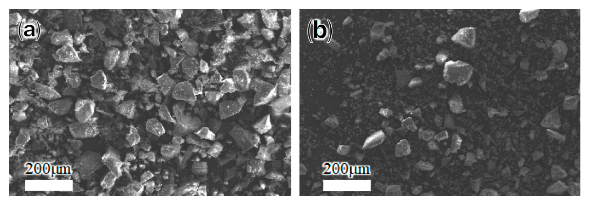
BSBG’s composition design was 6Na2O·8K2O·8MgO·16CaO·6SrO·36B2O3·2P2O5·18SiO2 by mole, and that of SBG was 6Na2O·8K2O·8MgO·22CaO·6SrO·2P2O5·48SiO2. Bioactive glasses were prepared via the melt-quenching method using carbonates, phosphates, boric acid, and silicon dioxide (AR), which were accurately weighed as designed and then thoroughly homogenized. The raw mixtures for BSBG and SBG were melted in a platinum crucible for 60 min at 1200 °C and 1400 °C, respectively, followed by quenching into ice water. The quenched glasses were then ball-milled and sieved through a 40 μm mesh, with the sieved fraction collected for subsequent experiments.
2.2. Atmospheric Plasma Spraying
In the animal experiments, Ti6Al4V disks (10 × 10 × 2 mm and Φ25.4 × 6 mm), tensile blocks (Φ25.4 × 25.4 mm), and metal plates (LCLP 12 III, 4 holes, both left and right, Double Medical Technology (Xiamen, China)) were used as research samples. All Ti6Al4V substrates were degreased, pickled, and then sandblasted to
Ra = (3.6~4.2) μm. Subsequently, bioactive glass powders were sprayed onto the roughened Ti6Al4V samples using an atmospheric plasma spraying system (Multicoat, Oerlikon Metco, Wohlen, Switzerland). To achieve appropriate droplet temperature and flight velocity for flying droplets, the spraying parameters were optimized based on our previous experimental experience, as listed in
Table 2.
2.3. Characterization of Materials
2.3.1. Structural Characterization
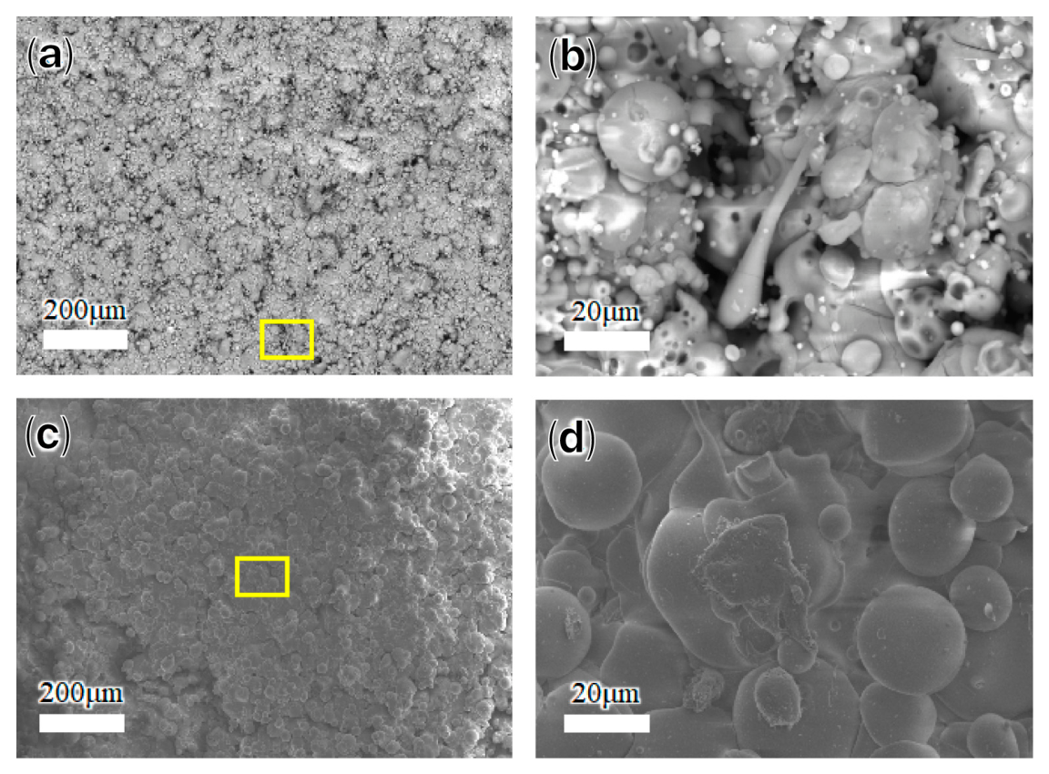
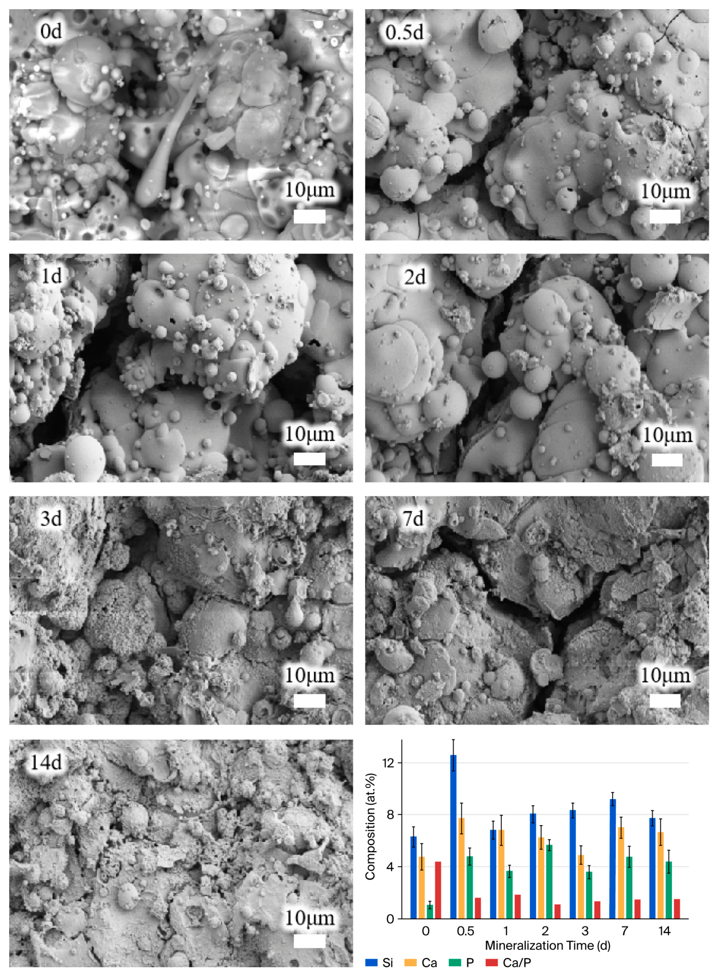
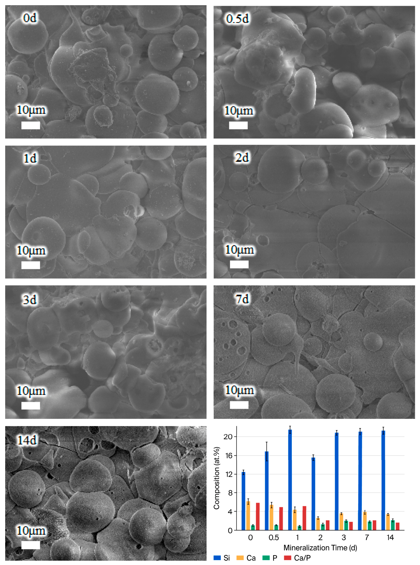
Micro-morphology was detected using a scanning electron microscope (SEM, Prisma E, Thermo Scientific, Waltham, MA, USA) with an energy-dispersed spectrometer (EDS, Ultim Max, Oxford Instruments, Abingdon, UK, automatic mode) for composition analysis. In composition tests, five random detection points were picked for each coating (n = 5).
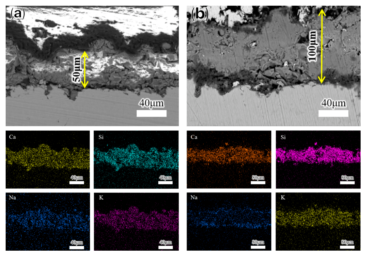
The thickness of coatings was measured by choosing five points from the cross-section morphology, and the porosity was analyzed using ImageJ software (version 1.46r) according to five random SEM images (n = 5).
The particle size distribution was analyzed via laser diffraction carried out using a laser diffraction particle size analyzer (Mastersizer 3000, Malvern, Malvern, UK).
The surface roughness of the coatings was measured via the stylus profilometry method (SJ-410, Mitutoyo Corporation, Kawasaki-shi, Japan). Five parallel pieces were tested (n = 5).
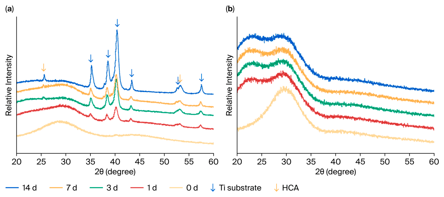
Coating structure was analyzed via X-ray diffraction (XRD, MiniFlex 600, Rigaku, Tokyo, Japan), with Cu Kα radiation (λ = 1.54 Å) at 40 kV and 30 mA. The test angle(2θ) ranged from 20° to 60° with a step size of 0.02° and a scanning speed of 5°/min.
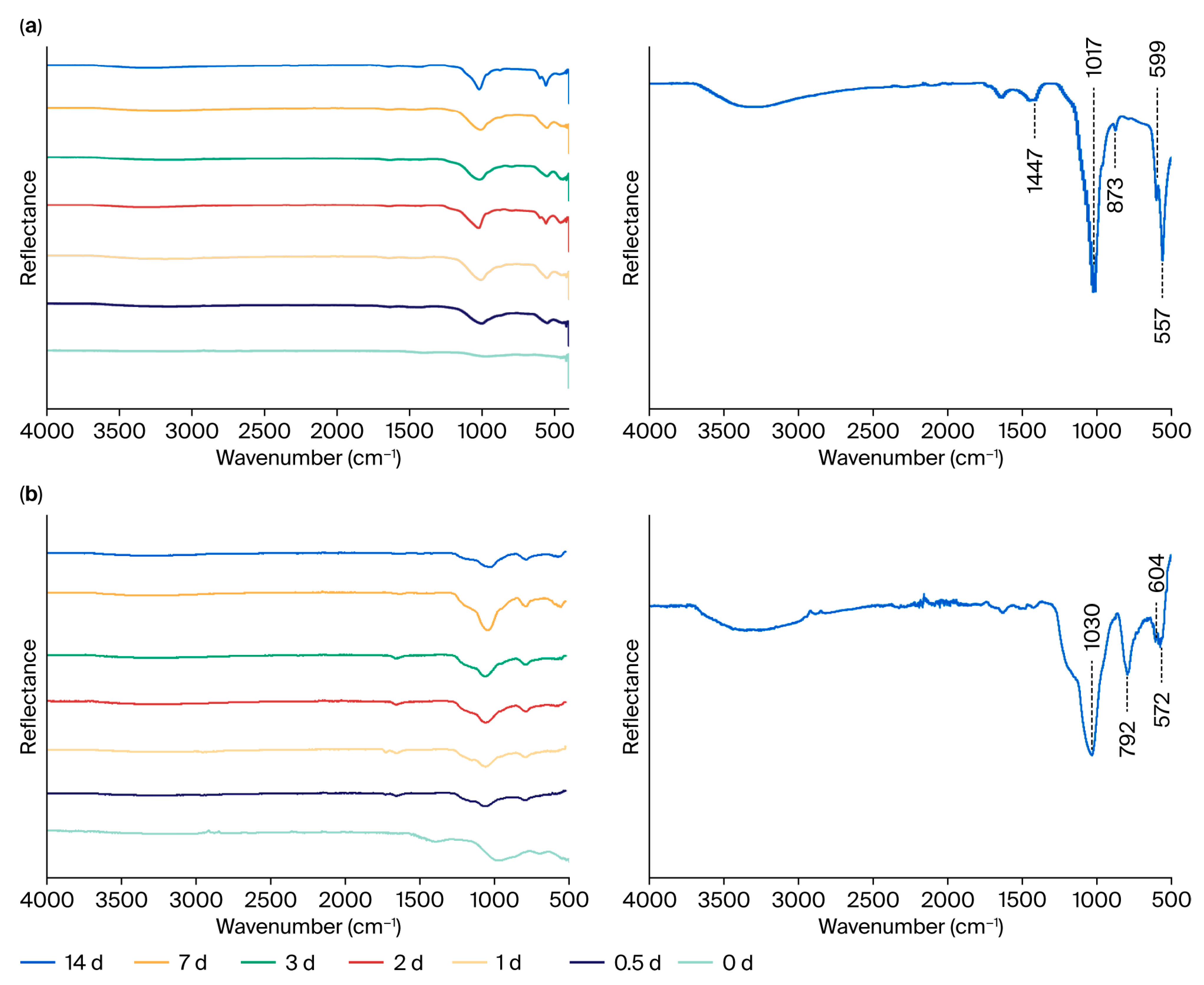
The infrared spectral characteristics of the coatings were recorded using a Fourier transform infrared spectrometer (FTIR, Nicolet iS50, Thermo Scientific, USA) equipped with an ATR diamond crystal, in the wavenumber range of 4000~500 cm−1.
2.3.2. Coefficient of Thermal Expansion Test
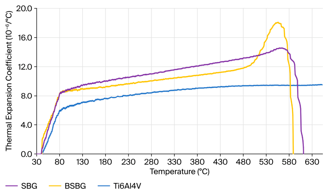
For the CTE test, BSBG and SBG powders were first sintered at 560 °C and 620 °C for 60 min, respectively, and then processed into Φ10 × 15 mm cylinders. The linear CTE of the cylinders was measured axially using a CTE tester (PCY, Xiangtan Instrument, Xiangtan, China), while Ti6Al4V cylinders with identical dimensions were simultaneously tested for comparison.
2.3.3. Bonding Strength
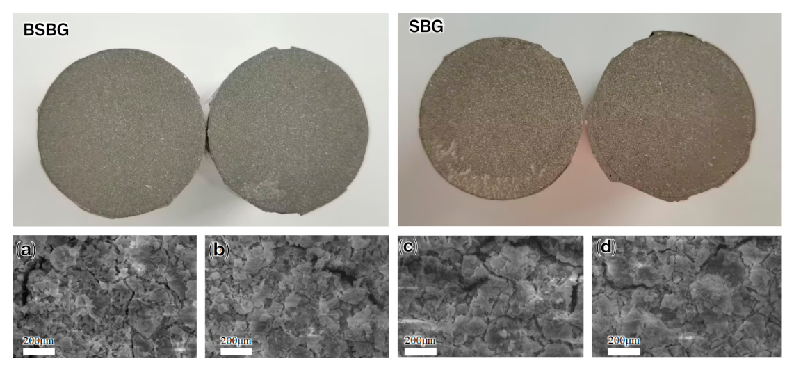
A tensile test was used for bonding strength and failure mode analysis. A Ti6Al4V tensile block was bonded to a coated surface using an FM 1000 polyamide–epoxy adhesive film and cured at 196 °C for 3 h. A tensile test of six parallel samples (n = 6) was performed on a universal material testing machine (LD26.205, Lishi (Shanghai) Instruments, Shanghai, China) at a constant crosshead speed of 2.5 mm/min until coating failure occurred.
2.3.4. In Vitro Self-Mineralization Test
Ti6Al4V disks of Φ25.4 × 6 mm with glass coatings were immersed in a Tris-HCl buffer solution (pH = 7.4) at 37 °C for 1, 2, 3, 7, and 14 days to evaluate in vitro self-mineralization ability. The formation rate of the bone-like apatite layer was determined to assess the bioactivity of the coatings.
2.3.5. Ion Release Test
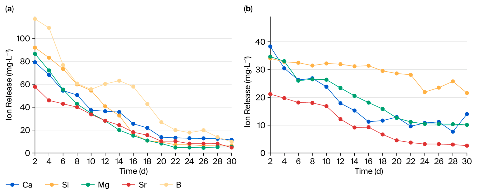
The degradation behavior of the bioactive glass coatings was evaluated by immersing samples in the Tris-HCl solution at an extraction ratio of 1 cm2/mL at 37 °C for 28 days. The solution was refreshed every two days during the experiment period. The concentration of the released ions was measured using inductively coupled plasma mass spectrometry (ICP-MS, Agilent 7800, Agilent, Santa Clara, CA, USA). Three parallel samples were established for each group (n = 3).
2.4. Cytological Experiments
The control groups were bare Ti6Al4V pieces (Φ25.4 × 6 mm), and the experimental groups were identical pieces with sprayed BSBG and SBG coatings. Three parallel samples were created for each group (n = 3) in these cytological tests. Rat bone marrow mesenchymal stem cells (rBMSCs, purchased from Baio Bowei Biotechnology Co., LTD, Beijing, China) were used for all cytological evaluations.
2.4.1. Cell Culture
The rBMSCs were cultured in cell culture flasks using DMEM-F12 medium (containing 10% fetal bovine serum, 100 U/mL penicillin, and 100 μg/mL streptomycin) at 37 °C under 5% CO2 conditions. When confluence reached 75~90%, cells were passaged using trypsin–EDTA digestion. All experiments utilized rBMSCs from the 3rd to 7th passages.
2.4.2. Biocompatibility Assay
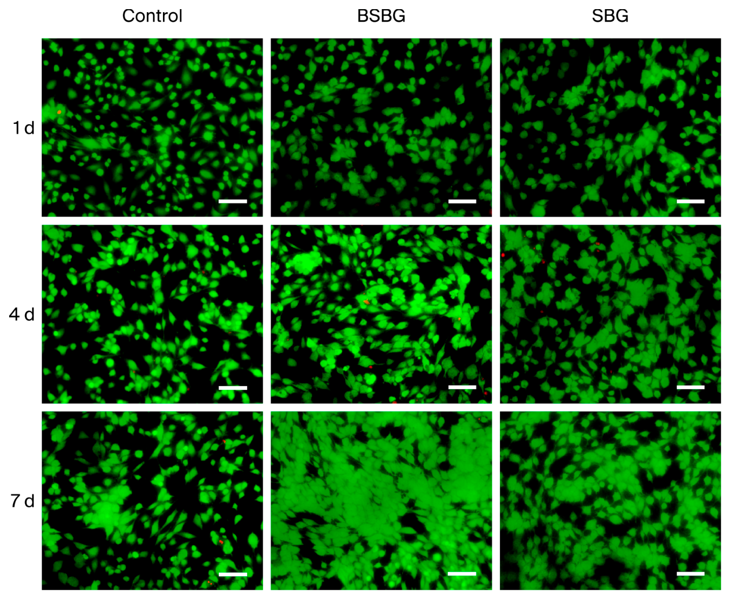
The cytotoxicity of the samples was tested via live/dead cell fluorescent staining. Sample extracts were prepared at an extraction ratio of 3 cm2/mL for 24 h. The rBMSCs were seeded in 6-well plates with 2 mL of cell suspension added to each well, and the plates were cultured at 37 °C in a 5% CO2 cell incubator for 24 h. The original culture medium was then replaced with the prepared extracts, and the cells were cultured for 1, 4, and 7 days. At each time point, the plates were taken out successively, and the extracts were discarded. Cells were rinsed with PBS, stained with live/dead reagent, and incubated in the dark for 20 min. Cell viability was observed and photographed under a fluorescence microscope.
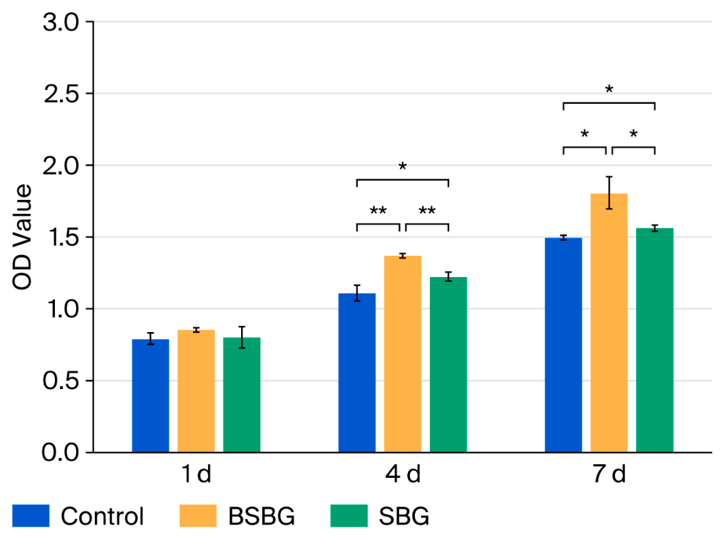
The rBMSCs were seeded into 24-well plates at a density of 5.0 × 10⁴ cells per well and cultured at 37 °C in a 5% CO2 atmosphere for 1, 4, and 7 days. At each time point, cells were transferred to new 96-well plates, cultured for another 24 h, and rinsed three times with phosphate-buffered saline (PBS). Cell proliferation was determined using the CCK-8 kit. Briefly, 180 μL of culture medium and 20 μL of CCK-8 solution were added to each well, followed by incubation for 1 h. The absorbance value at 450 nm was then measured using a microplate reader.
2.4.3. Cell Differentiation Assay
For the cell differentiation samples at the predetermined time points (7 and 14 days), TRIzol
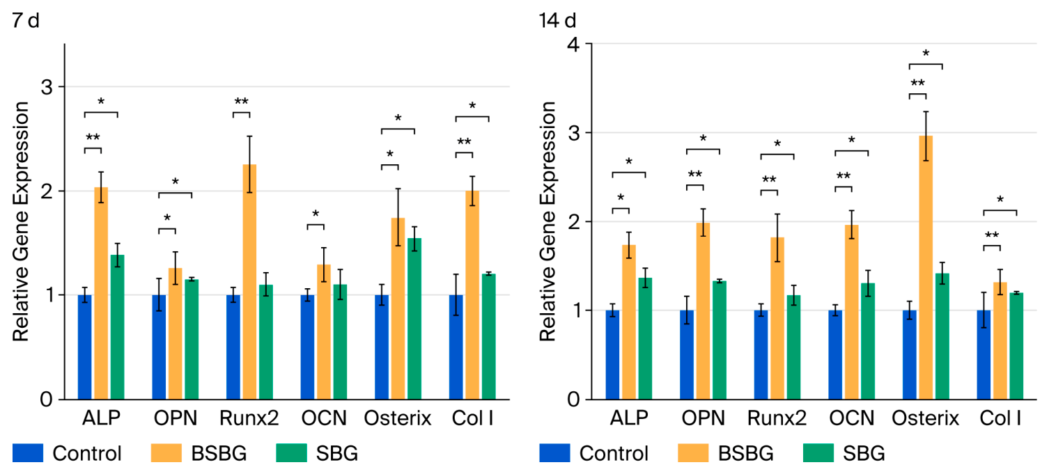
® reagent was added to lyse the cells on ice for 10 min. Total RNA was extracted, and a cDNA synthesis kit was subsequently used to synthesize cDNA templates for real-time quantitative reverse transcription polymerase chain reaction (RT-qPCR). The expression levels of osteogenic differentiation-related genes were detected via RT-qPCR, including Runt-related transcription factor 2 (Runx2), osteoblast-specific transcription factor (Osterix), osteopontin (OPN), alkaline phosphatase (ALP), osteocalcin (OCN), and type I collagen (Col I). The corresponding primer sequences are listed in
Table 3.
Regarding the statistical analysis, the relative quantitative method (2−ΔCT method) was used for the relative quantitative analysis of the amplification results to calculate the relative expression levels of each gene. One-way analysis of variance (ANOVA) combined with multiple comparisons was performed using statistical software SPSS (version 27.0.), and p < 0.05 indicated a statistically significant difference.
2.5. Animal Experiments and Surgical Procedure
A bone defect model was established in the tibia of Labrador Retrievers via osteotomy. The Ti6Al4V locking plate systems coated with BSBG coating were selected for fixation to simulate clinical application. Bare identical Ti6Al4V systems served as the control group. Six adult male Labrador Retrievers (~2.5 years old, 36.8 kg~41.6 kg) were randomly divided into 2 groups (n = 3 per group, randomized according to body weight and age). The dogs underwent overnight fasting or fasted for at least 6 h prior to animal anesthesia; all surgical procedures were performed under general anesthesia and strictly adhered to aseptic principles. Before anesthesia, intramuscular injection of Zoletil (3.5 mg/kg) and xylazine (0.2–1.0 mg/kg) was used to induce sedation and anesthesia in the animals, after which the surgical area was prepared and a venous channel was established. Before intubation, propofol was slowly administered intravenously to assist in anesthesia induction. The dosage of propofol should not exceed 6 mg/kg. After successful intubation of the animal, it was quickly transferred to the operating table, connected to a respiratory anesthesia machine, and maintained with 0.5% to 5% isoflurane inhalation anesthesia. The animal was placed in a prone position on the operating table and connected to a monitor. Depending on its needs, a ventilator can be used during the operation to change the breathing pattern of the animal.
Preoperative intramuscular injection of ceftriaxone sodium was administered at a dosage of 40–50 mg/kg to prevent infection, while preoperative intramuscular injection of sufentanil and meloxicam was administered at doses of 0.2–0.5 μg/kg and 0.1–0.2 mg/kg, respectively, for analgesia. Before surgery, the surgical area was disinfected and prepared, and sterile treatment towels were used.
During the surgery, physiological saline was administered intravenously to the animal, and the rate of intravenous fluid replacement was adjusted and recorded according to the animal’s condition. During the surgery, heart rate, respiratory rate, body temperature, end-expiratory CO2 concentration, and blood oxygen saturation were monitored and recorded at least once every 15 min. Animals were held by hand, and anesthesia was induced by an intravenous injection of propofol (5 mg/kg). Postoperatively, all animals received individual feeding with regular high-protein feed and daily intramuscular injections of cefotaxime sodium for 5 days until no abnormality was found during free movement.
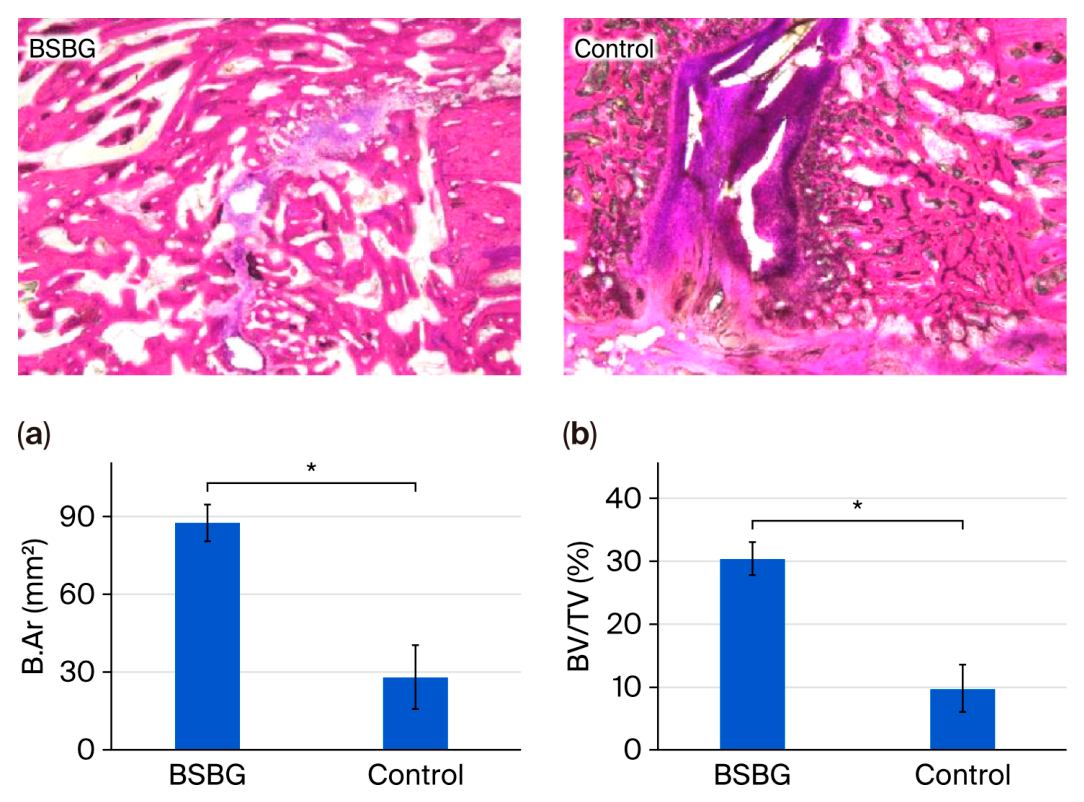
After the experiment period of 8 weeks, tibial specimens were harvested, fixed in 10% neutral buffered formalin, decalcified, embedded in paraffin, and longitudinally sectioned at a 5 μm thickness. Sections were stained with hematoxylin–eosin (HE) and Masson’s trichrome. The bone area (B.Ar.) was defined as the total new tissue area within the defect region, and bone volume fraction (BV/TV) was calculated as the ratio of mineralized bone area to total callus area. Three consecutive sections per sample were measured and averaged (n = 3), and the intergroup differences were analyzed using one-way ANOVA.
2.6. Statistical Analysis
Data are presented as mean ± standard deviation (SD). Statistical analyses were performed using SPSS (version 27.0.) Differences were considered statistically significant when * p < 0.05.