
Effect of Unit Cell Design and Volume Fraction of 3D-Printed Lattice Structures on Compressive Response and Orthopedics Screw Pullout Strength

Abstract
1. Introduction
2. Materials and Methods
2.1. Lattice Structure Design
2.2. Specimen Fabrication
2.3. Mechanical Tests
2.4. Screw Pullout Test
2.5. Statistical Analysis
3. Results
3.1. Quasi-Static Compression Test
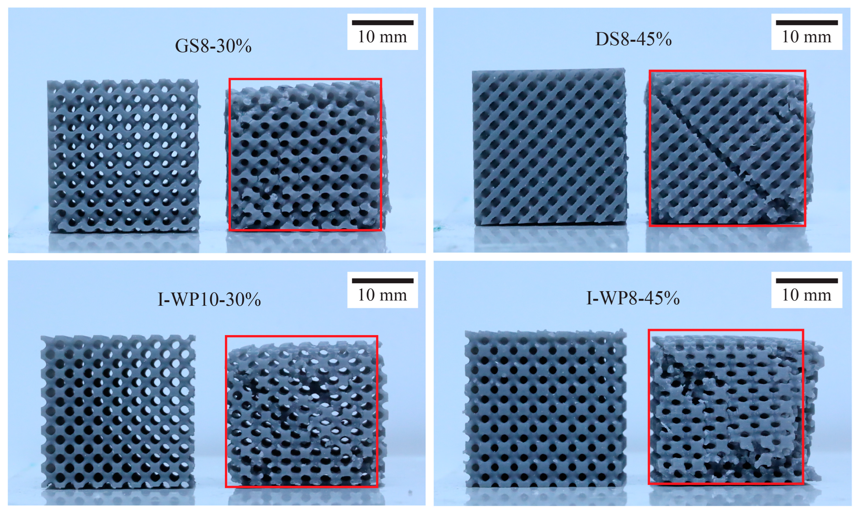
3.2. Screw Pullout Test
4. Discussion
4.1. Mechanical Responses of 3D-Printed Lattice Compared with Trabecular Bone
4.2. Screw Pullout Force
4.3. Limitations and Implications for Future Studies
5. Conclusions
Author Contributions
Funding
Institutional Review Board Statement
Informed Consent Statement
Data Availability Statement
Acknowledgments
Conflicts of Interest
Abbreviations
| AM | Additive manufacturing |
| BV/TV | Bone tissue volume to total volume |
| CAD | Computer-aided design |
| CBCT | Cone beam computed tomography |
| CT | Computed tomography |
| F-d | Force–displacement |
| FDM | Fused deposition modeling |
| SLA | Stereolithography (printing technology) |
| STL | Stereolithography (file type) |
| TPMS | Triply periodic minimal surfaces |
References
- Vandeweghe, S.; Koole, S.; Younes, F.; De Coster, P.; De Bruyn, H. Dental implants placed by undergraduate students: Clinical outcomes and patients’/students’ perceptions. Eur. J. Dent. Educ. Dent. 2014, 18, 60–69. [Google Scholar] [CrossRef] [PubMed]
- Wang, X.; Shujaat, S.; Shaheen, E.; Jacobs, R. Quality and haptic feedback of three-dimensionally printed models for simulating dental implant surgery. J. Prosthet. Dent. 2024, 131, 660–667. [Google Scholar] [CrossRef] [PubMed]
- Reymus, M.; Liebermann, A.; Diegritz, C.; Keßler, A. Development and evaluation of an interdisciplinary teaching model via 3D printing. Clin. Exp. Dent. Res. 2021, 7, 3–10. [Google Scholar] [CrossRef] [PubMed]
- Park, K.-M.; Min, K.-S.; Roh, Y.-S. Design optimization of lattice structures under compression: Study of unit cell types and cell arrangements. Materials 2021, 15, 97. [Google Scholar] [CrossRef]
- Ganguli, A.; Pagan-Diaz, G.J.; Grant, L.; Cvetkovic, C.; Bramlet, M.; Vozenilek, J.; Kesavadas, T.; Bashir, R. 3D printing for preoperative planning and surgical training: A review. Biomed. Microdevices 2018, 20, 65. [Google Scholar] [CrossRef]
- Lambrecht, J.T.; Berndt, D.; Christensen, A.M.; Zehnder, M. Haptic model fabrication for undergraduate and postgraduate teaching. Int. J. Oral Maxillofac. Surg. 2010, 39, 1226–1229. [Google Scholar] [CrossRef]
- Werz, S.M.; Zeichner, S.J.; Berg, B.I.; Zeilhofer, H.F.; Thieringer, F. 3D Printed Surgical Simulation Models as educational tool by maxillofacial surgeons. Eur. J. Dent. Educ. 2018, 22, e500–e505. [Google Scholar] [CrossRef]
- Feng, J.; Qi, W.; Duan, S.; Bao, C.; Zhang, X.; Cai, B.; Liu, X. Three-dimensional printed model of impacted third molar for surgical extraction training. J. Dent. Educ. 2021, 85, 1828–1836. [Google Scholar] [CrossRef]
- Narita, M.; Takaki, T.; Shibahara, T.; Iwamoto, M.; Yakushiji, T.; Kamio, T. Utilization of desktop 3D printer-fabricated ‘Cost-Effective’ 3D models in orthognathic surgery. Maxillofac. Plast. Reconstr. Surg. 2020, 42, 24. [Google Scholar] [CrossRef]
- Seifert, L.B.; Schnurr, B.; Herrera-Vizcaino, C.; Begic, A.; Thieringer, F.; Schwarz, F.; Sader, R. 3D printed patient individualised models versus cadaveric models in an undergraduate oral and maxillofacial surgery curriculum: Comparison of students’ perceptions. Eur. J. Dent. Educ. 2020, 24, 809–810. [Google Scholar] [CrossRef]
- Shujaat, S.; da Costa Senior, O.; Shaheen, E.; Politis, C.; Jacobs, R. Visual and haptic perceptibility of 3D printed skeletal models in orthognathic surgery. J. Dent. 2021, 109, 103660. [Google Scholar] [CrossRef] [PubMed]
- Van Dessel, J.; Huang, Y.; Depypere, M.; Rubira-Bullen, I.; Maes, F.; Jacobs, R. A comparative evaluation of cone beam CT and micro-CT on trabecular bone structures in the human mandible. Dento Maxillo Fac. Radiol. 2013, 42, 20130145. [Google Scholar] [CrossRef] [PubMed]
- Wu, D.; Spanou, A.; Diez-Escudero, A.; Persson, C. 3D-printed PLA/HA composite structures as synthetic trabecular bone: A feasibility study using fused deposition modeling. J. Mech. Behav. Biomed. Mater. 2020, 103, 103608. [Google Scholar] [CrossRef]
- Grzeszczak, A.; Lewin, S.; Eriksson, O.; Kreuger, J.; Persson, C. The potential of stereolithography for 3D printing of synthetic trabecular bone structures. Materials 2021, 14, 3712. [Google Scholar] [CrossRef] [PubMed]
- Gibson, L.; Ashby, M.F. Introduction. In Cellular Solids: Structure and Properties, 2nd ed.; Gibson, L.J., Ashby, M.F., Eds.; Cambridge University Press: Cambridge, UK, 1997; pp. 1–14. [Google Scholar]
- Gibson, L.J.; Ashby, M.F.; Karam, G.N.; Wegst, U.; Shercliff, H.R. The mechanical properties of natural materials. II. Microstructures for mechanical efficiency. Proc. R. Soc. Lond. A 1995, 450, 141–162. [Google Scholar]
- Ashby, M.; Gibson, L.; Olive, R. The mechanical properties of natural materials. I. Material property charts. Proc. R. Soc. A Math. Phys. Eng. Sci. 1995, 450, 123–140. [Google Scholar]
- Jäger, I.; Fratzl, P. Mineralized collagen fibrils: A mechanical model with a staggered arrangement of mineral particles. Biophys. J. 2000, 79, 1737–1746. [Google Scholar] [CrossRef]
- Gao, H.; Ji, B.; Jager, I.L.; Arzt, E.; Fratzl, P. Materials become insensitive to flaws at nanoscale: Lessons from nature. Proc. Natl. Acad. Sci. USA 2003, 100, 5597–5600. [Google Scholar] [CrossRef]
- Monkova, K.; Vasina, M.; Zaludek, M.; Monka, P.P.; Tkac, J. Mechanical vibration damping and compression properties of a lattice structure. Materials 2021, 14, 1502. [Google Scholar] [CrossRef]
- Al-Ketan, O.; Abu Al-Rub, R.K. Multifunctional mechanical metamaterials based on triply periodic minimal surface lattices. Adv. Eng. Mater. 2019, 21, 1900524. [Google Scholar] [CrossRef]
- Günther, F.; Wagner, M.; Pilz, S.; Gebert, A.; Zimmermann, M. Design procedure for triply periodic minimal surface based biomimetic scaffolds. J. Mech. Behav. Biomed. Mater. 2022, 126, 104871. [Google Scholar] [CrossRef] [PubMed]
- Deshpande, V.S.; Ashby, M.F.; Fleck, N.A. Foam topology: Bending versus stretching dominated architectures. Acta Mater. 2001, 49, 1035–1040. [Google Scholar] [CrossRef]
- Luxner, M.H.; Woesz, A.; Stampfl, J.; Fratzl, P.; Pettermann, H.E. A finite element study on the effects of disorder in cellular structures. Acta Biomater. 2009, 5, 381–390. [Google Scholar] [CrossRef]
- Abou-Ali, A.M.; Lee, D.-W.; Abu Al-Rub, R.K. On the effect of lattice topology on mechanical properties of SLS additively manufactured sheet-, ligament-, and strut-based polymeric metamaterials. Polymers Internet 2022, 14, 4583. [Google Scholar] [CrossRef]
- Maskery, I.; Sturm, L.; Aremu, A.O.; Panesar, A.; Williams, C.B.; Tuck, C.J.; Wildman, R.D.; Ashcroft, I.A.; Hague, R.J.M. Insights into the mechanical properties of several triply periodic minimal surface lattice structures made by polymer additive manufacturing. Polymer 2018, 152, 62–71. [Google Scholar] [CrossRef]
- Kim, J.E.; Shin, J.M.; Oh, S.O.; Yi, W.J.; Heo, M.S.; Lee, S.S.; Choi, S.C.; Huh, K.H. The three-dimensional microstructure of trabecular bone: Analysis of site-specific variation in the human jaw bone. Imaging Sci. Dent. 2013, 43, 227–233. [Google Scholar] [CrossRef]
- Lee, J.H.; Kim, H.J.; Yun, J.H. Three-dimensional microstructure of human alveolar trabecular bone: A micro-computed tomography study. J. Periodontal Implant. Sci. 2017, 47, 20–29. [Google Scholar] [CrossRef]
- Dekker, H.; Schulten, E.A.J.M.; ten Bruggenkate, C.M.; Bloemena, E.; van Ruijven, L.J.; Bravenboer, N. Regional differences in microarchitecture and mineralization of the atrophic edentulous mandible: A microcomputed tomography study. Arch. Oral Biol. 2022, 133, 105302. [Google Scholar] [CrossRef]
- Lim, E.L.; Ngeow, W.C.; Kadir, K.; Naidu, M. Facts to consider in developing materials that emulate the upper jawbone: A microarchitecture study showing unique characteristics at four different sites. Biomimetics Internet 2023, 8, 115. [Google Scholar] [CrossRef]
- Choël, L.; Last, D.; Duboeuf, F.; Seurin, M.J.; Lissac, M.; Briguet, A.; Guillot, G. Trabecular alveolar bone microarchitecture in the human mandible using high resolution magnetic resonance imaging. Dento Maxillo Fac. Radiol. 2004, 33, 177–182. [Google Scholar] [CrossRef]
- Monje, A.; Chan, H.L.; Galindo-Moreno, P.; Elnayef, B.; Suarez-Lopez del Amo, F.; Wang, F.; Wang, H.L. Alveolar bone architecture: A systematic review and meta-analysis. J. Periodontol. 2015, 86, 1231–1248. [Google Scholar] [CrossRef] [PubMed]
- Wang, J.; Zhou, B.; Liu, X.S.; Fields, A.J.; Sanyal, A.; Shi, X.; Adams, M.; Keaveny, T.M.; Guo, X.E. Trabecular plates and rods determine elastic modulus and yield strength of human trabecular bone. Bone 2015, 72, 71–80. [Google Scholar] [CrossRef] [PubMed]
- Maskery, I.; Parry, L.A.; Padrão, D.; Hague, R.J.M.; Ashcroft, I.A. FLatt Pack: A research-focussed lattice design program. Addit. Manuf. 2022, 49, 102510. [Google Scholar] [CrossRef]
- Martín-Montal, J.; Pernas-Sánchez, J.; Varas, D.J.P. Experimental characterization framework for SLA additive manufacturing materials. Polymers 2021, 13, 1147. [Google Scholar] [CrossRef]
- Cosmi, F.; Dal Maso, A. A mechanical characterization of SLA 3D-printed specimens for low-budget applications. Mater. Today Proc. 2020, 32, 194–201. [Google Scholar] [CrossRef]
- Zohdi, N.; Yang, R.C. Material anisotropy in additively manufactured polymers and polymer composites: A review. Polymers 2021, 13, 3368. [Google Scholar] [CrossRef]
- Yang, E.; Leary, M.; Lozanovski, B.; Downing, D.; Mazur, M.; Sarker, A.; Khorasani, A.; Jones, A.; Maconachie, T.; Bateman, S.; et al. Effect of geometry on the mechanical properties of Ti-6Al-4V Gyroid structures fabricated via SLM: A numerical study. Mater. Des. 2019, 184, 108165. [Google Scholar] [CrossRef]
- Misch, C.E.; Qu, Z.; Bidez, M.W. Mechanical properties of trabecular bone in the human mandible: Implications for dental implant treatment planning and surgical placement. J. Oral Maxillofac. Surg. 1999, 57, 700–706, discussion 6–8. [Google Scholar] [CrossRef]
- Lin, C.-Y.; Kang, J.-H. Mechanical properties of compact bone defined by the stress–strain curve measured using uniaxial tensile test: A concise review and practical guide. Materials 2021, 14, 4224. [Google Scholar] [CrossRef]
- Kumar, N.; Kumar, A.; Uniyal, P.; Ramalingaiah, B.; Sharma, S.; Goni, V.G.; Aggarwal, S.; Bhadada, S.K.; Bhushan, B. Mimicking high strength lightweight novel structures inspired from the trabecular bone microarchitecture. Philos. Trans. A Math. Phys. Eng. Sci. 2020, 378, 20190448. [Google Scholar] [CrossRef]
- Wang, T.-M.; Lin, Y.-C.; Lan, Y.-H.; Lin, L.-D. Evaluation of sawbones training protocol in bone quality classification using tactile sensation. J. Dent. Sci. 2022, 17, 897–902. [Google Scholar] [CrossRef] [PubMed]
- Devlin, H.; Horner, K.; Ledgerton, D. A comparison of maxillary and mandibular bone mineral densities. J. Prosthet. Dent. 1998, 79, 323–327. [Google Scholar] [CrossRef] [PubMed]
- Misch, C.E. Chapter 11. Bone density: A key determinant for treatment planning. In Dental Implant Prosthetics, 2nd ed.; Misch, C.E., Ed.; Mosby: St. Louis, MO, USA, 2015; pp. 237–252. [Google Scholar]
- Gibson, L.J. The mechanical behaviour of cancellous bone. J. Biomech. 1985, 18, 317–328. [Google Scholar] [CrossRef] [PubMed]
- Seebeck, J.; Goldhahn, J.; Städele, H.; Messmer, P.; Morlock, M.M.; Schneider, E. Effect of cortical thickness and cancellous bone density on the holding strength of internal fixator screws. J. Orthop. Res. 2004, 22, 1237–1242. [Google Scholar] [CrossRef]
- Seebeck, J.; Goldhahn, J.; Morlock, M.M.; Schneider, E. Mechanical behavior of screws in normal and osteoporotic bone. Osteoporos. Int. 2005, 16, S107–S111. [Google Scholar] [CrossRef]
- Halvorson, T.L.; Kelley, L.A.; Thomas, K.A.; Whitecloud, T.S., 3rd; Cook, S.D. Effects of bone mineral density on pedicle screw fixation. Spine 1994, 19, 2415–2420. [Google Scholar] [CrossRef]
- Bischoff, F.R.; Tille, E.; Beyer, F.; Bota, O.; Biewener, A.; Nowotny, J. Influence of bone density on stability in TBW. B.M.C. Musculoskelet. Disord. 2023, 24, 890. [Google Scholar] [CrossRef]
- Chapman, J.R.; Harrington, R.M.; Lee, K.M.; Anderson, P.A.; Tencer, A.F.; Kowalski, D. Factors affecting the pullout strength of cancellous bone screws. J. Biomech. Eng. 1996, 118, 391–398. [Google Scholar] [CrossRef]
- Procter, P.; Bennani, P.; Brown, C.J.; Arnoldi, J.; Pioletti, D.P.; Larsson, S. Variability of the pullout strength of cancellous bone screws with cement augmentation. Clin. Biomech. 2015, 30, 500–506. [Google Scholar] [CrossRef]
- Addevico, F.; Morandi, M.; Scaglione, M.; Solitro, G.F. Screw insertion torque as parameter to judge the fixation. Assessment of torque and pull-out strength in different bone densities and screw-pitches. Clin. Biomech. 2020, 72, 130–135. [Google Scholar] [CrossRef]
- Yakacki, C.M.; Poukalova, M.; Guldberg, R.E.; Lin, A.; Saing, M.; Gillogly, S.; Gall, K. The effect of the trabecular microstructure on the pullout strength of suture anchors. J. Biomech. 2010, 43, 1953–1959. [Google Scholar] [CrossRef]
- Wirth, A.J.; Goldhahn, J.; Flaig, C.; Arbenz, P.; Müller, R.; van Lenthe, G.H. Implant stability is affected by local bone microstructural quality. Bone 2011, 49, 473–478. [Google Scholar] [CrossRef]
- Pujari-Palmer, M.; Robo, C.; Persson, C.; Procter, P.; Engqvist, H. Influence of cement compressive strength and porosity on augmentation performance in a model of orthopedic screw pull-out. J. Mech. Behav. Biomed. Mater. 2018, 77, 624–633. [Google Scholar] [CrossRef]
- Wu, W.W.; Zhu, Y.B.; Chen, W.; Li, S.; Yin, B.; Wang, J.Z.; Zhang, X.J.; Liu, G.B.; Hu, Z.S.; Zhang, Y.Z. Bone hardness of different anatomical regions of human radius and its impact on the pullout strength of screws. Orthop. Surg. 2019, 11, 270–276. [Google Scholar] [CrossRef]
- Patel, P.S.D.; Shepherd, D.E.T.; Hukins, D.W.L. The effect of screw insertion angle and thread type on the pullout strength of bone screws in normal and osteoporotic cancellous bone models. Med. Eng. Phys. 2010, 32, 822–828. [Google Scholar] [CrossRef] [PubMed]
- Mueller, T.L.; Basler, S.E.; Müller, R.; van Lenthe, G.H. Time-lapsed imaging of implant fixation failure in human femoral heads. Med. Eng. Phys. 2013, 35, 636–643. [Google Scholar] [CrossRef] [PubMed]


| Cell Type | Design Group | Unit Cell Size (mm) | Avg. Thickness (mm) | Volume of the CAD Model (cm3) | ||
|---|---|---|---|---|---|---|
| ρ = 30 | ρ = 45 | ρ = 30 | ρ = 45 | |||
 Schwarz Diamond | Dia | 5.08 | 0.54 | 0.90 | 4.896 | 7.424 |
 Gyroid | Gyr | 5.08 | 0.53 | 0.82 | 4.900 | 7.425 |
 Skeletal Schwarz Diamond | DS5 DS8 DS10 | 5.08 3.175 2.54 | 1.27 0.79 0.63 | 1.51 0.94 0.75 | 4.898 4.894 4.902 | 7.426 7.421 7.434 |
 Skeletal Gyroid | GS5 GS8 GS10 | 5.08 3.175 2.54 | 1.30 0.82 0.65 | 1.75 1.10 0.87 | 4.906 4.914 4.903 | 7.426 7.403 7.416 |
 Skeletal Schoen I-Wrapped Package | I-WP5 I-WP8 I-WP10 | 5.08 3.175 2.54 | 1.64 1.02 0.82 | 2.10 1.32 1.05 | 4.837 4.851 4.837 | 7.429 7.452 7.420 |
| Properties | Photopolymer Resin | |
|---|---|---|
| Rigid 10k | Standard Grey | |
| Density (g/cm3) | 1.63 | 1.08 |
| Young’s modulus (GPa) | 11.6 * | 3.2 * |
| Tensile strength (MPa) | 81.7 * | 31.6 * |
| Unit Cell Design | Standard Grey | Rigid 10k | ||
|---|---|---|---|---|
| ρ = 30 | ρ = 45 | ρ = 30 | ρ = 45 | |
| Dia | 312.48 ± 28.4 a,b,c,d * | 709.42 ± 55.2 f,g | 1334.57 ± 120.5 c,d | 2706.90 ± 133.9 j |
| Gyr | 257.62 ± 15.1 a,b,c | 752.13 ± 33.7 f,g,h | 1087.80 ± 118.3 c | 2494.08 ± 196.4 i,j |
| DS-5 | 382.98 ± 20.4 c,d,e | 913.80 ± 92.3 j,k,l | N/A | N/A |
| GS-5 | 404.23 ± 69.2 d,e | 721.15 ± 80.8 f,g,h | N/A | N/A |
| I-WP-5 | 458.59 ± 47.0 e | 959.35 ± 87.0 k,l | N/A | N/A |
| DS-8 | 380.12 ± 53.9 c,d,e | 854.63 ± 41.0 i,j,k | 980.27 ± 116.2 c | 2337.70 ± 215.1 h,i,j |
| GS-8 | 255.09 ± 33.4 a,b,c | 996.24 ± 67.1 l | 236.22 ± 73.0 a,b | 1978.13 ± 225.7 e,f,g |
| I-WP-8 | 369.14 ± 36.2 b,c,d,e | 800.68 ± 55.3 g,h,i | 507.63 ± 93.0 b | 2295.74 ± 255.0 g,h |
| DS-10 | 215.44 ± 31.6 a | 703.53 ± 50.8 f,g | 222.56 ± 48.6 a,b | 1692.97 ± 142.6 d,e |
| GS-10 | 241.86 ± 15.4 a,b | 470.22 ± 27.6 e | 92.33 ± 30.0 a | 2089.84 ± 187.0 f,g |
| I-WP-10 | 224.17 ± 37.3 a | 659.15 ± 56.8 f | 358.60 ± 44.6 a,b | 1708.97 ± 203.9 d,e,f |
| Unit Cell Design | Standard Grey | Rigid 10k | ||
|---|---|---|---|---|
| ρ = 30 | ρ = 45 | ρ = 30 | ρ = 45 | |
| Dia | 362.15 ± 48.4 a * | 762.55 ± 37.0 c,d | 2378.63 ± 177.2 d | 3404.48 ± 250.8 e,f |
| Gyr | 290.53 ± 58.5 a | 923.89 ± 53.4 e,f | 2123.88 ± 57.5 d | 3619.18 ± 187.6 f |
| DS-5 | 605.83 ± 44.9 b | 1233.40 ± 91.1 g | N/A | N/A |
| GS-5 | 657.62 ± 50.2 b,c | 1026.53 ± 42.2 f | N/A | N/A |
| I-WP-5 | 538.73 ± 67.56 b | 1209.59 ± 34.5 g | N/A | N/A |
| DS-8 | 641.46 ± 40.0 b,c | 1293.08 ± 35.8 g,h | 2002.83 ± 209.2 d | 3391.50 ± 213.1 e,f |
| GS-8 | 353.41 ± 69.1 a | 1384.13 ± 77.5 h | 983.58 ± 165.4 b,c | 3368.95 ± 208.8 e,f |
| I-WP-8 | 630.40 ± 59.1 b,c | 1301.30 ± 74.5 g,h | 1188.82 ± 195.5 c | 3306.08 ± 197.3 e,f |
| DS-10 | 267.63 ± 24.8 a | 758.81 ± 67.5 c,d | 1169.59 ± 180.6 c | 3243.85 ± 161.4 e,f |
| GS-10 | 303.58 ± 23.6 a | 524.18 ± 39.2 b | 547.57 ± 30.9 a | 3398.13 ± 172.6 e,f |
| I-WP-10 | 313.21 ± 12.8 a | 826.42 ± 58.7 d,e | 644.57 ± 47.6 a,b | 3066.78 ± 57.2 e |
Disclaimer/Publisher’s Note: The statements, opinions and data contained in all publications are solely those of the individual author(s) and contributor(s) and not of MDPI and/or the editor(s). MDPI and/or the editor(s) disclaim responsibility for any injury to people or property resulting from any ideas, methods, instructions or products referred to in the content. |
© 2025 by the authors. Licensee MDPI, Basel, Switzerland. This article is an open access article distributed under the terms and conditions of the Creative Commons Attribution (CC BY) license (https://creativecommons.org/licenses/by/4.0/).
Share and Cite
Suksawang, B.; Chaijareenont, P.; Silthampitag, P. Effect of Unit Cell Design and Volume Fraction of 3D-Printed Lattice Structures on Compressive Response and Orthopedics Screw Pullout Strength. Materials 2025, 18, 1349. https://doi.org/10.3390/ma18061349
Suksawang B, Chaijareenont P, Silthampitag P. Effect of Unit Cell Design and Volume Fraction of 3D-Printed Lattice Structures on Compressive Response and Orthopedics Screw Pullout Strength. Materials. 2025; 18(6):1349. https://doi.org/10.3390/ma18061349
Chicago/Turabian StyleSuksawang, Boonyanuch, Pisaisit Chaijareenont, and Patcharawan Silthampitag. 2025. "Effect of Unit Cell Design and Volume Fraction of 3D-Printed Lattice Structures on Compressive Response and Orthopedics Screw Pullout Strength" Materials 18, no. 6: 1349. https://doi.org/10.3390/ma18061349
APA StyleSuksawang, B., Chaijareenont, P., & Silthampitag, P. (2025). Effect of Unit Cell Design and Volume Fraction of 3D-Printed Lattice Structures on Compressive Response and Orthopedics Screw Pullout Strength. Materials, 18(6), 1349. https://doi.org/10.3390/ma18061349