Article

Corticotomy Depth as a Modulator of Orthodontic Tooth Movement and PDL Stress—A Finite Element Study

1
Department of Dentofacial Orthopedics and Orthodontics, Wroclaw Medical University, Krakowska 26, 50-425 Wroclaw, Poland
2
Faculty of Mechanical Engineering, Military University of Technology, ul. gen. Sylwestra Kaliskiego 2, 00-908 Warsaw, Poland
3
Chair and Department of Oral Surgery, Medical University of Lublin, Doktora Witolda Chodźki 6 Street, 20-093 Lublin, Poland
4
Department of Periodontal and Oral Mucosa Diseases, Medical University of Białystok, ul. Waszyngtona 13, 15-269 Bialystok, Poland
5
Faculty of Law, University of Bialystok, Mickiewicza 1, 15-213 Bialystok, Poland
6
Department of Integrated Dentistry, Wroclaw Medical University, 50-425 Wroclaw, Poland
*
Authors to whom correspondence should be addressed.
Materials 2025, 18(23), 5290; https://doi.org/10.3390/ma18235290
Submission received: 15 October 2025 / Revised: 17 November 2025 / Accepted: 18 November 2025 / Published: 24 November 2025

Abstract

Introduction: The aim of this study was to evaluate the effect of corticotomy incision depth on tooth movement and stress distribution in the periodontal ligament (PDL) during orthodontic expansion using finite element analysis (FEA). The demand for accelerated and biologically safe orthodontic techniques has highlighted the importance of understanding biomechanical responses to surgical adjuncts like corticotomy. Objective: The aim of this study is to assess the effect of corticotomy depth on tooth movement and periodontal ligament stress distribution during orthodontic treatment using finite element analysis. Materials and methods: A 3D FEM model was developed based on CBCT and intraoral scans to replicate anatomical structures and simulate clinical orthodontic scenarios. Four conditions were analyzed: no corticotomy and corticotomy incisions of 1 mm, 2 mm, and 3 mm depths, applied between roots and above the apex region. Different cortical bone densities were tested using Young’s modulus values (12,500 MPa–27,500 MPa). Stress and displacement values were measured in both the crown and root regions. Results: The 3 mm corticotomy, penetrating through the cortical plate into the cancellous bone, significantly increased crown displacement (up to 26% in low-density bone) and altered root tipping patterns, reducing root movement relative to the crown. Shallower incisions (1–2 mm) had minimal effects. Despite increased movement, stress concentration in the cervical PDL region remained high across all scenarios, particularly in the premolar area, exceeding the 4.7 kPa threshold associated with tissue ischemia. Conclusions: Corticotomy depth is a critical factor for optimizing orthodontic tooth movement. Penetration into cancellous bone (3 mm) appears necessary to induce both: not only the Regional Acceleratory Phenomenon (RAP) but also to enhance displacement. However, this approach does not significantly reduce cervical PDL stress and offers limited periodontal protection. Individual planning based on bone density, morphology, and anatomical limitations is essential for balancing treatment efficiency and periodontal safety.

1. Introduction

In recent years, there has been a significant increase in the demand for orthodontic treatment, driven by both functional needs and increasing aesthetic awareness among patients. However, the choice of an appropriate therapeutic strategy, particularly the decision between treatment involving premolar extractions and non-extraction approaches, remains a subject of debate among orthodontic specialists. Research indicates that premolar extraction may facilitate better space management and occlusal stability, but concerns have been raised regarding its impact on temporomandibular joint health and facial profile deterioration [1,2,3,4,5,6,7,8].
One of the primary proponents of extraction was Charles H. Tweed, who in the 1940s introduced the Tweed Technique. This approach relied on the extraction of four first premolars to achieve stable, functional, and aesthetically pleasing occlusion. Tweed argued that extractions could eliminate anterior tooth protrusion, align incisors to enhance facial profile aesthetics, and prevent relapse and instability. The extraction approach gained widespread popularity, with the prevailing view that stable Class I occlusion could not be achieved without extractions. However, in the 1980s and 1990s, controversies emerged regarding the impact of premolar extractions on facial profile, including excessive lip retrusion and a premature ageing effect, as well as concerns about occlusal instability, relapse of crowding despite extractions, and adverse effects on airway function and speech. During this period, reports also suggested an increased risk of root resorption associated with extractions. These developments sparked growing interest in non-extraction techniques, such as arch expansion, interproximal reduction—also known as ‘stripping’, or the use of temporary anchorage devices (TADs) [9,10,11,12]. Non-extraction treatments, which often involve distalisation and arch expansion, may increase the risk of alveolar bone dehiscence and fenestration, particularly in patients with a thin gingival biotype or fragile bone morphology. Such structural changes may predispose patients to gingival recession, underscoring the need for individualised risk assessment prior to initiating orthodontic treatment [13,14,15,16,17,18,19,20,21,22]. Currently, the decision to extract first premolars is made more selectively and on a case-by-case basis. However, arch expansion may still be necessary, regardless of the extraction decision.
The biological mechanism of tooth movement relies on the periodontal tissues’ response to applied mechanical forces. In areas of compression, reduced blood flow in periodontal vessels and the displacement of periodontal ligament (PDL) fluid into bone canaliculi activate molecular pathways, such as the RANKL/OPG signalling cascade, leading to osteoclast activation and bone resorption. Conversely, in areas of tension, increased blood flow, fluid influx into the PDL space, and activation of osteoblast production promote bone formation. These processes are mediated by PDL cells, which respond to mechanical stimuli through activation of signalling pathways, such as RANKL/OPG, facilitating bone remodelling [23,24,25,26,27,28]. In vivo studies confirm that orthodontic forces upregulate RANKL expression in PDL fibroblasts and osteoblasts, with an increased presence of TRAP-positive cells (osteoclasts) within 24–72 h of force application. Animal model studies have demonstrated that blocking TNF-α inhibits bone resorption induced by orthodontic forces [29,30]. Prostaglandins, particularly PGE2, are produced by PDL and endothelial cells in response to mechanical pressure, upregulating RANKL and IL-1β expression. Together with pro-inflammatory cytokines IL-1β and IL-6, these enhance osteoclastogenesis on the compression side. In contrast, TGF-β (Transforming Growth Factor-β) may exert an anti-resorptive effect and support osteoblast differentiation on the tension side [31,32,33,34]. While these processes are desirable during tooth movement along the alveolar ridge, resorption of the buccal cortical plate during expansion can lead to undesirable outcomes, such as bone dehiscence, fenestration, and gingival recession [35,36].
For many years, corticotomy has been employed to leverage the Regional Acceleratory Phenomenon (RAP), a biological process involving accelerated remodelling of bone and surrounding tissues in response to surgical trauma or mechanical irritation. This procedure aims both to expedite orthodontic movement and protect periodontal health. However, its efficacy in the aspect of protecting periodontal health remains controversial due to the limited quality and heterogeneity of scientific studies and clinical protocols [37,38,39,40,41]. Understanding the biomechanics of tooth movement, including stress distribution in the PDL and the impact of various forces and adjunctive procedures, is critical for optimising orthodontic treatment. The finite element method (FEM) is a valuable tool for analysing these processes, enabling the simulation of various therapeutic scenarios and their potential effects on periodontal tissues [42,43]. The FEM study omitted the clinically studied RAP phenomenon affecting the acceleration of tooth movement and focused on the influence of corticotomy on the loading of the periodontium, which influences the process of remodeling and bone resorption during expansion.
In light of these considerations, this study aims to evaluate the influence of corticotomy depth on tooth movement and stress distribution in the periodontal ligament (PDL) during orthodontic treatment, using finite element analysis (FEM). The objective is to provide data that may assist clinicians in making informed therapeutic decisions, minimising the risk of periodontal complications, and enhancing the efficiency and stability of treatment outcomes.

2. Materials and Methods

To achieve the objective of the study, a research methodology based on the finite element method was used. This methodology allows for multiple studies to be conducted under exactly the same conditions. This makes it possible to accurately verify the impact of selected treatment parameters and patient conditions, e.g., on the state of stress and displacement in the stomatognathic system.
The numerical model used for the described research was constructed using two bone imaging methods: computed tomography and 3D scanning [44,45]. A CBCT was performed for the selected patient, resulting in DICOM files representing internal structures in grayscale. Based on these files, layer by layer, the compact bone, cancellous bone, teeth, and empty spaces were manually outlined. MIMICS 18.0 software (Materialise, Leuven, Belgium) was used for this purpose [46], in which masks were defined for each outline by filling them with a uniform color. This created surfaces on each layer of the DICOM image representing a cross-section of a given structure. Thanks to the high accuracy of CBCT, the boundary between compact and cancellous bone was clearly visible. However, in reality, these two structures may have different density ranges and, consequently, different stiffness ranges. In the study described, it was assumed that the entire compact bone would have one stiffness and the entire cancellous bone would have another, significantly lower stiffness. The bone thicknesses obtained were additionally consulted with orthodontic and oral surgeons.
After creating masks for each layer, a 3D model of the structures was determined. This process is carried out by extracting cross-sections of a given structure located on an adjacent layer. Therefore, the smaller the distance between the layers of the DICOM image, the more accurate the 3D model. The results of this process were saved in STL files (Figure 1).
CBCT imaging does not accurately reproduce enamel. Therefore, the geometry of the teeth and brackets was additionally determined using 3D intraoral scanning. It was also difficult to accurately determine the geometry of the PDL based on CBCT. Therefore, in accordance with the literature, a constant PDL thickness of 0.25 mm was assumed in the numerical model [47].
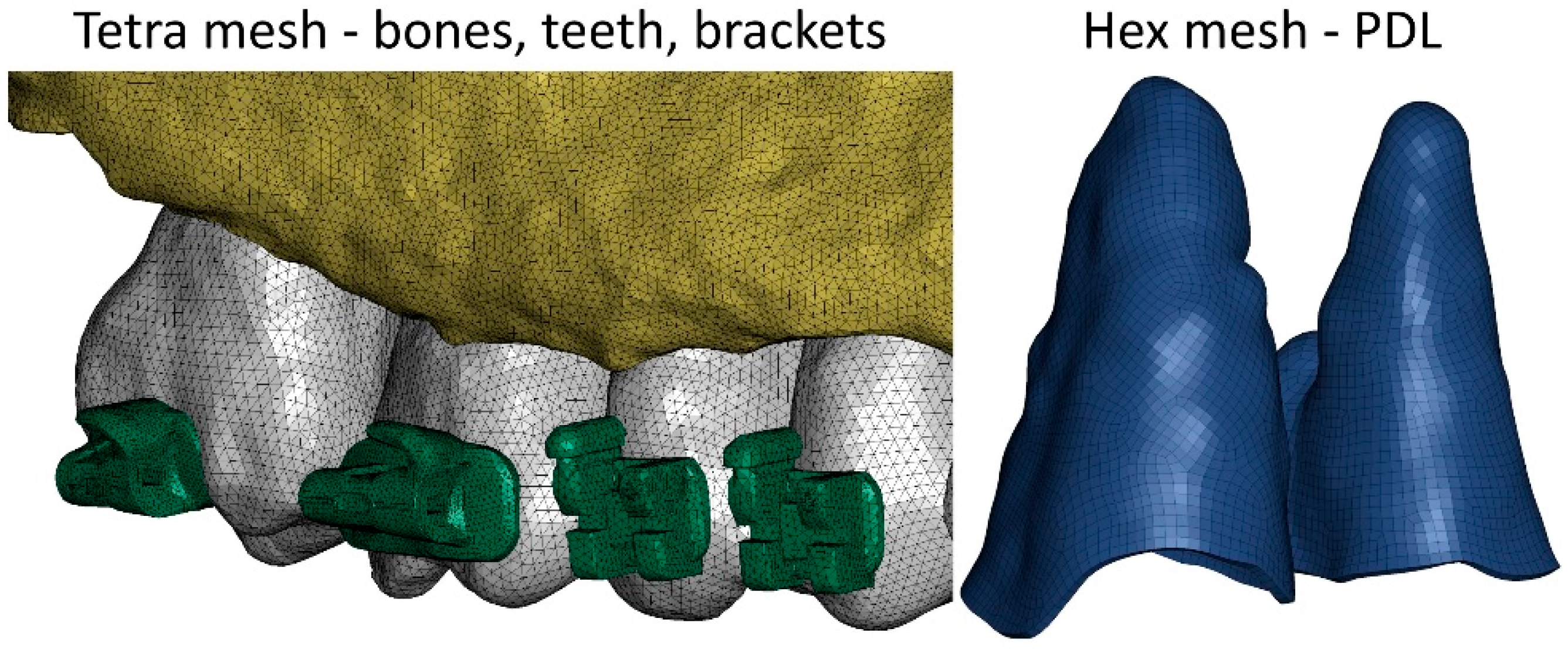
Due to the large volume of the skeletal system reproduced in the numerical model, it was very important to select the appropriate type and size of finite elements. The aim of the study was to determine, among other things, the loads acting in the PDL; hence, these structures were modeled using hexagonal elements. The mesh size was set to 0.25 mm. Other structures, such as bones, teeth, and brackets, were modeled using tetragonal elements. The average side length of the element was 0.3 mm (Figure 2). In total, the numerical model was constructed using over 4.5 million elements and over 1.2 million nodes. The entire mesh was meshed using Hypermesh 2023 (Altair, Troy, MI, USA).
The combination of all structures allowed for the reproduction of the complete structure of the patient’s skull without corticotomic incisions. Individual structures were connected either by sharing nodes or by gluing using TIED (LS-Dyna) contact (Figure 3).
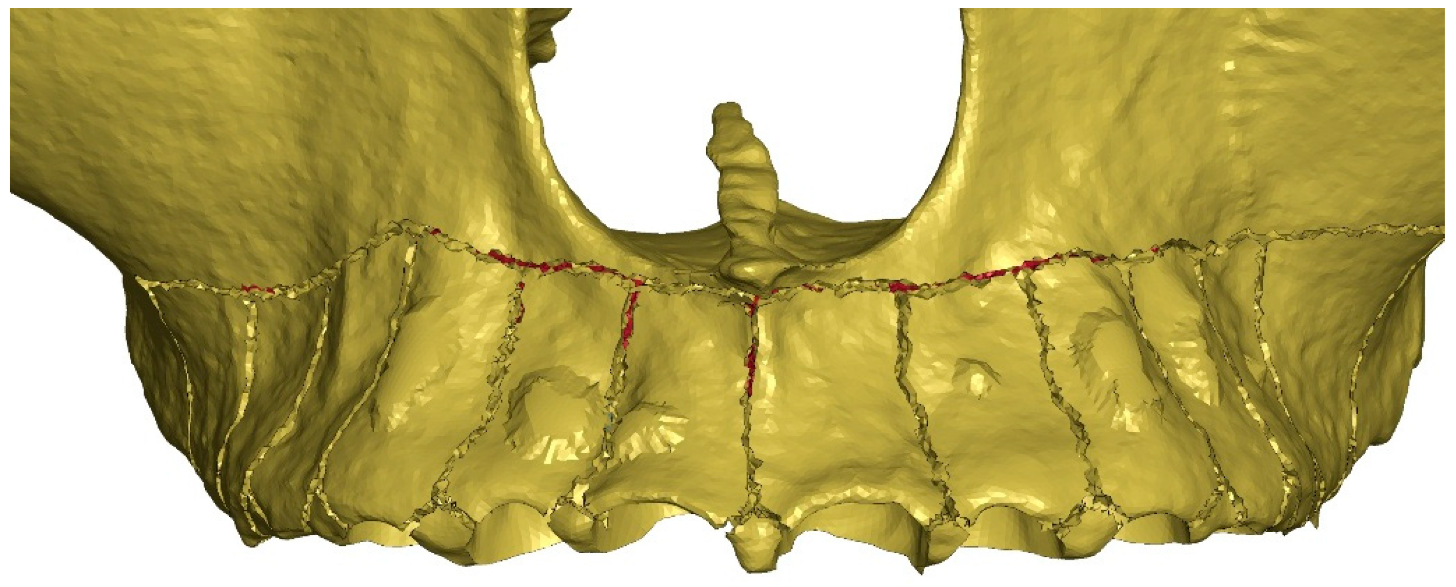
As part of the study, four clinical scenarios were analyzed, which differed in the depth of corticotomy incisions (G0 = 0 mm; G1 = 1 mm; G2 = 2 mm; G3 = 3 mm). For each scenario, six additional compact bone stiffness values were analyzed (E = 12,500 MPa; 15,500 MPa; 18,500 MPa; 21,500 MPa; 24,500 MPa; 27,500 MPa), giving a total of 24 variants. The incisions were introduced into the numerical model by removing elements to the appropriate depth according to the geometric pattern shown in the figure below (Figure 4). The incisions were made on the buccal side. The extension of the osteotomy was determined by the mesio-distal dimension of the tooth roots as well as by the position of the apices of the roots. The vertical cuts ended 5 mm apically from the crest and then Y-shape spread towards the neighboring teeth. The horizontal corticotomy was performed approximately 2–4 mm apically above the root apices. The depth of the incisions was limited by the thickness of the cortical plate at the maxillary sinus. Where cancellous bone was anatomically present, incisions were made to a depth of 1, 2, and 3 mm, respectively.
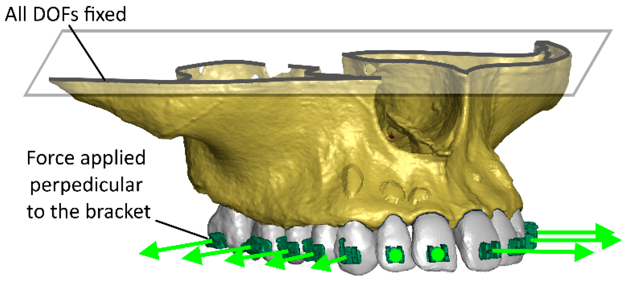
In each case, a conventional orthodontic expansion force of 100 g (F = 1 N) per lateral tooth was applied. To standardize the mechanical conditions across different bone stiffness levels, a simplified 1 N transverse load was applied instead of modeling the full archwire–bracket contact. This approach isolates the influence of corticotomy depth while avoiding additional variables such as wire stiffness, friction, or slot clearance. The authors acknowledge this as a simplification and plan to extend future models to include the full wire–bracket interaction for greater biomechanical realism. The fixation was defined by removing all degrees of freedom from the elements located on the upper surface of the model (Figure 5).
In the numerical model, it was verified that the brackets, teeth, and bones would be subjected to low loads and that there would be no significant stresses in these components. It was therefore assumed that these structures would be modeled using an isotropic, linear elastic constitutive model. The brackets were made of steel. The material data for the above components are given in Table 1.
During the research, the greatest emphasis was placed on modeling the behavior of the periodontal ligament (PDL). Ogden’s hyperelastic model was used to describe its strength properties. This model describes the behavior of the material using the strain energy function:
W = i = 1 3 j = 1 n μ j α j λ i α j 1 + K J 1 ln J
where W is the strain energy potential, λi is the main deviant stretches, µi and αi are material parameters, J is the determinant of the elastic strain gradient, and K is the volume modulus. The bulk modulus is calculated using the values of Poisson’s ratio and Young’s Modulus. The parameters presented in Table 2 [50] were used to describe the behavior of the PDL.
Displacement was measured at the mesiobuccal cusp centroid (crown) and at the root apex node set (root) of the first premolar (tooth 15), which represents the main site of expansion load transfer. The pressure values in Table 3 correspond to the maximum hydrostatic pressure (positive values indicate compression, negative values indicate tension) within the PDL for the same tooth. Clarification has been added to Table 3 to specify these reference points.

3. Results

Upon application of a 100 g expansive force to the maxillary posterior teeth in the finite element method (FEM) model without corticotomy, the stresses in the periodontal ligament (PDL) and the displacements of the crown and root vary depending on bone density (Table 3). However, the highest stress values are consistently located in the marginal periodontal region.
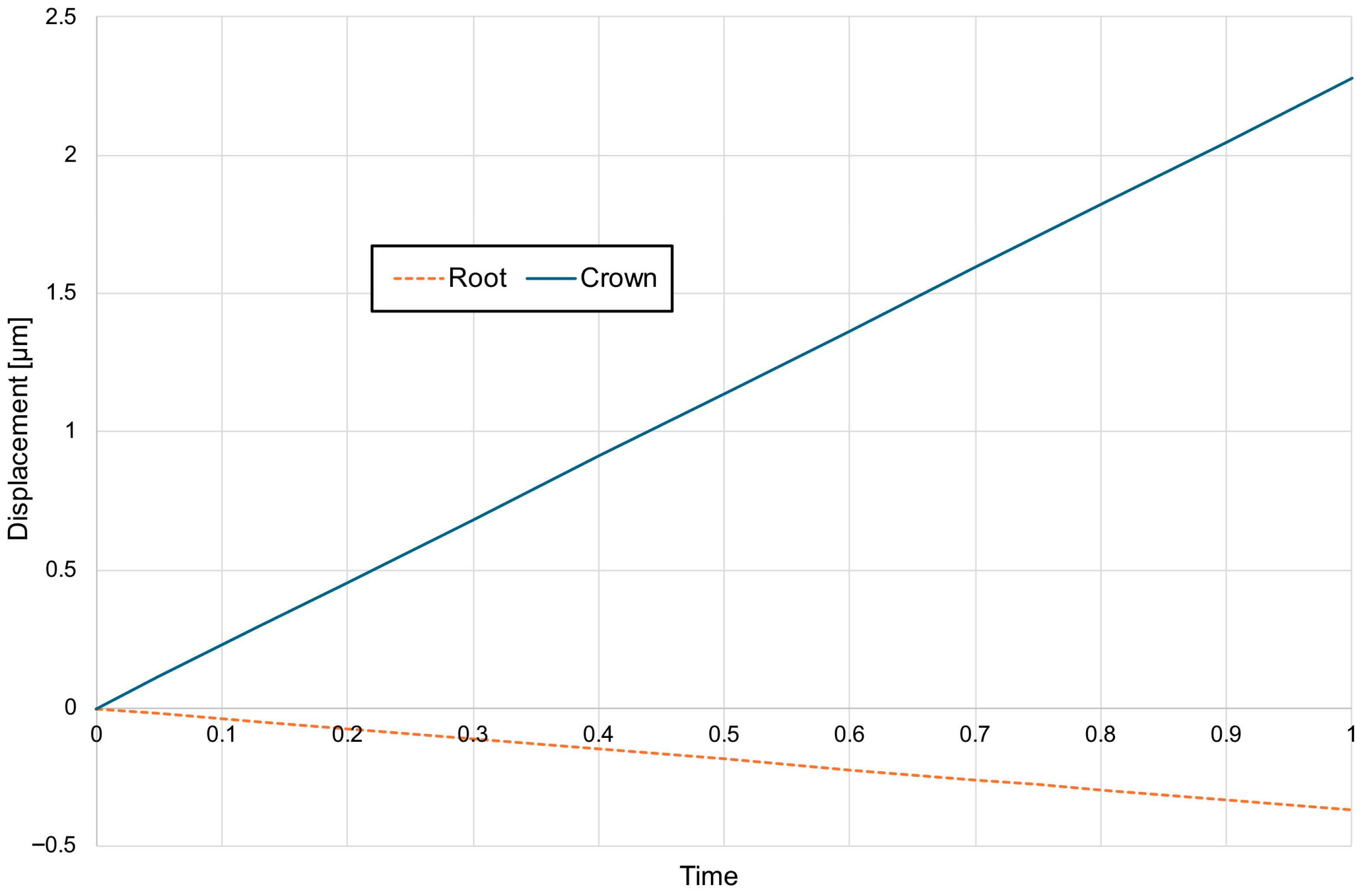
For a bone modulus of elasticity (E) of 12,500 MPa, the maximum compressive stresses reach approximately 7.69 kPa, with a crown displacement of 2.57 μm. At a higher E of 27,500 MPa, the stresses are slightly lower at 7.15 kPa, and the crown displacement is reduced to 1.98 μm. The observed movement is characterised as uncontrolled tipping, where the root displaces in the opposite direction to the crown, with the root displacement being approximately five times smaller than that of the crown (Figure 6, Figure 7 and Figure 8).
A corticotomy incision performed according to the protocol with a depth of 1 mm results in a minimal increase in tooth displacement of approximately 2%, without significantly affecting the stress distribution in the periodontal ligament (PDL) (Table 3).
A corticotomy incision of 2 mm increases the movement of the crown by about 12% in dense cortical bone and 8% in low-density cortical bone. The maximal hydrostatic pressure increases by about 3% while the minimum hydrostatic pressure is the same (Table 3)
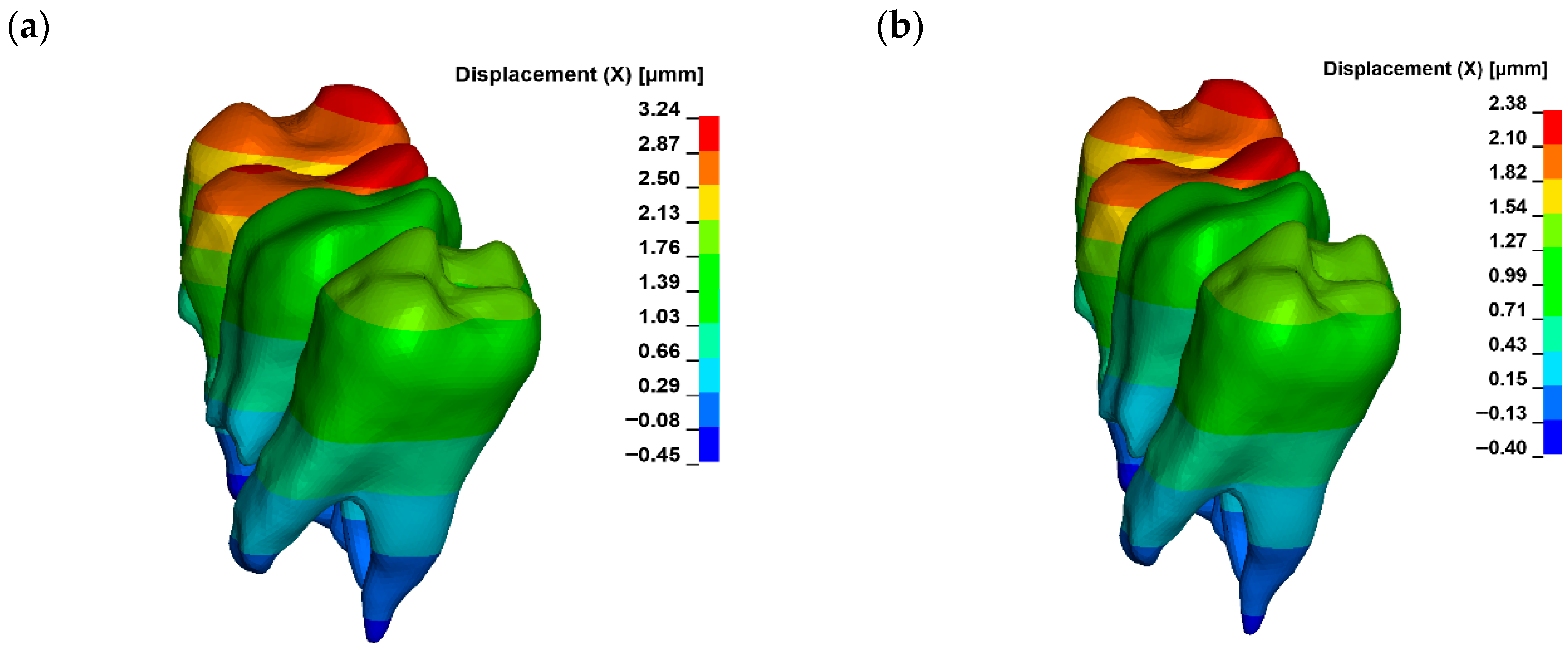
A corticotomy incision of 3 mm, which consistently penetrates the entire thickness of the cortical plate and reaches the cancellous bone, induces a significant change in the magnitude of displacement for both the crown and the root. For a bone modulus of elasticity (E) of 12,500 MPa, the displacement is 3.24 μm (26%), while for E = 27,500 MPa, it increases to 2.38 μm (20%) (Figure 9). The hydrostatic pressure values are similar and the change is clinically not significant (Figure 10). This also alters the extent of uncontrolled tipping, with the crown displacing 12 times more than the root (Figure 11). The root continues to move in the opposite direction to the crown, but the magnitude of root displacement is approximately two times smaller than that observed without corticotomy.

4. Discussion

The paper is a continuation of a series of publications on the assessment of loads occurring in the stomatognathic system [45,46]. Compared to previous works, the scientific and novel aspect of this paper involves the development of a series of numerical models that take into account different depths of corticotomy incisions and the stiffness of compact bone. The developed models were used to determine the impact of the above parameters on tooth displacement and PDL loads.
The results of this study indicate a significant increase in tooth movement after a 3-mm corticotomy, without a significant reduction in periodontal ligament (PDL) loading. These results are consistent with previous biomechanical studies that have shown that deeper corticotomies primarily increase tooth movement rather than reduce PDL loading. Although these studies primarily focus on other orthodontic movements, their results are comparable.
In a clinical study by Alighani et al. [51], micro-osteoperforations (MOPs) with a depth of 2–3 mm were employed to accelerate canine retraction. The study found that MOPs increased the rate of canine retraction by 2.3 times compared to the control group, suggesting that penetration into cancellous bone significantly enhances the efficiency of orthodontic treatment [52].
Similarly, in a finite element method (FEM) analysis by Uysal et al., the impact of corticotomy combined with zygomatic anchorage on maxillary molar intrusion was evaluated. The results indicated that a corticotomy depth of approximately 3 mm significantly reduced PDL stresses, potentially facilitating more effective tooth movement [53]. In contrast, a three-dimensional FEM analysis by Pacheco et al. comparing different corticotomy techniques during canine retraction found that the format of the corticotomy had minimal impact on PDL stress distribution, suggesting that depth and location may be more critical than the specific corticotomy pattern [54]. Animal studies have shown that tooth displacement increases significantly with greater numbers or depths of alveolar bone perforations. Both corticotomy (incision of the cortical plate) and osteotomy (complete transection of the cortical plate into cancellous bone) effectively accelerate tooth movement by promoting bone remodelling. Bone remodelling was faster and completed earlier following osteotomy compared to corticotomy, resulting in accelerated tooth movement in the early stages of orthodontic treatment [55].
The results of the present study indicate that only a corticotomy depth exceeding the cortical plate and reaching the cancellous bone (approximately 3 mm) significantly enhances tooth displacement, supporting the hypothesis of activation of the Regional Acceleratory Phenomenon (RAP) but without the justified protection mechanism on marginal periodontium because of no significant changes in PDL hydrostatic pressure. However, this raises questions about the impact of such interventions on periodontal tissue health and cortical bone integrity, particularly in the context of orthodontic expansion [55,56,57].
The literature reveals inconsistencies regarding the long-term safety of corticotomy. According to a recent systematic review by Kuc et al. [40], the effectiveness of corticotomy remains controversial. De Stefani et al. [58] emphasised that corticotomy should be limited to cases of moderate expansion and always accompanied by bone augmentation to ensure comprehensive root support. However, the high risk of systematic bias in their analysis limits the evidential value of these conclusions [40,58]. Dab et al. [59] noted that, despite low-level evidence of no adverse effects, a moderate increase in bone density (approximately 7%) can be expected following augmentation. Similar effects—increased bone density and alveolar ridge thickness—were reported by Gao et al. [60] in studies involving Periodontally Accelerated Osteogenic Orthodontics (PAOO) with additional bone augmentation, although the quality of the analysed studies was suboptimal. Apalimova et al. [61] compared various surgically assisted orthodontic techniques, concluding that classic corticotomy (with flap elevation) is more effective than piezocision, albeit more invasive. Notably, they found no significant differences in complications such as root resorption, gingival recession, or pulp vitality loss compared to conventional treatment. Regarding soft tissues, Al-Ibrahim et al. [62] demonstrated that piezocision combined with self-ligating appliances does not cause gingival recession, and the use of low-frequency vibrations or corticotomy does not adversely affect periodontal parameters. The moderate risk of systematic bias in their study lends credibility to these findings. Furthermore, Kamal et al. [63] and Alsino et al. [64] suggest that PAOO may not only accelerate treatment but also improve bone conditions and periodontal tissue stability by increasing alveolar ridge thickness. Wang et al. [65] reported that thickening the gingival biotype with soft tissue augmentation can provide clinical benefits, particularly in patients with a thin gingival biotype and unfavourable bone morphology.
Conversely, studies evaluating cortical bone changes following piezocision-assisted upper arch expansion found no significant adverse changes in the cortical plate, except in the premolar region, where a reduction in buccal cortical plate thickness was observed. However, due to the case series design and low evidential value, these findings carry a high risk of systematic bias. The reduction in cortical plate thickness in the premolar region aligns with the present study’s results, as the highest PDL stresses accumulate in this area, leading to bone resorption [66]. Corticotomy may result in minimal periodontal tissue changes, with outcomes depending on the technique used (e.g., incision method and depth). It is generally recommended to perform corticotomy incisions 3 mm below the bone crest and 3–5 mm away from root apices. Proper surgical management during flap preparation is also critical.
Sulewska et al. [67] highlighted this aspect, performing incisions that preserve interdental papillae in accordance with minimally invasive surgical principles to protect marginal periodontal tissues. Long-term observations (over 5 years) by the same research team [68] found no adverse effects of corticotomy-assisted orthodontic arch expansion on periodontal health in terms of soft tissue, focusing on the soft tissue aspect. However, the authors did not address incision depth, which could aid in interpreting the present study’s findings [67,68]. These results may explain the variability in research outcomes due to the lack of clear guidelines on incision depth. Comparing the effectiveness of a 1 mm corticotomy, which results in only a 2% change in displacement, to deeper 3 mm corticotomies, which produce approximately 40% greater displacement, without considering bone density—a critical factor—may lead to erroneous and controversial conclusions.
The differentiation of the periodontal ligament load after corticotomy depending on the initial bone density seems particularly interesting. This opens up further discussion regarding the potential reduction of the risk of root apical resorption and the risk of dehiscence. The study by Kuc et al. [45] showed that during incisor retraction, low bone density predisposes to a greater response and stress in the periodontal ligament, which significantly increases the risk of root resorption in patients with, e.g., high bone density or osteoporosis. While in the current study, on the contrary, a denser vestibular plate causes greater stress during expansion in the marginal periodontium and increases the risk of hyalinization and bone and soft tissue destruction. Detailed studies in this aspect are indicated to be able to draw deeper conclusions and clinical guidelines.
During expansion, PDL stresses may be particularly significant for periodontal tissue protection. Across all corticotomy depths, the cervical region, especially around premolars, is highly susceptible to undermining resorption due to stresses exceeding the 4.7 kPa threshold, which occludes blood vessels. This may lead to hyalinisation, tissue necrosis, and increased bone resorption, potentially causing bone dehiscence and gingival recession. These effects are linked to osteoclast and cementoclast activation in response to tissue ischaemia caused by prolonged PDL overloading. The 20–40% variation in PDL stresses depending on bone density, coupled with their accumulation in the marginal periodontal region, suggests that corticotomy as a standalone procedure may not significantly reduce marginal bone resorption compared to non-corticotomy cases. However, when performed, corticotomy should be deep enough to maximise positive outcomes [55,69,70,71,72,73,74,75].
A beneficial aspect of corticotomy is the reduced root movement in the opposite direction to the crown compared to non-corticotomy cases. This may decrease the risk of root resorption by reducing stresses on the limited PDL surface surrounding the root apex. Increased crown displacement, and thus significantly accelerated orthodontic movement, is observed following deep corticotomy. Notably, the lack of clear visibility of cancellous bone structure in CBCT scans necessitates caution when planning corticotomy. In such cases, limiting incision depth and considering additional imaging or consultation with an experienced specialist is recommended. In certain scenarios, cancellous bone may be virtually absent between tooth roots, particularly in specific anatomical regions or patient populations. This is especially relevant in the anterior mandible, where histological and imaging studies indicate that the space between roots may consist entirely of cortical bone without evident trabeculation. A similar issue may arise in patients with significantly reduced alveolar ridge height (e.g., due to periodontal disease), where sclerotisation may reduce or eliminate the cancellous bone layer [76]. Wilcko et al. [77], pioneers of the PAOO technique, described cases where incisions were made without penetrating cancellous bone due to its near absence. Kim et al. [78] noted that CBCT imaging revealed minimal or absent cancellous bone in the anterior mandible in some patients. Melsen [79] highlighted that the bone structure in the anterior maxilla and mandible may be almost entirely cortical, significantly influencing orthodontic responses. The depth of corticotomy incisions in the molar region warrants individual consideration. Depending on the technique, superficial corticotomy incisions in the maxillary molar region are recommended to minimise the risk of maxillary sinus damage [80,81,82,83]. However, this may reduce the procedure’s effectiveness and limit the extent of tooth displacement and PDL stresses. Current evidence suggests that incisions should target only the cortical bone layer without penetrating deeply into the cancellous bone. Excessively deep incisions may lead to sinus perforation or mucosal damage, resulting in serious complications (e.g., infection). Therefore, the risks must be weighed, and digitally navigated corticotomy is advisable to optimise outcomes safely. Preoperative imaging is critical. Modern techniques such as computed tomography (CT) or panoramic radiographs allow precise assessment of the anatomical relationship between tooth roots and the maxillary sinus, enabling accurate determination of incision depth to minimise complications. Contemporary approaches emphasise tailoring the procedure to the patient’s anatomy. In cases where the maxillary sinus is shallow or close to the root apices, incisions should be more conservative to avoid perforation [80,81,82,83].
From a regulatory perspective, surgical ancillaries such as corticotomy and piezocision are considered medical procedures under the European Medical Devices Regulation (MDR 2017/745) when specialized piezoelectric instruments or systems are used [84]. The regulation’s enhanced clinical evidence requirements and post-market surveillance obligations create documentation standards for device-related complications. Although this finite element model does not assess risk, soft tissue response, or device classification, it provides quantitative biomechanical data that can support preclinical assessments required by the evidence-based MDR framework. In this simulation, penetration into cancellous bone appears to exacerbate tooth displacement, but its clinical application should be assessed on a case-by-case basis, depending on bone morphology and anatomical safety. Therefore, the numerical results should be interpreted as supplementary scientific evidence rather than direct clinical guidance.
Considering all factors influencing procedure efficacy and anatomical constraints, corticotomy incisions should reach the cancellous bone where feasible and safe to maximise therapeutic benefits.
Limitations: The study used a numerical model developed on the basis of CBCT and scans performed on a single patient. The model accurately reproduced the geometry of bone structures and teeth. PDL thickness was assumed based on the literature. It should be noted that this model has not been validated by experimental tests, which means that the results obtained using it cannot be used to accurately predict changes during treatment of this particular patient or other patients. Due to the lack of accurate material characteristics, the study analyzed different stiffnesses of compact bone. Nevertheless, there may be significant differences in bone structure and the ratio of compact to cancellous bone between different patients. For this reason, the results presented in this article should be treated as results showing the trend of displacement and hydrostatic pressure changes in the case of changes in bone stiffness and the depth of corticotomy incisions.

5. Conclusions

The findings of this study, which demonstrate a reduction in periodontal ligament (PDL) stresses and a greater extent of tooth displacement only after penetration into cancellous bone, provide a biological rationale for controlled, selective, and deeply but individually planned corticotomy to assist orthodontic treatment. Such corticotomy accelerates tooth movement but offers limited protective function for periodontal tissues—provided that the periodontal phenotype is assessed beforehand.

Author Contributions

Conceptualization, A.E.K., K.S., G.H., M.S. (Magdalena Sulewska), S.S., B.K. and J.M.; Methodology, A.E.K., K.S., J.K., G.H., M.S. (Magdalena Sulewska), S.S., M.K., B.K., J.M. and M.S. (Michał Sarul); Software, K.S. and S.S.; Formal analysis, A.E.K., K.S., M.S. (Magdalena Sulewska) and J.E.K.-K.; Investigation, A.E.K., K.S. and M.S. (Magdalena Sulewska), S.S.; Resources, A.E.K., K.S., M.S. (Magdalena Sulewska), M.K. and B.K.; Data curation, K.S., S.S. and M.S. (Michał Sarul); Writing—original draft, A.E.K., K.S., J.K., G.H., M.S. (Magdalena Sulewska) and J.E.K.-K.; Writing—review & editing, A.E.K., K.S., J.K., M.S. (Magdalena Sulewska) and J.M.; Project administration, J.M.; Funding acquisition, K.S. All authors have read and agreed to the published version of the manuscript.

Funding

This work was supported by the Military University of Technology under project no. UGB 502-000013-W100-22.

Institutional Review Board Statement

Not applicable.

Informed Consent Statement

Not applicable.

Data Availability Statement

The original contributions presented in this study are included in the article. Further inquiries can be directed to the corresponding authors.

Conflicts of Interest

The authors declare no conflict of interest.

References

  1. Iared, W.; da Silva, E.M.K.; Iared, W.; Macedo, C.R. Esthetic perception of changes in facial profile resulting from orthodontic treatment with extraction of premolars: A systematic review. J. Am. Dent. Assoc. 2017, 148, 9–16. [Google Scholar] [CrossRef] [PubMed]
  2. Freitas, B.V.; Rodrigues, V.P.; Rodrigues, M.F.; de Melo, H.V.F.; Dos Santos, P.C.F. Soft tissue facial profile changes after orthodontic treatment with or without tooth extractions in Class I malocclusion patients: A comparative study. J. Oral Biol. Craniofac. Res. 2019, 9, 172–176. [Google Scholar] [CrossRef] [PubMed] [PubMed Central]
  3. Allgayer, S.; Mezomo, M.B. Do premolar extractions necessarily result in a flat face? No, when properly indicated. Dent. Press J. Orthod. 2018, 23, 82–92. [Google Scholar] [CrossRef] [PubMed] [PubMed Central]
  4. Elias, K.G.; Sivamurthy, G.; Bearn, D.R. Extraction vs nonextraction orthodontic treatment: A systematic review and meta-analysis. Angle Orthod. 2024, 94, 83–106. [Google Scholar] [CrossRef] [PubMed] [PubMed Central]
  5. Moon, S.; Mohamed, A.M.A.; He, Y.; Dong, W.; Yaosen, C.; Yang, Y. Extraction vs. Nonextraction on Soft-Tissue Profile Change in Patients with Malocclusion: A Systematic Review and Meta-Analysis. Biomed. Res. Int. 2021, 2021, 7751516. [Google Scholar] [CrossRef] [PubMed] [PubMed Central]
  6. Drobocky Oles, B.; Smith Richard, J. Changes in facial profile during orthodontic treatment with extraction of four first premolars. Am. J. Orthod. Dentofac. Orthop. 1989, 95, 220–230. [Google Scholar] [CrossRef]
  7. Young, T.M.; Smith, R.J. Effects of orthodontics on the facial profile: A comparison of changes during nonextraction and four premolar extraction treatment. Am. J. Orthod. Dentofac. Orthop. 1993, 103, 452–458. [Google Scholar] [CrossRef] [PubMed]
  8. Verma, S.L.; Sharma, V.P.; Tandon, P.; Singh, G.P.; Sachan, K. Comparison of esthetic outcome after extraction or non-extraction orthodontic treatment in class II division 1 malocclusion patients. Contemp. Clin. Dent. 2013, 4, 206–212. [Google Scholar] [CrossRef] [PubMed] [PubMed Central]
  9. Cross, J.J. The Tweed philosophy: The Tweed years. Semin. Orthod. 1996, 2, 231–236. [Google Scholar] [CrossRef] [PubMed]
  10. Altamash, S.; Sakrani, H.; Ahmed, N.; Marya, A.; Heboyan, A. Non-extraction orthodontic treatment for severe dental crowding using miniscrew-assisted rapid maxillary expansion. J. Surg. Case Rep. 2022, 2022, rjac509. [Google Scholar] [CrossRef] [PubMed]
  11. Raghis, T.R.; Alsulaiman, T.M.A.; Mahmoud, G.; Youssef, M. Efficiency of maxillary total arch distalization using temporary anchorage devices (TADs) for treatment of Class II-malocclusions: A systematic review and meta-analysis. Int. Orthod. 2022, 20, 100666. [Google Scholar] [CrossRef] [PubMed]
  12. Oğuz, F.; Özden, S.; Cicek, O. Distalization Methods for Maxillary Molars Utilizing Temporary Anchorage Devices (TADs): A Narrative Review. Appl. Sci. 2024, 14, 11333. [Google Scholar] [CrossRef]
  13. Wishney, M. Potential risks of orthodontic therapy: A critical review and conceptual framework. Aust. Dent. J. 2017, 62 (Suppl. S1), 86–96. [Google Scholar] [CrossRef] [PubMed]
  14. Johal, A.; Katsaros, C.; Kiliaridis, S.; Letao, P. State of the science on controversial topics: Orthodontic therapy and gingival recession (a report of the Angle Society of Europe 2013 meeting). Prog. Orthod. 2013, 14, 16. [Google Scholar] [CrossRef]
  15. Jati, A.S.; Furquim, L.Z.; Consolaro, A. Gingival recession: Its causes and types, and the importance of orthodontic treatment. Dent. Press J. Orthod. 2016, 21, 18–29. [Google Scholar] [CrossRef] [PubMed] [PubMed Central]
  16. Sözen, Ö.; Erolu, E.G.; Ünal, B.K. Gingival Recession Due to Orthodontic Treatment: Prevention and Treatment Methods. J. Oral Med. Dent. Res. 2025, 6, 1–6. [Google Scholar] [CrossRef] [PubMed]
  17. Yagci, A.; Veli, I.; Uysal, T.; Ucar, F.I.; Ozer, T.; Enhos, S. Dehiscence and fenestration in skeletal Class I, II, and III malocclusions assessed with cone-beam computed tomography. Angle Orthod. 2012, 82, 67–74. [Google Scholar] [CrossRef] [PubMed] [PubMed Central]
  18. Sawan, N.M.; Ghoneima, A.; Stewart, K.; Liu, S. Risk factors contributing to gingival recession among patients undergoing different orthodontic treatment modalities. Interv. Med. Appl. Sci. 2018, 10, 19–26. [Google Scholar] [CrossRef] [PubMed] [PubMed Central]
  19. Kalina, E.; Grzebyta, A.; Zadurska, M. Bone Remodeling during Orthodontic Movement of Lower Incisors-Narrative Review. Int. J. Environ. Res. Public Health 2022, 19, 15002. [Google Scholar] [CrossRef] [PubMed] [PubMed Central]
  20. Kim, J.-W.; Baik, H.-S.; Mo, S.-S.; Giap, H.-V.; Lee, K.-J. Age-related osteogenesis on lateral force application to rat incisor—Part II: Bony recession and cortical remodeling. APOS Trends Orthod. 2021, 11, 174–182. [Google Scholar] [CrossRef]
  21. Sendyk, M.; Linhares, D.S.; Pannuti, C.M.; Paiva, J.B.; Neto, J.R. Effect of orthodontic treatment on alveolar bone thickness in adults: A systematic review. Dent. Press J. Orthod. 2019, 24, 34–45. [Google Scholar] [CrossRef] [PubMed] [PubMed Central]
  22. Gorbunkova, A.; Pagni, G.; Brizhak, A.; Farronato, G.; Rasperini, G. Impact of Orthodontic Treatment on Periodontal Tissues: A Narrative Review of Multidisciplinary Literature. Int. J. Dent. 2016, 2016, 4723589. [Google Scholar] [CrossRef] [PubMed] [PubMed Central]
  23. Yamaguchi, M. RANK/RANKL/OPG during orthodontic tooth movement. Orthod. Craniofac. Res. 2009, 12, 113–119. [Google Scholar] [CrossRef] [PubMed]
  24. Nakai, Y.; Praneetpong, N.; Ono, W.; Ono, N. Mechanisms of Osteoclastogenesis in Orthodontic Tooth Movement and Orthodontically Induced Tooth Root Resorption. J. Bone Metab. 2023, 30, 297–310. [Google Scholar] [CrossRef] [PubMed] [PubMed Central]
  25. Tsolakis, I.A.; Christopoulou, I.; Sitaras, S.; Lyros, I.; Rontogianni, A.; Dalampira, M.; Tsolakis, A.I. Molecular and Biological Aspects of Orthodontic Tooth Movement: Possibilities for Bioengineering Intervention: A Narrative Review. Bioengineering 2023, 10, 1275. [Google Scholar] [CrossRef] [PubMed] [PubMed Central]
  26. Otero, L.; García, D.A.; Wilches-Buitrago, L. Expression and Presence of OPG and RANKL mRNA and Protein in Human Periodontal Ligament with Orthodontic Force. Gene Regul. Syst. Biol. 2016, 10, 15–20. [Google Scholar] [CrossRef] [PubMed] [PubMed Central]
  27. Meeran, N.A. Biological response at the cellular level within the periodontal ligament on application of orthodontic force—An update. J. Orthod. Sci. 2012, 1, 2–10. [Google Scholar] [CrossRef] [PubMed] [PubMed Central]
  28. Zhou, J.; Guo, L.; Yang, Y.; Liu, Y.; Zhang, C. Mechanical force regulates root resorption in rats through RANKL and OPG. BMC Oral. Health 2022, 22, 290. [Google Scholar] [CrossRef] [PubMed] [PubMed Central]
  29. Karaduman, B.; Uraz, A.; Altan, G.; Tuncer, B.B.; Alkan, Ö.; Gönen, S.; Pehlivan, S.; Çetiner, D. Changes of tumor necrosis factor-α, interleukin-10, and tartrate-resistant acid phosphatase5b in the crevicular fluid in relation to orthodontic movement. Eur. J. Inflamm. 2015, 13, 3–13. [Google Scholar] [CrossRef]
  30. Ogawa, S.; Kitaura, H.; Kishikawa, A.; Qi, J.; Shen, W.R.; Ohori, F.; Noguchi, T.; Marahleh, A.; Nara, Y.; Ochi, Y.; et al. TNF-α is responsible for the contribution of stromal cells to osteoclast and odontoclast formation during orthodontic tooth movement. PLoS ONE 2019, 14, e0223989. [Google Scholar] [CrossRef] [PubMed] [PubMed Central]
  31. Kanzaki, H.; Chiba, M.; Shimizu, Y.; Mitani, H. Periodontal ligament cells under mechanical stress induce osteoclastogenesis by receptor activator of nuclear factor kappaB ligand up-regulation via prostaglandin E2 synthesis. J. Bone Miner. Res. 2002, 17, 210–220. [Google Scholar] [CrossRef] [PubMed]
  32. Kawatsu, M.; Takeshita, N.; Takimoto, A.; Yoshimoto, Y.; Seiryu, M.; Ito, A.; Kimura, S.; Kawamoto, T.; Hiraki, Y.; Shukunami, C.; et al. Scleraxis upregulated by transforming growth factor-β1 signaling inhibits tension-induced osteoblast differentiation of priodontal ligament cells via ephrin A2. Bone 2021, 149, 115969. [Google Scholar] [CrossRef] [PubMed]
  33. Alghamdi, B.; Jeon, H.H.; Ni, J.; Qiu, D.; Liu, A.; Hong, J.J.; Ali, M.; Wang, A.; Troka, M.; Graves, D.T. Osteoimmunology in Periodontitis and Orthodontic Tooth Movement. Curr. Osteoporos. Rep. 2023, 21, 128–146. [Google Scholar] [CrossRef] [PubMed] [PubMed Central]
  34. Nakao, K.; Goto, T.; Gunjigake, K.K.; Konoo, T.; Kobayashi, S.; Yamaguchi, K. Intermittent force induces high RANKL expression in human periodontal ligament cells. J. Dent. Res. 2007, 86, 623–628. [Google Scholar] [CrossRef] [PubMed]
  35. Kim, Y.H.; Park, Y.G.; Kim, J.W.; Kyung, H.M. Assessment of alveolar bone remodeling after rapid maxillary expansion using cone-beam computed tomography. Am. J. Orthod. Dentofac. Orthop. 2010, 138, 24–35. [Google Scholar] [CrossRef]
  36. Garrett, B.J.; Caruso, J.M.; Rungcharassaeng, K.; Farrage, J.R.; Kim, J.S.; Taylor, G.D. Skeletal effects to the maxilla after rapid maxillary expansion assessed with cone-beam computed tomography. Am. J. Orthod. Dentofac. Orthop. 2008, 134, 8–9. [Google Scholar] [CrossRef] [PubMed]
  37. Wilcko, M.T.; Wilcko, W.M.; Pulver, J.J.; Bissada, N.F.; Bouquot, J.E. Accelerated osteogenic orthodontics technique: A 1-stage surgically facilitated rapid orthodontic technique with alveolar augmentation. J. Oral Maxillofac. Surg. 2009, 67, 2149–2159. [Google Scholar] [CrossRef] [PubMed]
  38. Murphy, K.G.; Wilcko, M.T.; Wilcko, W.M.; Ferguson, D.J. Periodontal accelerated osteogenic orthodontics: A description of the surgical technique. J. Oral Maxillofac. Surg. 2009, 67, 2160–2166. [Google Scholar] [CrossRef]
  39. Ren, A.; Maltha, J.C.; Kuijpers-Jagtman, A.M. Effectiveness of surgical methods for accelerating orthodontic tooth movement: A systematic review. Orthod. Craniofacial Res. 2010, 13, 221–231. [Google Scholar] [CrossRef]
  40. Firat, E.; Yuksel, S.; Yagci, A.; Can, M. Effects of corticotomy and piezocision on the rate of orthodontic tooth movement: A systematic review and meta-analysis. Eur. J. Orthod. 2019, 41, 544–558. [Google Scholar] [CrossRef]
  41. Kuc, A.E.; Kulgawczyk, M.; Sulewska, M.E.; Kuc, N.; Kawala, B.; Lis, J.; Sarul, M.; Kotuła, J. The Effect of Corticotomy-Assisted Orthodontic Therapy (CAOT) or Periodontally Accelerated Osteogenic Orthodontics (PAOO) on Bone Remodeling and the Health of Periodontium: A Systematic Review of Systematic Reviews. J. Clin. Med. 2024, 13, 5726. [Google Scholar] [CrossRef] [PubMed]
  42. Cattaneo, P.M.; Dalstra, M.; Melsen, B. Orthodontic tooth movement and mechanical stress—A finite element analysis. Am. J. Orthod. Dentofac. Orthop. 2005, 127, 280–287. [Google Scholar] [CrossRef]
  43. Rajgopal, N. Finite Element Analysis in Orthodontics, Dentistry. In Current Trends in Orthodontics; IntechOpen: Rijeka, Croatia, 2022. [Google Scholar] [CrossRef]
  44. Kuc, A.E.; Sybilski, K.; Kotuła, J.; Piątkowski, G.; Kowala, B.; Lis, J.; Saternus, S.; Sarul, M. The Hydrostatic Pressure Distribution in the Periodontal Ligament and the Risk of Root Resorption—A Finite Element Method (FEM) Study on the Nonlinear Innovative Model. Materials 2024, 17, 1661. [Google Scholar] [CrossRef] [PubMed]
  45. Kuc, A.E.; Sybilski, K.; Stankiewicz, M.; Kotuła, J.; Kuc, N.; Hajduk, G.; Małachowski, J.; Sarul, M. The Influence of Bone Density on Stresses in the Periodontal Ligament During Orthodontic Movement-Finite Element Study on Innovative Model. Materials 2025, 18, 776. [Google Scholar] [CrossRef]
  46. Pietron, K.; Mazurkiewicz, Ł.; Sybilski, K.; Małachowski, J. Correlation of bone material model using voxel mesh and parametric optimization. Materials 2022, 15, 5163. [Google Scholar] [CrossRef]
  47. Martinez, S. A variable finite element model of the human masticatory system for different loading conditions, AIF—Zebris cooperation project. In Proceedings of the 5th GACM Colloquium on Computational Mechanics, Hamburg, Germany, 30 September–2 October 2010. [Google Scholar]
  48. Liu, X.; Liu, M.; Tang, W. A visco-hyperelastic constitutive model of human periodontal ligament and the verification with finite element method. J. Phys. Conf. Ser. 2022, 2321, 012001. [Google Scholar] [CrossRef]
  49. Choy, M.; Ernesto, S. A Comprehensive Finite Element Model of the Human Masticatory System. Master’s Thesis, Karlsruher Institut für Technologie (KIT), Berlin, Germany, 2018. [Google Scholar] [CrossRef]
  50. Simon, M.; Lenz, J.; Karl, S.; Hans, S. A Variable Finite Element Model of the Overall Human Masticatory System for Evaluation of Stress Distributions During Biting and Bruxism. In Proceedings of the 10th European LS-DYNA Conference, Würzburg, Germany, 15–17 June 2015. [Google Scholar]
  51. Mani, A.; Raptis, M.; Zoldan, B.; Sangsuwon, C.; Lee, Y.B.; Alyami, B.; Corpodian, C.; Barrera, L.M.; Alansari, S.; Khoo, E.; et al. Effect of micro-osteoperforations on the rate of tooth movement. Am. J. Orthod. Dentofac. Orthop. 2013, 144, 639–648. [Google Scholar] [CrossRef]
  52. Patatou, A.; Iacovou, N.; Zaxaria, P.; Vasoglou, M.; Vasoglou, G. Corticotomy-Assisted Orthodontic Treatment: A Literature Review. Oral 2023, 3, 389–401. [Google Scholar] [CrossRef]
  53. Uysal, C.; Baloş Tuncer, B.; Tuncer, C. Maxillary posterior intrusion with corticotomy-assisted approaches with zygomatic anchorage—A finite element stress analysis. Prog. Orthod. 2019, 20, 8. [Google Scholar] [CrossRef]
  54. Pacheco, A.A.; Saga, A.Y.; de Lima, K.F.; Paese, V.N.; Tanaka, O.M. Stress Distribution Evaluation of the Periodontal Ligament in the Maxillary Canine for Retraction by Different Alveolar Corticotomy Techniques: A Three-dimensional Finite Element Analysis. J. Contemp. Dent. Pract. 2016, 17, 32–37. [Google Scholar] [CrossRef]
  55. Pathomkulmai, T.; Chanmanee, P.; Samruajbenjakun, B. Effect of Extending Corticotomy Depth to Trabecular Bone on Accelerating Orthodontic Tooth Movement in Rats. Dent. J. 2022, 10, 158. [Google Scholar] [CrossRef]
  56. Lee, W. Corticotomy for orthodontic tooth movement. J. Korean Assoc. Oral Maxillofac. Surg. 2018, 44, 251–258. [Google Scholar] [CrossRef]
  57. Zhang, W.; Wang, C.; Yang, C. Finite Element Analysis on Corticotomy-facilitated Orthodontic Tooth Movement in Rat. Yiyong Shengwu Lixue/J. Med. Biomech. 2018, 33, 48–54. [Google Scholar] [CrossRef]
  58. DE Stefani, A.; Bruno, G.; Irlandese, G.; Gracco, A. Is the corticotomy assisted orthodontic treatment efficient in the expansion of narrow arches in adult patients? Minerva Dent. Oral Sci. 2021, 70, 44–48. [Google Scholar] [CrossRef] [PubMed]
  59. Dab, S.; Chen, K.; Flores-Mir, C. Short- and long-term potential effects of accelerated osteogenic orthodontic treatment: A systematic review and meta-analysis. Orthod. Craniofac. Res. 2019, 22, 61–68. [Google Scholar] [CrossRef]
  60. Gao, J.; Nguyen, T.; Oberoi, S.; Oh, H.; Kapila, S.; Kao, R.T.; Lin, G.H. The Significance of Utilizing a Corticotomy on Periodontal and Orthodontic Outcomes: A Systematic Review and Meta-Analysis. Biology 2021, 10, 803. [Google Scholar] [CrossRef] [PubMed]
  61. Apalimova, A.; Roselló, À.; Jané-Salas, E.; Arranz-Obispo, C.; Marí-Roig, A.; López-López, J. Corticotomy in orthodontic treatment: Systematic review. Heliyon 2020, 6, e04013. [Google Scholar] [CrossRef] [PubMed]
  62. Al-Ibrahim, H.M.; Hajeer, M.Y.; Burhan, A.S.; Sultan, K.; Ajaj, M.A.; Mahaini, L. The Efficacy of Accelerating Orthodontic Tooth Movement by Combining Self-Ligating Brackets With One or More Acceleration Methods: A Systematic Review. Cureus 2022, 14, e32879. [Google Scholar] [CrossRef]
  63. Kamal, A.T.; Malik, D.E.S.; Fida, M.; Sukhia, R.H. Does periodontally accelerated osteogenic orthodontics improve orthodontic treatment outcome? A systematic review and meta-analysis. Int. Orthod. 2019, 17, 193–201. [Google Scholar] [CrossRef] [PubMed]
  64. Alsino, H.I.; Hajeer, M.Y.; Burhan, A.S.; Alkhouri, I.; Darwich, K. The Effectiveness of Periodontally Accelerated Osteogenic Orthodontics (PAOO) in Accelerating Tooth Movement and Supporting Alveolar Bone Thickness During Orthodontic Treatment: A Systematic Review. Cureus 2022, 14, e24985. [Google Scholar] [CrossRef]
  65. Wang, C.W.; Yu, S.H.; Mandelaris, G.A.; Wang, H.L. Is periodontal phenotype modification therapy beneficial for patients receiving orthodontic treatment? An American Academy of Periodontology best evidence review. J. Periodontol. 2020, 91, 299–310. [Google Scholar] [CrossRef] [PubMed]
  66. Pietruska, M.J.; Waszkiewicz, E.; Skurska, A.; Sajewicz, E.; Dolińska, E.; Pietruska, M. The Cone Beam Computed Tomography Evaluation of Cortical Bone Plate after Piezocision-Assisted Orthodontic Upper Arch Expansion: A Case Series. Materials 2021, 14, 6967. [Google Scholar] [CrossRef]
  67. Sulewska, M.; Duraj, E.; Bugała-Musiatowicz, B.; Waszkiewicz-Sewastianik, E.; Milewski, R.; Pietruski, J.K.; Sajewicz, E.; Pietruska, M. Assessment of the effect of the corticotomy-assisted orthodontic treatment on the maxillary periodontal tissue in patients with malocclusions with transverse maxillary deficiency: A case series. BMC Oral Health 2018, 18, 162. [Google Scholar] [CrossRef]
  68. Sulewska, M.E.; Baczewska, A.; Bugała-Musiatowicz, B.; Waszkiewicz-Sewastianik, E.; Pietruski, J.K.; Pietruska, M. Long-Term Assessment of Periodontal Tissues after Corticotomy-Assisted Orthodontic Arch Expansion. J. Clin. Med. 2021, 10, 5588. [Google Scholar] [CrossRef] [PubMed] [PubMed Central]
  69. Jain, A.; Prasantha, G.S.; Mathew, S.; Sabrish, S. Analysis of stress in periodontium associated with orthodontic tooth movement: A three dimensional finite element analysis. Comput. Methods Biomech. Biomed. Eng. 2021, 24, 1841–1853. [Google Scholar] [CrossRef]
  70. Moga, R.A.; Buru, S.M.; Olteanu, C.D.; Botez, M.; Cosgarea, R. Assessment of the Maximum Amount of Orthodontic Force for Dental Pulp and Apical Neuro-Vascular Bundle in Intact and Reduced Periodontium on Bicuspids (Part II). Int. J. Environ. Res. Public Health 2023, 20, 1179. [Google Scholar] [CrossRef]
  71. Hohmann, A.; Wolfram, U.; Geiger, M.; Boryor, A.; Faltin, R.; Faltin, K.; Sander, F.G. Periodontal ligament hydrostatic pressure with areas of root resorption after application of a continuous torque moment. Angle Orthod. 2007, 77, 653–659. [Google Scholar] [CrossRef]
  72. Hohmann, A.; Wolfram, U.; Geiger, M.; Boryor, A.; Kober, C.; Sander, C.; Sander, F.G. Correspondences of hydrostatic pressure in periodontal ligament with regions of root resorption: A clinical and a finite element study of the same human teeth. Comput. Biol. Med. 2009, 93, 155–161. [Google Scholar] [CrossRef]
  73. Nazeri, A.; Castillo, J.A., Jr.; Ghaffari-Rafi, A. Impact of Molar Distalization with Clear Aligners on Periodontal Ligament Stress and Root Resorption Risk: A Systematic Review of 3D Finite Element Analysis Studies. Dent. J. 2025, 13, 65. [Google Scholar] [CrossRef] [PubMed] [PubMed Central]
  74. Montevecchi, M.; Parrilli, A.; Fini, M.; Gatto, M.R.; Muttini, A.; Checchi, L. The influence of root surface distance to alveolar bone and periodontal ligament on periodontal wound healing. J. Periodontal Implant. Sci. 2016, 46, 303–319. [Google Scholar] [CrossRef] [PubMed] [PubMed Central]
  75. Yang, C.; Wang, C.; Deng, F.; Fan, Y. Biomechanical effects of corticotomy approaches on dentoalveolar structures during canine retraction: A 3-dimensional finite element analysis. Am. J. Orthod. Dentofac. Orthop. 2015, 148, 457–465. [Google Scholar] [CrossRef] [PubMed]
  76. Alqerban, A.; Willems, G.; Bernaerts, C.; Vangastel, J.; Politis, C.; Jacobs, R. Orthodontic treatment planning for impacted maxillary canines using conventional records versus 3D CBCT. Eur. J. Orthod. 2014, 36, 698–707. [Google Scholar] [CrossRef] [PubMed]
  77. Wilcko, M.T.; Wilcko, W.M.; Bouquot, J.E. Periodontally accelerated osteogenic orthodontics: A novel surgical approach for accelerated tooth movement. Int. J. Periodontics Restor. Dent. 2001, 21, 423–433. [Google Scholar]
  78. Kim, Y.; Park, J.; Kim, S. Combining virtual model and cone beam computed tomography to assess periodontal changes after anterior tooth movement. BMC Oral Health 2011, 18, 180. [Google Scholar] [CrossRef]
  79. Melsen, B. Biological reactions of alveolar bone to orthodontic loading of oral implants. Clin. Oral Implant. Res. 2001, 12, 144–152. [Google Scholar] [CrossRef]
  80. Lyu, M.; Xu, D.; Zhang, X.; Yuan, Q. Maxillary sinus floor augmentation: A review of current evidence on anatomical factors and a decision tree. Int. J. Oral Sci. 2023, 15, 41. [Google Scholar] [CrossRef] [PubMed] [PubMed Central]
  81. Tadinada, A.; Fung, K.; Thacker, S.; Mahdian, M.; Jadhav, A.; Schincaglia, G.P. Radiographic evaluation of the maxillary sinus prior to dental implant therapy: A comparison between two-dimensional and three-dimensional radiographic imaging. Imaging Sci. Dent. 2015, 45, 169–174. [Google Scholar] [CrossRef] [PubMed] [PubMed Central]
  82. Rios, H.F.; Tyndall, D.A.; Ludlow, J.B. The use of cone-beam computed tomography in management of patients requiring dental implants: An American Academy of Periodontology best evidence review. J. Periodontol. 2017, 88, 803–815. [Google Scholar] [CrossRef]
  83. Rahpeyma, A.; Khajehahmadi, S. Open sinus lift surgery and the importance of preoperative cone-beam computed tomography scan: A review. Int. J. Oral Health 2015, 7, 127–133. [Google Scholar] [PubMed] [PubMed Central]
  84. European Parliament and Council. Regulation (EU) 2017/745 of the European Parliament and of the Council of 5 April 2017 on Medical Devices, Amending Directive 2001/83/EC, Regulation (EC) No 178/2002 and Regulation (EC) No 1223/2009 and Repealing Council Directives 90/385/EEC and 93/42/EEC. Available online: https://eur-lex.europa.eu/eli/reg/2017/745/oj/eng (accessed on 17 November 2025).
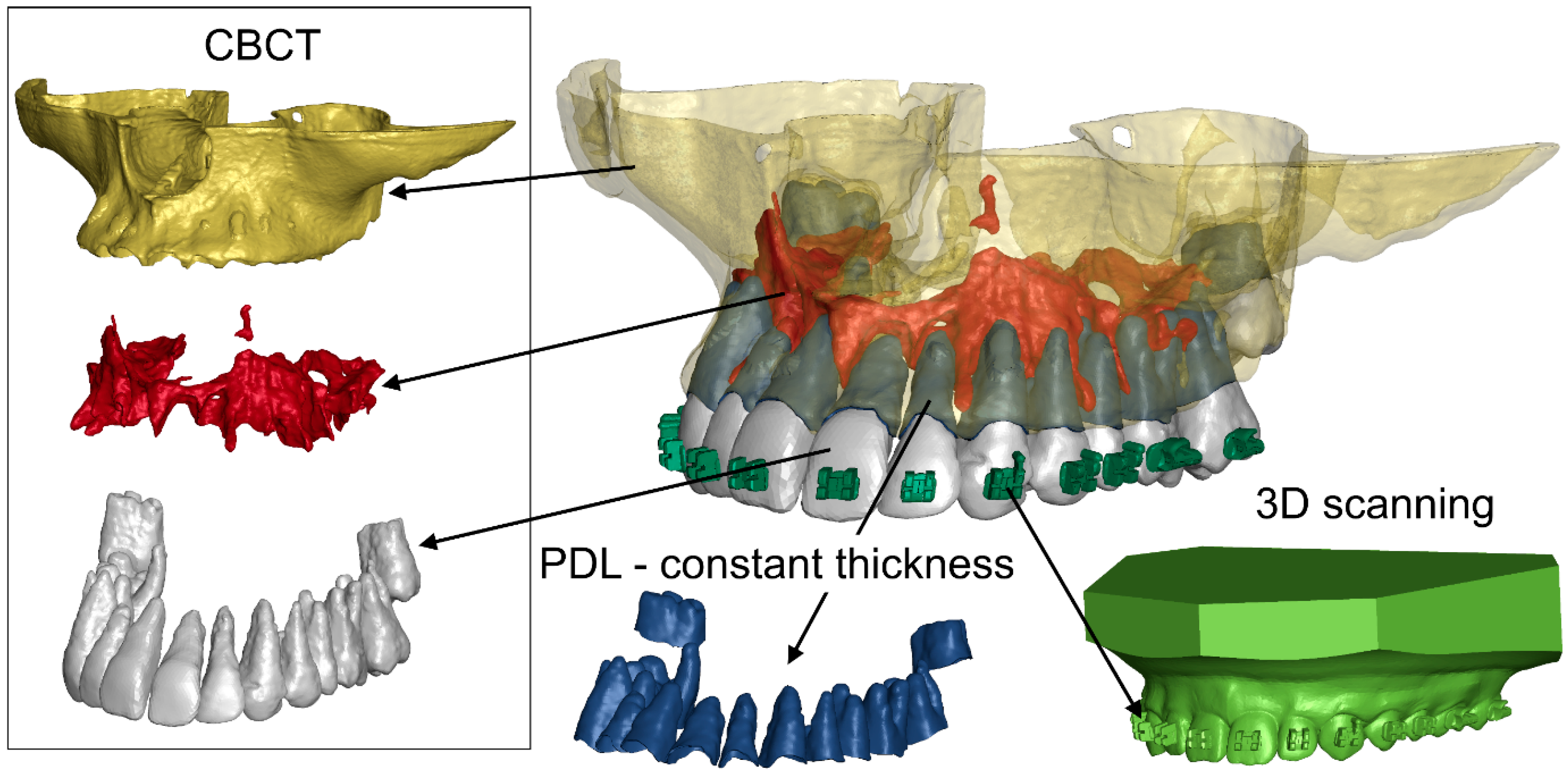
Figure 1. Geometries of structures reproduced in the FEM model.
Materials 18 05290 g001
Figure 2. Finite element mesh used in the model.
Materials 18 05290 g002
Figure 3. Cross-section showing the representation of internal structures in the model.
Materials 18 05290 g003
Figure 4. Corticotomy incision pattern (G3 = 3 mm).
Materials 18 05290 g004
Figure 5. Initial and boundary conditions in the FEM model.
Materials 18 05290 g005
Figure 6. Maximal hydrostatic pressure in PDL without corticotomy G0 ((a) E = 12,500, (b) E = 27,500).
Materials 18 05290 g006
Figure 7. Chart of displacement of the crown—blue and the root—orange without corticotomy G0.
Materials 18 05290 g007
Figure 8. Displacement without corticotomy G0 ((a) E = 12,500, (b) E = 27,500).
Materials 18 05290 g008
Figure 9. Displacement with corticotomy 3 mm G3 ((a) E = 12,500, (b) E = 27,500).
Materials 18 05290 g009
Figure 10. Maximal hydrostatic pressure in PDL corticotomy 3 mm G3 ((a) E = 12,500, (b) E = 27,500).
Materials 18 05290 g010
Figure 11. Chart of displacement of the crown—blue and the root—orange with 3 mm corticotomy G3 E = 12,500.
Materials 18 05290 g011
Table 1. Material data used to describe the material behavior [44].
ComponentYoung’s Modulus [MPa]Poisson’s Ratio [ ]
Steel210,0000.30
Tooth [48] 18,6000.31
Cancellous [49]20000.30
Table 2. Parameters used for describing the PDL [44].
µ1 [MPa]α1 [MPa]Poisson’s Ratio [ ]
2.5 × 10−31500.46
Table 3. The values of displacement and minimum and maximum hydrostatic pressure depending on the bone density and depth of incision: G0—without corticotomy, G1—1 mm corticotomy, G2—2 mm corticotomy, G3—3 mm corticotomy.
Displacement [µm]
Young Modulus [MPa]
12,50015,50018,50021,50024,50027,500
G02.572.372.232.132.041.98
G12.632.422.272.162.082.01
G22.892.642.472.342.242.15
G33.242.952.752.592.482.38
Hydrostatic Pressure Min [kPa]
Young Modulus [MPa]
12,50015,50018,50021,50024,50027,500
G0−5.95−5.84−5.76−5.69−5.63−5.58
G1−5.95−5.85−5.76−5.69−5.63−5.58
G2−5.95−5.84−5.76−5.69−5.63−5.58
G3−5.88−5.79−5.72−5.66−5.61−5.56
Hydrostatic Pressure Max [kPa]
Young Modulus [MPa]
12,50015,50018,50021,50024,50027,500
G07.697.537.47.37.227.15
G17.697.537.417.37.227.15
G27.947.777.647.547.457.38
G37.717.667.627.587.547.5
Disclaimer/Publisher’s Note: The statements, opinions and data contained in all publications are solely those of the individual author(s) and contributor(s) and not of MDPI and/or the editor(s). MDPI and/or the editor(s) disclaim responsibility for any injury to people or property resulting from any ideas, methods, instructions or products referred to in the content.