Article

Cold-Plasma Method in Counteracting Prosthetic Stomatitis: Analysis of the Influence of Cold Plasma on Prosthetic Materials

by
Agnieszka Mazur-Lesz
1,*,
Joanna Pawłat
2,
Piotr Terebun
2,
Dawid Zarzeczny
2,
Elżbieta Grządka
3,*,
Agnieszka Starek-Wójcicka
4,
Michał Kwiatkowski
2,
Irena Malinowska
3,
Magdalena Mnichowska-Polanowska
5 and
Monika Machoy
6
1
Private Dental Office, Witkiewicza 49u/14 Street, 71-124 Szczecin, Poland
2
Department of Electrical Engineering and Smart Technologies, Lublin University of Technology, Nadbystrzycka Street 38A, 20-618 Lublin, Poland
3
Faculty of Chemistry, Institute of Chemical Sciences, Maria Curie-Skłodowska University, M. Skłodowskiej-Curie 3 Sq., 20-031 Lublin, Poland
4
Department of Biological Bases of Food and Feed Technologies, Faculty of Production Engineering, University of Life Sciences in Lublin, 20-612 Lublin, Poland
5
Department of Microbiology, Immunology and Laboratory Medicine, Pomeranian Medical University, Powstańców Wielkopolskich 72 Street, SPSK 2, 70-111 Szczecin, Poland
6
Department of Periodontology, Faculty of Medicine and Dentistry, Pomeranian Medical University, Powstańców Wielkopolskich 72 Street, SPSK 2, 70-111 Szczecin, Poland
*
Authors to whom correspondence should be addressed.
Materials 2025, 18(17), 4162; https://doi.org/10.3390/ma18174162
Submission received: 31 July 2025 / Revised: 29 August 2025 / Accepted: 1 September 2025 / Published: 4 September 2025
(This article belongs to the Special Issue Advances in Plasma Treatment of Materials)

Abstract

The aim of this study was to determine the possibilities of using cold-plasma technology in counteracting the development of denture stomatitis (DS) in patients using different kinds of prosthetic restorations. The study focused mainly on the effect of cold atmospheric plasma on prosthetic materials, such as acryl (AR), acetal (AT), and a prosthetic metal alloy (MA). The materials were tested in terms of the effect of the plasma exposure time (5, 10, and 20 min) on changes in the chemical composition, morphology, and surface topography (FT-IR, SEM-EDS, optical profilometer) as well as changes in the color and contact angle (spectrophotometer, goniometer) after the plasma process. Furthermore, the ability of reference fungi (C. albicans and C. glabrata) to adhere to non-modified and cold atmospheric plasma (CAP)-modified dental materials was examined to evaluate the susceptibility of dental material surfaces to 12 h fungal contamination. The obtained results demonstrate that CAP appears viable for the surface modification of the acetal resin and the metal alloy, not compromising their structural integrity while variably limiting fungal overgrowth involved in the development of DS, whereas its application to the acrylic resin may be inadvisable due to morphological and optical alterations.

Graphical Abstract

1. Introduction

According to the World Health Organization (WHO), severe tooth loss, recognized as a disability, affects over 250 million people worldwide [1]. One of the most common means of oral rehabilitation are removable dentures, which, due to relatively low costs and government funding in many European countries, remain accessible to a broad patient population. Removable dentures play a key role in restoring both esthetics and masticatory function. The most widely used materials for their fabrication are acrylic resin, acetal, and cobalt–chromium–molybdenum (Co-Cr-Mo) alloys.
Acrylic resin (AR), based on methyl methacrylate, is typically available in powder or liquid form and can be polymerized thermally, chemically, or using light [2]. Despite its popularity, AR is prone to rapid absorption, which favors fungal colonization, lowers denture stability, and increases the risk of inflammation, discoloration, and allergic reactions [3,4]. Acetal (AT), or polyoxymethylene, offers superior esthetics, biocompatibility, chemical stability, and comfort compared with acrylics, while showing reduced adhesion of Candida spp. [5,6,7,8]. However, AT is more fracture-prone than metal frameworks. The Co-Cr-Mo alloy, widely used in skeletal dentures and partial denture elements, provides high strength, rigidity, corrosion resistance, and good casting fluidity [9,10,11,12,13].
However, using a dental prosthesis carries the risk of denture stomatitis (DS). DS, primarily associated with Candida spp., affects 15–70% of denture wearers [14,15]. It is a recurrent local infection, often necessitating new prostheses. Conventional therapies remain insufficient, which emphasizes the importance of alternative methods such as cold atmospheric plasma (CAP) treatment. The antimicrobial effect of CAP was first described in the late 1990s [16]. This method generates a mixture of reactive oxygen and nitrogen species (ROS/RNS), UV radiation, and electric fields, which effectively damage microbial cells and biofilm matrices, leading to significant reductions in viable bacteria and fungi on prosthetic and implant materials. Moreover, it does not damage surrounding healthy tissues [17], and its ability to limit antibiotic use makes it a promising tool against microbial resistance [18]. In dentistry, CAP has been investigated for periodontitis and peri-implantitis therapy, caries management, bleaching, endodontics, and biofilm decontamination [19,20,21]. Numerous reports demonstrate that CAP exhibits antifungal efficacy against Candida albicans on denture materials, particularly acrylic resin [22,23,24,25,26,27]. Delben et al. showed that CAP substantially decreased colony-forming units (CFU) of Candida albicans and Staphylococcus aureus while causing no toxic effects on in vitro reconstructed oral epithelium, confirming the biological safety of short CAP exposures [28]. Laboratory and clinical studies on dentures demonstrated that CAP reduces early C. albicans adherence to acrylic surfaces and increases hydrophilicity without major chemical degradation of the material in most protocols, although its efficacy against older, mature biofilms remains limited; for example, it was highly effective against 2-day biofilms but exerted little impact on 7–16-day biofilms [29]. It is also known that CAP increases surface hydrophilicity and reduces early fungal adherence without significantly altering the chemical structure of AR, although some studies suggest possible changes [25,26]. While CAP frequently increases surface roughness, this does not appear to promote fungal adhesion [24]. In endodontics, systematic reviews and meta-analyses highlight the strong potential of CAP in root canal disinfection, particularly against Enterococcus faecalis, although its efficacy depends on the device parameters, carrier gas, exposure time, and application distance; standardized protocols are required before clinical implementation [20]. In the case of implants and titanium surfaces, studies conducted by Kamionka et al. demonstrated that CAP can effectively remove biofilms and may enhance host cell adhesion after decontamination, suggesting its promising role in peri-implantitis and periodontal therapy [30]. A systematic review compiled by Jungbauer et al. [31] summarized robust in vitro evidence of broad antimicrobial activity of CAP against oral pathogens but emphasized methodological heterogeneity and the need for further clinical trials.
Simultaneous studies on the effect of CAP on three different types of prosthetic materials (acryl, acetal, and metal alloy) conducted so far have not analyzed the use of metal alloys or presented contradictory results with respect to acryl. Moreover, the use of different types of cold plasma and different plasma treatment times significantly complicates the comparison of the effect of plasma on different prosthetic materials. It is also hypothesized that cold-plasma treatment improves the hygienic conditions of oral tissues and dental prosthetics by impairing fungal adhesion and biofilm formation, i.e., processes involved in the development of DS.
Based on the above findings, the aim of this study was to evaluate the effect of cold atmospheric plasma (CAP) with helium and oxygen on three prosthetic materials: acrylic resin, acetal resin, and a metal alloy (Co-Cr-Mo) at distinct exposure times. Specifically, we analyzed changes in their chemical composition, surface morphology, surface roughness, contact angle, and color as well as the adhesion of Candida albicans ATCC 10231 and Candida glabrata ATCC 90030 fungi.

2. Materials and Methods

2.1. Materials

The research material comprised acrylic and acetal plates (5 × 5 × 2 mm) and disks (Ø5 × 1 mm) cut from a prosthetic metal alloy. These sample dimensions and shapes were selected to ensure adequate mechanical strength, compatibility with the measuring apparatus, and reproducibility of surface preparation. Four samples were prepared for each test type.

2.1.1. Acrylic Resin

Polymerization, a standard dental laboratory procedure, requires precise control to ensure optimal material properties [32]. In this study, polymethyl methacrylate (PMMA) powder was mixed with methyl methacrylate (MMA) monomer to obtain acrylic dough for denture base fabrication. The process proceeded through the characteristic stages: wet sand, unsticking, thread, dough, and polymerization, followed by the typical reaction phases of initiation, propagation, termination, and chain transfer. After thermal polymerization, the material was processed in a dental laboratory and cut into acrylic plates (5 × 5 × 2 mm). The acrylic resin used was Villacryl H Plus® (Zhermack, Badia Polesine, Italy), with the chemical composition given in Table 1.

2.1.2. Acetal Resin

Acetal resin, supplied as a finished product and virtually free of residual monomers, was thermally conditioned in an injection molding machine prior to prosthetic application. The sample fabrication required strict control of time, pressure, and temperature during all stages of injection and cooling. The process parameters were as follows: melting point 220 °C, melting time 20 min, application time 2–5 min, cooling time 20–40 min, and application pressure 4–6 bar. Samples were then prepared in a dental laboratory following the same procedures as for acrylic. The material used was Dental D® acetal resin (Pressing Dental, Falciano, San Marino), with the chemical composition provided in Table 1.

2.1.3. Metal Alloy

In this study, metal-alloy blocks were processed with CAD/CAM and cut into disks (Ø5 × 2 mm). The alloy used was Biosil F® (DeguDent, Hanau, Germany), with the chemical composition shown in Table 1.

2.2. Methods

2.2.1. Cold Atmospheric Plasma Treatment

The samples were treated with an atmospheric pressure plasma jet generated in a reactor consisting of a 5 mm glass tube with two ring electrodes spaced 10 mm apart (Figure 1). Plasma formed inside the nozzle was transported by forced gas flow to the sample positioned 5 mm from the nozzle tip. The working gas was helium (purity 4.0, 1667 sccm) with a small oxygen admixture (purity 5.0, 13 sccm), where helium facilitated discharge ignition. The reactor was powered by a 28.5 kHz VLF signal at 3.25 kV RMS and 10 W. The treatment times were 5, 10, and 20 min, and the sample surface temperature, measured with a K-type thermocouple, did not exceed 32.6 °C.

2.2.2. FT-IR

The FT-IR analysis was conducted using a FT-IR-4200 type A spectrometer (JASCO Corporation, Tokyo, Japan) with a ATR PRO ONE accessory (JASCO Corporation, Tokyo, Japan) equipped with a diamond crystal (Jasco, Tokyo, Japan). Spectra were recorded in the 4000–500 cm−1 range, at 4 cm−1 resolution, and averaged over 32 scans.

2.2.3. SEM-EDS

Morphological changes after plasma treatment (air plasma, 5, 10, or 20 min) and the elemental composition (EDS) were analyzed using a scanning electron microscope (SEM, Quanta 3D FEG, FEI, Hillsboro, OR, USA) operated at 5 kV. Micrographs were acquired at 500× and 5000× magnifications. Quantitative results represent the mean of four replicates with standard deviations.

2.2.4. Surface Roughness

Surface topography was analyzed using an optical profilometer (Contour GT-K1, Bruker, Ettlingen Germany) based on light interference and fringe imaging. Three-dimensional surface images were acquired to determine roughness parameters (Ra, Rq, Rt, Rv). Measurements were taken at three sites within a 58 × 44 μm2 area per sample, averaged, and compared before and after plasma treatment at identical locations to minimize natural variability. Data analysis was performed with Veeco Vision 4.20 software (Veeco Instruments Inc., Plainview, New York, USA).

2.2.5. Contact Angle

The contact angle was measured using a DSA25E goniometer (KRÜSS, Hamburg, Germany) applying the sessile drop method. For each condition, 3 contact angle measurements were taken (one at the center of the sample and two at a distance of 10 mm), each time placing 0.5 µL of distilled water. In the case of plasma processing, measurements were performed 10 s after each treatment. The contact angle was determined in the KRÜSS Advance software, version 1.3.1.0 (KRÜSS GmbH, Hamburg, Germany) using the ellipse (tangent − 1) fitting method.

2.2.6. Color Measurements

Color measurements were performed with an SV-100 spectrophotometer (Tri-Color, Narama, Poland) (aperture 3 × 1 mm) in the CIE Lab color space. The device was calibrated with a white standard prior to testing. Measurements were carried out under D65 illumination with d/8° geometry, at multiple points on each sample surface. Recorded parameters included L* (lightness), a* (red–green), and b* (blue–yellow).

2.2.7. The 12 h Adhesion of Single-Species Candida albicans and Candida glabrata to Dental Materials

Candida albicans (ATCC 10231) and Candida glabrata (ATCC 90030) were cultured on Sabouraud dextrose agar (SDA;, bioMérieux Polska, Warsaw, Poland) at 30 °C for 24 h. Inocula were prepared in Sabouraud dextrose broth (SDB;bioMérieux Polska, Warsaw, Poland) and adjusted to 0.5 McFarland standard, corresponding to 1 × 10⁶ CFU/mL for C. albicans and 5 × 10⁶ CFU/mL for C. glabrata, confirmed by serial dilutions. Dental material samples (AR, AT, MA) were immersed in standardized suspensions. For each material, unexposed controls and CAP-treated samples (5, 10, 20 min) were tested for Candida early biofilm formation, (measured as 12 h adhesion). The 12 h adhesion of the single-species C. albicans ATCC 101231 and C. glabrata ATCC 90030 to three CAP-treated prosthetic materials, each subjected to three distinct CAP exposure times in salivary pellicle-free conditions, was evaluated. The study assessed C. albicans adhesion at the 12 h time point, because hallmark structural components of early biofilm were present. C. glabrata adhesion was also measured at the 12 h time point to compare it with that of C. albicans. After 12 h incubation at 30 °C, the samples were washed five times in phosphate-buffered saline (PBS) to remove non-adherent cells. Adherent cells were detached using a vortex–sonication–vortex protocol (30 s vortex, 45 s sonication at 30 kHz, room temperature, followed by 30 s vortex) as described previously [33]. The suspensions were serially diluted (1:100, 1:1000, 1:10,000), and 100 µL were plated on SDA for CFU determination. Colonies were manually counted Scan®100, (Interscience, Saint-Nom-la-Breteche, France); eCount™ colony counter pen (Heathrow Scientific, Vernon Hills, IL, USA), and the results were expressed as CFU/mm2. The reduction in C. albicans and C. glabrata growth on the CAP-exposed samples was calculated relative to the controls. Each material was tested in triplicate in four conditions (three CAP exposures and one control).

3. Results and Discussion

3.1. Influence of CAP on Physico-Chemical Properties of Prosthetic Materials

3.1.1. Changes in the Chemical Composition of the CAP-Treated Surfaces of the Prosthetic Materials

Table 1 presents the analysis of the surface composition of the tested prosthetic materials before the plasma procedure and after 5, 10, and 20 min of cold-plasma exposure of the surface. The first of the tested materials, acryl (AR), is a material based on the methyl ester of methacrylic acid, i.e., methyl methacrylate (monomer). According to the obtained SEM-EDS data, the tested acrylic consisted of about 65% carbon and about 35% oxygen. Moreover, its composition did not change as a result of the cold-plasma action regardless of the duration of the procedure, which allowed us to conclude that this material was chemically resistant to the action of CAP. The second of the tested materials, acetal, is chemically a product of formaldehyde polymerization. This material consisted of approximately 47% carbon and 53% oxygen. The plasma treatment did not change the chemical composition of the material regardless of the treatment time. The third type of samples was a prosthetic metal alloy consisting mainly of cobalt (about 50%) and chromium (about 24%) and containing admixtures of other chemical elements, such as oxygen, carbon, molybdenum, silicon, aluminum, and iron. In this case, the plasma treatment resulted in insignificant changes in the chemical composition regardless of the time. Based on the above findings, it can be concluded that cold-plasma treatment is chemically safe for use with prosthetic materials, as it does not adversely affect their composition.
The normalized FT-IR spectra of the AR samples are shown in Figure 2, where the wavelength axis has been clipped to areas depicting characteristic bands for better readability. The obtained spectra are similar to those obtained by other authors studying acrylic materials [34,35,36,37]. The band at 2950 cm−1 can be correlated with the characteristic methylene C–H stretch bands. The ester group can be seen as intense carbonyl C=O stretching vibrations occurring at 1722 cm−1. The asymmetric stretching of CH2 is shown by two bands at 1478 cm−1 and 1437 cm−1, while the CH3 is visible at the 1444 cm−1 band. The band visible at 1386 cm−1 can be attributed to the O–CH3 deformation and the asymmetric peak at 1144 cm−1 can be assigned to the C-O-C group. The other CH2 vibrations can be associated with the bands at 995 cm−1 and 750 cm−1. No changes were observed in the spectra in any of the treatment times tested, which could indicate that no new chemical compounds or functional groups were formed at the penetration depth tested. Comparing this with analyses performed using other methods, which showed a change in the contact angle and color, it can be assumed that changes induced by the plasma treatment occur only in a thin layer near the surface of the tested materials. The results may also have been influenced by the shape of the spectra of the untreated samples, which overlap with the bands of potential new functional groups formed after the treatment in air, such as the C=O bands for wavelengths of 1740–1710 cm−1, or their low intensity.
A comparison of normalized FT-IR spectra of AT is shown in Figure 3. Four bands at wavelengths 2920 cm−1 [38], 1468 cm−1 [39], 1235 cm−1 [38], and 625 cm−1 [38] can be related to the CH2 stretching [38,39]. The band at wavelength 1235 cm−1 can also be related to the C-O-C stretching which is observed for the band at a wavelength of 1088 cm−1 and a broad band around 900 cm−1 [40]. As before, no significant changes were observed after the plasma treatment.

3.1.2. Changes in the Morphology of the CAP-Treated Surfaces of the Prosthetic Materials

Figure 4 shows the effect of the plasma treatment time on the surface morphology of the tested prosthetic materials. In the case of the acrylic materials, the CAP treatment caused significant surface changes, which were more visible at the longer CAP treatment time. As can be seen, the surface lost its homogeneity. There are visible inhomogeneities of the round shape on the surface, which are deeper and more distinct with the extended plasma exposure. These findings suggest that the plasma treatment altered the acrylic surface, underscoring the importance of optimizing the exposure time to ensure microbiological efficacy without significantly impairing the structural integrity or durability of the material. In the case of the tested acetal and prosthetic alloy, no morphological changes were observed after the plasma treatment. This proves that cold plasma can be successfully used to process these materials without the risk of deterioration of their morphological properties and therefore their strength and service life.
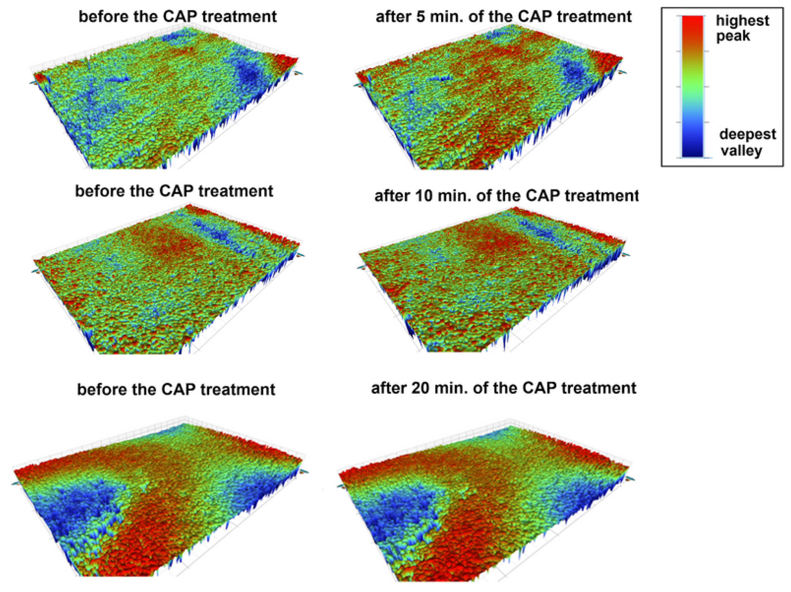
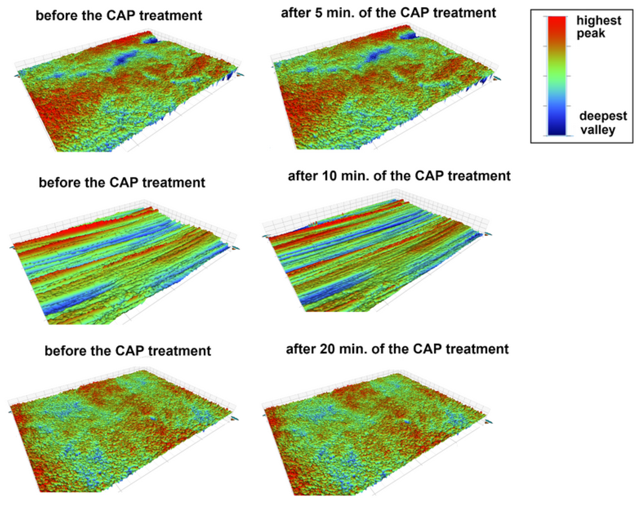
Table 2 presents the surface roughness parameters of the studied materials before and after the CAP treatment. The main roughness parameters: average roughness (Ra), root-mean-square roughness (Rq), peak-to-valley difference (Rt) and the value of the largest profile depression (Rv) were measured. As can be seen, the surface properties of acrylic changed during the plasma treatment. The Ra and Rq parameters did not change, but the surface after the plasma treatment exhibited a greater difference between peaks and valleys (higher Rt values) and deeper valleys (more negative Rv values). This observation was confirmed by the results presented in Figure 5. A comparison of the surface profiles at the same points before and after the plasma treatment clearly indicated the simultaneous appearance of higher and lower areas on the surface (intensification of red and blue colors), which was consistent with the data obtained in the SEM micrographs. In the case of the other two materials, i.e., acetal and prosthetic metal alloy, no differences in their surface roughness were observed. The differences in the roughness parameters were insignificant. These observations were also in accordance with the obtained 3D profiles of the material surfaces before and after the plasma treatment (Figure 5, Figure 6 and Figure 7) and the SEM measurement results.

3.1.3. Changes in the Wetting Properties of the CAP-Treated Surfaces of the Prosthetic Materials

Images of droplets on the surface of the materials before and after the plasma treatment are shown in Figure 8. As in the previous studies using the tested reactor and helium as the main working gas [41,42], the treatment caused a reduction in the water contact angle. For all the processing times and materials, the contact angle decreased by at least 40%, with the effect being most visible for acrylic (50%, 52%, and 58% for 5, 10, and 20 min of treatment, respectively). The results between the individual plasma treatment times were usually within the error limit determined as a standard deviation, which could be related to both the relatively long treatment times and the differences on the surface of the control samples themselves (Figure 9). The lack of further significant variation in the contact angle after 5 min of treatment suggests that the surface reached a modification threshold, implying that this exposure time may represent the optimal duration for altering this parameter [43].
Reductions in the contact angle after treatment of similar materials have also been observed by other research groups. Vukušić et al. studied the effect of low-pressure oxygen plasma on acryl-coated polypropylene foils for food packaging, indicating that reducing the angle below 40 degrees can be difficult and causes etching of the material, worsening its functionality, in contrast to mild treatment, where the change in the contact angle occurred as a result of surface functionalization [44]. Guschl et al. studied the effect of radiofrequency plasma treatment of an acetal copolymer using a mixture of helium with a small amount of oxygen, after which an increase in surface energy (and thus a decrease in the contact angle) was observed. It was associated with etching or chain scission of the polymer [45]. The results obtained by Terpiłowski et al. showed that the reduction in the contact angle of metal alloys depended on their composition, but a reduction in all of them was observed after using a low-pressure reactor with argon or air as the working gas [46]. The observed effect was linked to modifications in surface topography and polarity, specifically due to the incorporation of polar oxygen-containing functional groups onto the material surface.

3.1.4. Changes in the Color of the CAP-Treated Surfaces of the Prosthetic Materials

Spectrophotometric measurements on the CIE Lab scale are a precise method for assessing the color of dental materials. This method ensures color consistency, which is important for the aesthetics of prosthetic restorations and dental reconstructions. The AR-C control sample was characterized by an initial color lightness level L* (before the plasma treatment) of 36.86. The 5 min cold-plasma treatment increased the average value to 38.19, which indicates a brightening of the material surface. The CAP treatment of the acrylic plate for 10 min led to a further brightening of the surface, suggesting an intensification of the process and clear changes in its optical properties. On the other hand, prolonging the plasma exposure to 20 min led to a partial darkening of the sample relative to its condition at 10 min, potentially reflecting either the stabilization of plasma-induced surface effects or continued structural alterations at the surface level (Table 3).
The initial value of the L* parameter of 59.66 for the AT-C control sample gradually decreased with the extension of the CAP treatment time. The systematic decrease in the L* parameter suggested that the plasma affected the acetal surface structure, leading to its darkening. Possible mechanisms of these changes include oxidation and/or changes in reflective properties. The initial value of 53.09, corresponding to the MA-C control sample, increased slightly to 53.69 after the 5 min plasma treatment. The CAP treatment of the sample for 10 min caused the value of the described parameter to decrease to 52.47, which could indicate increased light absorption by the surface or the formation of new layers with reduced reflectivity. In turn, the increase in the MA-20 brightness to 65.17 could indicate significant surface transformations of the sample. A comparable enhancement of brightness upon plasma surface modification has been observed in ITO layers, where plasma treatment led to markedly improved luminance performance in OLED devices [47]. This could be the effect of oxidation and changes in reflective properties [48]. Such a significant change suggested that the longer exposure to plasma induced more intensive chemical and physical processes, such as removal of lower reflective surface layers, exposing more optically active regions of the material, reorganization of the surface structure leading to increased light reflectivity, and intensification of oxidation processes resulting in the formation of layers with high reflectivity. The data presented in Table 3 also show the effect of the plasma process on the value of the a* parameter of the three tested materials. AR showed dynamic changes in this value, which suggested surface modifications under the influence of CAP. After 5 min, a small increase in the parameter value was observed (from 8.25 to 8.38); however, after 10 min, there was a clear decrease in the chromaticity index. After 20 min, the value increased again to 8.48, which could indicate the reconstruction of the surface layer or stabilization of new optical properties of the material. The acetal plate was characterized by a relatively stable course of changes in the a* value during the first 10 min of plasma exposure (from 0.97 to 0.86). A significant decrease to 0.64 was observed only after 20 min, which could suggest slight degradation of the surface or changes in the chemical structure of the material. In turn, the metal-alloy plate showed a clearly different characteristic of changes in the a* value. During the first 10 min, the parameter value increased (from 0.66 to 0.95), which could be the effect of oxidation processes or adsorption of plasma molecules on the material surface. The decrease in the parameter to 0.18 in the MA-20 sample may indicate potential removal of the formed surface layers as a result of further plasma interaction.
The b* color parameter tests showed significant differences in the chromatic characteristics, which can have a significant impact on the visual properties of the analyzed materials (Table 3). The b* parameter value reflects a shift in the color towards yellow (for positive values) or blue (for negative values), which allows determining the dominant color component. In the samples from the AR group, small deviations of the parameter values were observed, except for the sample exposed to plasma for 10 min, where a clear shift towards blue color occurred (−2.16). The other samples from this group demonstrated relative color neutrality. Materials made of acetal were characterized by positive values of the parameter (dominance of a yellowish shade). The greatest shift towards yellow was observed in the AT-5 sample (6.04), while AT-20 was characterized by a lower intensity of this shade (4.59), which may indicate differences in the surface structure or the material composition. All the samples of the prosthetic metal alloy had negative b* values, corresponding to the dominance of cool, bluish tones. A particularly pronounced shift towards blue occurred in the material treated with a plasma jet for 20 min (−6.83). The effect of CAP on the color could be an effect of chemical reactions occurring on the surface, such as oxidation or changes in the structure. Plasma electrolytic oxidation (PEO) is an advanced technique for modifying metal surfaces, in which the application of high voltage in the electrolyte leads to the initiation of electric discharges generating plasma. This process induces metal oxidation, resulting in the formation of an oxide coating with diverse physicochemical properties. Process parameters, such as electrolytic composition, treatment time, and intensity, determine the structure, thickness, and optical properties of the formed layer, which can significantly affect the chromatic characteristics of the surface [49,50]. Plasma treatment with different plasma gases (argon, oxygen, and air), significantly modified the hydrophobic properties of such materials as polystyrene [51] or polycarbonate and polymethyl methacrylate [52], potentially affecting their optical characteristics. This process led to the introduction of oxygen-containing functional groups, which resulted in increased hydrophilicity of the material, as evidenced by reduced contact angles, also observed in our work. The color change in plastics could also be the effect of modifying their chemical structure and absorption properties [53,54]. As indicated by Bhatt et al. [55] and Wang et al. [56], plasma treatment improved adhesion by modifying the surface of materials, increasing their surface energy and wettability and introducing new functional groups.

3.2. Influence of CAP on the 12 h Fungal Adhesion to the Prosthetic Materials

In order to investigate the effect of the plasma treatment on the adhesion of denture stomatitis-causing fungi to prosthetic materials, a set of microbiological studies involving both C. albicans (pseudo/hypha producer, azole-susceptible species) and C. glabrata (non-pseudo/hypha producer, azole-resistant species) was carried out (Figure 10, Figure 11 and Figure 12).

3.2.1. Effect of CAP on 12 h Adhesion of Reference C. albicans to the Prosthetic Materials

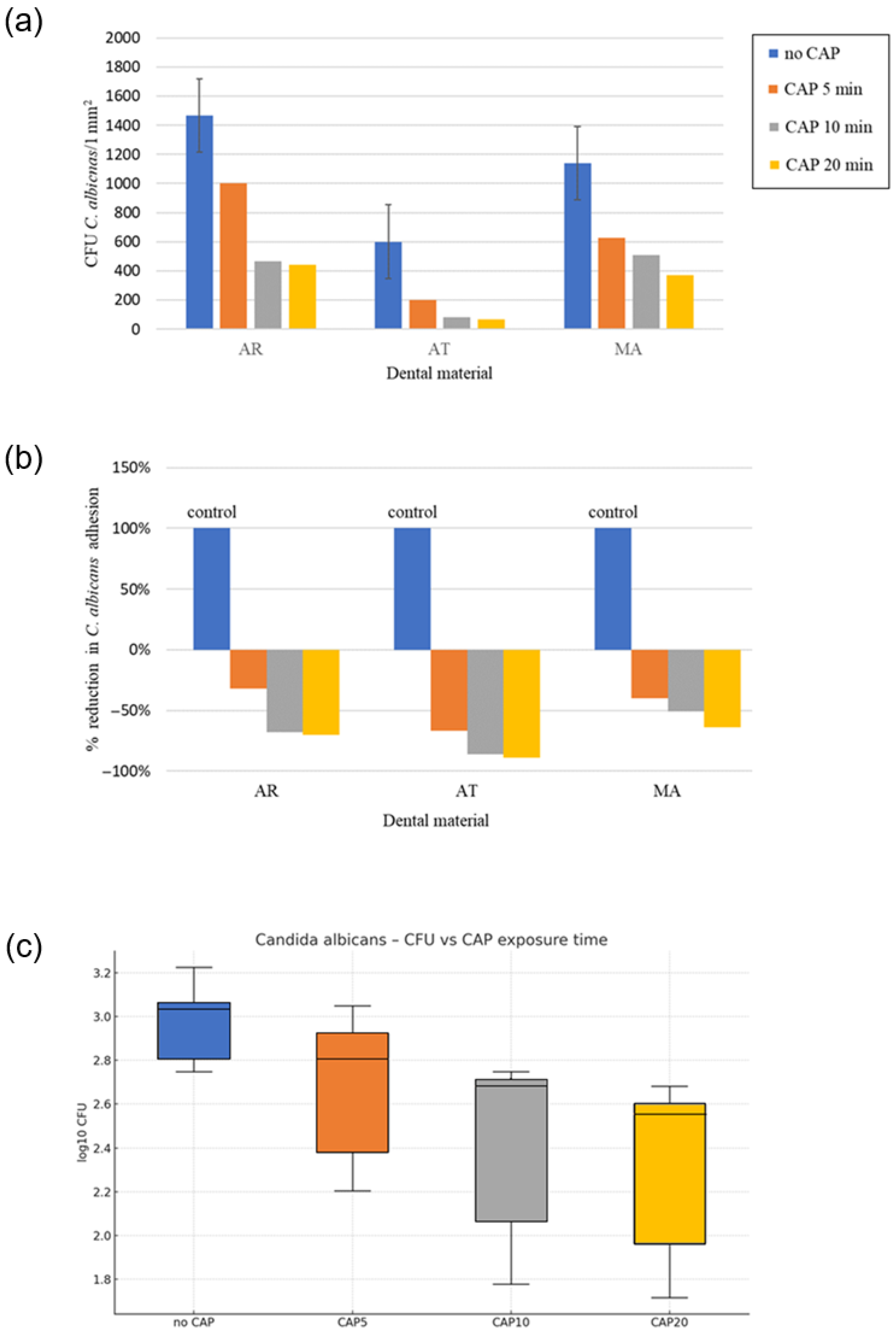
A significant decrease in C. albicans adhesion (expressed as CFU/1 mm2) was observed following the CAP treatment, with the strongest reduction recorded on AT ≈ 89% at 20 min (Table 4; Figure 10b). CAP decreased the 12 h adhesion of C. albicans on every denture resin (Figure 10c), a result which supports the current large body of literature describing ≥ 1 log antimicrobial effects within clinically relevant exposure times [57,58].
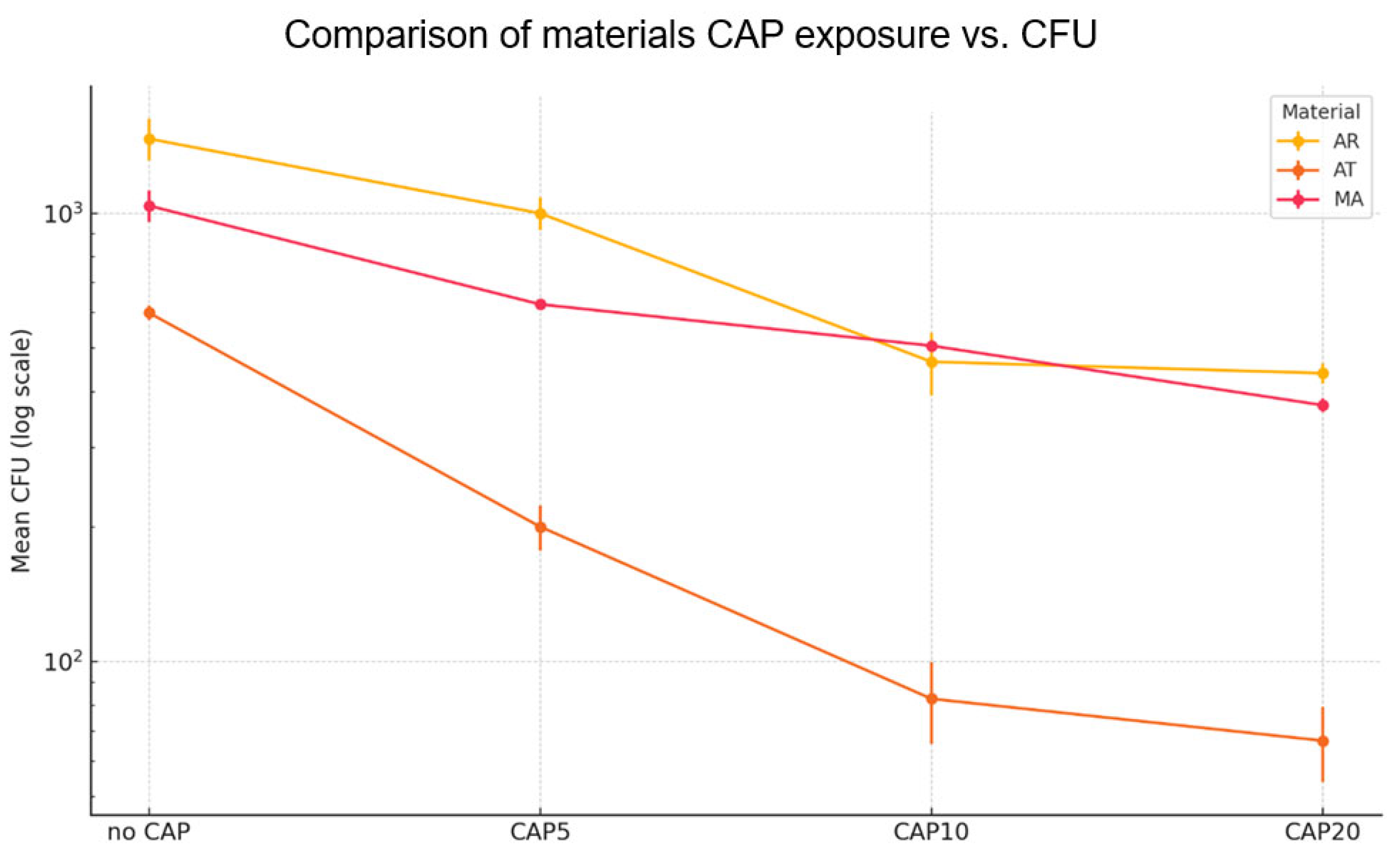
The acetal resin (AT) was characterized by smoother baseline topography, and the greater polar component of surface free energy seems to enhance the hydrophilization generated by CAP, thus generating conditions unfavorable for the initial Candida cell attachment, which accords with earlier findings that AT is more sensitive to short-term plasma functionalization than highly cross-linked polymethyl methacrylate (AR) [58]. In contrast, the metal alloy (MA) exhibited the lowest absolute decrease in the adhesion of fungi to the denture base materials (Figure 10, Figure 11 and Figure 12), However, even on MA, the log transformed counts decreased by more than 0.5 following the 10 min protocol (Figure 11).
A two-way ANOVA of log10–transformed colony-forming units (CFU mm−2) showed significant main effects of the material (η2 = 0.53, ω2 = 0.52), CAP exposure time (−η2 = 0.38, ω2 = 0.38), and material × time interaction (η2 = 0.06, ω2 = 0.05). While the material × time interaction was statistically significant in the two-way ANOVA, its effect size was minimal (ω2 ≈ 0.05). Practically, this implies that the downward trend in CFU was monotonic for all the resins, and one 10 min setting is likely to be sufficient across the range of materials routinely used for removable prostheses. The extension of the CAP exposure from 10 to 20 min did not lead to any further significant reduction in the 12 h adhesion of C. albicans on any of the tested materials (Figure 11). Tukey HSD post-hoc comparisons confirmed that every CAP treatment (5, 10, 20 min) reduced adhesion versus the unexposed control (p < 0.01) and that the 10 min and 20 min variants did not differ from each other (p > 0.05). Prolonging the CAP exposure to 20 min achieved only incremental additional killing (<3% compared to 10 min) and did not enhance any of the physicochemical parameters assayed in parallel (roughness, wettability, color stability). The lack of a clinically significant advantage beyond 10 min is consistent with the kinetic plateau described by Trebulova [59] and is promising from a chairside perspective, since extended treatment times would detract from workflow and patient comfort without proportional increases in efficacy. The degree of response was a function of both the type of material and the plasma treatment time, but a single 10 min cycle in all the instances was sufficient to produce a biologically significant reduction in colony forming units of C. albicans.

3.2.2. Effect of CAP on 12 h Adhesion of Reference C. glabrata to the Prosthetic Materials

Similarly, the acetal resin (AT) CAP treatment resulted in the steepest decline in the 12 h adhesion of C. glabrata (≈98% at 20 min) (Figure 12, Table 5). The ≥10-min CAP treatment produced ≥75% reduction across all the resins. C. glabrata exhibited lower baseline adhesion compared to C. albicans on all the tested dental materials prior to the CAP exposure (Figure 12a,b). The lower baseline adhesion and higher percentage reduction in adhesion observed for C. glabrata is likely related to its morphological characteristics, as it produces only round blastospores in contrast to C. albicans, which is capable of forming both blastospores and filamentous hyphae. The supporting experiment with C. glabrata, which is inherently less susceptible to azole treatment, strengthened these findings. Unfortunately, only a single measurement was available for each material × time combination. We pooled the three materials and performed a one-way ANOVA on log10 CFU by the CAP exposure time (per level). The effect of the CAP exposure time was significant (strong impact: η2 = 0.66, ω2 = 0.51) (Figure 12c). The one-way ANOVA on log10 CFU revealed a robust time-dependent decrease (η2 = 0.66), once more plateauing after 10 min. Although the percentage reduction continued to rise numerically up to 20 min, the additional gain was ≤ 18 percentage points and therefore clinically marginal (Figure 10a–c).
The more pronounced slope on the studied materials might be attributed not merely to the surface chemistry but also to the thinner β glucan layer of C. glabrata, which may have allowed deeper penetration of reactive oxygen and nitrogen species formed in the plasma [59,60]. Notably, the rank order of susceptibility among the materials was identical to that of C. albicans, implying that a common protocol can be viable even for mixed biofilms.
Based on the presented results, it is clearly visible that the plasma-based technology reduced early biofilm growth of reference Candida species on all the tested material surfaces. The 10 min CAP exposure was sufficient to significantly reduce the 12 h adhesion of the single species C. albicans and C. glabrata. Cumulatively, these results endorse the implementation of a 10 min helium jet CAP cycle as a practical adjunct to denture hygiene. This quick and non-thermal treatment seems to preserve the optical and mechanical integrity of the polymer whilst conferring a significant antifungal advantage. This 65% threshold was used as a pragmatic cut-off for clinically relevant early biofilm control, echoing reports that a two-third reduction in denture biofilm is associated with symptomatic relief [57,61].

4. Conclusions

Cold atmospheric plasma (CAP) is a rapid non-thermal adjunct to denture base decontamination. One 10 min helium-jet cycle reduced Candida loads on all the resins by ≥ 1 log, with large effect sizes for both the material factor (η2 ≈ 0.53) and the exposure time (η2 ≈ 0.38). Prolonging the treatment to 20 min yielded only marginal further killing (<3%), demonstrating a distinct efficacy plateau.
In both Candida species, the AT resin was most responsive, reaching up to 98 % adhesion reduction for C. albicans and 97–98% for C. glabrata, while the traditional AR resin exhibited the lowest but still clinically significant reduction. The same hierarchy of material susceptibility and the strong time-dependent response found for C. glabrata2 = 0.66) suggest that the same 10 min protocol can be used confidently on mixed Candida biofilms. One 10 min CAP cycle decreased C. albicans and C. glabrata loads on the conventional denture base resins by at least one logarithmic unit. This study demonstrates that cold atmospheric plasma (CAP) treatment holds promise as a method for maintaining the hygiene of prosthetic materials. CAP was found to be chemically safe for all the acrylic, acetal, and metal-alloy materials, without significantly altering their elemental composition. However, distinct material-specific responses were observed under the CAP treatment. The acetal resin and the metal alloy exhibited stability in surface morphology, roughness, and color following the CAP exposure together with the ability to reduce the water contact angle and potential fungal adhesion. This supports the use of CAP for cleaning and decontaminating these materials. In turn, the acrylic resin showed substantial topographical and optical changes, including increased surface roughness and altered brightness. These modifications may compromise its structural integrity and aesthetic qualities, making CAP unsuitable for acrylic-based prostheses in clinical use. Overall, CAP treatment emerges as a promising adjunctive technology for enhancing prosthetic hygiene and reducing Candida albicans and Candida glabrata colonization, provided material-specific limitations are considered. Plasma treatment can be used to clean and activate surfaces of certain materials by removing adsorbed contaminants and introducing polar functional groups (e.g., hydroxyl, carbonyl, or carboxyl groups). As the population ages, denture stomatitis prevalence will increase; therefore, there is an increasing demand for new preventive strategies, and the current results offer an encouraging proof of principle.
Limitations. The in vitro study used a static 12 h fungal adhesion model, capturing early biofilm growth, using single-species reference strains and investigated only one CAP parameter setting (helium jet, 25 kHz, 7 W). Since only early biofilm growth was assessed and neither mature nor mixed-species biofilms were tested, the anti-adhesion/anti-biofilm effect of CAP may be overestimated relative to more complex, clinically relevant conditions. Another partial limitation of the study is that only 2D roughness parameters were analyzed. According to ISO 25178 [62], 3D areal parameters and sampling from clinically relevant fields would be necessary for a more representative characterization of prosthetic surfaces.
Future work. Subsequent research should address mixed, dual, and multispecies dynamic biofilms in salivary-pellicle conditions and optimize plasma parameters (gas composition, power, and exposure time) to maximize antifungal efficacy in prosthetics in conditions resembling those in the oral cavity. Incorporation of CAP application into clinical workflow to prevent denture stomatitis caused by yeast overgrowth remains a challenge.

Author Contributions

Conceptualization, A.M.-L., M.M., J.P. and E.G.; methodology, M.M., J.P., A.S.-W., E.G. and M.M.-P.; validation, M.K. and M.M.-P.; formal analysis, I.M.; investigation, A.M.-L., E.G., P.T., A.S.-W., D.Z. and M.M.-P.; resources, A.M.-L., M.M. and J.P.; data curation, A.M.-L.; writing—original draft preparation, A.M.-L., E.G., A.S.-W., M.M.-P. and P.T.; writing—review and editing, M.M., M.M.-P., J.P., E.G. and I.M.; visualization, A.M.-L., E.G., A.S.-W., P.T., M.M. and M.M.-P.; supervision, M.M., J.P. and E.G.; project administration, J.P.; funding acquisition, J.P. All authors have read and agreed to the published version of the manuscript.

Funding

This research received no external funding.

Data Availability Statement

The original contributions presented in this study are included in the article. Further inquiries can be directed to the corresponding author(s).

Acknowledgments

We are grateful for the fruitful discussions in the following cooperative initiatives: CA20114 (Therapeutical applications of Cold Plasmas); CA23104 (Mainstreaming water reuse into the circular economy paradigm) and CEEPUS CIII-AT-0063. The authors would like to acknowledge that the Graphical Abstract was created using BioRender.com.

Conflicts of Interest

Author Agnieszka Mazur-Lesz is employed by the company Private Dental Office. The remaining authors declare that the research was conducted in the absence of any commercial or financial relationships that could be construed as a potential conflict of interest.

References

  1. Li, P.; Krämer-Fernandez, P.; Klink, A.; Xu, Y.; Spintzyk, S. Repairability of a 3D Printed Denture Base Polymer: Effects of Surface Treatment and Artificial Aging on the Shear Bond Strength. J. Mech. Behav. Biomed. Mater. 2021, 114, 104227. [Google Scholar] [CrossRef]
  2. Nejatian, T.; Pezeshki, S.; Yaqin Syed, A.U. 5—Acrylic Denture Base Materials. In Advanced Dental Biomaterials; Khurshid, Z., Najeeb, S., Zafar, M.S., Sefat, F., Eds.; Woodhead Publishing: Cambridge, UK, 2019; pp. 79–104. ISBN 978-0-08-102476-8. [Google Scholar]
  3. Reynolds, I.R. A Review of Direct Orthodontic Bonding. Br. J. Orthod. 1975, 2, 171–178. [Google Scholar] [CrossRef]
  4. Tardelli, J.D.C.; dos Reis, A.C. Biomechanical, Esthetic, and Hygienic Considerations of Materials for Overdenture Bars: A Systematic Review. Dent. Rev. 2024, 4, 100082. [Google Scholar] [CrossRef]
  5. Thomas, S.; Nandini, V. Acetal Resin—A Quantum Leap in Aesthetic Restorative Dentistry: A Review. Int. J. Clin. Dent. Sci. 2011, 2, 56–59. [Google Scholar]
  6. Campo, E.A. 1—Polymeric Materials and Properties. In Selection of Polymeric Materials; Campo, E.A., Ed.; Plastics Design Library; William Andrew Publishing: Norwich, UK, 2008; pp. 1–39. ISBN 978-0-8155-1551-7. [Google Scholar]
  7. Bogucki, Z.A.; Kownacka, M. Elastic Dental Prostheses—Alternative Solutions for Patients Using Acrylic Prostheses: Literature Review. Adv. Clin. Exp. Med. 2018, 27, 1441–1445. [Google Scholar] [CrossRef] [PubMed]
  8. Sobolewska, E.; Fraczak, B.; Czarnomysy-Furowicz, D.; Ey-Chmielewska, H.; Karakulska, J. Bacteria Adhesion to the Surface of Various Prosthetics Materials. Ann. Acad. Med. Stetin. 2007, 53, 68–71. [Google Scholar]
  9. Timbó, I.C.G.; Oliveira, M.S.C.S.; Regis, R.R. Effect of Sanitizing Solutions on Cobalt Chromium Alloys for Dental Prostheses: A Systematic Review of in Vitro Studies. J. Prosthet. Dent. 2024, 132, 704–713. [Google Scholar] [CrossRef]
  10. Al Jabbari, Y.S. Physico-Mechanical Properties and Prosthodontic Applications of Co-Cr Dental Alloys: A Review of the Literature. J. Adv. Prosthodont. 2014, 6, 138–145. [Google Scholar] [CrossRef]
  11. Bates, J.F.; Knapton, A.G. Metals and Alloys in Dentistry. Int. Met. Rev. 1977, 22, 39–60. [Google Scholar] [CrossRef]
  12. Wataha, J.C. Alloys for Prosthodontic Restorations. J. Prosthet. Dent. 2002, 87, 351–363. [Google Scholar] [CrossRef]
  13. Vaicelyte, A.; Janssen, C.; Le Borgne, M.; Grosgogeat, B. Cobalt–Chromium Dental Alloys: Metal Exposures, Toxicological Risks, CMR Classification, and EU Regulatory Framework. Crystals 2020, 10, 1151. [Google Scholar] [CrossRef]
  14. Skupien, J.A.; Valentini, F.; Boscato, N.; Pereira-Cenci, T. Prevention and Treatment of Candida Colonization on Denture Liners: A Systematic Review. J. Prosthet. Dent. 2013, 110, 356–362. [Google Scholar] [CrossRef] [PubMed]
  15. Qian, K.; Pan, H.; Li, Y.; Wang, G.; Zhang, J.; Pan, J. Time-Related Surface Modification of Denture Base Acrylic Resin Treated by Atmospheric Pressure Cold Plasma. Dent. Mater. J. 2016, 35, 97–103. [Google Scholar] [CrossRef] [PubMed]
  16. Laroussi, M. Plasma Medicine: A Brief Introduction. Plasma 2018, 1, 47–60. [Google Scholar] [CrossRef]
  17. Braný, D.; Dvorská, D.; Halašová, E.; Škovierová, H. Cold Atmospheric Plasma: A Powerful Tool for Modern Medicine. Int. J. Mol. Sci. 2020, 21, 2932. [Google Scholar] [CrossRef]
  18. Izadjoo, M.; Zack, S.; Kim, H.; Skiba, J. Medical Applications of Cold Atmospheric Plasma: State of the Science. J. Wound Care 2018, 27, S4–S10. [Google Scholar] [CrossRef]
  19. Stornelli, G.; Petrucci, G.; Caponio, V.C.A.; Sardella, E.; Balice, G.; Femminella, B.; Paolantonio, M.; Cela, I.; Acharya, T.R.; Jha, N.; et al. Translational application of cold atmospheric plasma in periodontology and implantology: Where are we? A systematic review of in vivo studies in human and animal models. J. Evid.-Based Dent. Pract. 2025, 25, 102096. [Google Scholar] [CrossRef]
  20. Sanesi, L.; Puca, V.; Caponio, V.C.A.; Pinti, M.; Balice, G.; Femminella, B.; Paolantonio, M.; Cela, I.; Kaushik, N.K.; Choi, E.H.; et al. Disinfection of Dental Root Canals by Cold Atmospheric Plasma: A Systematic Review and Meta-Analysis of Dental Biofilm. Front. Oral. Health 2024, 5, 1483078. [Google Scholar] [CrossRef]
  21. Davis, C.C.; Dias Panariello, F.; Panariello, B. Exploring the Efficacy of Low-Temperature Plasmas on Oral Biofilms: A Scoping Review. Med. Sci. 2025, 13, 79. [Google Scholar] [CrossRef]
  22. Kim, J.-H.; Lee, M.-A.; Han, G.-J.; Cho, B.-H. Plasma in Dentistry: A Review of Basic Concepts and Applications in Dentistry. Acta Odontol. Scand. 2014, 72, 1–12. [Google Scholar] [CrossRef]
  23. Kolb, J.F.; Mohamed, A.-A.H.; Price, R.O.; Swanson, R.J.; Bowman, A.; Chiavarini, R.L.; Stacey, M.; Schoenbach, K.H. Cold Atmospheric Pressure Air Plasma Jet for Medical Applications. Appl. Phys. Lett. 2008, 92, 241501. [Google Scholar] [CrossRef]
  24. Kang, S.-H.; Lee, H.-J.; Hong, S.-H.; Kim, K.-H.; Kwon, T.-Y. Influence of Surface Characteristics on the Adhesion of Candida Albicans to Various Denture Lining Materials. Acta Odontol. Scand. 2013, 71, 241–248. [Google Scholar] [CrossRef] [PubMed]
  25. Pan, H.; Wang, G.; Pan, J.; Ye, G.; Sun, K.; Zhang, J.; Wang, J. Cold Plasma-Induced Surface Modification of Heat-Polymerized Acrylic Resin and Prevention of Early Adherence of Candida Albicans. Dent. Mater. J. 2015, 34, 529–536. [Google Scholar] [CrossRef] [PubMed]
  26. Qanber, L.M.; Hamad, T.I. Effect of Plasma Treatment on the Bond of Soft Denture Liner to Conventional and High Impact Acrylic Denture Materials. J. Baghdad Coll. Dent. 2021, 33, 9–17. [Google Scholar] [CrossRef]
  27. Wang, G.M.; Sun, P.P.; Pan, H.; Ye, G.P.; Sun, K.; Zhang, J.; Pan, J.; Fang, J. Inactivation of Candida Albicans Biofilms on Polymethyl Methacrylate and Enhancement of the Drug Susceptibility by Cold Ar/O2 Plasma Jet. Plasma Chem. Plasma Process 2016, 36, 383–396. [Google Scholar] [CrossRef]
  28. Delben, J.A.; Zago, C.E.; Tyhovych, N.; Duarte, S.; Vergani, C.E. Effect of Atmospheric-Pressure Cold Plasma on Pathogenic Oral Biofilms and In Vitro Reconstituted Oral Epithelium. PLoS ONE 2016, 11, e0155427. [Google Scholar] [CrossRef]
  29. Matthes, R.; Jablonowski, L.; Koban, I.; Quade, A.; Hübner, N.-O.; Schlueter, R.; Weltmann, K.-D.; von Woedtke, T.; Kramer, A.; Kocher, T. In Vitro Treatment of Candida Albicans Biofilms on Denture Base Material with Volume Dielectric Barrier Discharge Plasma (VDBD) Compared with Common Chemical Antiseptics. Clin. Oral. Investig. 2015, 19, 2319–2326. [Google Scholar] [CrossRef]
  30. Kamionka, J.; Matthes, R.; Holtfreter, B.; Pink, C.; Schlüter, R.; von Woedtke, T.; Kocher, T.; Jablonowski, L. Efficiency of Cold Atmospheric Plasma, Cleaning Powders and Their Combination for Biofilm Removal on Two Different Titanium Implant Surfaces. Clin. Oral. Investig. 2022, 26, 3179–3187. [Google Scholar] [CrossRef]
  31. Jungbauer, G.; Moser, D.; Müller, S.; Pfister, W.; Sculean, A.; Eick, S. The Antimicrobial Effect of Cold Atmospheric Plasma against Dental Pathogens—A Systematic Review of In-Vitro Studies. Antibiotics 2021, 10, 211. [Google Scholar] [CrossRef]
  32. Darvell, B.W. Chapter 5—Acrylic. In Materials Science for Dentistry, 10th ed.; Darvell, B.W., Ed.; Woodhead Publishing Series in Biomaterials; Woodhead Publishing: Cambridge, UK, 2018; pp. 121–142. ISBN 978-0-08-101035-8. [Google Scholar]
  33. Silva, N.D.S.; De Melo, B.S.T.; Oliva, A.; de Araújo, P.S.R. Sonication Protocols and Their Contributions to the Microbiological Diagnosis of Implant-Associated Infections: A Review of the Current Scenario. Front. Cell. Infect. Microbiol. 2024, 14, 1398461. [Google Scholar] [CrossRef]
  34. Al-Ali, A.; Kassab-Bashi, T. Fourier Transform Infra Red (FTIR) Spectroscopy of New Copolymers of Acrylic Resin Denture Base Materials. IJERSTE 2015, 4, 172–180. [Google Scholar]
  35. Torrisi, L.; Roszkowska, A.M.; Silipigni, L.; Cutroneo, M.; Torrisi, A. Effects of 365 Nm UV Lamp Irradiation of Polymethylmethacrylate (PMMA). Radiat. Eff. Defects Solids 2024, 179, 264–274. [Google Scholar] [CrossRef]
  36. Bakr, A.M.; Darwish, A.; Azab, A.A.; El Awady, M.E.; Hamed, A.A.; Elzwawy, A. Structural, Dielectric, and Antimicrobial Evaluation of PMMA/CeO2 for Optoelectronic Devices. Sci. Rep. 2024, 14, 2548. [Google Scholar] [CrossRef] [PubMed]
  37. Huszank, R.; Szilágyi, E.; Szoboszlai, Z.; Szikszai, Z. Investigation of Chemical Changes in PMMA Induced by 1.6 MeV He+ Irradiation by Ion Beam Analytical Methods (RBS-ERDA) and Infrared Spectroscopy (ATR-FTIR). Nucl. Instrum. Methods Phys. Res. Sect. B Beam Interact. Mater. At. 2019, 450, 364–368. [Google Scholar] [CrossRef]
  38. Lim, K.; Hayat, M.D.; Jena, K.D.; Zhang, W.; Cao, P. On Amine Treated Polyoxymethylene (POM) Blends with Low Formaldehyde Emission for Metal Injection Moulding (MIM). J. Mater. Sci. 2022, 57, 15160–15170. [Google Scholar] [CrossRef]
  39. Liu, D.; Zhang, S.; Cheng, H.; Peng, R.; Luo, Z. Thermally Triggered Vanishing Bulk Polyoxymethylene for Transient Electronics. Sci. Rep. 2019, 9, 18107. [Google Scholar] [CrossRef]
  40. Pang, Y.-J.; Xu, W.-S.; Yang, B.-T.; Ni, H.-Y.; Chen, J. Influence of Early Thermal-Oxidative Ageing on the Structure and Properties of Polyoxymethylene Copolymer. R. Soc. Open Sci. 2021, 8, 210034. [Google Scholar] [CrossRef]
  41. Kwiatkowski, M.; Terebun, P.; Mazurek, P.; Pawlat, J. Wettability of Polymeric Materials after Dielectric Barrier Discharge Atmospheric-Pressure Plasma Jet Treatment. Sens. Mater. 2018, 30, 1207–1212. [Google Scholar] [CrossRef]
  42. Kwiatkowski, M.; Pawłat, J.; Starek-Wójcicka, A.; Krajewska, M.; Terebun, P.; Zarzeczny, D.; Machoy, M.; Mazur-Lesz, A.; Matsuyama, N.; Murakami, T.; et al. Impact of DBD Plasma Jet Treatment on the Enamel Surface of Primary Teeth. Materials 2024, 17, 5173. [Google Scholar] [CrossRef]
  43. Correia, D.M.; Nunes-Pereira, J.; Alikin, D.; Kholkin, A.L.; Carabineiro, S.A.C.; Rebouta, L.; Rodrigues, M.S.; Vaz, F.; Costa, C.M.; Lanceros-Méndez, S. Surface Wettability Modification of Poly(Vinylidene Fluoride) and Copolymer Films and Membranes by Plasma Treatment. Polymer 2019, 169, 138–147. [Google Scholar] [CrossRef]
  44. Vukušić, T.; Vesel, A.; Holc, M.; Ščetar, M.; Jambrak, A.R.; Mozetič, M. Modification of Physico-Chemical Properties of Acryl-Coated Polypropylene Foils for Food Packaging by Reactive Particles from Oxygen Plasma. Materials 2018, 11, 372. [Google Scholar] [CrossRef]
  45. Guschl, P.C.; Hicks, R.F.; MacDavid, S. Atmospheric Oxygen-Helium Plasma Surface Modification of Medical Plastics. In Proceedings of the 2008 IEEE 35th International Conference on Plasma Science, Karlsruhe, Germany, 15–19 June 2008; p. 1. [Google Scholar]
  46. Terpilowski, K.; Rymuszka, D.; Hołysz, L.; Ilnicki, M. Surface Properties of Metal Alloys Used in Aviation after Plasma Treatment. Surf. Interface Anal. 2017, 49, 647–653. [Google Scholar] [CrossRef]
  47. Wu, C.C.; Wu, C.I.; Sturm, J.C.; Kahn, A. Surface Modification of Indium Tin Oxide by Plasma Treatment: An Effective Method to Improve the Efficiency, Brightness, and Reliability of Organic Light Emitting Devices. Appl. Phys. Lett. 1997, 70, 1348–1350. [Google Scholar] [CrossRef]
  48. Onderwaater, W.G.; Taranovskyy, A.; van Baarle, G.C.; Frenken, J.W.M.; Groot, I.M.N. In Situ Optical Reflectance Difference Observations of CO Oxidation over Pd(100). J. Phys. Chem. C 2017, 121, 11407–11415. [Google Scholar] [CrossRef] [PubMed]
  49. Wu, T.; Blawert, C.; Serdechnova, M.; Karlova, P.; Dovzhenko, G.; Florian Wieland, D.C.; Stojadinovic, S.; Vasilic, R.; Mojsilovic, K.; Zheludkevich, M.L. Formation of Plasma Electrolytic Oxidation Coatings on Pure Niobium in Different Electrolytes. Appl. Surf. Sci. 2022, 573, 151629. [Google Scholar] [CrossRef]
  50. Fernández-López, P.; Alves, S.A.; San-Jose, J.T.; Gutierrez-Berasategui, E.; Bayón, R. Plasma Electrolytic Oxidation (PEO) as a Promising Technology for the Development of High-Performance Coatings on Cast Al-Si Alloys: A Review. Coatings 2024, 14, 217. [Google Scholar] [CrossRef]
  51. Lau, Y.T.; Chin, O.H.; Lee, H.C.; Chiu, W.S.; Woo, H.J. Plasma Surface Treatment of Polystyrene in a Low Power Low Frequency Argon Glow Discharge. Appl. Surf. Sci. 2022, 578, 151963. [Google Scholar] [CrossRef]
  52. Gizer, S.G.; Bhethanabotla, V.R.; Ayyala, R.S.; Sahiner, N. Low-Pressure Plasma Treated Polycarbonate and Polymethyl Methacrylate (PMMA) Sheets with Different Surface Patterns to Change Their Surface Properties. Surf. Interfaces 2023, 37, 102646. [Google Scholar] [CrossRef]
  53. Basyigit, Z.O. Enhancing the Coloration of Polypropylene Surfaces through Low-Frequency Plasma Modification with Disperse Dyes. Int. J. Adhes. Adhes. 2024, 135, 103837. [Google Scholar] [CrossRef]
  54. Duangkanya, K.; Kopwitthaya, A.; Chanhorm, S.; Infahsaeng, Y. Oxygen Plasma Treatment Time Induced Hydrophilicity of Polydimethylsiloxane (PDMS) Thin Films for Liquid Lenses Application. Mater. Today Proc. 2022, 65, 2442–2445. [Google Scholar] [CrossRef]
  55. Bhatt, P.; Kumar, V.; Subramaniyan, V.; Nagarajan, K.; Sekar, M.; Chinni, S.V.; Ramachawolran, G. Plasma Modification Techniques for Natural Polymer-Based Drug Delivery Systems. Pharmaceutics 2023, 15, 2066. [Google Scholar] [CrossRef]
  56. Wang, D.; Li, Y.; Zou, T.; Fu, J.; Liu, Z. Increasing Strength and Fracture Toughness of Carbon Fibre-Reinforced Plastic Adhesively Bonded Joints by Combining Peel-Ply and Oxygen Plasma Treatments. Appl. Surf. Sci. 2023, 612, 155768. [Google Scholar] [CrossRef]
  57. Eslami, H.; Fakhrzadeh, V.; Nazari, E.; Katebi, K.; Alinejad, M. Cold Plasma Treatment for Candida Biofilm on Resin Base Dentures: A Systematic Review. J. Lasers Med. Sci. 2025, 16, e10. [Google Scholar] [PubMed]
  58. Zamperini, C.A.; Machado, A.L.; Vergani, C.E.; Pavarina, A.C.; Giampaolo, E.T.; da Cruz, N.C. Adherence in Vitro of Candida Albicans to Plasma Treated Acrylic Resin. Effect of Plasma Parameters, Surface Roughness and Salivary Pellicle. Arch. Oral. Biol. 2010, 55, 763–770. [Google Scholar] [CrossRef] [PubMed]
  59. Trebulová, K.; Krčma, F.; Skoumalová, P.; Kozáková, Z.; Machala, Z. Effects of Different Cold Atmospheric-Pressure Plasma Sources on the Yeast Candida Glabrata. Plasma Process. Polym. 2023, 20, e2300048. [Google Scholar] [CrossRef]
  60. Chew, S.Y.; Ho, K.L.; Cheah, Y.K.; Sandai, D.; Brown, A.J.P.; Than, L.T.L. Physiologically Relevant Alternative Carbon Sources Modulate Biofilm Formation, Cell Wall Architecture, and the Stress and Antifungal Resistance of Candida Glabrata. Int. J. Mol. Sci. 2019, 20, 3172. [Google Scholar] [CrossRef]
  61. Gad, M.M.; Fouda, S.M. Current Perspectives and the Future of Candida Albicans-Associated Denture Stomatitis Treatment. Dent. Med. Probl. 2020, 57, 95–102. [Google Scholar] [CrossRef]
  62. ISO 25178-2:2021; Geometrical Product Specifications (GPS)—Surface Texture: Areal—Part 2: Terms, Definitions and Surface Texture Parameters. ISO: Geneva, Switzerland, 2021. Available online: https://www.iso.org/obp/ui/en/#iso:std:iso:25178:-2:ed-2:v1:en (accessed on 31 August 2025).
Figure 1. Scheme of the plasma processing system.
Materials 18 04162 g001
Figure 2. Normalized FT-IR spectra of AR samples for different plasma treatment times.
Materials 18 04162 g002
Figure 3. Normalized FT-IR spectra of AT samples for different plasma treatment times.
Materials 18 04162 g003
Figure 4. SEM micrographs showing the effect of the plasma treatment time (5, 10, 20 min) on the prosthetic materials: acryl (AR), acetal (AT), and prosthetic alloy (MA), C-control samples (in red square): (a) AR-C; (b) AR-5; (c) AR-10; (d) AR-20; (e) AT-C; (f) AT-5; (g) AT-10; (h) AT-20; (i) MA-C; (j) MA-5; (k) MA-10; (l) MA-20.
Materials 18 04162 g004
Figure 5. Images of the surface of acryl (AR) before and after the CAP treatment (5, 10, 20 min), C-control/untreated sample (the colors represent the topography of the surface; the higher the peaks—the more red, the deepest the valleys—the more navy blue).
Materials 18 04162 g005
Figure 6. Images of the surface of acetal (AT) before and after the CAP treatment (5, 10, 20 min), C-control/untreated sample (the colors represent the topography of the surface; the higher the peaks—the more red, the deepest the valleys—the more navy blue).
Materials 18 04162 g006
Figure 7. Images of the surface of the metal alloy (MA) before and after the CAP treatment (5, 10, 20 min), C-control/untreated sample (the colors represent the topography of the surface; the higher the peaks—the more red, the deepest the valleys—the more navy blue).
Materials 18 04162 g007
Figure 8. Images of water droplets on the surface of the materials before and after the plasma treatment.
Materials 18 04162 g008
Figure 9. Average values of the contact angle of the material surfaces before and after the plasma treatment.
Materials 18 04162 g009
Figure 10. Effect of plasma treatment on 12 h adhesion of C. albicans ATCC to denture base materials (AR = acrylic resin; AT = acetal resin; MA = metal alloy) following standardized cold atmospheric plasma (CAP) exposure (5, 10, and 20 min). Control (no CAP) refers to the corresponding dental material samples not exposed to CAP: (a) Mean colony-forming units (CFU) of C. albicans per 1 mm2 after the CAP exposure; (b) Mean percentage reduction relative to the unexposed control; (c) Box-and-whisker distribution of log10 CFU for C. albicans versus the CAP exposure time. Control = material not exposed to CAP; bars show (a) mean CFU and (b) mean % reduction; (c) the box-plot depicts log10 CFU for C. albicans as a function of the CAP exposure time.
Materials 18 04162 g010
Figure 11. Material-specific response of C. albicans to the CAP exposure: mean ± sem log10 CFU plotted for acrylic resin (AR), acetal resin (AT), and metal alloy (MA) across the four treatment times.
Materials 18 04162 g011
Figure 12. Comparison between the effect of the plasma treatment on the 12 h adhesion of single species C. albicans ATCC 10231 and C. glabrata ATCC 90030 to the prosthetic materials (AR—acrylic resin; AT—acetal resin; MA—metal alloy) following standardized cold atmospheric plasma (CAP) exposure (5, 10, and 20 min). Control (no CAP) refers to the corresponding dental material samples not exposed to CAP: (a) Mean colony-forming unit (CFU) count of C. albicans and C. glabrata per 1 mm2 of different dental materials; (b) Mean percentage reduction in C. albicans and C. glabrata growth (%) on three different dental materials; (c) Effect of the CAP exposure time on log10 CFU of C. glabrata ATCC 90030 adhering to the denture base resins. The box-and-whisker plot shows the distribution of log10 CFU at each exposure point.
Materials 18 04162 g012
Table 1. Surface composition (wt.%) of the prosthetic materials before and after cold atmospheric plasma treatment: C-control; 5 min treatment; 10 min treatment, and 20 min treatment, (mean ± SD).
AR-C [wt.%]AR-5 [wt.%]AR-10 [wt.%]AR-20 [wt.%]
C65.17 ± 0.7864.93 ± 0.7865.82 ± 0.8266.56 ± 0.91
O34.83 ± 0.7735.07 ± 0.7334.18 ± 0.8533.44 ± 0.81
AT-C [wt.%]AT-5 [wt.%]AT-10 [wt.%]AT-20 [wt.%]
C46.95 ± 0.4647.47 ± 0.4947.74 ± 0.8646.59 ± 0.53
O53.05 ± 0.5352.53 ± 0.4152.26 ± 0.7853.41 ± 0.67
MA-C [wt.%]MA-5 [wt.%]MA-10 [wt.%]MA-20 [wt.%]
Co49.69 ± 0.7249.78 ± 0.6150.14 ± 0.6250.07 ± 0.63
Cr24.47 ± 0.3924.53 ± 0.4224.27 ± 0.3524.82 ± 0.48
O9.72 ± 0.769.96 ± 0.459.77 ± 0.529.69 ± 0.64
C6.55 ± 0.655.54 ± 0.455.55 ± 0.445.29 ± 0.31
Mo4.06 ± 0.084.06 ± 0.014.46 ± 0.474.07 ± 0.04
Si3.14 ± 0.093.22 ± 0.063.53 ± 0.073.18 ± 0.03
Al1.73 ± 0.272.21 ± 0.341.54 ± 0.121.89 ± 0.13
Fe0.64 ± 0.040.70 ± 0.010.74 ± 0.050.69 ± 0.06
Table 2. Roughness parameters of the studied materials before and after the CAP treatment. SD ≤ 8%.
AR-5before CAPafter 5 min of CAP
Ra1.061.08
Rq1.391.42
Rt22.6240.41
Rv−15.23−33.48
AR-10before CAPafter 10 min of CAP
Ra0.710.73
Rq0.920.96
Rt26.2743.03
Rv−20.84−37.85
AR-20before CAPafter 20 min of CAP
Ra1.761.82
Rq2.232.31
Rt23.4632.47
Rv−15.85−34.36
AT-5before CAPafter 5 min of CAP
Ra5.695.63
Rq7.187.12
Rt57.3156.36
Rv−30.13−29.34
AT-10before CAPafter 10 min of CAP
Ra4.234.17
Rq5.365.98
Rt47.9347.52
Rv−18.72−20.57
AT-20before CAPafter 20 min of CAP
Ra5.325.27
Rq7.437.49
Rt77.9976.17
Rv−32.75−31.32
MA-5before CAPafter 5 min of CAP
Ra0.530.42
Rq0.690.54
Rt9.188.38
Rv−5.88−5.15
MA-10before CAPafter 10 min of CAP
Ra1.751.82
Rq2.182.28
Rt19.8720.03
Rv−7.51−8.74
MA-20before CAPafter 20 min of CAP
Ra0.420.41
Rq0.550.54
Rt8.868.83
Rv−5.10−5.63
Table 3. Lab* parameter values for dental composite samples before and after the exposure to cold plasma for a specified time: C- control; 5 min treatment; 10 min treatment, and 20 min treatment (mean ± SD).
Color CoordinatesAR-CAR-5AR-10AR-20
Brightness (L)36.86 ± 0.29 a38.19 ± 0.35 b39.26 ± 0.50 c37.85 ± 0.10 b
Chromatic component a8.25 ± 0.07 b8.38 ± 0.18 b6.68 ± 0.32 a8.48 ± 0.27 b
Chromatic component b0.59 ± 0.09 b0.93 ± 0.13 c−2.16 ± 0.08 a0.56 ± 0.08 b
AT-CAT-5AT-10AT-20
Brightness (L)59.66 ± 0.49 b59.00 ± 0.20 ab58.91 ± 0.28 ab58.66 ± 0.03 a
Chromatic component a0.97 ± 0.03 b0.96 ± 0.06 b0.86 ± 0.03 b0.64 ± 0.14 a
Chromatic component b5.69 ± 0.11 b6.04 ± 0.19 c4.86 ± 0.37 ab4.59 ± 0.61 a
MA-CMA-5MA-10MA-20
Brightness (L)53.09 ± 0.80 a53.69 ± 1.08 a52.47 ± 1.02 a65.17 ± 0.89 b
Chromatic component a0.66 ± 0.12 b0.79 ± 0.21 b0.95 ± 0.09 b0.18 ± 0.01 a
Chromatic component b−5.45 ± 1.42 ab−4.81 ± 0.81 ab−3.86 ± 0.07 b−6.83 ± 0.06 a
a,b,c—values in the rows marked with different letters are statistically significantly different at p ≤ 0.05.
Table 4. Data showing mean ± sem (sem—standard error of the mean) of CFU and mean % reduction in C. albicans ATCC 10231 adhesion vs. control.
Mean ± Sem CFU and %-Reduction vs. Control
MaterialTreatmentMean CFUSem CFU% Reduction
ARno CAP1467157.20
ARCAP5100083.332
ARCAP1046774.268
ARCAP2044023.170
ATno CAP60023.10
ATCAP520023.167
ATCAP10831786
ATCAP206712.789
MAno CAP104083.30
MACAP562713.340
MACAP1050713.351
MACAP2037313.364
Table 5. Data showing mean ± sem (sem—standard error of the mean; sem is not calculable when n = 1) of CFU and mean % reduction in C. glabrata ATCC 90030 adhesion vs. control.
Mean ± Sem CFU and %-Reduction vs. Control–C. glabrata
MaterialTreatmentMean CFUSem% Reduction
ARno CAP6000
ARCAP544027
ARCAP1012080
ARCAP204093
ATno CAP3600
ATCAP54488
ATCAP101297
ATCAP20898
MAno CAP4800
MACAP528042
MACAP1012075
MACAP203693
Disclaimer/Publisher’s Note: The statements, opinions and data contained in all publications are solely those of the individual author(s) and contributor(s) and not of MDPI and/or the editor(s). MDPI and/or the editor(s) disclaim responsibility for any injury to people or property resulting from any ideas, methods, instructions or products referred to in the content.