Article

Amorphous MoTex Nanomaterials Promote Visible-Light Co-Catalytic Degradation of Methylene Blue

by
Zhen Zhang
1,
Bin Liu
2,3,
Jian Zhou
1 and
Zhimei Sun
1,*
1
School of Materials Science and Engineering, Beihang University, Beijing 100191, China
2
National Key Laboratory of Spintronics, Hangzhou International Innovation Institute, Beihang University, Hangzhou 311115, China
3
State Key Laboratory of Materials for Integrated Circuits, Shanghai Institute of Microsystem and Information Technology, Chinese Academy of Sciences, 865 Changning Road, Shanghai 200050, China
*
Author to whom correspondence should be addressed.
Materials 2025, 18(14), 3388; https://doi.org/10.3390/ma18143388
Submission received: 20 June 2025 / Revised: 14 July 2025 / Accepted: 17 July 2025 / Published: 18 July 2025

Abstract

To investigate the application potential of amorphous transition metal chalcogenides in catalysis, this study successfully synthesized amorphous molybdenum telluride (MoTex) materials and systematically explored their structural characteristics, compositional modulation, and catalytic performance. Experimental results indicate that the synthesized amorphous system consists of particles of approximately 200–300 nm in size. This distinct microstructure facilitates the exposure of abundant active sites and enhances physical adsorption capacity. The amorphous MoTe2/MoTe3 catalysts achieve an approximately 30%/40% degradation of methylene blue (MB) within 90 min, demonstrating significantly enhanced photocatalytic efficiency compared to that of crystalline MoTe2 (≈20% degradation under identical conditions). Furthermore, when integrated with titanium dioxide (TiO2), the composite exhibits exceptional co-catalytic performance, achieving a 90% degradation of MB within 90 min under visible-light irradiation, representing a catalytic efficiency improvement exceeding 160% compared to the results for pristine TiO2. Furthermore, through comparative analysis of the catalytic behavior and microstructural variations between amorphous MoTe3 (a-MoTe3) and MoTe2 (a-MoTe2), we observed that the catalytic activity of molybdenum tellurides exhibits a weak correlation with the tellurium content, with co-catalytic efficacy jointly governed by the density of the active sites and the physical adsorption properties. This research provides new methods and insights for the study and improvement of catalytic performance in chalcogenide materials.

Graphical Abstract

1. Introduction

With the rapid development of modern industry, textile and paper manufacturing sectors generate substantial volumes of pollution containing methylene blue (MB) [1,2,3,4]. Due to its severe ecotoxicity and biodegradation resistance, the development of effective treatment strategies for MB-contaminated effluents is imperative to lessen long-term ecological impacts [5]. Therefore, the photocatalytic degradation of pollutants stands out as a highly promising strategy due to its environmental compatibility, high efficiency, and sustainability [6]. Among various photocatalysts, titanium dioxide (TiO2), a prototypical wide-bandgap semiconductor, has long dominated photocatalytic degradation research due to its exceptional stability, low toxicity, and cost-effectiveness [7,8,9,10]. However, owing to its broad bandgap structure, its practical catalytic efficiency remains constrained by limited visible-light utilization and rapid charge carrier recombination [11].
Recent advances have substantiated that heterojunction co-catalytic systems—incorporating metal oxides, ionic species, and multi-composite materials—constitute a viable approach for transcending the performance constraints inherent to single-component catalysts [11,12,13,14,15,16,17]. Thus, chalcogenide compounds have garnered widespread attention from researchers due to their unique electronic structure and exceptional catalytic performance [18]. Specifically, transition metal chalcogenides with a layered structure, such as MoS2 and MoSe2, are emerging as a new class of highly efficient co-catalysts [19,20]. Compared to their S- and Se-based materials, Te-based MoTe2 is a layered narrow-bandgap semiconductor (bandgap = 1.59 [21]) that has also attracted considerable interest in recent years due to its unique catalytic potential [22,23,24,25].
Building on research concerning MoS2 and similar materials, edges and basal planes containing vacancy defects are now generally considered highly active and can serve as centers for co-catalytic reactions [26,27,28,29,30,31]. However, the scarcity of catalytic sites has always been a key factor limiting the performance enhancement of transition metal chalcogenides [32,33,34,35]. Therefore, in recent years, research has been focused on the development and utilization of edge structures in MoS2 and other materials, aiming to further improve their catalytic efficiency. And amorphous materials demonstrate a promising direction [36,37,38].
Compared to long-range ordered crystalline materials, amorphous materials possess a unique disordered structure [39,40], which often leads to lattice distortions and unsaturated bonds within their structure [41,42]. The disordered atomic arrangement also enhances their reactivity and flexibility [43,44,45,46]. Amorphous materials can be regarded as the result of an interconnection of numerous edge states, which in turn provides a multitude of active sites and unique electronic properties [18,38,47]. In this context, amorphous chalcogenides emerge as a particularly promising candidate: their tunable bandgaps enable not only spectral response extension into the visible range but also directional interfacial charge migration that effectively mitigates recombination losses [6]. Moreover, compared to their crystalline chalcogenide counterparts, the amorphous phases can exhibit a higher density of catalytically active sites and distinct surface/interface architectures, which hold significant potential to synergistically enhance the photocatalytic efficiency of TiO2-based composite systems.
In this study, we developed an innovative synthesis strategy to obtain amorphous molybdenum telluride (MoTex) materials through a room-temperature liquid-phase ion-mediated reaction. Subsequently, we composited the synthesized MoTex with TiO2 and systematically evaluated the catalytic/co-catalytic performance of the composite using methylene blue (MB) as a representative substrate. Additionally, the study investigated the factors contributing to the superior co-catalytic performance of amorphous molybdenum telluride materials through compositional tuning and pore structure analysis. The study not only achieved a significant enhancement of the visible-light photocatalytic degradation efficiency of TiO2 through a streamlined and efficient approach but also offers new insights for the design of future transition metal chalcogenide catalysts.

2. Materials and Methods

2.1. Materials

All the chemicals used were of analytical grade. Tellurium (Te) of 99.99% purity, ammonium molybdate ((NH4)2MoO4) of 99% purity, sodium-borohydride, NaBH4 of 99% purity, ethylenediaminetetraacetic acid (EDTA) of 99.5% purity, titanium dioxide (TiO2) of 99.99% purity, ethanol absolute of ≥99.5% purity was obtained from Shanghai Macklin (Shanghai, China), the crystalline MoTe2 reference material were purchased from Xinxi Technology (Shenzhen, China); All materials were used as received without further purification. Deionized water was employed throughout the study.

2.2. Preparation of the MoTex Materials

The synthetic strategy developed in this study presents distinct advantages compared to those of conventional protocols. Traditional amorphous-phase fabrication typically depends on high-temperature quenching processes, which not only demand energy-intensive conditions but also frequently involve toxic or hazardous precursor systems.
The product is obtained by combining tellurium anions with ammonium molybdate. The reaction mechanism is shown in the following formulas:
2 T e + 4 B H 4 + 7 H 2 O 2 H T e + B 4 O 7 2 + 14 H 2
3 H T e + ( N H 4 ) 2 M o O 4 3 H + M o T e 3 + 2 N H 3 + 4 H 2 O
Firstly, 20 mL of deionized water was added to a 100 mL three-neck flask, followed by the gradual addition of 2.0 g of NaBH4. While stirring, 30 mmol of elemental Te was slowly added, and the reaction mixture was stirred continuously for 1 h to ensure complete reaction. To the obtained telluride ion solution, 10 mmol of (NH4)2MoO4 was added, and the mixture was stirred at room temperature for an additional 1 h to ensure thorough reaction. After the reaction was complete, the mixture was transferred to a centrifuge tube and centrifuged at 5000 rpm for 10 min to separate the solid product. The washed solid product was then carefully transferred to a vacuum drying oven and dried at 60 °C for 12 h until completely dry. For specific details of the process, please refer to the Supplementary Materials.

2.3. Fabrication of MoTex@TiO2 Nanocomposites

We took 50 mg of the prepared MoTex products and dispersed it in 20 mL of ultrapure water via ultrasound. We added the obtained suspension into 0.45 g of anatase TiO2 dispersion and stirred the mixture evenly. Then, we slowly poured the mixed suspension into the vacuum suction bottle for suction filtration. After suction filtration, we transferred the obtained film to a vacuum-drying oven and dried it at room temperature for 12 h to obtain MoTex@TiO2 nanocomposites.

2.4. Characterizations of Materials

The morphological features and elemental compositions of the MoTex and MoTex@TiO2 nanocomposites were analyzed using field emission scanning electron microscopy (FESEM, JEOL JSM7001F, Tokyo, Japan) equipped with energy-dispersive spectroscopy (EDS), and transmission electron microscopy (TEM, JEM-2010, Tokyo, Japan) was used to determine the amorphous state of the synthesized MoTex. The zeta potential and particle size distribution of the MoTe3 dispersions were characterized via a nanoparticle size and zeta potential analyzer (Zetasizer Nano ZS, Malvern Panalytical, Malvern, UK). The phase transformation behavior of MoTe3 was characterized using differential scanning calorimetry (DSC). The phase identification and lattice parameters were determined through X-ray diffraction (XRD, Rigaku-D MAX-2550PC diffractometer using Cu Kα radiation with scanning steps of 0.02°, Tokyo, Japan). The bonding properties were analyzed using X-ray photoelectron spectroscopy (XPS, Thermo SCIENTIFIC ESCALAB Xi+, Waltham, MA, USA) equipped with a monochromatic Al Kα (1486.6 eV). The XPS data analysis was performed using Avantage software (version 5.967, Thermo Fisher Scientific, Waltham, MA, USA). Raman spectroscopy was employed for testing, utilizing an NRS-7100 Laser Raman Spectrometer (JASCO, Tokyo, Japan). The specific surface area and pore distribution of the samples were assessed using the Brunauer–Emmett–Teller (BET) method.
The photocatalytic performance of MoTex@TiO2 was evaluated using MB. A 500 W Xe lamp (GXZ500, Shanghai Jiguang Special Lighting Electric Appliance Factory, Shanghai, China) was used for simulating sunlight. The distance from the lamp to the MB suspension liquid level was 15 cm. As is typical, 10.0 mg of MoTex@TiO2 were added into an aqueous MB solution (50 mL, 10 mg/L), and then the MB photocatalytic degradation experiment was carried out under the irradiation of the Xe lamp. All photocatalytic experiments in this study incorporated a 60 min dark adsorption process to eliminate potential errors. At a certain time interval, a 3 mL suspension was withdrawn from the reaction, and the MB concentration was determined by a UV–VIS spectrophotometer (UV-1240 Shimadzu, Kyoto, Japan) at the wavelength of 664 nm. To reveal the photocatalytic mechanism, typical radical scavenger ethylene diamine tetra acetic acid (EDTA) was used in the reactions to verify the active species.

3. Results and Discussion

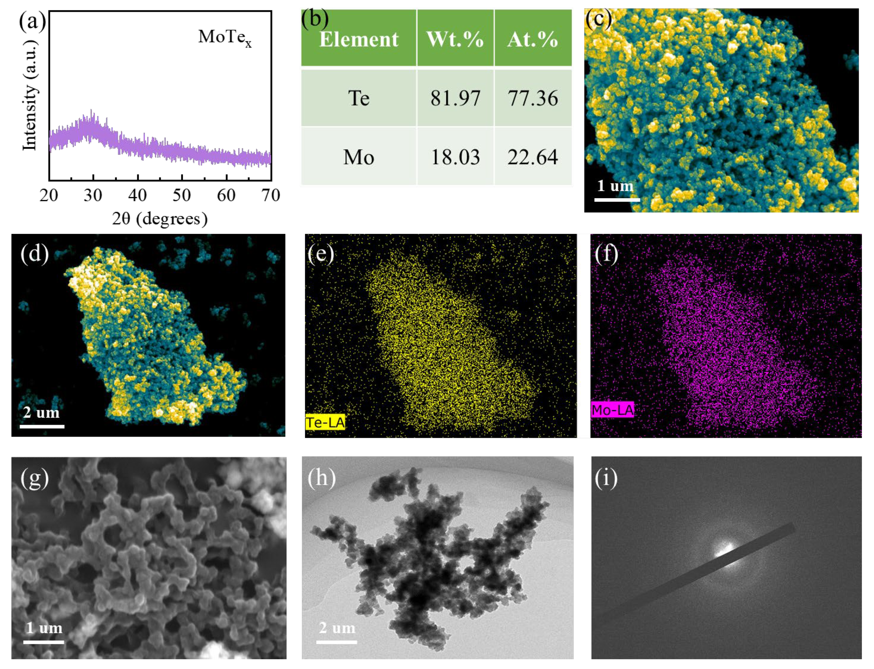
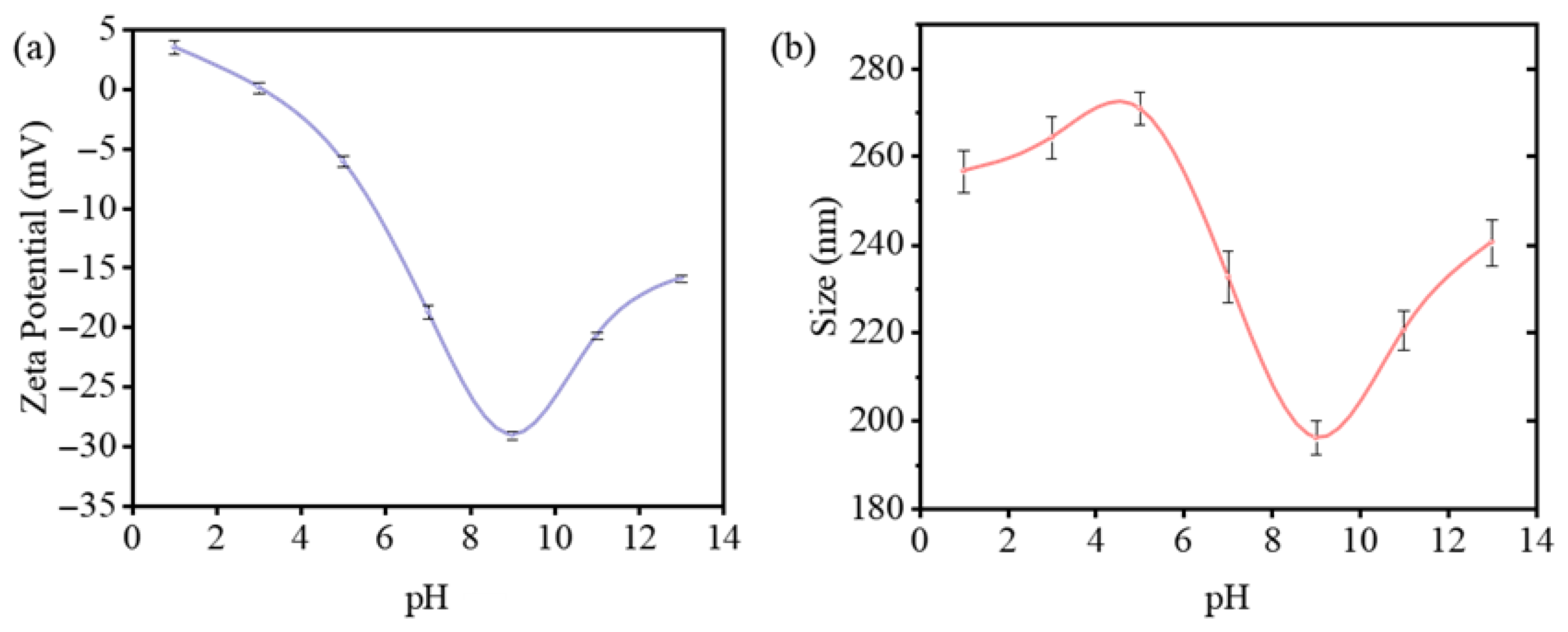
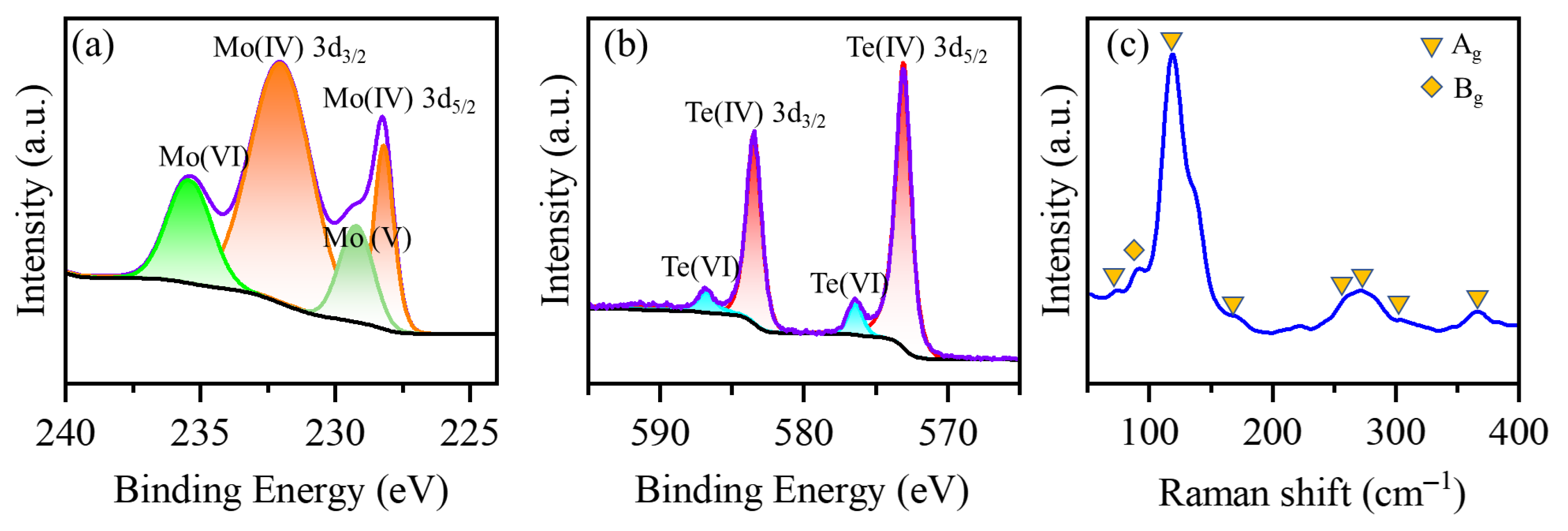
3.1. Morphology and Structure Characterization

To confirm the phase and composition of the reaction products, we conducted a characterization and analysis. The obtained powder XRD pattern exhibited distinct amorphous characteristics, with a pronounced peak around 30 degrees, which is highly similar to that of other amorphous chalcogenides (Figure 1a). The absence of any sharp peaks in the XRD suggests that the product is a homogeneous amorphous material. Additionally, the suspended MoTex product manifested as particles, with some aggregation (Figure 1c). Energy-dispersive X-ray spectroscopy (EDS) was also employed to assess the elemental distribution within the materials (Figure 1b). It was observed that the distribution of Te and Mo elements in the sample was very uniform (Figure 1d–f), with a ratio close to 3:1. This stoichiometry is different from that of typical molybdenum telluride (crystalline MoTe2) and is more reflective of the characteristics of amorphous materials. Consequently, we designate this phase as a-MoTe3 in subsequent discussions. This preliminary characterization indicates that the synthesized amorphous MoTe3 exhibits characteristics typical of amorphous materials. Notably, this stoichiometry is related to the ionic valences of the two elements (Te (−2) and Mo (+6)), reflecting the credibility of the reaction mechanism in Formula (2).
After ultrasonic dispersion, the MoTe3 samples more intuitively exhibit an interconnected morphology (Figure 1g,h), in contrast to the typical monodispersed state of the amorphous particles [39]. The electron diffraction patterns further confirm the amorphous characteristics of the material. (Figure 1i) The microstructure of MoTex resembles a network composed of interconnected clusters, which demonstrates the uniqueness of the Mo–Te bonding in the material. It is likely to possess a bridging structure similar to that of α-MoSx. To further characterize the properties of the obtained MoTe3 particles, we performed characterization using a Zetasizer Nano ZS nanoparticle size analyzer (Malvern, UK).
The results (Figure 2) show that the material’s zeta potential changes significantly with changes in pH. At pH 9.0, the zeta potential reaches approximately −30 mV, indicating the formation of a relatively stable dispersion. As pH decreases, the absolute zeta potential value rapidly approaches zero, accompanied by a clear decrease in system stability—consistent with our experimental observations. At pH 14.0, while stability decreases (zeta potential ≈ −15 mV), rapid aggregation would not occur. The characterization also revealed particle size distribution in the liquid phase. The particle size fluctuates with pH changes, reaching a minimum of about 190 nm at pH 9.0. The maximum diameter under different pH conditions remains below 300 nm, which is generally consistent with our SEM and TEM observations, confirming that the material exhibits typical nanoparticle properties.
Based on previous studies, it is generally believed that amorphous chalcogenide compounds often possess a polymer-like cross-linked network structure [37]. It is reasonable to speculate that the MoTe3 we synthesized may also possess similar properties. To address this, we characterized the chemical states of the Mo and Te elements in the samples using X-ray photoelectron spectroscopy (XPS) and Raman spectroscopy.
The peak analysis for the Mo element revealed that the Mo 3d5/2 peak occurs at 228.2 eV, and the Mo 3d3/2 peak is observed at 232.1 eV, characteristic of Mo(IV) (Figure 3a, orange curve). The presence of Mo(IV) indicates that amorphous MoTe3 contains Mo elements with chemical states similar to those in crystalline MoTe2. However, a distinct Mo(VI) peak is observed at 235.4 eV (Figure 3a, green curve). Although the Mo in our starting material (ammonium molybdate) was already in the +6 oxidation state, the appearance of this peak typically suggests the oxidation of Mo in the material [48]. This phenomenon is commonly observed in two-dimensional MoTe2. In our obtained nanoparticles, due to their amorphous nature, Mo elements are more exposed at the surface and thus more susceptible to oxidation, which may affect subsequent catalytic performance. Additionally, we note the presence of a Mo(V) peak at 229.2 eV, which, according to literature reports, indicates the existence of Mo–O bonds [49], further confirming the partial oxidation of the material. However, compared to typical MoTe2, the 3d5/2 peak of Mo shifts towards higher binding energy, indicating an increased trend for Mo to donate electrons. Considering that the intrinsic electronegativity of the elements does not differ, this suggests an increased Mo–Te bond formation. For the Te element, the XPS spectrum exhibits two distinct doublets. The first doublet at 573 eV and 583.9 eV corresponds to Te(IV), representing characteristic Mo–Te bonding [49], confirming that the product is predominantly composed of Mo–Te bonds. Additionally, a weaker doublet is observed at 577.0 eV and 587 eV, which, after analysis, is attributed to Te(VI) (Te–O). As previously discussed regarding the partial oxidation of Mo, the Te element also shows signs of oxidation. This is particularly evident considering that XPS is a surface-sensitive technique that is more responsive to surface oxidation in nanomaterials due to their high specific surface area. Combining the XPS results of both Mo and Te, we conclude that the obtained MoTe3 material primarily consists of Mo–Te bonding. However, the high surface area of the nanomaterial leads to some degree of surface oxidation, which is a typical characteristic of small-sized materials. Based on XPS analysis, we also calculated the atomic ratio of Te to Mo elements, which is in good agreement with the results from EDS (Table S1).
Raman spectroscopy with a 532 nm excitation laser was employed to further evaluate the chemical states of MoTe3 (Figure 3c). The Raman spectrum exhibited characteristic peaks at 84.3, 102.7, 130.3, 161.3, 252.2, and 264.8 cm−1, which are relatively close to the reported Raman modes of monoclinic MoTe2. However, it is noteworthy that the peaks at typical Raman positions (e.g., 161.3 cm−1) all appear broad rather than sharp, indicating the low crystallinity that is characteristic of amorphous materials [48,50]. These Raman results demonstrate that the obtained MoTe3 maintains a bonding configuration dominated by Mo–Te interactions, yet distinctly differs from typical crystalline MoTe2. The data analysis above indicates that the amorphous MoTe3 we prepared displays distinct Mo–Te bonding, but compared to regular MoTe2, the number of bonds has increased. Considering its morphological characteristics, it is sufficient to conclude that it also possesses the chain-like structure typical of amorphous chalcogenide compounds [51]. Such long-range disordered structural configurations, coupled with high specific surface areas, undoubtedly confer inherent advantages to their catalytic performance.
Leveraging the compositional flexibility inherent to amorphous materials, we attempted to synthesize amorphous MoTe2 from the MoTe3 precursor, thereby enabling direct performance comparisons with that of crystalline MoTe2. This comparative approach facilitates systematic investigation into the role of chalcogenides within co-catalytic systems, particularly their influence on active site density. Firstly, we conducted differential scanning calorimetry (DSC) characterization on the amorphous MoTe3 (Figure S1). A distinct endothermic peak was observed around 450 °C, indicating a significant change in the properties of the amorphous MoTe3 at this temperature. Above 500 °C, the material exhibited significant weight loss, which, in conjunction with the characteristics of Te, suggests that the volatilization of Te elements began to occur. To further validate our hypothesis regarding the transition process, we selected several typical time points to analyze the morphology and XRD patterns of the samples.
The XRD patterns at 150 °C still maintained the typical characteristics of amorphous materials, and the microstructure of the samples showed almost no change (Figure 4a,f). This indicates that the amorphous material we prepared exhibits excellent thermal stability. At 250 °C, Te nanocrystals began to precipitate from the sample (red circles). The XRD patterns also showed distinct sharp peaks corresponding to those of elemental Te (Figure 4b,g). At 350 °C and 400 °C, the precipitation of Te became more pronounced, with an increasing quantity and growing size of the nanocrystals. The XRD patterns still contained a significant number of patterns from elemental Te (Figure 4c,d,h,i). When the temperature reached 600 °C, the microstructure of the sample returned to a spherical cluster form, and the elemental Te nanocrystals had almost completely volatilized (Figure 4e). The XRD tests also confirmed this, with the sharp peaks almost disappearing and the overall pattern again exhibiting significant amorphous characteristics (Figure 4j). EDS analysis revealed a Mo:Te atomic ratio of 1:2, confirming the formation of amorphous MoTe2 (Table S2). The transformation characteristics are consistent with our expectations, and it is interesting to note that the MoTe2 product remains amorphous. This also confirms our inference regarding the structure of MoTe3. Given the discussion on the structure of amorphous MoTe3, it is indeed irrational for the cross-linked structure, which loses some Te atoms after the phase change, to directly transform into the typical crystalline MoTe2.

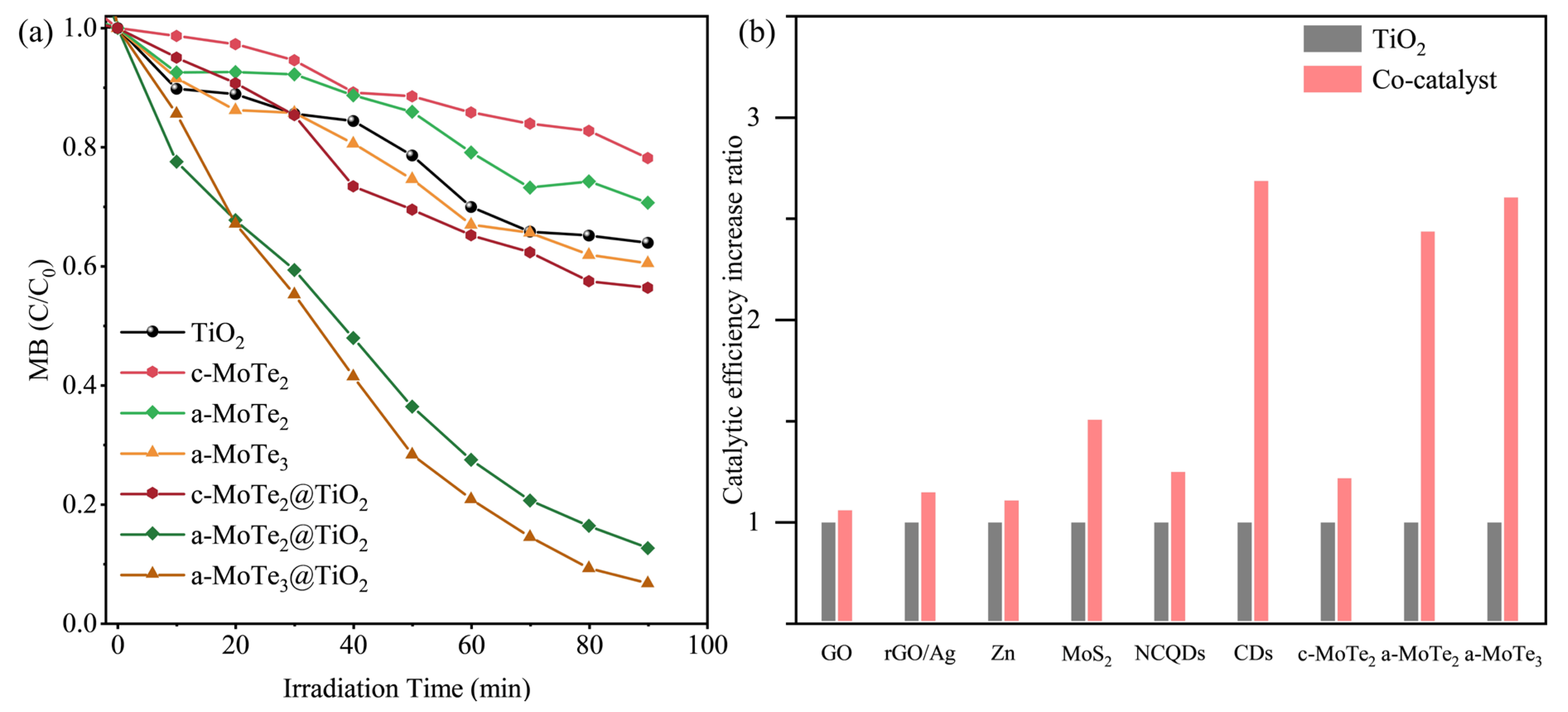
3.2. Photocatalyst Degradation of Methylene Blue

Researchers generally believe that in transition metal chalcogenides, particularly in their two-dimensional forms, the edge states possess a significant catalytic advantage, which is attributed to the exposed metal atoms [30]. Meanwhile, molybdenum chalcogenides demonstrate strong adsorption and enrichment capabilities toward specific organic dyes, rendering them particularly promising for photocatalytic degradation applications [52]. Therefore, the amorphous MoTe3 and MoTe2 materials synthesized in this study exhibit dual structural advantages: their unique disordered structure not only generates abundant microscopic “edge states” but also contributes to increase specific surface area and adsorption capacity. These synergistic characteristics theoretically predict superior catalytic performance. To verify the speculation regarding the catalytic properties of amorphous MoTex, we selected typical methylene blue (MB) [53] as the photocatalytic degradation substrate. Simultaneously, based on previous literature studies suggesting the superior co-catalytic performance of molybdenum chalcogenides [52,54,55], we prepared MoTex@TiO2 composite materials for catalytic testing (Figures S2 and S3) and benchmarked these against analogous systems to assess the enhancement of TiO2 photocatalytic performance induced by amorphous chalcogenides.
To investigate whether co-catalytic systems can extend the spectral response range of TiO2, catalytic experiments were conducted under visible light irradiation. Initial observations revealed that amorphous MoTe3 (a-MoTe3) and amorphous MoTe2 (a-MoTe2) exhibited activity in methylene blue (MB) degradation, achieving approximately 40% MB decomposition within 90 min, significantly surpassing the performance of crystalline MoTe2 (c-MoTe2). (Figure 5a) This efficiency level was nearly comparable to that of intrinsic anatase TiO2. Notably, however, the catalytic efficiency of TiO2 under visible light was substantially reduced compared to that under ultraviolet light, a limitation attributed to its inherent wide bandgap nature [56]. However, when MoTex is combined with TiO2, the catalytic efficiency of the composite significantly improves. While c-MoTe2@TiO2 exhibited comparatively inferior catalytic performance, it nonetheless achieved 40% methylene blue (MB) degradation within 90 min. Meanwhile, both a-MoTe2@TiO2 and a-MoTe3@TiO2 successfully decomposed about 90% of the MB substrate within 90 min, which demonstrates at least a 140% enhancement in catalytic degradation efficiency compared to that of isolated TiO2. To systematically evaluate the enhancement capabilities of various co-catalytic materials on TiO2, we conducted a comprehensive comparative analysis of analogous systems. Given the significant variations in the catalytic performance of TiO2 across different experimental systems, direct comparisons of degradation efficiency fail to isolate the intrinsic contributions of co-catalysts. To address this limitation, we normalized the literature-derived data by defining the intrinsic catalytic degradation efficiency of pristine TiO2 as 1.0 under every different experimental condition to achieve unity. Subsequently, the catalytic enhancement factor of each co-catalyst was calculated as the relative efficiency ratio compared to this normalized baseline (Figure 5b). The results demonstrate that conventional co-catalysts—including metallic species [57], two-dimensional materials [58], and crystalline MoS2 [59]—yielded less than a 50% improvement in the catalytic efficiency of TiO2. Notably, carbon nanodots (CDs) achieved a performance enhancement exceeding 160%, comparable to that of amorphous MoTex (a-MoTex) [60]. However, compared to the intricate synthesis and composite integration processes required for CDs, a-MoTex emerges as a far more practical co-catalytic material, offering superior cost-to-performance efficiency in scalable manufacturing. Foreseeably, with optimized application conditions, this catalyst series will exhibit more significant potential. The improvement in performance, based on research into transition metal chalcogenides, should be attributed to the unique bonding structure of MoTex, which can better absorb photons and separate the photo-produced carriers [61]. Supplementary experiments substantiated the viewpoint (Table S3). The addition of EDTA (a typical OH· scavenger) most significantly reduced the MB degradation efficiency across all three composite catalysts when compared to the effects of CHOONa and p-benzoquinone, indicating that the degradation of MB occurred through hole-mediated pathways via hydroxyl radical (OH) generation [60]. Thus, we acknowledge that the enhanced catalytic performance of TiO2 facilitated by MoTex arises primarily from the increased yield of hydroxyl radical (OH) species. Since the radical-mediated oxidative processes are fundamentally critical in air purification and antimicrobial disinfection, it is reasonable to believe that the MoTex@TiO2 co-catalytic system could also demonstrate considerable potential in the aforementioned application domains.
The comparative analysis reveals that amorphous MoTex demonstrates substantial advantages over its crystalline MoTe2 counterparts, not only in regards to its intrinsic catalytic activity but particularly concerning its co-catalytic performance. Given their analogous degradation mechanisms (predominantly hydroxyl radical-mediated pathways), this enhanced efficacy fundamentally originates from its unique disordered structure, which provides increased active site density at equivalent loadings, thereby significantly amplifying overall catalytic capacity. It is also noticeable that the catalytic performance of a-MoTe2@TiO2 is slightly lower compared to that of a-MoTe3@TiO2. This suggests that the difference in Te content has an impact on the co-catalytic performance of MoTe. Given that hole scavengers exhibit comparable attenuation effects across different MoTex systems, the performance discrepancies likely originate from structural discrepancies at the microscopic level. These disparities fundamentally stem from differential adsorption capacities inherent to distinct amorphous compositions. To validate this hypothesis, we conducted systematic analyses of the composite catalysts’ specific surface areas and pore size distributions (Figure 6).

3.3. Specific Surface and Pore Size Analysis of MoTex@TiO2

The nitrogen adsorption–desorption isotherms for both composite materials are relatively consistent, lacking a saturated adsorption platform, indicating that they are typical macroporous adsorbents. However, when comparing the specific surface areas of the two, it is observed that the surface area of a-MoTe2@TiO2 has decreased compared to that of a-MoTe3@TiO2, which aligns with our hypothesis (Figure 6a,b). Due to the reduction in Te atoms required for the amorphous network, the disordered structure of the a-MoTe2@TiO2 composite samples is inevitably simplified compared to that of a-MoTe3@TiO2, leading to a tendency to aggregate and stack, which in turn reduces the corresponding specific surface area. The distribution of pore quantity also supports a similar conclusion: compared to a-MoTe3@TiO2, a-MoTe2@TiO2 shows a significant reduction in the number of micropores (<2 nm), while there is almost no difference in the content of mesopores (Figure 6c,d), which is consistent with the changes in the material’s microstructure and composition.
It is well established that the specific surface area of a catalyst can significantly influences the rate of a reaction [64]. Given that the specific surface area difference between the two composite materials is about 15%, and their catalytic efficiencies after 90 min differ by less than 10%, we can infer that the performance differences between the two catalysts are likely more attributable to their microstructural differences rather than to changes in the chemical state or electronic structure due to the differences in the Te elements. This finding substantiates that for MoTex materials, both active site increase and specific surface area enhancement (directly governing adsorption capacity) constitute dual determinants of catalytic efficacy. Crucially, the distinctive disordered amorphous structure simultaneously amplifies both parameters through structural disordering. Thus, amorphous-based chalcogenides emerge as a promising frontier for next-generation catalytic material development.

4. Conclusions

In this study, amorphous MoTex materials with polymeric networks were synthesized via a room-temperature ionic approach. Hybridization with TiO2 revealed exceptional co-catalytic performance in the MoTex composites. Systematic comparison between a series of crystalline and amorphous MoTex materials further elucidated the mechanistic basis for their catalytic enhancement. The amorphous structure was demonstrated to concurrently increase both active site density and substrate adsorption capacity through its cross-linked configuration, thereby synergistically boosting photocatalytic degradation efficiency. These findings establish the use of amorphous chalcogenides as a developmental pathway for further optimizing transition metal dichalcogenide catalysts.

Supplementary Materials

The following supporting information can be downloaded at: https://www.mdpi.com/article/10.3390/ma18143388/s1, Experimental details; Table S1: The elemental distribution of MoTe3 based on XPS; Figure S1: DSC characterization of amorphous MoTe3 (the blue areas and numbers indicate the detection points selected); Table S2: The Mo–Te stoichiometric ratio in MoTex materials under varying temperatures; Figure S2: Scanning electron microscopy (SEM) characterization and energy dispersive spectrometer (EDS) mapping of the synthesized a-MoTe3/TiO2; Figure S3: Scanning electron microscopy (SEM) characterization and energy dispersive spectrometer (EDS) mapping of the synthesized a-MoTe2/TiO2. Table S3: Degradation efficiency comparison between composites catalyst series incorporating EDTA, CHOONa, and PBQ sacrificial agent and blank controls at 90 min. Table S4: Supplementary experimental conditions for different co-catalysts.

Author Contributions

Z.Z.: conceptualization, investigation, visualization, writing—original draft, data curation, and formal analysis. B.L.: investigation, visualization, writing—original draft, data curation, and software. J.Z.: conceptualization, investigation, data curation, and writing—review and editing. Z.S.: conceptualization, supervision, funding acquisition, and writing—review and editing. All authors have read and agreed to the published version of the manuscript.

Funding

This research was funded by the National Key Research and Development Program of China (Grant No. 2022YFB3807200) and the National Natural Science Foundation of China (Grant No. 52332005).

Institutional Review Board Statement

Not applicable.

Informed Consent Statement

Not applicable.

Data Availability Statement

The original contributions presented in this study are included in the article/Supplementary Materials. Further inquiries can be directed to the corresponding author.

Conflicts of Interest

The authors declare no conflicts of interest.

References

  1. Wu, J.; Qu, J.; Yin, G.; Zhang, T.; Zhao, H.-Y.; Jiao, F.-Z.; Liu, J.; Li, X.; Yu, Z.-Z. Omnidirectionally irradiated three-dimensional molybdenum disulfide decorated hydrothermal pinecone evaporator for solar-thermal evaporation and photocatalytic degradation of wastewaters. J. Colloid Interface Sci. 2023, 637, 477–488. [Google Scholar] [CrossRef] [PubMed]
  2. Qian, X.; Peng, Y.; Guo, S.; Hou, M.; Liang, Y.; Jiang, G.; Yang, L.; Ye, X.; Sun, H.; Chen, G. Fabrication of porous boron-doped diamond/Si electrodes for the electrocatalytic degradation of methylene blue. Appl. Surf. Sci. 2025, 684, 161944. [Google Scholar] [CrossRef]
  3. Zhang, J.; Li, Y.; Wang, X.; Zhao, S.; Du, Q.; Pi, X.; Jing, Z.; Jin, Y. Polydopamine coating for enhanced electrostatic adsorption of methylene blue by multiwalled carbon nanotubes in alkaline environments. J. Colloid Interface Sci. 2024, 675, 263–274. [Google Scholar] [CrossRef] [PubMed]
  4. Wang, S.; Wang, K.; Cao, W.; Qiao, L.; Peng, X.; Yu, D.; Wang, S.; Li, C.; Wang, C. Degradation of methylene blue by ellipsoidal β-FeOOH@MnO2 core-shell catalyst: Performance and mechanism. Appl. Surf. Sci. 2023, 619, 156667. [Google Scholar] [CrossRef]
  5. Chen, S.H.; Zhang, J.; Zhang, C.L.; Yue, Q.Y.; Li, Y.; Li, C. Equilibrium and kinetic studies of methyl orange and methyl violet adsorption on activated carbon derived from Phragmites australis. Desalination 2010, 252, 149–156. [Google Scholar] [CrossRef]
  6. Yang, R.J.; Fan, Y.Y.; Zhang, Y.F.; Mei, L.; Zhu, R.S.; Qin, J.Q.; Hu, J.G.; Chen, Z.X.; Ng, Y.H.; Voiry, D.; et al. 2D Transition Metal Dichalcogenides for Photocatalysis. Angew. Chem. Int. Ed. 2023, 62, e202218016. [Google Scholar] [CrossRef] [PubMed]
  7. Singh, N.; Jana, S.; Singh, G.P.; Dey, R.K. Graphene-supported TiO2: Study of promotion of charge carrier in photocatalytic water splitting and methylene blue dye degradation. Adv. Compos. Hybrid Mater. 2020, 3, 127–140. [Google Scholar] [CrossRef]
  8. Zhang, Y.; Hao, X.; Lu, M.; Zhang, L.; Guo, B.; Liao, L.; Zhang, K.; Qin, A. CdSe/TiO2 nanowire arrays heterojunction enhanced self-driven photoelectrocatalytic degradation of mixed organic pollutants. Nano Energy 2025, 136, 110762. [Google Scholar] [CrossRef]
  9. Liu, S.; Lou, H.; Luo, J.; Albashir, D.; Shi, Y.; Chen, Q. A Novel In Situ Biosynthesized Bacterial Cellulose/MoS2/TiO2 Composite Film for Efficient Removal of Dyes and Pathogenic Bacteria from Industrial Wastewater under Sunlight Illumination. ACS Appl. Mater. Interfaces 2025, 17, 19543–19561. [Google Scholar] [CrossRef] [PubMed]
  10. Chawraba, K.; Medlej, H.; Toufaily, J.; Lalevee, J.; Hamieh, T. TiO2 Sensitized by Natural Dye Extracted from Cinnamon Bark for Photodegradation of Methylene Blue in Water Under LED Irradiation. Chem. Afr. 2024, 7, 2087–2101. [Google Scholar] [CrossRef]
  11. Geldasa, F.T.; Kebede, M.A.; Shura, M.W.; Hone, F.G. Experimental and computational study of metal oxide nanoparticles for the photocatalytic degradation of organic pollutants: A review. RSC Adv. 2023, 13, 18404–18442. [Google Scholar] [CrossRef] [PubMed]
  12. Mohamed, R.M.; Mkhalid, I.A.; Baeissa, E.S.; Al-Rayyani, M.A. Photocatalytic Degradation of Methylene Blue by Fe/ZnO/SiO2 Nanoparticles under Visiblelight. J. Nanotechnol. 2012, 2012, 329082. [Google Scholar] [CrossRef]
  13. Liang, P.; Yang, W.; Peng, H.; Zhao, S. Efficient Degradation of Methylene Blue in Industrial Wastewater and High Cycling Stability of Nano ZnO. Molecules 2024, 29, 5584. [Google Scholar] [CrossRef] [PubMed]
  14. Paredes, P.; Rauwel, E.; Wragg, D.S.; Rapenne, L.; Estephan, E.; Volobujeva, O.; Rauwel, P. Sunlight-Driven Photocatalytic Degradation of Methylene Blue with Facile One-Step Synthesized Cu-Cu2O-Cu3N Nanoparticle Mixtures. Nanomaterials 2023, 13, 1311. [Google Scholar] [CrossRef] [PubMed]
  15. Parvees, A.; Priyadarshini, U.; Remya, N. Visible light induced photocatalytic degradation of methylene blue using carbon fibre cloth—Bismuth oxybromide—Water hyacinth derived silver nanocomposite. Environ. Res. 2024, 262, 119787. [Google Scholar] [CrossRef] [PubMed]
  16. Eswaran, P.; Madasamy, P.D.; Pillay, K.; Brink, H. Sunlight-driven photocatalytic degradation of methylene blue using ZnO/biochar nanocomposite derived from banana peels. Biomass Convers. Biorefin. 2025, 15, 12347–12367. [Google Scholar] [CrossRef]
  17. Pan, G.; Xu, M.; Zhou, K.; Meng, Y.; Chen, H.; Guo, Y.; Wu, T. Photocatalytic Degradation of Methylene Blue Over Layered Double Hydroxides Using Various Divalent Metal Ions. Clays Clay Miner. 2019, 67, 340–347. [Google Scholar] [CrossRef]
  18. Lee, S.C.; Benck, J.D.; Tsai, C.; Park, J.; Koh, A.L.; Abild-Pedersen, F.; Jaramillo, T.F.; Sinclair, R. Chemical and phase evolution of amorphous molybdenum sulfide catalysts for electrochemical hydrogen production. ACS Nano 2016, 10, 624–632. [Google Scholar] [CrossRef] [PubMed]
  19. Zhang, L.; Ji, X.; Ren, X.; Ma, Y.; Shi, X.; Tian, Z.; Asiri, A.M.; Chen, L.; Tang, B.; Sun, X. Electrochemical ammonia synthesis via nitrogen reduction reaction on a MoS2 catalyst: Theoretical and experimental studies. Adv. Mater. 2018, 30, 1800191. [Google Scholar] [CrossRef] [PubMed]
  20. Wu, M.H.; Lee, J.T.; Chung, Y.J.; Srinivaas, M.; Wu, J.M. Ultrahigh efficient degradation activity of single- and few-layered MoSe2 nanoflowers in dark by piezo-catalyst effect. Nano Energy 2017, 40, 369–375. [Google Scholar] [CrossRef]
  21. Nirjhar, A.R.; Tan-Ema, S.J.; Sahriar, M.A.; Ahsan Dipon, M.N.; Hasan Abed, M.R.; Gainza, D.B.; Koneru, A.; Nishat, S.S.; Shorowordi, K.M.; Ahmed, S. Vacancy-induced spontaneous H2 evolution by overall water splitting on MoTe2/Ti2CO2: A two-dimensional direct Z scheme heterostructure. Int. J. Hydrogen Energy 2023, 48, 37273–37285. [Google Scholar] [CrossRef]
  22. Hellstern, T.R.; Kibsgaard, J.; Tsai, C.; Palm, D.W.; King, L.A.; Abild-Pedersen, F.; Jaramillo, T.F. Investigating catalyst–support interactions to improve the hydrogen evolution reaction activity of thiomolybdate [Mo3S13]2− nanoclusters. ACS Catal. 2017, 7, 7126–7130. [Google Scholar] [CrossRef]
  23. Sahu, A.; Steinmann, S.N.; Raybaud, P. Size-dependent structural, energetic, and spectroscopic properties of MoS3 polymorphs. Cryst. Growth Des. 2020, 20, 7750–7760. [Google Scholar] [CrossRef]
  24. Qu, D.; Liu, X.; Huang, M.; Lee, C.; Ahmed, F.; Kim, H.; Ruoff, R.S.; Hone, J.; Yoo, W.J. Carrier-type modulation and mobility improvement of thin MoTe2. Adv. Mater. 2017, 29, 1606433. [Google Scholar] [CrossRef] [PubMed]
  25. Lee, Y.; Lee, S.H.; Han, S.K.; Park, J.; Lee, D.; Preston, D.J.; Kim, I.S.; Hersam, M.C.; Kwon, Y.; Shong, B.; et al. Strain-Enabled Local Phase Control in Layered MoTe2 for Enhanced Electrocatalytic Hydrogen Evolution. ACS Energy Lett. 2023, 8, 4716–4725. [Google Scholar] [CrossRef]
  26. Zhao, X.; Sun, W.; Geng, D.; Fu, W.; Dan, J.; Xie, Y.; Kent, P.R.; Zhou, W.; Pennycook, S.J.; Loh, K.P. Edge segregated polymorphism in 2D molybdenum carbide. Adv. Mater. 2019, 31, 1808343. [Google Scholar] [CrossRef] [PubMed]
  27. Zhao, X.; Fu, D.; Ding, Z.; Zhang, Y.-Y.; Wan, D.; Tan, S.J.; Chen, Z.; Leng, K.; Dan, J.; Fu, W. Mo-terminated edge reconstructions in nanoporous molybdenum disulfide film. Nano Lett. 2018, 18, 482–490. [Google Scholar] [CrossRef] [PubMed]
  28. Sang, X.; Li, X.; Zhao, W.; Dong, J.; Rouleau, C.M.; Geohegan, D.B.; Ding, F.; Xiao, K.; Unocic, R.R. In situ edge engineering in two-dimensional transition metal dichalcogenides. Nat. Commun. 2018, 9, 2051. [Google Scholar] [CrossRef] [PubMed]
  29. Han, B.; Hu, Y.H. MoS2 as a co-catalyst for photocatalytic hydrogen production from water. Energy Sci. Eng. 2016, 4, 285–304. [Google Scholar] [CrossRef]
  30. Zhou, H.; Chen, Z.; Kountoupi, E.; Tsoukalou, A.; Abdala, P.M.; Florian, P.; Fedorov, A.; Müller, C.R. Two-dimensional molybdenum carbide 2D-Mo2C as a superior catalyst for CO2 hydrogenation. Nat. Commun. 2021, 12, 5510. [Google Scholar] [CrossRef] [PubMed]
  31. Hu, J.; Yu, L.; Deng, J.; Wang, Y.; Cheng, K.; Ma, C.; Zhang, Q.; Wen, W.; Yu, S.; Pan, Y.; et al. Sulfur vacancy-rich MoS2 as a catalyst for the hydrogenation of CO2 to methanol. Nat. Catal. 2021, 4, 242–250. [Google Scholar] [CrossRef]
  32. Wang, B.; Zhang, S.-Y.; Ye, L.-H.; Zhang, X.-F.; Zhang, Y.-F.; Chen, W.-J. Exploring the reaction mechanism of H2S decomposition with MS3 (M = Mo, W) clusters. ACS Omega 2020, 5, 13324–13332. [Google Scholar] [CrossRef] [PubMed]
  33. Voiry, D.; Yang, J.; Chhowalla, M. Recent strategies for improving the catalytic activity of 2D TMD nanosheets toward the hydrogen evolution reaction. Adv. Mater. 2016, 28, 6197–6206. [Google Scholar] [CrossRef] [PubMed]
  34. Wang, X.; Zhang, Y.; Wu, J.; Zhang, Z.; Liao, Q.; Kang, Z.; Zhang, Y. Single-atom engineering to ignite 2D transition metal dichalcogenide based catalysis: Fundamentals, progress, and beyond. Chem. Rev. 2021, 122, 1273–1348. [Google Scholar] [CrossRef] [PubMed]
  35. Er, D.; Ye, H.; Frey, N.C.; Kumar, H.; Lou, J.; Shenoy, V.B. Prediction of enhanced catalytic activity for hydrogen evolution reaction in Janus transition metal dichalcogenides. Nano Lett. 2018, 18, 3943–3949. [Google Scholar] [CrossRef] [PubMed]
  36. Nguyen, Q.T.; Nguyen, P.D.; Nguyen, D.N.; Truong, Q.D.; Kim Chi, T.T.; Ung, T.T.D.; Honma, I.; Liem, N.Q.; Tran, P.D. Novel amorphous molybdenum selenide as an efficient catalyst for hydrogen evolution reaction. ACS Appl. Mater. Interfaces 2018, 10, 8659–8665. [Google Scholar] [CrossRef] [PubMed]
  37. Yu, H.; Xiao, P.; Wang, P.; Yu, J. Amorphous molybdenum sulfide as highly efficient electron-cocatalyst for enhanced photocatalytic H2 evolution. Appl. Catal. B Environ. 2016, 193, 217–225. [Google Scholar] [CrossRef]
  38. Yang, Z.; Hao, J.; Lau, S.P. Synthesis, properties, and applications of 2D amorphous inorganic materials. J. Appl. Phys. 2020, 127, 220901. [Google Scholar] [CrossRef]
  39. Kang, J.; Yang, X.; Hu, Q.; Cai, Z.; Liu, L.-M.; Guo, L. Recent progress of amorphous nanomaterials. Chem. Rev. 2023, 123, 8859–8941. [Google Scholar] [CrossRef] [PubMed]
  40. Lu, P.; Sun, Y.; Xiang, H.; Liang, X.; Yu, Y. 3D Amorphous Carbon with Controlled Porous and Disordered Structures as a High-Rate Anode Material for Sodium-Ion Batteries. Adv. Energy Mater. 2018, 8, 1702434. [Google Scholar] [CrossRef]
  41. Li, X.; Cai, W.; Li, D.-S.; Xu, J.; Tao, H.; Liu, B. Amorphous alloys for electrocatalysis: The significant role of the amorphous alloy structure. Nano Res. 2021, 16, 4277–4288. [Google Scholar] [CrossRef]
  42. Wu, Y.; Liu, Z.; Zhong, X.; Cheng, X.; Fan, Z.; Yu, Y. Amorphous Red Phosphorus Embedded in Sandwiched Porous Carbon Enabling Superior Sodium Storage Performances. Small 2018, 14, 1703472. [Google Scholar] [CrossRef] [PubMed]
  43. Tian, H.; Ma, Y.; Li, Z.; Cheng, M.; Ning, S.; Han, E.; Xu, M.; Zhang, P.-F.; Zhao, K.; Li, R. Disorder-tuned conductivity in amorphous monolayer carbon. Nature 2023, 615, 56–61. [Google Scholar] [CrossRef] [PubMed]
  44. Finkelstein-Zuta, G.; Arnon, Z.A.; Vijayakanth, T.; Messer, O.; Lusky, O.S.; Wagner, A.; Zilberman, G.; Aizen, R.; Michaeli, L.; Rencus-Lazar, S. A self-healing multispectral transparent adhesive peptide glass. Nature 2024, 630, 368–374. [Google Scholar] [CrossRef] [PubMed]
  45. Rosu-Finsen, A.; Davies, M.B.; Amon, A.; Wu, H.; Sella, A.; Michaelides, A.; Salzmann, C.G. Medium-density amorphous ice. Science 2023, 379, 474–478. [Google Scholar] [CrossRef]
  46. Liu, A.; Kim, Y.-S.; Kim, M.G.; Reo, Y.; Zou, T.; Choi, T.; Bai, S.; Zhu, H.; Noh, Y.-Y. Selenium alloyed tellurium oxide for amorphous p-channel transistors. Nature 2024, 629, 798–802. [Google Scholar] [CrossRef] [PubMed]
  47. Escalera-López, D.; Lou, Z.; Rees, N.V. Benchmarking the activity, stability, and inherent electrochemistry of amorphous molybdenum sulfide for hydrogen production. Adv. Energy Mater. 2019, 9, 1802614. [Google Scholar] [CrossRef]
  48. Ma, L.; Zhu, J.; Li, W.; Huang, R.; Wang, X.; Guo, J.; Choi, J.-H.; Lou, Y.; Wang, D.; Zou, G. Immobilized Precursor Particle Driven Growth of Centimeter-Sized MoTe2 Monolayer. J. Am. Chem. Soc. 2021, 143, 13314–13324. [Google Scholar] [CrossRef] [PubMed]
  49. Zong, J.; Liang, Y.; Liu, F.; Zhang, M.; Feng, J.; Xi, B.; Xiong, S. Effect of Combination Model of MoTe2 and MXene Layers on Sodium Ion Storage. Adv. Mater. 2025, 2503252. [Google Scholar] [CrossRef] [PubMed]
  50. Tan, Y.; Luo, F.; Zhu, M.; Xu, X.; Ye, Y.; Li, B.; Wang, G.; Luo, W.; Zheng, X.; Wu, N.; et al. Correction and removal of expression of concern: Controllable 2H-to-1T′ phase transition in few-layer MoTe2. Nanoscale 2019, 11, 23498–23501. [Google Scholar] [CrossRef] [PubMed]
  51. Hung, N.T.; Yin, L.-C.; Tran, P.D.; Saito, R. Simultaneous Anionic and Cationic Redox in the Mo3S11 Polymer Electrode of a Sodium-Ion Battery. J. Phys. Chem. C 2019, 123, 30856–30862. [Google Scholar] [CrossRef]
  52. Li, X.; Li, B.; Ji, D.; Guan, Q.; Thirumalraj, B.; Palmisano, G.; Mohamed, S.; Zheng, L. A highly efficient bifunctional Z-scheme photocatalyst with spatially-separated and synergistically-enhanced adsorption and photodegradation. Appl. Catal. B Environ. Energy 2025, 362, 124758. [Google Scholar] [CrossRef]
  53. Begum, R.; Najeeb, J.; Sattar, A.; Naseem, K.; Irfan, A.; Al-Sehemi, A.G.; Farooqi, Z.H. Chemical reduction of methylene blue in the presence of nanocatalysts: A critical review. Rev. Chem. Eng. 2020, 36, 749–770. [Google Scholar] [CrossRef]
  54. Zazpe, R.; Sopha, H.; Charvot, J.; Krumpolec, R.; Rodriguez-Pereira, J.; Michalička, J.; Mistrík, J.; Bača, D.; Motola, M.; Bureš, F.; et al. 2D MoTe2 nanosheets by atomic layer deposition: Excellent photo- electrocatalytic properties. Appl. Mater. Today 2021, 23, 101017. [Google Scholar] [CrossRef]
  55. Sabarinathan, M.; Bharathi, P.; Harish, S.; Hayakawa, Y. Two-dimensional layered MoS2@CuS nanostructures decorated over TiO2 spheres for the decomposition of organic pollutant under visible light. Inorg. Chem. Commun. 2023, 153, 110828. [Google Scholar] [CrossRef]
  56. Armaković, S.J.; Savanović, M.M.; Armaković, S. Titanium Dioxide as the Most Used Photocatalyst for Water Purification: An Overview. Catalysts 2023, 13, 26. [Google Scholar] [CrossRef]
  57. Vasilaki, E.; Georgaki, I.; Vernardou, D.; Vamvakaki, M.; Katsarakis, N. Ag-loaded TiO2/reduced graphene oxide nanocomposites for enhanced visible-light photocatalytic activity. Appl. Surf. Sci. 2015, 353, 865–872. [Google Scholar] [CrossRef]
  58. Yunarti, R.T.; Safitri, T.N.; Dimonti, L.C.C.; Aulia, G.; Khalil, M.; Ridwan, M. Facile synthesis of composite between titania nanoparticles with highly exposed (001) facet and coconut shell-derived graphene oxide for photodegradation of methylene blue. J. Phys. Chem. Solids 2022, 160, 110357. [Google Scholar] [CrossRef]
  59. Ibukun, O.; Evans, P.E.; Dowben, P.A.; Kyung Jeong, H. Titanium dioxide-molybdenum disulfide for photocatalytic degradation of methylene blue. Chem. Phys. 2019, 525, 110419. [Google Scholar] [CrossRef]
  60. Cong, S.; Cai, J.; Li, X.; You, J.; Wang, L.; Wang, X. Direct Z-Scheme Xylan-Based Carbon Dots@TiO2-x Nanocomposites for Visible Light Driven Photocatalytic of Dye Degradation and Antibacterial. Adv. Funct. Mater. 2024, 34, 2401540. [Google Scholar] [CrossRef]
  61. Vikraman, D.; Hussain, S.; Hussain, T.; Karuppasamy, K.; Santhoshkumar, P.; Kim, K.-Y.; Manikandan, R.; Jung, J.; Kim, H.-S. Diverse chalcogen bonded molybdenum dichalcogenide alloy for the efficient photo- and electro-catalytic activity to eradicate the methylene blue and Congo red dyes. J. Clean. Prod. 2023, 426, 139127. [Google Scholar] [CrossRef]
  62. Jin, Y.; Tang, W.; Wang, J.; Ren, F.; Chen, Z.; Sun, Z.; Ren, P.-G. Construction of biomass derived carbon quantum dots modified TiO2 photocatalysts with superior photocatalytic activity for methylene blue degradation. J. Alloys Compd. 2023, 932, 167627. [Google Scholar] [CrossRef]
  63. Karuppasamy, P.; Ramzan Nilofar Nisha, N.; Pugazhendhi, A.; Kandasamy, S.; Pitchaimuthu, S. An investigation of transition metal doped TiO2 photocatalysts for the enhanced photocatalytic decoloration of methylene blue dye under visible light irradiation. J. Environ. Chem. Eng. 2021, 9, 105254. [Google Scholar] [CrossRef]
  64. Chawraba, K.; Medlej, H.; Hamieh, M.; Lalevée, J.; Hamieh, T.; Toufaily, J. SBA-15 Loaded with Methylene Blue Dye for Efficient and Low-Cost Decontamination of Water under Visible Led Light. Preprints 2024. [Google Scholar] [CrossRef]
Figure 1. The characterization of MoTex using X-ray diffraction (XRD) and scanning electron microscopy (SEM). (a) XRD pattern of MoTe3. (b) The elemental distribution of MoTex based on energy-dispersive X-ray spectroscopy (EDS). (c) SEM image of MoTe3 particles. (df) The distribution of Te and Mo elements in MoTe3 particles. (g) Ultrasonically dispersed polymer-like MoTe3 particles. (h) Transmission electron microscopy (TEM) images of MoTe3. (i) Transmission electron microscopy electron diffraction pattern of MoTe3.
Materials 18 03388 g001
Figure 2. Characterization of MoTe3 particles: (a) zeta potential; (b) particle size distribution.
Materials 18 03388 g002
Figure 3. (a) XPS spectrum of Mo 3d. Mo(VI): bright green curve; Mo(IV): orange curve; Mo(V): green curve. (b) XPS spectrum of Te 3d. Te(VI): bright blue curve; Te(IV): red curve. (c) Raman spectrum of MoTe3.
Materials 18 03388 g003
Figure 4. SEM images of MoTex materials at different temperatures: (a) 150 °C; (b) 250 °C; (c) 350 °C; (d) 400 °C; (e) 600 °C. XRD pattern of MoTex materials at different temperatures: (f) 150 °C; (g) 250 °C; (h) 350 °C; (i) 400 °C; (j) 600 °C. The red vertical lines in the XRD pattern correspond to the reference diffraction peaks of elemental tellurium (ICSD No. 98-002-3067), which serve as standard markers for phase identification.
Materials 18 03388 g004
Figure 5. (a) The photocatalytic degradation efficiency of different catalytic materials for methylene blue (MB). (b) Comparative enhancement capabilities of different co-catalytic systems for the photocatalytic degradation efficiency of TiO2 [57,58,59,60,62,63]. More supplementary experimental conditions are shown in Table S4.
Materials 18 03388 g005
Figure 6. The nitrogen adsorption–desorption isotherms and pore size distributions of a-MoTe3@TiO2 and a-MoTe2@TiO2. Nitrogen adsorption–desorption isotherms of (a) a-MoTe3@TiO2 and (b) a-MoTe2@TiO2. Pore size distribution diagrams of (c) a-MoTe3@TiO2 and (d) a-MoTe2@TiO2.
Materials 18 03388 g006
Disclaimer/Publisher’s Note: The statements, opinions and data contained in all publications are solely those of the individual author(s) and contributor(s) and not of MDPI and/or the editor(s). MDPI and/or the editor(s) disclaim responsibility for any injury to people or property resulting from any ideas, methods, instructions or products referred to in the content.

