
Analysis of Different Lithium Disilicate Ceramics According to Their Composition and Processing Technique—A Systematic Review and Meta-Analysis

 ,
,  , ,
, ,
Abstract
1. Introduction
2. Objectives
3. Materials and Methods
3.1. Study Selection and Criteria
- “P” (patients) refers to LDS bars, discs, and crowns.
- “I” (intervention) is the influence of the composition and processing technique on ceramic properties.
- “C” (comparison) includes different ceramic groups (LDS, ZLS, and ALD) and processing techniques available in the market.
- “O” (outcome) comprises the properties of each material: Young’s modulus, flexural strength, fracture resistance, hardness, surface roughness (Ra and Rz), wear, and translucency.
3.2. Search Strategy
3.3. Inclusion and Exclusion Criteria
- Published within the last ten years;
- In vitro experimental studies focusing on LDS, ZLS, and ALD ceramics;
- Data presented in numerical form.
- The exclusion criteria included the following:
- In vivo studies;
- Studies analyzing materials other than lithium silicate ceramics;
- Studies with non-numeric or graphical data representation.
3.4. Risk of Bias Assessment
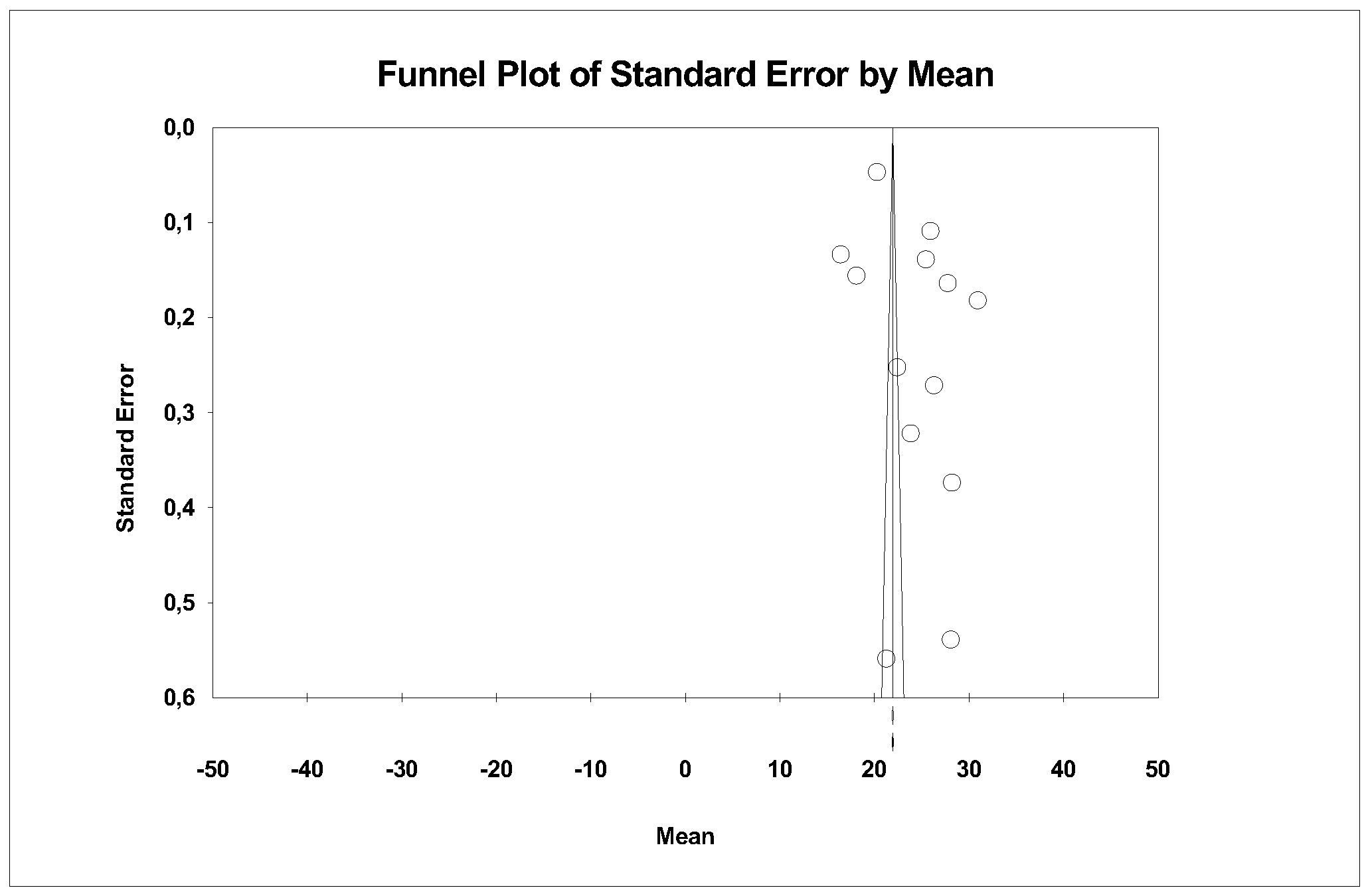
3.5. Meta-Analysis and Meta-Regression Design
- A random-effects model was applied.
- Subgroup analysis was performed (LDS, ZLS, and ALD), with significance set at p < 0.05.
- Meta-regressions were used to estimate the moderator effect of ceramic type and processing technique on analyzed variables.
4. Results
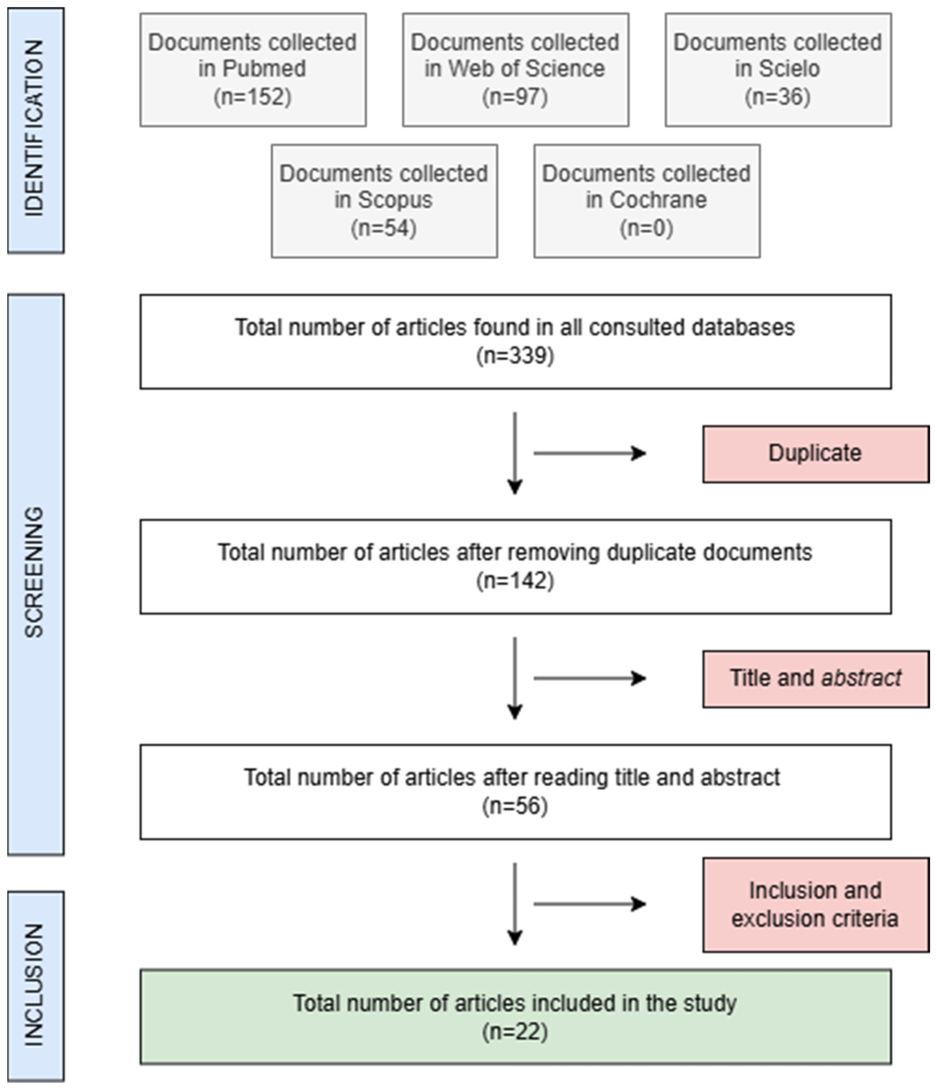
4.1. Study Selection and Description
4.2. Methodological Quality
4.3. Results of the Meta-Analysis and Meta-Regression
4.3.1. Young’s Modulus (Stiffness/Elasticity)
4.3.2. Fracture Resistance
4.3.3. Flexural Strength
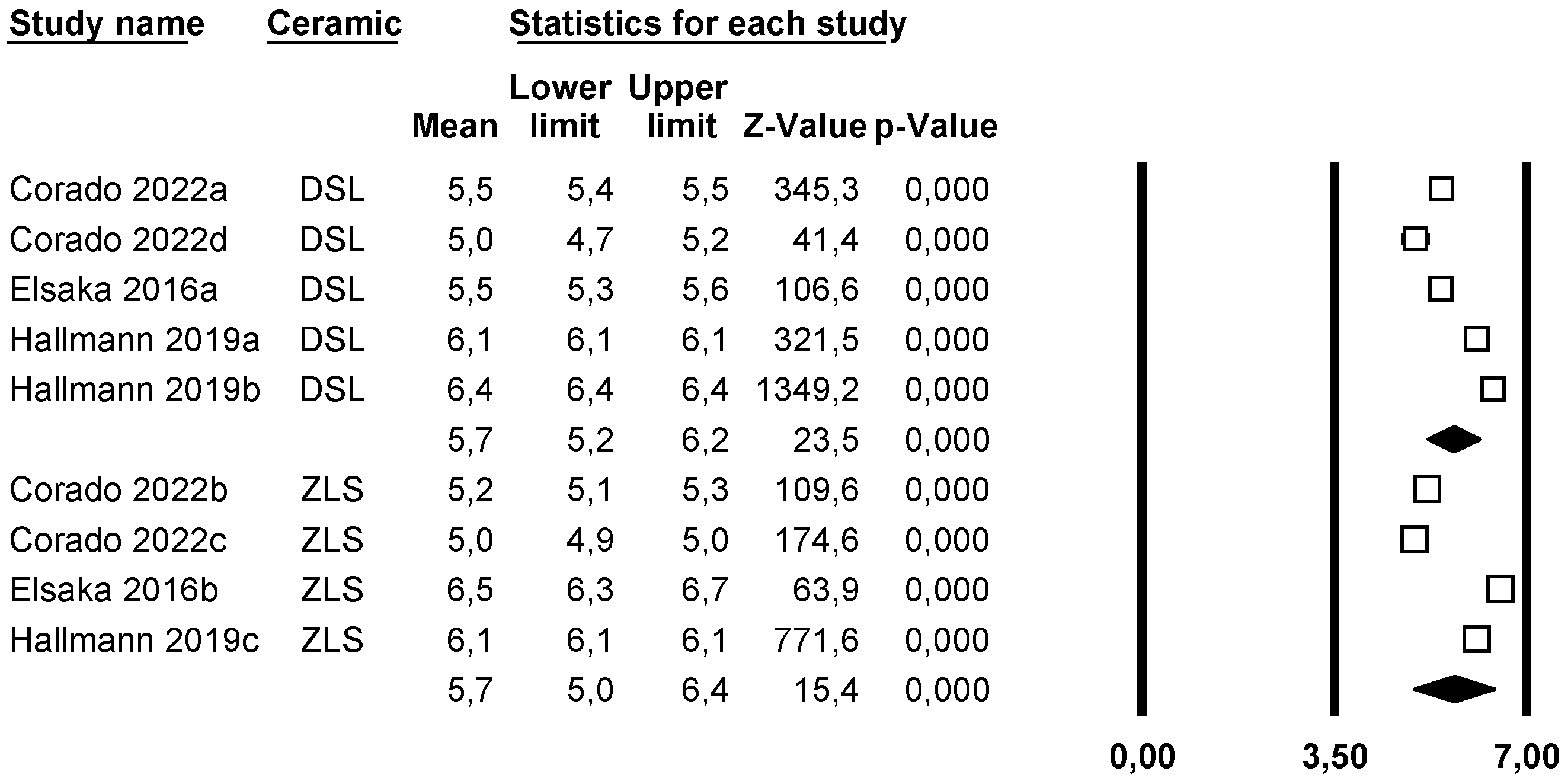
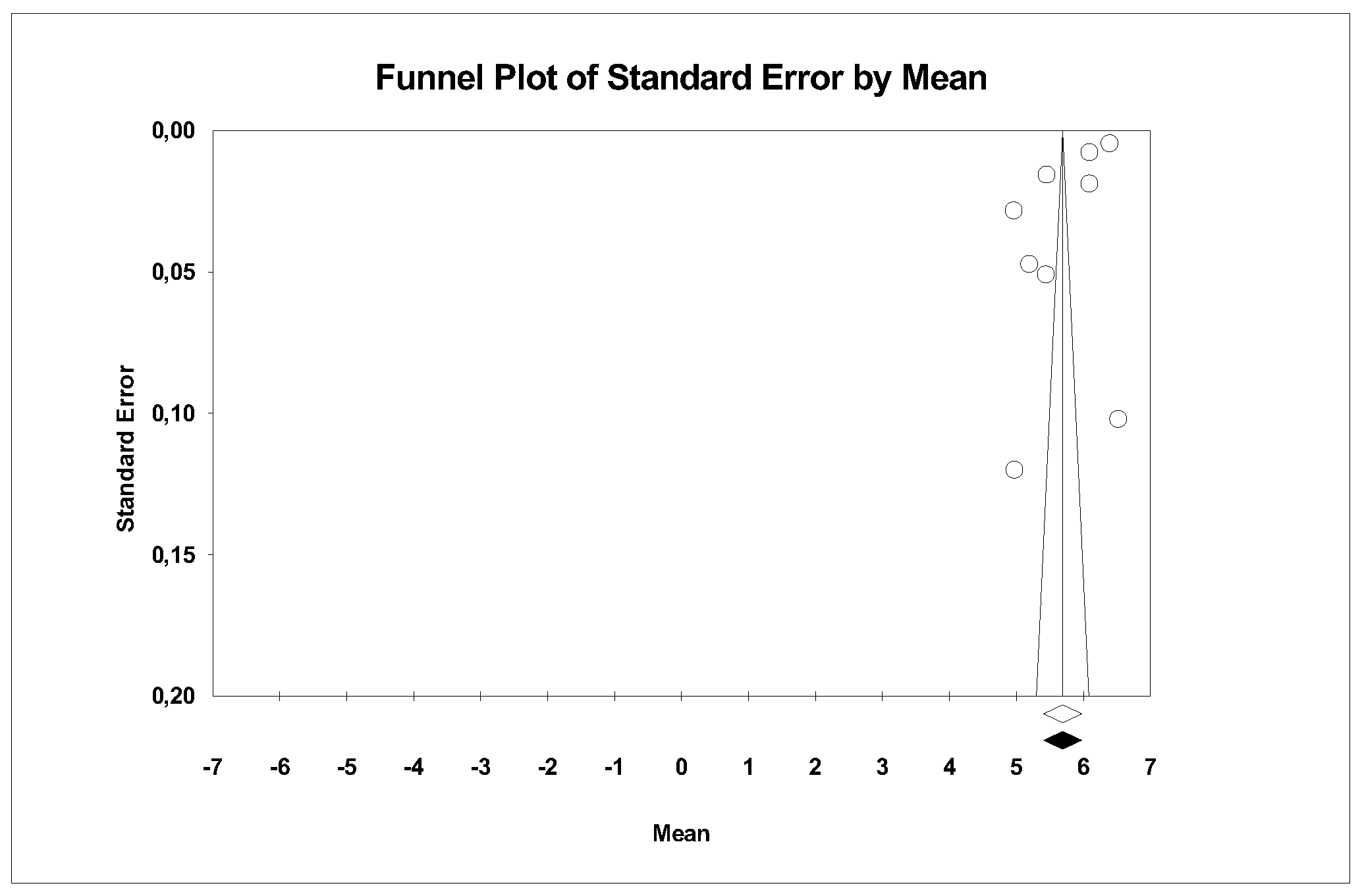
4.3.4. Hardness
Roughness (Ra) (Average Roughness)
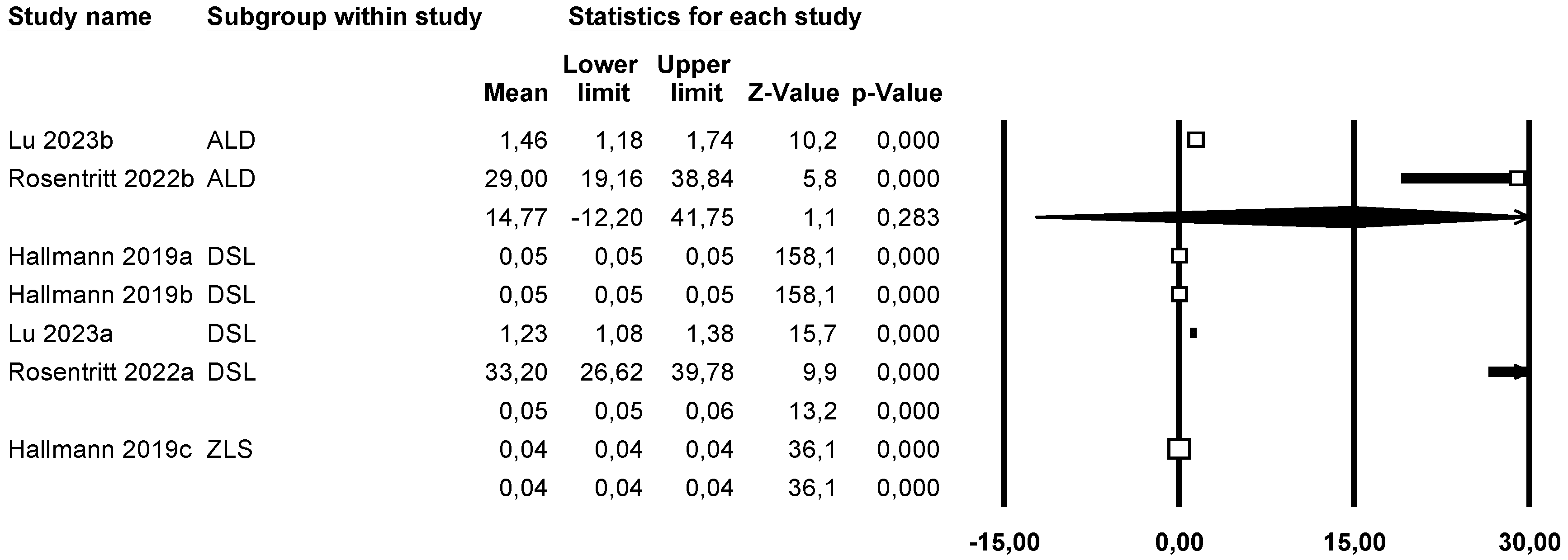
Roughness (Rz) (Mean Roughness Depth)
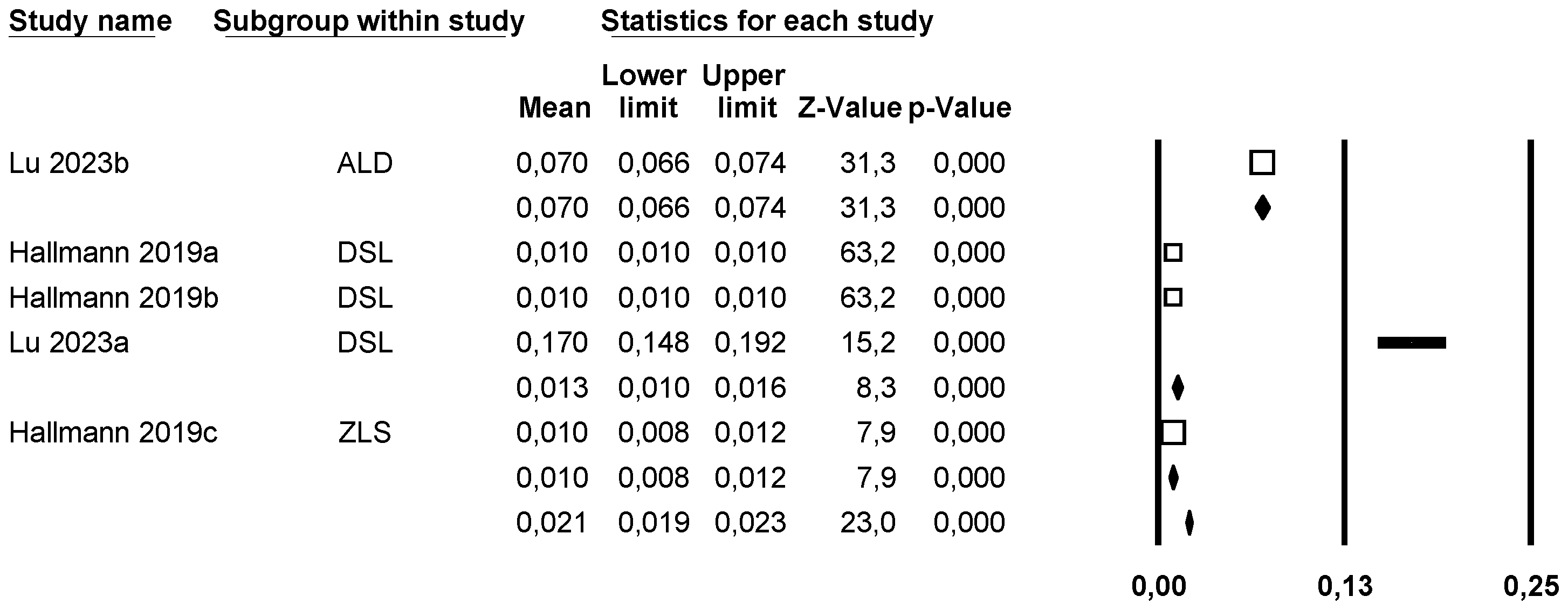
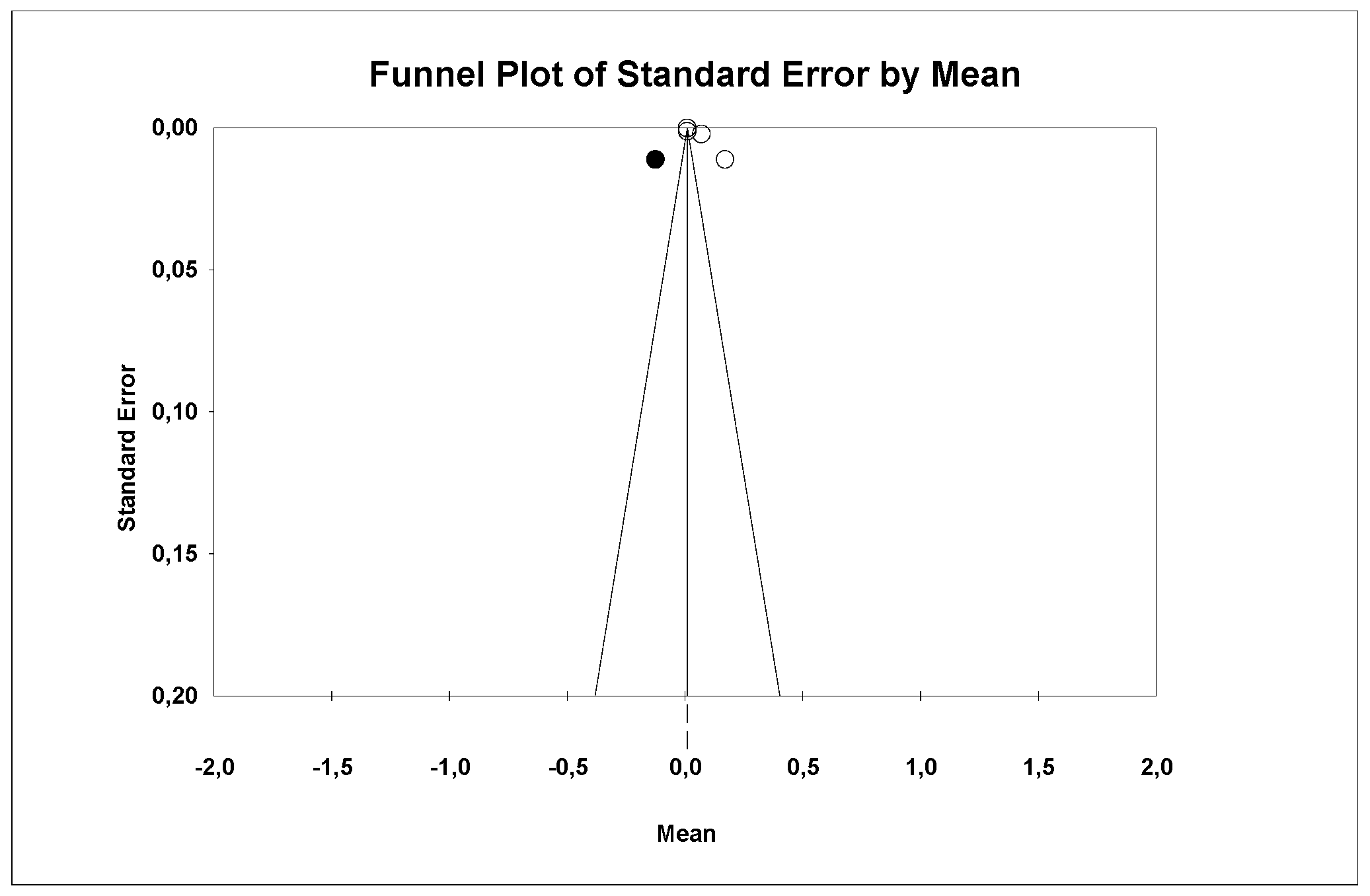
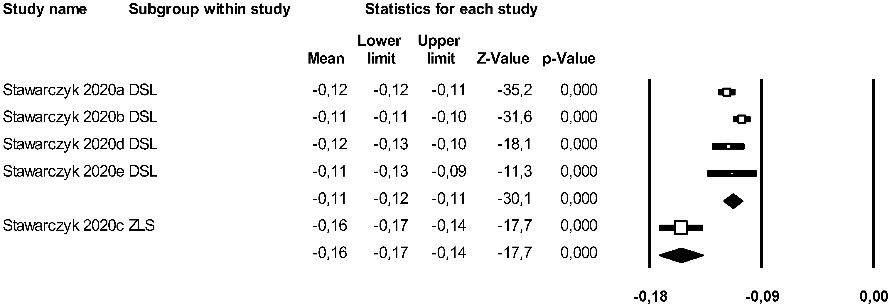
4.3.5. Wear of the Restoration
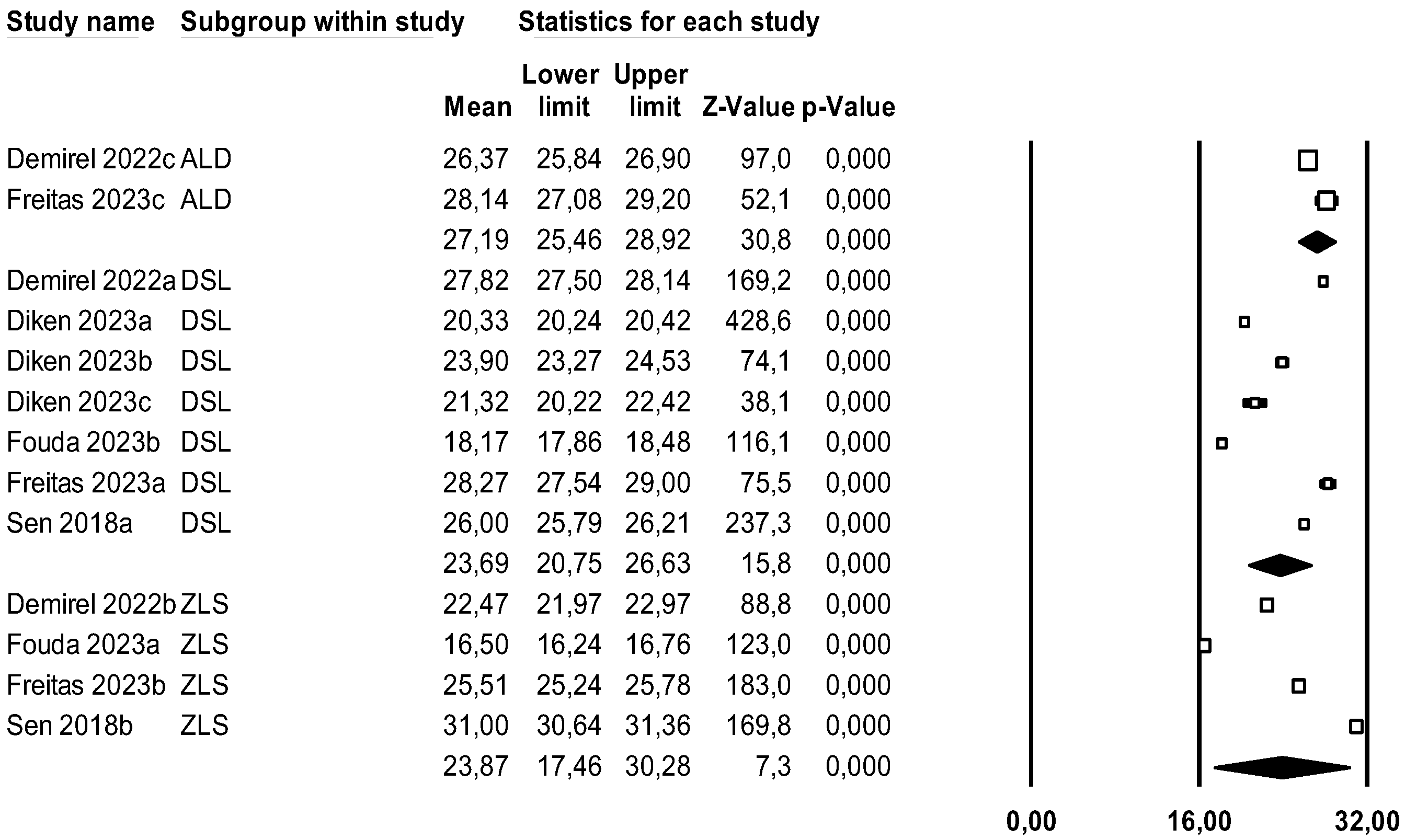
4.3.6. Translucency
5. Discussion
5.1. Young’s Modulus (Stiffness/Elasticity)
5.2. Fracture Resistance
5.3. Flexural Strength
5.4. Hardness
5.5. Roughness (Ra) (Mean Roughness)
Roughness (Rz) (Mean Depth of Roughness)
5.6. Wear
5.7. Translucency
5.8. Clinical Relevance
6. Conclusions
- The LDS presented the highest fracture resistance of the three groups studied (LDS, ZLS, and ALD) and the least wear. No significant differences were found in flexural strength, hardness, stiffness, translucency, or mean roughness; however, the roughness depth was greater in ALD.
- The processing technique in the dental laboratory does influence the properties of the ceramics. The press technique increases hardness and decreases roughness, while the CAD-CAM technique showed better results in flexural strength.
- The most suitable ceramics for clinical use, based on the results obtained and considering the limitations of the study, would be those of LDS due to their physical properties, particularly their high fracture resistance.
Supplementary Materials
Author Contributions
Funding
Data Availability Statement
Conflicts of Interest
References
- Zhang, Y.; Kelly, J.R. Dental ceramics for restoration and metal-veneering. Dent. Clin. N. Am. 2017, 61, 797–819. [Google Scholar] [CrossRef] [PubMed]
- Hallmann, L.; Ulmer, P.; Kern, M. Effect of microstructure on the mechanical properties of lithium disilicate glass-ceramics. J. Mech. Behav. Biomed. Mater. 2018, 82, 355–370. [Google Scholar] [CrossRef]
- Phark, J.; Duarte, S. Microstructural considerations for novel lithium disilicate glass ceramics: A review. J. Esthet. Restor. Dent. 2022, 34, 92–103. [Google Scholar] [CrossRef]
- Willard, A.; Gabriel Chu, T. The science and application of IPS e.Max dental ceramic. Kaohsiung J. Med. Sci. 2018, 34, 238–242. [Google Scholar] [CrossRef]
- Sorrentino, R.; Ruggiero, G.; Di Mauro, M.I.; Breschi, L.; Leuci, S.; Zarone, F. Optical behaviors, surface treatment, adhesion, and clinical indications of zirconia-reinforced lithium silicate (ZLS): A narrative review. J. Dent. 2021, 112, 103722. [Google Scholar] [CrossRef]
- Lubauer, J.; Belli, R.; Peterlik, H.; Hurle, K.; Lohbauer, U. Grasping the Lithium hype: Insights into modern dental Lithium Silicate glass-ceramics. Dent. Mater. 2022, 38, 318–332. [Google Scholar] [CrossRef]
- Page, M.J.; McKenzie, J.E.; Bossuyt, P.M.; Boutron, I.; Hoffmann, T.C.; Mulrow, C.D.; Shamseer, L.; Tetzlaff, J.M.; Akl, E.A.; Brennan, S.E.; et al. The PRISMA 2020 statement: An updated guideline for reporting systematic reviews. BMJ 2021, 372, n71. [Google Scholar] [CrossRef] [PubMed]
- Al-Thobity, A.M.; Alsalman, A. Flexural properties of three lithium disilicate materials: An in vitro evaluation. Saudi Dent. J. 2021, 33, 620–627. [Google Scholar] [CrossRef]
- Hallmann, L.; Ulmer, P.; Gerngross, M.; Jetter, J.; Mintrone, M.; Lehmann, F.; Kern, M. Properties of hot-pressed lithium silicate glass-ceramics. Dent. Mater. 2019, 35, 713–729. [Google Scholar] [CrossRef]
- Stawarczyk, B.; Dinse, L.; Eichberger, M.; Jungbauer, R.; Liebermann, A. Flexural strength, fracture toughness, three-body wear and Martens parameters of pressable lithium-X-silicate ceramics. Dent. Mater. 2020, 36, 420–430. [Google Scholar] [CrossRef]
- Salem, B.O.; Elshehawi, D.M.; Elnaggar, G.A. Fracture resistance of pressed ZLS crowns versus pressed LD crowns under thermo-mechanical cycling. Braz. Dent. J. 2022, 33, 103–109. [Google Scholar] [CrossRef] [PubMed]
- Alkadi, L.; Ruse, N.D. Fracture toughness of two lithium disilicate dental glass ceramics. J. Prosthet. Dent. 2016, 116, 591–596. [Google Scholar] [CrossRef]
- Fonzar, R.F.; Carrabba, M.; Sedda, M.; Ferrari, M.; Goracci, C.; Vichi, A. Flexural resistance of heat-pressed and CAD-CAM lithium disilicate with different translucencies. Dent. Mater. 2017, 33, 63–70. [Google Scholar] [CrossRef] [PubMed]
- Elsaka, S.E.; Elnaghy, A.M. Mechanical properties of zirconia reinforced lithium silicate glass-ceramic. Dent. Mater. 2016, 32, 908–914. [Google Scholar] [CrossRef]
- Sen, N.; Us, Y.O. Mechanical and optical properties of monolithic CAD-CAM restorative materials. J. Prosthet. Dent. 2018, 119, 593–599. [Google Scholar] [CrossRef]
- Sieper, K.; Wille, S.; Kern, M. Fracture strength of lithium disilicate crowns compared to polymer-infiltrated ceramic-network and zirconia reinforced lithium silicate crowns. J. Mech. Behav. Biomed. Mater. 2017, 74, 342–348. [Google Scholar] [CrossRef]
- Rosentritt, M.; Schmid, A.; Huber, C.; Strasser, T. In Vitro Mastication Simulation and Wear Test of Virgilite and Advanced Lithium Disilicate Ceramics. Int. J. Prosthodont. 2022, 35, 770–776. [Google Scholar] [CrossRef]
- Demirel, M.; Diken Türksayar, A.A.; Donmez, M.B. Translucency, color stability, and biaxial flexural strength of advanced lithium disilicate ceramic after coffee thermocycling. J. Esthet. Restor. Dent. 2022, 35, 390–396. [Google Scholar] [CrossRef] [PubMed]
- Hamza, T.A.; Sherif, R.M. Fracture Resistance of Monolithic Glass-Ceramics Versus Bilayered Zirconia-Based Restorations. J. Prosthodont. 2019, 28, 259–264. [Google Scholar] [CrossRef]
- Corado, H.P.R.; da Silveira Pedro, H.P.M.; Ortega, V.L.; Ramos, G.G.; Elias, C.N. Flexural Strength of Vitreous Ceramics Based on Lithium Disilicate and Lithium Silicate Reinforced with Zirconia for CAD/CAM. Int. J. Biomater. 2022, 25, 96–115. [Google Scholar] [CrossRef]
- Shono, N.; Elhejazi, A.; Maawadh, A.; Al Nahedh, H. Ball-on-three-balls biaxial flexural strength of bonded and unbonded CAD/CAM materials. J. Cer. Sil. 2022, 66, 66–77. [Google Scholar] [CrossRef]
- Fouda, A.M.; Atta, O.; Özcan, M.; Stawarczyk, B.; Glaum, R.; Bourauel, C. An investigation on fatigue, fracture resistance, and color properties of aesthetic CAD/CAM monolithic ceramics. Clin. Oral Investig. 2023, 27, 2653–2665. [Google Scholar] [CrossRef] [PubMed]
- Lu, Y.; Dal Piva, A.M.O.; Nedeljkovic, I.; Tribst, J.P.M.; Feilzer, A.J.; Kleverlaan, C.J. Effect of glazing technique and firing on surface roughness and flexural strength of an advanced lithium disilicate. Clin. Oral Investig. 2023, 27, 3917–3926. [Google Scholar] [CrossRef]
- Attar, E.A.; Aldharrab, A.; Ajaj, R. Flexural Strength Properties of Five Different Monolithic Computer-Aided Design/Computer-Aided Manufacturing Ceramic Materials: An In Vitro Study. Cureus 2023, 15, 368. [Google Scholar] [CrossRef]
- Diken Türksayar, A.A.; Demirel, M.; Donmez, M.B. Optical properties, biaxial flexural strength, and reliability of new-generation lithium disilicate glass-ceramics after thermal cycling. J. Prosthodont. 2023, 32, 815–820. [Google Scholar] [CrossRef]
- Freitas, J.S.; Souza, L.F.B.; Dellazzana, F.Z.; Silva, T.M.R.D.; Ribeiro, L.; Pereira, G.K.R.; May, L.G. Advanced lithium disilicate: A comparative evaluation of translucency and fatigue failure load to other ceramics for monolithic restorations. J. Mech. Behav. Biomed. Mater. 2023, 148, 106192. [Google Scholar] [CrossRef]
- Murillo-Gómez, F.; Murillo-Alvarado, F.; Vásquez-Sancho, F.; Avendaño, E.; Urcuyo, R. Effect of “fast”-crystallization and simultaneous glazing on physicochemical properties of lithium-disilicate CAD/CAM ceramic. J. Dent. 2024, 148, 157. [Google Scholar] [CrossRef]
- Zaniboni, J.F.; Silva, A.S.; Silva, A.M.; Besegato, J.F.; Muñoz-Chávez, O.F.; de Campos, E.A. Microstructural and flexural strength of various CAD-CAM lithium disilicate ceramics. J. Prosthodont. 2024; Online ahead of print. [Google Scholar] [CrossRef]
- Martínez Rus, F.; Pradíes Ramiro, G.; Suárez García, M.J.; Rivera Gómez, B. Cerámicas dentales: Clasificación y criterios de selección. RCOE 2007, 12, 253–263. [Google Scholar] [CrossRef]
- Font, A.F.; Panadero, R.A.; Ruiz, M.F.S. Prostodoncia Fija. Fundamentos Y Procedimientos Clínicos; Lisermed Editorial SL: Lisermed, Valencia, 2021. [Google Scholar]
- Zarone, F.; Di Mauro, M.I.; Ausiello, P.; Ruggiero, G.; Sorrentino, R. Current status on lithium disilicate and zirconia: A narrative review. BMC Oral Health 2019, 19, 134. [Google Scholar] [CrossRef]
- Faggion, C.M. Guidelines for reporting pre-clinical in vitro studies on dental materials. J. Evid. Based Dent. Pract. 2012, 12, 182–189. [Google Scholar] [CrossRef]
- Jurado, C.A.; Pinedo, F.; Trevino, D.A.C.; Williams, Q.; Marquez-Conde, A.; Irie, M.; Tsujimoto, A. CAD/CAM lithium disilicate ceramic crowns: Effect of occlusal thickness on fracture resistance and fractographic analysis. Dent. Mater J. 2022, 41, 705–709. [Google Scholar] [CrossRef] [PubMed]
- Turkyilmaz, I.; Benli, M.; Yun, S. Evaluation of marginal and internal fit of lithium disilicate and zirconia all-ceramic CAD-CAM crowns using digital impressions: A systematic review. Prim. Dent. J. 2023, 12, 88–95. [Google Scholar] [CrossRef]
- Edelhoff, D.; Sorensen, J.A. Tooth structure removal associated with various preparation designs for anterior teeth. J. Prosthet. Dent. 2002, 87, 503–509. [Google Scholar] [CrossRef] [PubMed]
- Pilecco, R.O.; Machry, R.V.; Baldi, A.; Tribst, J.P.M.; Sarkis-Onofre, R.; Valandro, L.F.; Kleverlaan, C.J.; Scotti, N.; Pereira, G.K.R. Influence of CAD-CAM milling strategies on the outcome of indirect restorations: A scoping review. J. Prosthet. Dent. 2024, 131, 811.e1–811.e10. [Google Scholar] [CrossRef] [PubMed]
- Abdulrahman, S.; Von See Mahm, C.; Talabani, R.; Abdulateef, D. Evaluation of the clinical success of four different types of lithium disilicate ceramic restorations: A retrospective study. BMC Oral Health 2021, 21, 625. [Google Scholar] [CrossRef]
- Gerritsen, A.E.; Allen, P.F.; Witter, D.J.; Bronkhorst, E.M.; Creugers, N.H. Tooth loss and oral health-related quality of life: A systematic review and meta-analysis. Health Qual Life Outcomes 2010, 8, 126. [Google Scholar] [CrossRef]
- Ferrairo, B.M.; Piras, F.F.; Lima, F.F.; Honório, H.M.; Duarte, M.A.H.; Borges, A.F.S.; Rubo, J.H. Comparison of marginal adaptation and internal fit of monolithic lithium disilicate crowns produced by 4 different CAD/CAM systems. Clin Oral Investig. 2021, 25, 2029–2036. [Google Scholar] [CrossRef]


| Inclusion Criteria | Exclusion Criteria |
|---|---|
| In vitro experimental studies | In vivo experimental studies |
| Lithium disilicate | Materials other than lithium disilicate |
| Numerical data | Lack of numerical data or data presented in graphs |
| Nº | Author | Year | Title | Journal | Type of Study |
|---|---|---|---|---|---|
| 1 [6] | Lubauer, J., Belli, R., Peterlik, H., Hurle, K., Lohbauer, U. | 2022 | Grasping the Lithium hype: Insights into modern dental Lithium Silicate glass-ceramics. | Dental Materials | In vitro study |
| 2 [8] | Al-Thobity, A.M., Alsalman, A. | 2021 | Flexural properties of three lithium disilicate materials: An in vitro evaluation. | The Saudi dental journal | In vitro study |
| 3 [9] | Hallmann, L., Ulmer, P., Gerngross, M., Jetter, J., Mintrone, M., Lehmann, F. | 2019 | Properties of hot-pressed lithium silicate glass-ceramics. | Dental Materials | In vitro study |
| 4 [10] | Stawarczyk, B., Dinse, L., Eichberger, M., Jungbauer, R., Liebermann, A. | 2020 | Flexural strength, fracture toughness, three-body wear, and Martens parameters of pressable lithium-X-silicate ceramics. | Dental Materials | In vitro study |
| 5 [11] | Salem, B.O., Elshehawi, D.M., Elnaggar, G.A. | 2022 | Fracture resistance of pressed ZLS crowns versus pressed LD crowns under thermo-mechanical cycling. | Brazilian Dental Journal | In vitro study |
| 6 [12] | Alkadi, L., Ruse, N.D. | 2016 | Fracture toughness of two lithium disilicate dental glass ceramics. | The Journal of prosthetic dentistry | In vitro study |
| 7 [13] | Fabian Fonzar, R., Carrabba, M., Sedda, M., Ferrari, M., Goracci, C., Vichi, A. | 2017 | Flexural resistance of heat-pressed and CAD-CAM lithium disilicate with different translucencies. | Dental Materials | In vitro study |
| 8 [14] | Elsaka, S.E., Elnaghy, A.M. | 2016 | Mechanical properties of zirconia reinforced lithium silicate glass-ceramic. | Dental Materials | In vitro study |
| 9 [15] | Sen, N., Us, Y.O. | 2018 | Mechanical and optical properties of monolithic CAD-CAM restorative materials. | The Journal of prosthetic dentistry | In vitro study |
| 10 [16] | Sieper, K., Wille, S., Kern, M. | 2017 | Fracture strength of lithium disilicate crowns compared to polymer-infiltrated ceramic-network and zirconia reinforced lithium silicate crowns. | Journal of the mechanical behavior of biomedical materials | In vitro study |
| 11 [17] | Rosentritt, M., Schmid, A., Huber, C., Strasser, T. | 2022 | In Vitro Mastication Simulation and Wear Test of Virgilite and Advanced Lithium Disilicate Ceramics. | The International journal of prosthodontics | In vitro study |
| 12 [18] | Demirel, M., Diken Türksayar, A.A., Donmez, M.B. | 2022 | Translucency, color stability, and biaxial flexural strength of advanced lithium disilicate ceramic after coffee thermocycling. | Journal of Esthetic and Restorative Dentistry | In vitro study |
| 13 [19] | Hamza, T.A., Sherif, R.M. | 2019 | Fracture Resistance of Monolithic Glass-Ceramics Versus Bilayered Zirconia-Based Restorations. | Journal of Prosthodontics | In vitro study |
| 14 [20] | Corado, H.P.R., da Silveira, P.H.P.M., Ortega, V.L., Ramos, G.G., Elias, C.N. | 2022 | Flexural Strength of Vitreous Ceramics Based on Lithium Disilicate and Lithium Silicate Reinforced with Zirconia for CAD/CAM. | International Journal of Biomaterials | In vitro study |
| 15 [21] | Shono, N., Elhejazi, A., Maawadh, A., Al Nahedh, H. | 2022 | Ball-on-three-balls biaxial flexural strength of bonded and unbonded CAD/CAM materials. | Journal Ceramics-Silikáty | In vitro study |
| 16 [22] | Fouda, A.M., Atta, O., Özcan, M., Stawarczyk, B., Glaum, R., Bourauel, C. | 2023 | An investigation on fatigue, fracture resistance, and color properties of aesthetic CAD/CAM monolithic ceramics. | Clinical Oral Investigations | In vitro study |
| 17 [23] | Lu, Y., Dal Piva, A.M.O., Nedeljkovic, I., Tribst, J.P.M., Feilzer, A.J., Kleverlaan, C.J. | 2023 | Effect of glazing technique and firing on surface roughness and flexural strength of an advanced lithium disilicate. | Clinical Oral Investigations | In vitro study |
| 18 [24] | Attar, E.A., Aldharrab, A., Ajaj, R. | 2023 | Flexural Strength Properties of Five Different Monolithic Computer-Aided Design/Computer-Aided Manufacturing Ceramic Materials: An In Vitro Study. | The Cureus Journal of Medical Science | In vitro study |
| 19 [25] | Diken Türksayar, A.A., Demirel, M., Donmez, M.B. | 2023 | Optical properties, biaxial flexural strength, and reliability of new-generation lithium disilicate glass-ceramics after thermal cycling. | Journal of Prosthodontics | In vitro study |
| 20 [26] | Freitas, J.S., Souza, L.F.B., Dellazzana, F.Z., Silva, T.M.R.D., Ribeiro, L., Pereira, G.K.R., May, L.G. | 2023 | Advanced lithium disilicate: A comparative evaluation of translucency and fatigue failure load to other ceramics for monolithic restorations. | Journal of the Mechanical Behavior of Biomedical Materials | In vitro study |
| 21 [27] | Murillo-Gómez, F., Murillo-Alvarado, F., Vásquez-Sancho, F., Avendaño, E., Urcuyo, R. | 2024 | Effect of “fast”-crystallization and simultaneous glazing on physicochemical properties of lithium-disilicate CAD/CAM ceramic. | Journal of Dentistry | In vitro study |
| 22 [28] | Zaniboni, J.F., Silva, A.S., Silva, A.M., Besegato, J.F., Muñoz-Chávez, O.F., de Campos, E.A. | 2024 | Microstructural and flexural strength of various CAD-CAM lithium disilicate ceramics. | Journal of Prosthodontics | In vitro study |
| Material | Commercial Brand | Composition | Ceramic Group | E-Study | N | Young’s Modulus (GPa) | Flexural Strength (MPa) | Fracture Strength (kic) (MPa·m1/2) | Vickers Hardness (GPa) | Roughness (µm) | Wear (mm3) | Translucency |
|---|---|---|---|---|---|---|---|---|---|---|---|---|
| IPS e.max® Press | Ivoclar Vivadent (Amherst, NY, USA) | SiO2 (64.2%) Li2O (26%) K2O (2.5%) ZnO (1.47%) Al2O3 (1.30%) P2O5 (1.76%) Otros | DSL | 1 [6] | n = 15 blocks | 100.8 | 2.25 ± 0.17 | |||||
| 2 [8] | n = 15 blocks | 79.77 ± 9.76 | 249.59 ± 75.08 | |||||||||
| 3 [9] | n = 40 discs | 446 ± 81 | 1.03 ± 0.05 | 6.1 ± 0.12 | Ra 0.01 ± 0.001 Rz 0.05 ± 0.002 | |||||||
| 4 [10] | n = 15 bars | 303 ± 56 | 2.76 ± 0.4 | −0.118 ± 0.013 | ||||||||
| 5 [11] | n = 7 crowns | (Newton) 1706.01 ± 154.32 N | ||||||||||
| 6 [12] | n = 20 blocks | 2.50 ±0.31 | ||||||||||
| 7 [13] | n = 60 blocks | 344.35 ± 65.94 | ||||||||||
| IPS e.max® CAD | Ivoclar Vivadent | SiO2 (68.3%) Li2O (24.3%) K2O (2.42%) Al2O3 (1.97%) P2O5 (1.33%) Otros | DSL | 1 [6] | n = 15 blocks | 102.5 | 2.13 ± 0.05 | |||||
| 2 [8] | n = 15 blocks | 79.33 ± 17.39 | 364.64 ± 66.51 | |||||||||
| 4 [10] | n = 60 blocks | 345.74 ± 68 | ||||||||||
| 6 [12] | n = 20 blocks | 1.79 ± 0.26 | ||||||||||
| 8 [14] | n = 30 bars | 60.6 ± 1.64 | 348.33 ± 28.69 | 2.01 ± 0.13 | 5.45 ± 0.28 | |||||||
| 9 [15] | n = 30 discs | 415 ± 26 | 26.0 ± 0.6 | |||||||||
| 10 [16] | n = 32 crowns | (Newtons) 2648 N | ||||||||||
| 11 [17] | n = 8 crowns | 648 | (Newtons) 2529 ± 468.7 N | Ra: 4.4 ± 1.1 Rz: 33.2 ± 9.5 | ||||||||
| 12 [18] | n = 10 discs | 424.3 ± 52.26 | 27.82 ± 0.52 | |||||||||
| 13 [19] | n = 5 crowns | (Newtons) 1565.2 ± 89.7 | ||||||||||
| 14 [20] | n = 10 blocks | 418.22 ± 53.98 | 5.46 ± 0.05 | |||||||||
| 15 [21] | n = 20 discs | 605 ± 104.3 | ||||||||||
| 16 [22] | n = 20 crowns | (Newtons) 1794 ± 288 | 15.6 ± 0.4 | |||||||||
| 17 [23] | n = 20 bars | 370.6 ± 59.3 | Ra: 0.17 ± 0.05 Rz: 1.23 ± 0.35 | |||||||||
| 18 [24] | n = 10 blocks | 372.68 ± 24.10 | ||||||||||
| 19 [25] | n = 10 discs | 560.56 ± 48.17 | 20.33 ± 0.15 | |||||||||
| 20 [26] | n = 15 discs | Ra: 0.36 Rz: 2.38 | 28.27 ± 1.45 | |||||||||
| 21 [27] | n = 30 bars | 427.48 ± 42.41 | ||||||||||
| 22 [28] | n = 15 blocks | 371.26 ± 109.17 | ||||||||||
| Initial™ LiSi Press | GC (Houston, TX, USA) | SiO2 (66.8%) Li2O (24.3%) Al2O3 (2.93%) K2O (1.29%) Na2O (1.33%) Otros | DSL | 1 [6] | n = 15 blocks | 102.9 | 2.11 ± 0.10 | |||||
| 2 [8] | n = 15 blocks | 76.97 ± 7.20 | 203.54 ± 38.68 | |||||||||
| 3 [9] | n = 40 discs | 520 ± 100 | 1.02 ± 0.04 | 6.4 ± 0.03 | Ra 0.01 ± 0.001 Rz 0.05 ± 0.002 | |||||||
| 4 [10] | n = 15 bars | 251 ± 47 | 2.38 ± 0.4 | −0.106 ± 0.013 | ||||||||
| Initial™ LiSi Block | GC | SiO2 (68%) Li2O (22%) Al2O3 (2.08%) K2O (1.49%) Na2O (1.19%) Otros | DSL | 1 [6] | n = 15 blocks | 95.6 | 1.50 ± 0.04 | |||||
| 16 [23] | n = 20 crowns | (Newtons) 1237 ± 263 | 18.17 ± 0.7 | |||||||||
| 19 [26] | n = 10 discs | 458.50 ± 16.09 | 21.32 ± 1.77 | |||||||||
| Vita Suprinity® | Vita Zahnfabrik (Bad Säckingen, Germany) | SiO2 (54.7%) Li2O (34.09%) ZrO2 (4.52%) P2O5 (2.31%) K2O (1.14%) Al2O3 (1.13%) Otros | ZLS | 1 [6] | n = 15 blocks | 102.9 | 1.57 ± 0.04 | |||||
| 8 [14] | n = 30 bars | 70.44 ± 1.97 | 443.63 ± 38.90 | 2.31 ± 0.17 | 6.53 ± 0.56 | |||||||
| 9 [15] | n = 30 discs | 510 ± 43 | 31.0 ± 1 | |||||||||
| 10 [16] | n = 32 crowns | (Newtons) 2923 N | ||||||||||
| 12 [18] | n = 10 discs | 549.4 ± 79.71 | 22.47 ± 0.8 | |||||||||
| 13 [19] | n = 5 crowns | (Newtons) 1742.9 ± 102.7 | ||||||||||
| 14 [20] | n = 10 blocks | 281.23 ± 49.43 | 5.20 ± 0.15 | |||||||||
| 15 [21] | n = 20 discs | 330.7 ± 58.4 | ||||||||||
| 18 [25] | n = 10 blocks | 428.48 ± 12.39 | ||||||||||
| 20 [27] | n = 15 discs | Ra: 0.10 Rz: 1.22 | 25.51 ± 0.54 | |||||||||
| Celtra® Press | Dentsply (Woodbridge, ON, Canada) | Li2Si2O5 (58–65%) ZrO2 (10%) Óxidos de aluminio y de otros metales | ZLS | 3 [9] | n = 40 discs | 458 ± 113 | 0.74 ± 0.03 | 6.1 ± 0.05 | Ra 0.01 ± 0.008 Rz 0.04 ± 0.007 | |||
| 4 [10] | n = 15 bars | 320 ± 63 | 2.36 ± 0.4 | −0.155 ± 0.034 | ||||||||
| 5 [11] | n = 7 crowns | (Newtons) 1550.67 ± 196.71 N | ||||||||||
| Celtra® Duo | Dentsply | SiO2 (54.7%) Li2O (34.9%) ZrO2 (4.52%) P2O5 (2.31%) K2O (1.14%) Al2O3 (1.13%) Otros | ZLS | 1 [6] | n = 15 blocks | 107.6 | 1.51 ± 0.06 | |||||
| 14 [20] | n = 10 blocks | 246 ± 39.81 | 4.97 ± 0.09 | |||||||||
| 16 [23] | n = 20 crowns | (Newtons) 1176 ± 323 | 16.5 ± 0.6 | |||||||||
| CEREC Tessera™ | Dentsply | Li2Si2O5 (90.0%) Li3PO4 (5.0%) Li0.5Al0.5Si2.5O6 (virgilite): 5% | ALD | 1 [6] | n = 15 blocks | 103.1 | 1.45 ± 0.10 | |||||
| 11 [17] | n = 8 crowns | >700 | (Newtons) 2101.4 ± 752.6 | Ra: 4.1 ± 1.6 Rz: 29 ± 14.2 | ||||||||
| 12 [18] | n = 20 discs | 463.22 ± 48.55 | 26.37 ± 0.86 24.91 ± 0.86 | |||||||||
| 17 [24] | n = 20 bars | 313.6 ± 52.5 | Ra: 0.07 ± 0.01 Rz: 1.46 ± 0.64 | |||||||||
| 20 [27] | n = 15 discs | Ra: 0.04 Rz:0.66 | 28.14 ± 2.09 | |||||||||
| Amber® Mill | HASS (Oxnard, CA, USA) | SiO2 (69.8%) Li2O (23%) K2O (1.77%) Al2O3 (1.68%) P2O5 (1.50%) Otros | DSL | 1 [6] | n = 15 blocks | 98.3 | 1.71 ± 0.04 | |||||
| 19 [26] | n = 10 discs | 514.08 ± 33.03 | 23.90 ± 1.02 | |||||||||
| Amber® Press | HASS | SiO2 (62.9%) Li2O (28.1%) K2O (2.50%) ZnO (1.31%) Al2O3 (1.21%) P2O5 (1.47%) Otros | DSL | 1 [6] | n = 15 blocks | 105.5 | 2.29 ± 0.08 | |||||
| 4 [19] | n = 15 bars | 324 ± 43 | 2.86 ± 0.3 | −0.117 ± 0.025 | ||||||||
| N!CE® | Straumann AG (Basel, Switzerland) | SiO2 (63.2%) Li2O (22.8%) Al2O3 (6.22%) Na2O (2.49%) P2O5 (2.42%) CaO (1.51%) Otros | DSL | 1 [6] | n = 15 blocks | 91.7 | 1.53 ± 0.05 | |||||
| Obsidian® | Glidewell (Newport Beach, CA, USA) | SiO2 (56.6%) Li2O (30%) K2O (2.66%) B2O3 (1.94%) Al2O3 (1.62%) P2O5 (1.12%) Otros | DSL | 1 [6] | n = 15 blocks | 100.04 | 1.84 ± 0.06 | |||||
| Livento Press | Cendres + Metaux (Biel, Switzerland) | Li2Si2O5 (60–65%) SiO2 (55–65%) K2O2-Na2O (3–5%) Al2O2 (<1%) Otros | DSL | 4 [19] | n = 15 bars | 301 ± 22 | 2.67 ± 0.2 | −0.114 ± 0.039 | ||||
| Rosetta SM | HASS | Li2Si2O5 (60–65%) SiO2 (55–65%) Al2O2 (<1%) Otros | DSL | 14 [20] | n = 10 blocks | 369.59 ± 74.86 | 4.98 ± 0.38 | |||||
| 22 [28] | n = 15 blocks | 315.27 ± 94.17 |
| E-Studies | 1 | 2a | 2b | 3 | 4 | 5 | 6 | 7 | 8 | 9 | 10 | 11 | 12 | 13 | 14 |
|---|---|---|---|---|---|---|---|---|---|---|---|---|---|---|---|
| Lubauer, J., et al. [6] | * | * | * | * | * | * | * | ||||||||
| Al-Thobity, A.M., et al. [8] | * | * | * | * | * | * | * | * | * | ||||||
| Hallmann, L., et al. [9] | * | * | * | * | * | * | * | ||||||||
| Stawarczyk, B., et al. [10] | * | * | * | * | * | * | * | * | * | ||||||
| Salem, B.O., et al. [11] | * | * | * | * | * | * | * | ||||||||
| Alkadi, L., et al. [12] | * | * | * | * | * | * | * | * | |||||||
| Fabian Fonzar, R., et al. [13] | * | * | * | * | * | * | * | ||||||||
| Elsaka, S.E., et al. [14] | * | * | * | * | * | * | * | * | |||||||
| Sen, N., et al. [15] | * | * | * | * | * | * | * | ||||||||
| Sieper, K., et al. [16] | * | * | * | * | * | * | * | ||||||||
| Rosentritt, M., et al. [17] | * | * | * | * | * | * | * | * | |||||||
| Demirel, M., et al. [18] | * | * | * | * | * | * | * | * | * | ||||||
| Hamza, T.A., et al. [19] | * | * | * | * | * | * | * | * | * | ||||||
| Corado, H.P.R., et al. [20] | * | * | * | * | * | * | * | ||||||||
| Shono, N., et al. [21] | * | * | * | * | * | * | * | * | * | ||||||
| Fouda, A.M., et al. [22] | * | * | * | * | * | * | * | * | |||||||
| Lu, Y., et al. [23] | * | * | * | * | * | * | * | * | |||||||
| Attar, E.A., et al. [24] | * | * | * | * | * | * | * | * | |||||||
| Diken Türksayar, A.A., et al. [25] | * | * | * | * | * | * | * | * | * | ||||||
| Freitas, J.S., et al. [26] | * | * | * | * | * | * | * | * | |||||||
| Murillo-Gómez, F., et al. [27] | * | * | * | * | * | * | * | * | * | ||||||
| Zaniboni, J.F., et al. [28] | * | * | * | * | * | * | * | * |
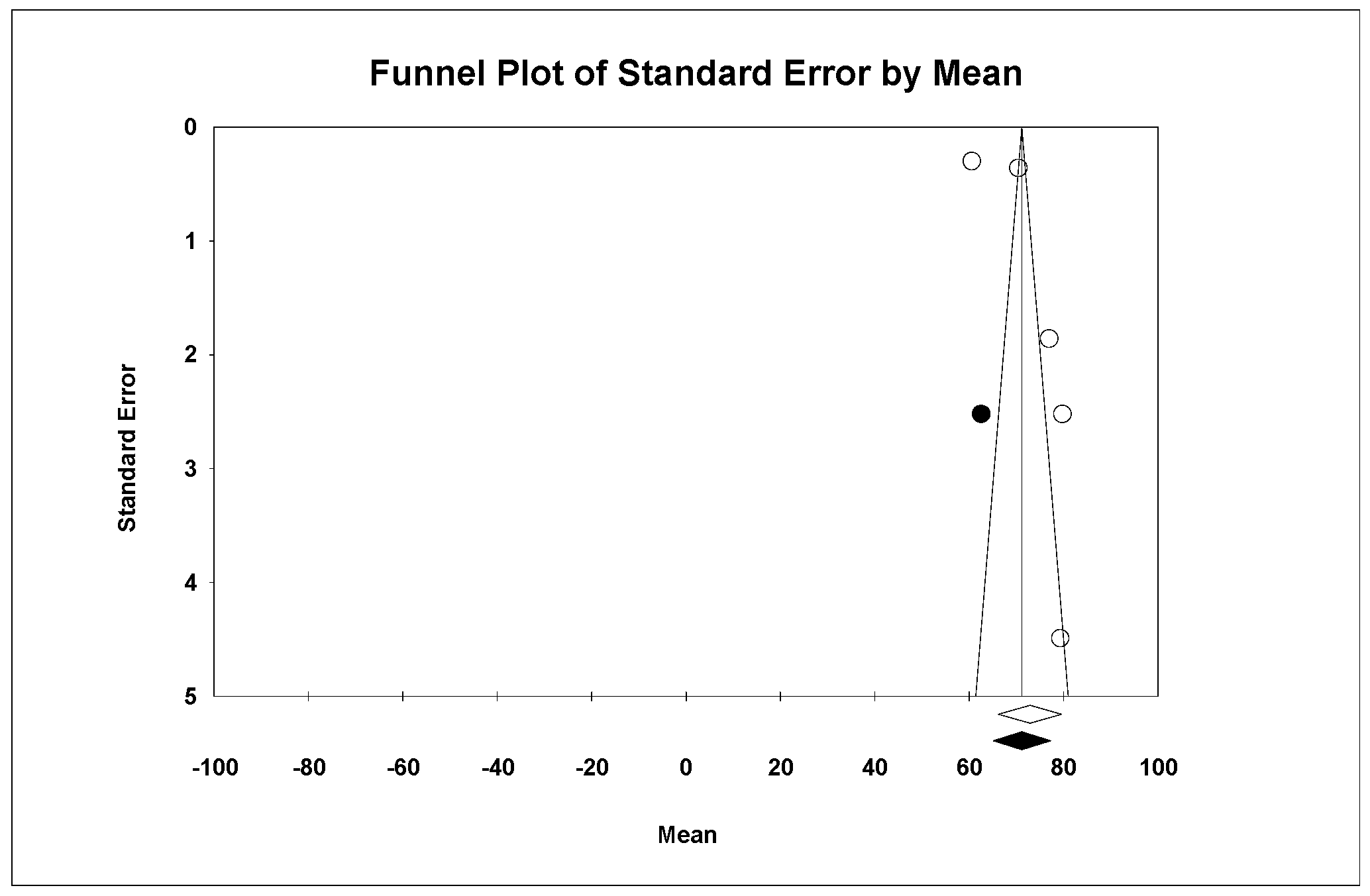
| Properties | Ceramic Group | Mean (IC 95%) | Q-Test for the Difference Between Groups | Independent Variables of the Meta-Regression |
|---|---|---|---|---|
| Young modulus | LDS | 73.9 (61.7–86.2) | Q = 0.31 p < 0.577 | No significance |
| ZLS | 70.4 (69.7–71.1) | |||
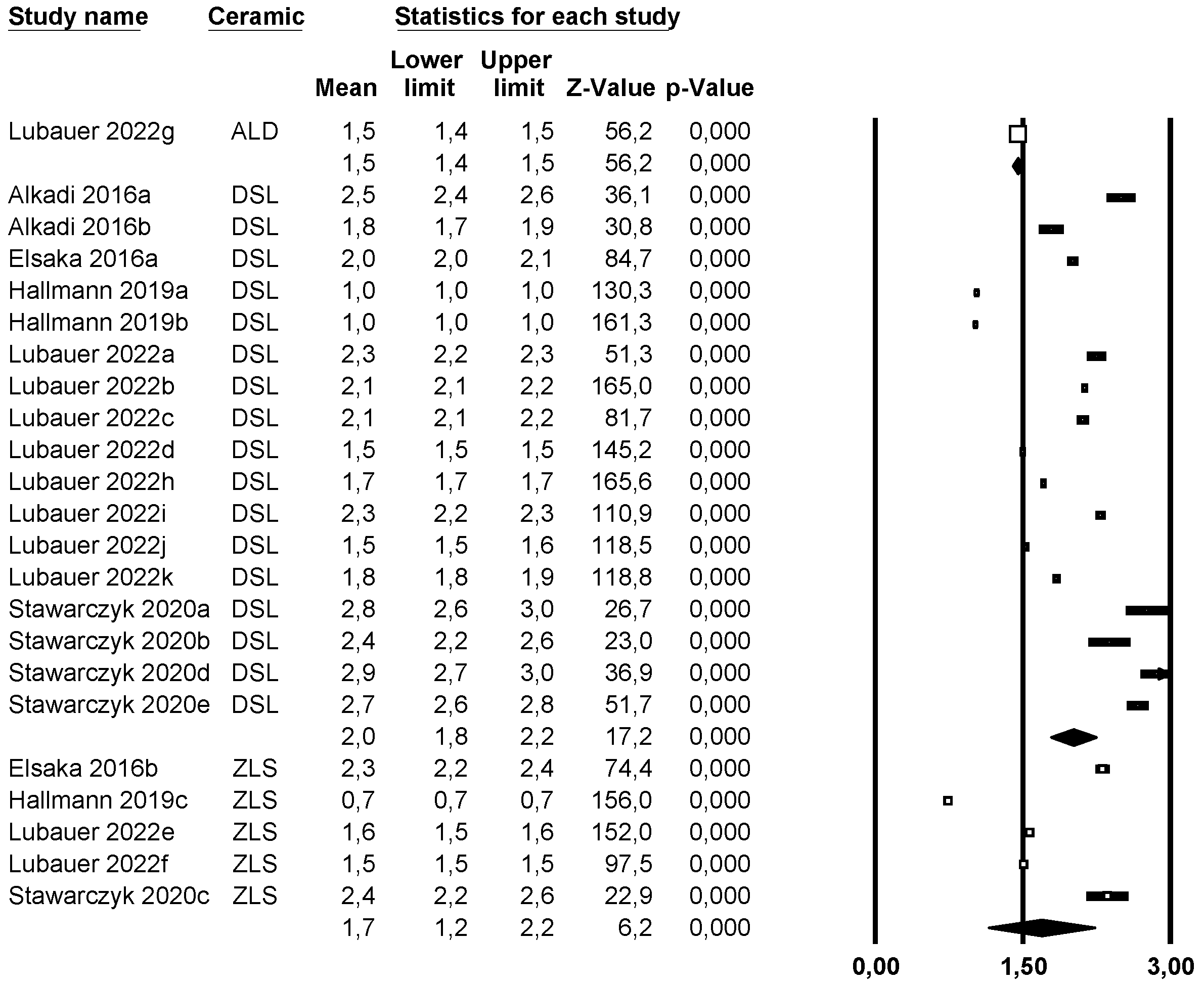
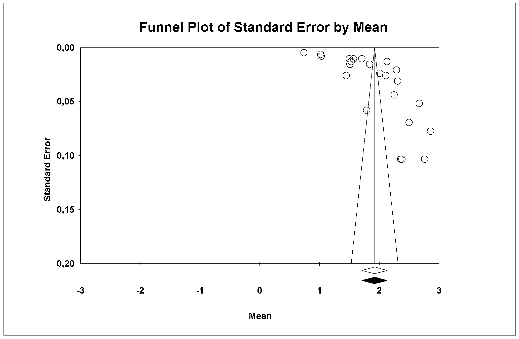
| Fracture resistance | ALD | 1.5 (1.4–1.5) | Q = 22.97 p < 0.05 | No significance |
| LDS | 2 (1.8–2.2) | |||
| ZLS | 1.7 (1.2–2.2) | |||
| Flexural resistance | ALD | 388.1 (241.5–534.7) | Q = 0.126 p = 0.939 | Press technique (p < 0.05) |
| LDS | 384.7 (353–416.3) | |||
| ZLS | 395.7 (343.9–447.5) | |||
| Hardness | LDS | 5.7 (5.2–6.2) | Q = 0.001 p = 0.979 | Pressed technique (p < 0.05) |
| ZLS | 5.7 (5–6.4) | |||
| Wear | LDS | −0.11 (−0.12–(−0.11)) | Q = 19.15 p < 0.05 | ZLS (p < 0.05) |
| ZLS | −0.16 (−0.17–(−0.14)) | |||
| Roughness (Ra) | ALD | 2.04 (−1.903–5.994) | Q = 5.345 p = 0.069 | ALD (p < 0.05) Pressed technique (p < 0.05) |
| LDS | 0.015 (0.011–0.019) | |||
| ZLS | 0.01 (0.008–0.012) | |||
| Roughness (Rz) | ALD | 14.8 (−12.2–41.7) | Q = 11.102 p < 0.05 | ZLS (p < 0.05) Pressed technique (p < 0.05) |
| LDS | 0.05 (0.05–0.06) | |||
| ZLS | 0.04 (0.038–0.042) | |||
| Wear | LDS | −0.11 (−0.12–(−0.11)) | Q = 0.001 p = 0.979 | ZLS (p < 0.05) |
| ZLS | −0.16 (−0.17–(−0.14)) | |||
| Translucency | ALD | 27.2 (25.4–28.1) | Q = 4.576 p = 0.101 | NS |
| LDS | 23.7 (20.7–26.6) | |||
| ZLS | 23.9 (17.4–30.3) |
Disclaimer/Publisher’s Note: The statements, opinions and data contained in all publications are solely those of the individual author(s) and contributor(s) and not of MDPI and/or the editor(s). MDPI and/or the editor(s) disclaim responsibility for any injury to people or property resulting from any ideas, methods, instructions or products referred to in the content. |
© 2025 by the authors. Licensee MDPI, Basel, Switzerland. This article is an open access article distributed under the terms and conditions of the Creative Commons Attribution (CC BY) license (https://creativecommons.org/licenses/by/4.0/).
Share and Cite
Guaita-Sáez, R.; Montiel-Company, J.M.; Agustín-Panadero, R.; Fons-Badal, C.; Serra-Pastor, B.; Solá-Ruiz, M.F. Analysis of Different Lithium Disilicate Ceramics According to Their Composition and Processing Technique—A Systematic Review and Meta-Analysis. Materials 2025, 18, 2709. https://doi.org/10.3390/ma18122709
Guaita-Sáez R, Montiel-Company JM, Agustín-Panadero R, Fons-Badal C, Serra-Pastor B, Solá-Ruiz MF. Analysis of Different Lithium Disilicate Ceramics According to Their Composition and Processing Technique—A Systematic Review and Meta-Analysis. Materials. 2025; 18(12):2709. https://doi.org/10.3390/ma18122709
Chicago/Turabian StyleGuaita-Sáez, Rubén, Jose María Montiel-Company, Rubén Agustín-Panadero, Carla Fons-Badal, Blanca Serra-Pastor, and María Fernanda Solá-Ruiz. 2025. "Analysis of Different Lithium Disilicate Ceramics According to Their Composition and Processing Technique—A Systematic Review and Meta-Analysis" Materials 18, no. 12: 2709. https://doi.org/10.3390/ma18122709
APA StyleGuaita-Sáez, R., Montiel-Company, J. M., Agustín-Panadero, R., Fons-Badal, C., Serra-Pastor, B., & Solá-Ruiz, M. F. (2025). Analysis of Different Lithium Disilicate Ceramics According to Their Composition and Processing Technique—A Systematic Review and Meta-Analysis. Materials, 18(12), 2709. https://doi.org/10.3390/ma18122709