Article

Mg–Zn–Ca Alloy with Ultra-High Ductility and Strength Processed by Screw Rolling

1
Aerospace and Mechanical College, Shandong University of Aeronautics, Binzhou 256600, China
2
Shandong Provincial Key Laboratory of Advanced Technology and Equipment for Laser Additive Manufacturing, Binzhou 256600, China
3
Shandong Key Laboratory of Advanced Engine Piston Assembly, Binzhou 256600, China
4
Magnestum Technology lnnovation Center, School of Materials Science and Engineering, Seoul National University, Seoul 08826, Republic of Korea
*
Authors to whom correspondence should be addressed.
Materials 2025, 18(11), 2586; https://doi.org/10.3390/ma18112586
Submission received: 8 May 2025 / Revised: 26 May 2025 / Accepted: 29 May 2025 / Published: 1 June 2025

Abstract

:
Mg alloys are highly attractive for biodegradable surgical clips because of their low density and good biocompatibility; however, their limited strength and ductility restrict their widespread application. To overcome this limitation, this study employed screw rolling (SR) to produce a Mg–3Zn–0.2Ca alloy with a fine microstructure and an average grain size of 1.6 µm. Experimental results showed that the SR process improved the comprehensive tensile properties of the alloy, increasing the yield strength, ultimate tensile strength, and elongation from 192.6, 234.4 MPa, and 21.7% for the pre-extruded alloy to 252.3, 289 MPa, and 39.5%, respectively. Quantitative analysis of the strengthening behaviour identified grain refinement as the primary strengthening mechanism, along with considerable contributions from Orowan and dislocation strengthening. The ultra-high-tensile ductility was primarily attributed to the low internal stress, nano-sized precipitates, texture weakening, and activation of multiple slip systems. These findings provide a strategy for simultaneously increasing the ductility and strength of Mg alloys and lay a foundation for applying them as biodegradable clips.

Graphical Abstract

1. Introduction

Biodegradable clips for laparoscopic surgery have attracted widespread attention because they can avoid additional pain and injury caused by removal. Among the different types of surgical clips, the mechanical strength of biodegradable polymer clips is relatively low, and their structure is relatively complex, which limits their application [1,2]. In recent years, Mg and its alloys have garnered growing attention in the realm of biomedical materials because of their excellent biocompatibility and biodegradability [3,4,5,6]. However, pure Mg has limited clinical viability because of its low ductility and strength, which are attributed to its hexagonal close-packed (HCP) structure [7]. Adding alloying elements or imposing severe plastic deformation are both effective methods for improving these mechanical properties [8].
Research has shown that Mg–Zn–Ca alloys are suitable for such biodegradable clips [9]. Zn and Ca are important minerals for the human body, offering superior compatibility with human tissues compared to Al and rare-earth elements [10,11]. Incorporating Zn and Ca can strengthen Mg alloys through both grain refinement and solid-solution mechanisms; additionally, they form uniformly distributed secondary-phase distributions, which further boost the alloy’s mechanical properties [12]. Moreover, when Zn and Ca are incorporated into Mg alloys, together with the internal stress concentration caused by extrusion, they activate the <c + a> slip, thereby enhancing the ductility of the resultant Mg–Zn–Ca alloy [13].
Meanwhile, some researchers have plastically deformed Mg–Zn–Ca alloys to improve their mechanical properties. For example, a Mg–1.0Zn–0.5Ca alloy extruded at 370 °C demonstrated a high elongation of 44%, but its yield strength (YS, 105 MPa) and ultimate tensile strength (UTS, 205 MPa) were low [14]. By contrast, a Mg–0.6Zn–0.5Ca alloy, processed via double equal channel angular pressing (D-ECAP) at 280 °C, exhibited a higher YS of 372 MPa but a lower elongation of 7% [15]. In addition, significant residual stress occurs at the apex of surgical clips during closure, easily leading to cracking [16]. Therefore, optimising the processing method to improve the plasticity and overcome the strength–ductility trade-off of alloys for such clips is important.
Screw rolling (SR) is an emerging deformation processing method with considerable application potential that can refine alloy grains by inducing a large plastic deformation [17,18,19,20,21]. Notably, SR always produces parts with smooth surfaces, which is a considerable advantage of this method. Further, the area reduction achieved by one rolling pass of an SR mill is approximately equal to that obtained by 6–8 rolling passes in a traditional mill, indicating its higher efficiency [22]. The SR deformation process differs from extrusion or rolling but is similar to torsion deformation. As the rolls rotate to deform the billet, it also moves forward in a spiralling motion, which enables the mill to greatly reduce the billet’s cross-sectional area. The heat generated by this large plastic deformation and friction may cause dynamic recrystallisation (DRX). Thus, the SR process enables the production of materials with fine-grained structures and enhanced mechanical properties [23].
However, to the best of our knowledge, the SR technique has only rarely been applied to Mg alloys [24,25,26]. When pure Mg [24] was processed using SR technology, its YS increased from 55 to 146 MPa after 11 SR passes, demonstrating the efficacy of SR for strengthening Mg. In another study, SR processing increased the YS of a pre-extruded Mg–3.71Zn–2.04Al–0.63Ca–0.62Mn alloy [25] from 225.3 to 272.7 MPa because of grain refinement and the dissolution of the secondary phases; however, its elongation decreased from 23.9 to 8.3%. Meanwhile, the fracture elongation of a Mg–5.56%Zn–0.82%Mn alloy [26] increased from 15.2% for the as-received samples to 27.8% after nine SR passes at 300 °C; this increase of 82.9% was primarily attributed to its microstructure and weakened texture. In addition, SR promoted DRX and decreased the elevated dislocation density that was initially induced by work hardening in the as-received sample. Moreover, the YS increased from 115 to 189 MPa through grain refinement and precipitation strengthening. Notably, the final microstructure and texture of a Mg alloy are influenced not just by the alloying elements but also by the processing parameters, such as the number of processing passes and the processing temperature.
Our previous studies showed that the Mg–3Zn–0.2Ca (wt.%) alloy has good biocompatibility, and its YS and elongation were 205 MPa and 17.85%, respectively [27,28]. Therefore, in this study, a Mg–3Zn–0.2Ca alloy was pre-extruded and then subjected to SR to improve its strength and plasticity. An analysis was conducted on the microstructure, texture, secondary-phase, and mechanical performance of the SR-processed Mg–Zn–Ca alloy. The outcomes offer a fresh strategy for boosting the mechanical properties of Mg-based alloys.

2. Experimental Details

This study employed a pure Mg (99.99%) ingot, Zn particles (99.99%, Ke Wei company of Tianjin University Co. Ltd., Tianjin, China), and a Mg–25Ca intermediate alloy as the raw materials. The Mg–3Zn–0.2Ca alloy was synthesized using a vacuum induction melting furnace (Shanghai Chenhua Science Technology Co. Ltd., ZG-10, Shanghai, China) in an Ar atmosphere. The raw materials were melted and stirred at 720 °C for 5 min to homogenise the composition before casting in a graphite mould at 690 °C. After cooling naturally in the furnace, rod billets with a 60 mm diameter were produced. Next, they underwent a homogenization process at 400 °C for 24 h. Following this, a hot extrusion was carried out at 320 °C using a YQ32-315 extruder (Shangdong DaYin Industry Machine Co., Ltd., Jinan, China), with an extrusion ratio of 6:1, resulting in a pre-extruded bar measuring ∅25 mm. The SR process [29] was then performed, with a 30-min anneal before each pass in the sequence of 320, 300, 280, 260, 240, 220, and 200 °C [24]. Finally, a ∅16 mm SR bar was obtained. Figure 1 schematically illustrates the process for preparing the alloy. Hereafter, the specimens identified as 3ZX and SR-3ZX correspond to the pre-extruded and SR Mg–3Zn–0.2Ca (wt%) samples, respectively.
To observe the microstructures of the alloy specimens, the test specimens were sectioned perpendicular to the direction of extrusion. The microstructure was examined using optical microscopy (OM; OLYMPUS, U-TV0.5XC-3, Tokyo, Japan), scanning electron microscopy (SEM; Helios NanoLab 460HP, Hillsboro, OR, USA), transmission electron microscopy (TEM; Talos F200X, Thermo Fisher Scientific, Waltham, MA, USA), and electron backscatter diffraction (EBSD; Oxford Instruments, NordlysNano, Oxford, UK). For the EBSD analysis, OIM (https://www.edax.com/products/ebsd/oim-analysis, accessed on 8 May 2025) and ATEX software were employed. The grain size was determined by applying the linear intercept method to the obtained EBSD maps.
The hardness values of the specimens were measured using a Vickers hardness tester (HMV-2T, Shimadzu, Kyoto, Japan) with a load of 9.8 N for 20 s. Each specimen was randomly tested in at least six locations, and the values were averaged. Specimens for tensile testing were crafted from a location 0.4 mm from the edge of the bar in the extrusion direction, following the dimensions depicted in Figure 2. The mechanical performance was assessed by tensile tests using a DNS universal testing machine with a strain rate of 10−4 s−1 at ambient temperature. To ensure accuracy, three identical samples were tested. The fracture surface was observed via SEM, and energy-dispersive X-ray spectroscopy (EDS) was used to analyse the chemical composition of the sample and fracture surfaces. A typical TEM dual-beam analysis was carried out to identify the types of dislocation activation.

3. Results

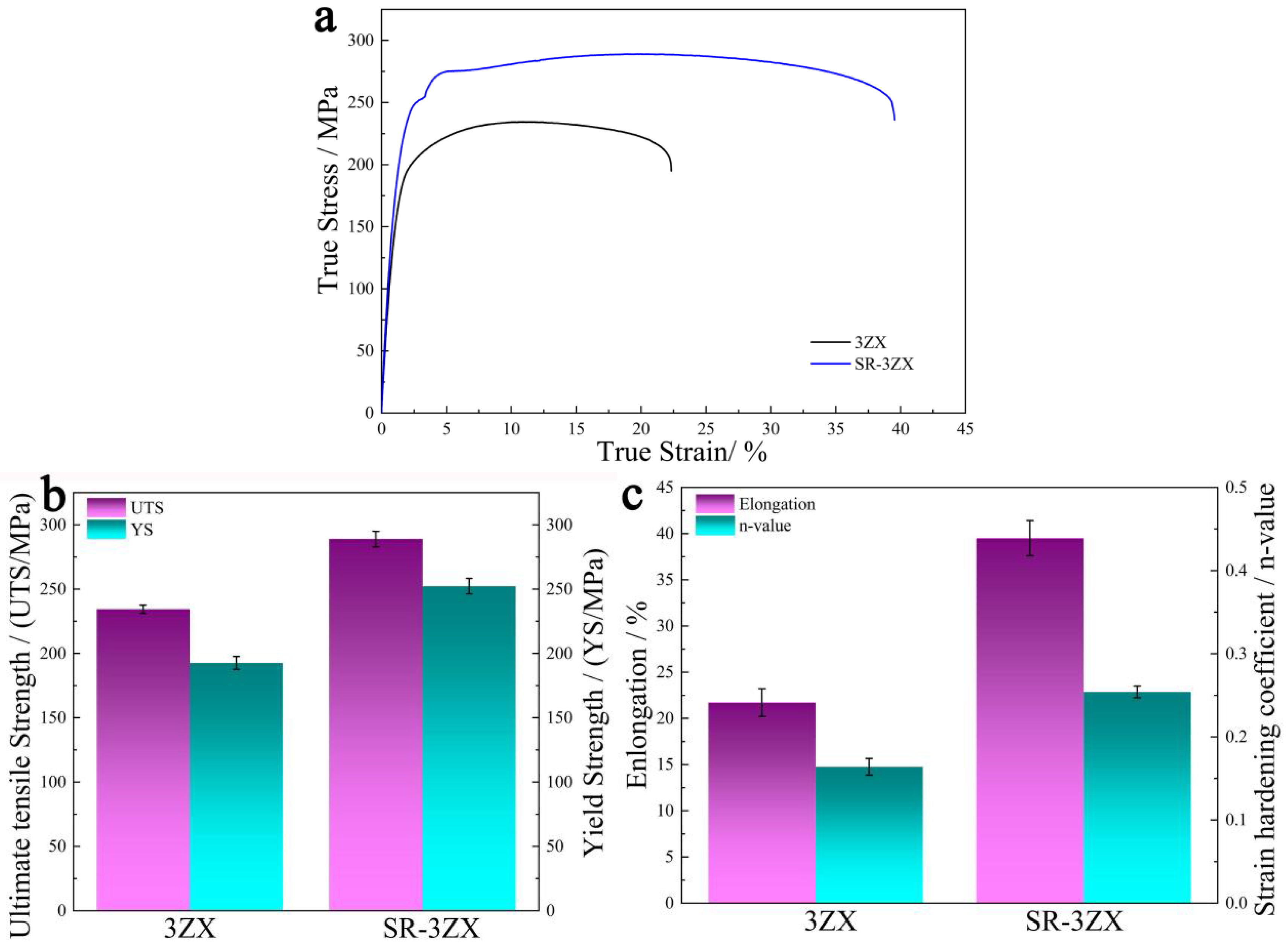
3.1. Tensile Properties

Figure 3a illustrates the true stress–strain curves for 3ZX and SR-3ZX, and Figure 3b,c plot their corresponding mechanical performance parameters. The 3ZX specimen exhibits a low UTS of ~234.4 MPa, YS of ~192.6 MPa, and elongation of ~21.7%. Remarkably, for SR-3ZX, these values increase simultaneously to ~289 MPa, ~252.3 MPa, and ~39.5%, respectively. Furthermore, the strain hardening coefficients (n) of 3ZX and SR-3ZX are 0.16 and 0.25, respectively. An increased value of n indicates that the material is capable of withstanding greater strain hardening [30,31]; thus, SR-3ZX also exhibits superior plasticity. In addition, the hardness of 3ZX increased from ~70 to ~82 HV after SR. Overall, SR-3ZX demonstrates favourable mechanical performance, successfully balancing robust strength with remarkable ductility.
Figure 4 shows the fracture surfaces of 3ZX and SR-3ZX following tensile tests conducted at room temperature. As shown in Figure 4a,b, the 3ZX fracture surface exhibits quasi-cleavage fracture characteristics, containing cleavage planes (blue arrows) and a few dimples (yellow arrows). However, as shown in Figure 4c,d, the fracture morphology of SR-3ZX exhibits microvoid coalescence, as evidenced by the presence of many dimples and the absence of typical cleavage planes. Compared with those of SR-3ZX, the 3ZX dimples are smaller and shallower. These phenomena confirm that the SR process significantly increases the ductility of the alloy, as demonstrated by these typical features of a ductile fracture surface. In addition, the green arrow in Figure 4d shows that the precipitates are uniformly distributed at the bottoms of the dimples in the SR-3ZX fracture surface. Further, EDS analysis reveals that the chemical composition of point 1 indicated by the green arrow is 69.87 at.% Mg, 17.62 at.% Zn, and 12.51 at.% Ca, implying that these particles are Ca2Mg6Zn3 precipitates. Such nanoscale Ca2Mg6Zn3 precipitates not only hinder dislocation movement but also promote dislocation multiplication [32], which facilitates dimple formation and improves the material toughness. Overall, the fracture surface clearly indicates the remarkable ductility of the SR-3ZX alloy.

3.2. Microstructures

Figure 5 shows optical images and transverse cross-sectional SEM images of the microstructures of 3ZX and SR-3ZX. Figure 5a,b reveal that, unlike SR-3ZX, which comprises ultra-fine equiaxed DRX grains, the 3ZX structure contains both the original coarse grains and fine equiaxed grains. This indicates that the shear strain induced by SR provides sufficient energy for DRX. As shown in Figure 5c,d, many precipitates are uniformly distributed in both alloys. By combining the phase diagram with the EDS results from points 1–4 in Figure 5c,d, which are summarised in Table 1, these precipitates are determined to be Ca2Mg6Zn3 and Mg2Zn11.
On average, the SR-3ZX grains are significantly smaller and more uniform in size than the 3ZX grains. To further observe the SR-3ZX microstructure, TEM images were obtained (Figure 6). The bright-field (BF) TEM image of SR-3ZX in Figure 6a displays lattice distortion within the grains, which appears black in this image. The high-angle annular dark-field (HAADF) image of SR-3ZX in Figure 6b reveals a large amount of nanoscale precipitates with an average diameter of ~34 nm. The average interparticle spacing was determined to be ~338 nm by the linear intercept method. In SR-3ZX, these precipitates are evenly spread throughout both the grain boundaries and the interiors of the grains. Selected area electron diffraction (SAED) patterns were recorded from points A–C in Figure 6c, which identified the phases as α-Mg, Mg2Zn11, and Ca2Mg6Zn3, respectively (Figure 6d–f). These findings are consistent with the SEM results in Figure 5.
Figure 7 shows the EBSD results for 3ZX and SR-3ZX. The inverse pole figure (IPF) maps of 3ZX and SR-3ZX in Figure 7a,b, respectively, reveal that SR reconfigures the grain boundaries. The 3ZX specimen exhibits a distinct bimodal microstructure comprising equiaxed recrystallised grains with diameters of several micrometres and non-recrystallised regions with coarse elongated grains. By contrast, the SR-3ZX sample is fully recrystallised. In addition, the grains are more randomly orientated in the SR sample, as evidenced by the more-randomly coloured grains in its IPF (Figure 7a). Furthermore, as shown in Figure 7e,f, the average grain sizes of 3ZX and SR-3ZX are 32.5 and 1.6 μm, respectively, corresponding to a reduction of 95.1% after SR. The corresponding kernel average misorientation (KAM) map in Figure 7c indicates that, in the non-recrystallised region of 3ZX, more severe lattice distortion is retained than in the recrystallised region; this contributes to a high geometrically necessary dislocation (GND) density of 1.21 × 10¹⁴ m−2, which was calculated using ATEX software based on the EBSD data. For SR-3ZX (Figure 7d), however, the residual stress distribution is more uniform, and the high-stress regions are smaller. Further, the GND density decreases to 5.35 × 10¹³ m−2. This indicates that the internal stress and high density of dislocations in the material are released via recrystallisation during the SR process, making the internal stress in SR-3ZX lower than that of 3ZX. The IPF for 3ZX (Figure 7g) reveals a bimodal texture (similar to a rare-earth texture), indicating the formation of a non-basal texture during the hot extrusion procedure. Following SR, however, the grain boundary misorientation angle of SR-3ZX (Figure 7h) is deflected, forming a similar <10−12>//RD texture [33,34]. Compared with that of 3ZX, the maximum texture intensity of SR-3ZX decreases from 7.80 to 1.99, implying superior plasticity.
Figure 8 shows the Schmid factors (SFs) of different slip systems in 3ZX and SR-3ZX. In 3ZX, the SF of the basal <a> slip is lower than those of the prismatic <a>, pyramidal <a>, and pyramidal <a + c> slips, indicating that the activation of the non-basal slip systems is more likely. By contrast, the SFs of the different slip systems in SR-3ZX do not differ significantly, indicating that more slip systems may have been activated. In addition, the average SF of the basal slip increases after SR, which improves the material ductility.

4. Discussion

Figure 9 presents a comparison of the mechanical properties of the materials assessed in this study with those of other biomedical Mg–Zn alloys [35,36,37,38,39,40,41]. Notably, the SR-3ZX alloy produced in this work is considerably more ductile than all the other Mg–Zn alloys (Figure 9a), but its strength is comparable to many of the high-strength Mg–Zn alloys (Figure 9b). Thus, the SR processing of these alloys overcomes the strength–ductility trade-off found in previously documented Mg–Zn alloys. The elevated ductility indicates the enhanced formability of the Mg alloy at ambient temperature, highlighting the significant potential of SR-3ZX as a Mg-based biodegradable clip.
During SR plastic deformation, the bar experiences uniform stress in three directions and moves forward while spiralling due to the frictional force between the roll and the bar. Throughout several passes, the bar undergoes torsion deformation, leading to a decrease in diameter and an increase in length. As deformation occurs, the external shear and compressive stresses gradually infiltrate the metal bar, causing metal flow and plastic deformation, which subsequently refine the grains within the microstructure. In particular, the strain energy generated by SR encourages grain nucleation for DRX, leading to notable grain refinement of SR-3ZX under the influence of dual-stress conditions.
The influence of grain refinement on the YS can be assessed through the Hall–Petch relationship. In addition, residual dislocations can enhance the strength. The presence of GNDs impedes dislocation activation during plastic deformation, thereby enhancing the YS of SR-3ZX. Furthermore, the Orowan mechanism, which refers to dislocations bowing around obstacles such as precipitates, can also strengthen the material. In SR-3ZX, the high number fraction of nanoscale secondary phases and the short distance between these precipitates substantially increased the degree of Orowan strengthening.
To further quantify the contributions of the grain boundary strengthening (σGB), Orowan strengthening (σOrowan), and dislocation strengthening (σDislocation) mechanisms to the YS of SR-3ZX, the theoretical YS value of each possible strengthening mechanism was calculated using the following equations [42,43,44,45,46,47,48]:
σGB = σ0 + kd−1/2,
σ O r o w a n = 0.4 M G b π λ l n d 1 b / 1 ν M g   ,
σ D i s l o c a t i o n = M α G b ρ D i s l o c a t i o n ,
where σ0 is 44 MPa for the Mg–Zn–Ca alloy [13]; k represents the slope of the Hall–Petch relation, with a value of 153 MPa μm1/2; d refers to the average grain size of SR-3ZX; the value of the Taylor factor, symbolized as M, is 3.5; the shear modulus, indicated by G, is 17 GPa for the Mg–Zn–Ca alloy; the Burgers vector, symbolized as b, has a magnitude of 0.32 nm; λ refers to the average interparticle spacing; and the effective diameter of the precipitates is indicated by d1. For magnesium, Poisson’s ratio, denoted as νMg, is 0.35. The numerical constant α, which is 0.2, assesses the efficiency of dislocation strengthening. The total dislocation density, represented by ρDislocation, is approximately 1.5 times greater than the GND density.
The contributions of σGB, σOrowan, and σDislocation to the YS of SR-3ZX were calculated to be 165.0, 41.5, and 34.1 MPa, respectively. The contributions of the various strengthening mechanisms and a comparison with the experimental results are shown in Figure 10. The predicted YS was 240.6 MPa, which is slightly lower than the experimental YS value of ~252.3 MPa. This discrepancy can possibly be attributed to texture strengthening, as the texture was not high and therefore was not considered in the analysis. Nonetheless, the three aforementioned strengthening mechanisms could be concluded to synergistically strengthen SR-3ZX [49,50,51]. Grain refinement was the main mechanism for strengthening in SR-3ZX; however, the contributions of the Orowan and dislocation strengthening mechanisms were also not trivial.
Additionally, SR-3ZX showed exceptional ductility, achieving a fracture elongation reaching 39.5%. To explore the properties underlying this exceptional ductility, the TEM results used to identify the activated dislocations of SR-3ZX under the double-beam condition are shown in Figure 11. Figure 11a,b present BF and dark-field (DF) images under the two-beam condition of g = [–1011], respectively. Numerous <a> dislocations (marked in red) and a small number of <c> dislocations (yellow) are clearly identified. The <c> and <c + a> dislocations (green) are observed under g = [–110–1] (Figure 11b). As shown in Figure 11c,f, under the g = [–2–110] vector, <a> dislocations are mainly observed, accompanied by a small number of <c + a> dislocations. These results indicate that <a>, <c>, and <c + a> dislocations can all be activated in SR-3ZX. For HCP metals, the scarcity of slip systems often results in insufficient ductility. Therefore, the high plasticity of SR-3ZX is due to the widespread activation of slip systems. The BF TEM images (Figure 11d–f) reveal that a large number of ultra-fine nanoprecipitates pin the dislocations, and the activation of various slip systems increases the number of dislocation intersections caused by different dislocation movements. These enhance the resistance to dislocation movement through SR-3ZX, thereby improving its strength.
Under SR, the bimodal texture of 3ZX changed to a more uniform <10−12>//RD texture in SR-3ZX because of the large plastic deformation and DRX induced by the SR process. Additionally, the texture weakened. Therefore, the stress concentration could be more easily released, as evidenced by the higher fracture elongation of SR-3ZX. Texture weakening, therefore, contributed to improving the plasticity of SR-3ZX. Moreover, uniformly distributed nanoprecipitates in SR-3ZX (Figure 6) promoted dislocation accumulation, yielding a high ductility [52,53,54]. In addition, the lower dislocation density in SR-3ZX (Figure 7d) provided additional space for dislocation sliding and accumulation during deformation, which may have reduced the work-hardening capability and improved ductility [55,56]. Finally, the initiation of basal and non-basal slip systems provided more dislocations (Figure 11) to promote plastic deformation; thus, the SR-3ZX strain hardening (Figure 3) and ductility were improved by the SR-induced deformation.
This study demonstrated that SR-3ZX has excellent ductility, showing great promise for fabricating biodegradable surgical clips. However, we only studied its mechanical properties; hence, future studies will focus on carefully investigating its corrosion resistance and biocompatibility as well as optimising its structure.

5. Conclusions

The microstructure, grain refinement, precipitates, texture, and tensile properties of a biomedical Mg–Zn–Ca alloy processed by SR were examined in this study. Under SR, the average grain size decreased significantly from 32.5 μm for 3ZX to 1.6 μm for SR-3ZX. Moreover, SR-3ZX exhibited an ultra-high elongation of ~39.5%, an excellent YS of ~252.3 MPa, and a UTS of ~289 MPa, which were 82.0%, 31%, and 23.3% higher, respectively, than those of 3ZX prior to SR. The high YS of SR-3ZX primarily stemmed from grain boundary strengthening (~165 MPa), with the considerable involvement of Orowan strengthening (~41.5 MPa) induced by precipitates, as well as dislocation strengthening (~34.1 MPa). The weak internal stress, nanoscale precipitates, texture weakening, and activation of multiple slip systems were the main factors generating the ultra-high tensile ductility of SR-3ZX up to ~39.5%. These metrics make SR-3ZX a promising Mg alloy for application in biodegradable clips.

Author Contributions

H.Z.: writing—original draft, data curation, methodology. W.S.: writing—review and editing, investigation. L.D.: formal analysis, investigation. L.Z.: methodology, investigation. K.S.S.: conceptualization, resources. J.Z.: writing—review and editing, supervision. All authors have read and agreed to the published version of the manuscript.

Funding

The authors acknowledge the financial support for this work from the Natural Science Foundation of Shandong Province (Grants No. ZR2023QE119 and ZR2023QE222) and Shandong University of Aeronautics Doctoral Research Initiation Fund Project (No. 2021Y38).

Institutional Review Board Statement

Not Applicable.

Informed Consent Statement

Not Applicable.

Data Availability Statement

The raw/processed data required to reproduce these findings cannot be shared.

Conflicts of Interest

The authors declare no conflict of interest.

References

  1. Yu, X.; Li, D.; Liu, Y.; Ding, P.; He, X.; Zhao, Y.; Chen, M.; Liu, D. In vitro and in vivo studies on the degradation and biosafety of Mg-Zn-Ca-Y alloy hemostatic clip with the carotid artery of SD rat model. Mater. Sci. Eng. C 2020, 115, 111093. [Google Scholar] [CrossRef] [PubMed]
  2. Yoshida, T.; Fukumoto, T.; Urade, T.; Kido, M.; Toyama, H.; Asari, S.; Ajiki, T.; Ikeo, N.; Mukai, T.; Ku, Y. Development of a new biodegradable operative clip made of a magnesium alloy: Evaluation of its safety and tolerability for canine cholecystectomy. Surgery 2017, 161, 1553–1560. [Google Scholar] [CrossRef] [PubMed]
  3. Li, D.; Zhang, D.; Yuan, Q.; Liu, L.; Li, H.; Xiong, L.; Guo, X.; Yan, Y.; Yu, K.; Dai, Y.; et al. In vitro and in vivo assessment of the effect of biodegradable magnesium alloys on osteogenesis. Acta Biomater. 2022, 141, 454–465. [Google Scholar] [CrossRef]
  4. Liu, Y.; Liu, L.; Chu, X.; Zhang, Z.; Fu, Z.; Pan, B.; Wang, J.; Tian, H.; Liu, Y.; Yan, Y.; et al. Mechanical properties and bio-corrosion behavior of Mg-Zn-Zr alloy influenced by rotary swaging. Mater. Today Commun. 2024, 39, 108808. [Google Scholar] [CrossRef]
  5. Peng, B.; Xu, H.; Song, F.; Wen, P.; Tian, Y.; Zheng, Y. Additive manufacturing of porous magnesium alloys for biodegradable orthopedic implants: Process, design, and modification. J. Mater. Sci. Technol. 2024, 182, 79–110. [Google Scholar] [CrossRef]
  6. Tsakiris, V.; Tardei, C.; Clicinschi, F.M. Biodegradable Mg alloys for orthopedic implants—A review. J. Magnes. Alloys 2021, 9, 1884–1905. [Google Scholar] [CrossRef]
  7. Zhao, T.; Zhang, Q.; Wang, R.; Wu, J.; Hu, Y.; Jiang, B.; Pan, F. Achieving high strength-ductility synergy in Mg-Gd-Zn-Zr alloy by controlling extrusion processes parameters. Mater. Sci. Eng. A 2024, 916, 147342. [Google Scholar] [CrossRef]
  8. Zhang, X.; He, Y.; Chen, T.; Bi, G.; Li, Y.; Tang, D.; Wang, X. Coordinating the deformation of a low-alloyed magnesium alloy for a superior combination of strength and ductility through core-shell structured reinforcements. Acta Mater. 2024, 281, 120365. [Google Scholar] [CrossRef]
  9. Ikeo, N.; Nakamura, R.; Naka, K.; Hashimoto, T.; Yoshida, T.; Urade, T.; Fukushima, K.; Yabuuchi, H.; Fukumoto, T.; Ku, Y.; et al. Fabrication of a magnesium alloy with excellent ductility for biodegradable clips. Acta Biomater. 2016, 29, 468–476. [Google Scholar] [CrossRef]
  10. Holweg, P.; Berger, L.; Cihova, M.; Donohue, N.; Clement, B.; Schwarze, U.; Sommer, N.G.; Hohenberger, G.; van den Beucken, J.J.J.P.; Seibert, F.; et al. A lean magnesium–zinc–calcium alloy ZX00 used for bone fracture stabilization in a large growing-animal model. Acta Biomater. 2020, 113, 646–659. [Google Scholar] [CrossRef]
  11. Cho, D.H.; Avey, T.; Nam, K.H.; Dean, D.; Luo, A.A. In vitro and in vivo assessment of squeeze-cast Mg-Zn-Ca-Mn alloys for biomedical applications. Acta Biomater. 2022, 150, 442–455. [Google Scholar] [CrossRef] [PubMed]
  12. Chen, Y.; Xu, Z.; Smith, C.; Sankar, J. Recent advances on the development of magnesium alloys for biodegradable implants. Acta Biomater. 2014, 10, 4561–4573. [Google Scholar] [CrossRef]
  13. Zhang, H.; Ding, Y.; Li, R.; Gao, Y. Enhanced strength-ductility synergy and activation of non-basal slip in as-extruded Mg–Zn–Ca alloy via heterostructure. J. Mater. Res. Technol. 2024, 28, 1841–1851. [Google Scholar] [CrossRef]
  14. Zhang, B.P.; Geng, L.; Huang, L.J.; Zhang, X.X.; Dong, C.C. Enhanced mechanical properties in fine-grained Mg-1.0Zn-0.5Ca alloys prepared by extrusion at different temperatures. Scr. Mater. 2010, 63, 1024–1027. [Google Scholar] [CrossRef]
  15. Horky, J.; Bryta, K.; Krystian, M.; Mozdzen, G.; Mingler, B.; Sajti, L. Improving mechanical properties of lean Mg–Zn–Ca alloy for absorbable implants via Double Equal Channel Angular Pressing (D-ECAP). Mater. Sci. Eng. A 2021, 826, 142002. [Google Scholar] [CrossRef]
  16. Mao, L.; Zhou, Y.; Zheng, X.; Cai, X.; Chen, Y.; Yang, W.; Wang, J.; Zhang, J.; Song, C. Structural optimization and in vitro corrosion analysis of biodegradable Mg-Nd-Zn-Zr alloy clip. J. Mech. Behav. Biomed. Mater. 2025, 161, 106790. [Google Scholar] [CrossRef] [PubMed]
  17. Han, Y.; Zhang, X.B.; Wang, Z.X.; Xiao, Y.; Gao, X.L.; Zhao, Y.Q. Study on cooling process of copper tube after three-roll planetary rolling. Int. Commun. Heat Mass 2020, 110, 104393. [Google Scholar] [CrossRef]
  18. Shih, C.K.; Hung, C.; Hsu, R.Q. The fnite element analysis on planetary rolling process. J. Mater. Process. Technol. 2001, 113, 115–123. [Google Scholar] [CrossRef]
  19. Shih, C.K.; Hung, C. Experimental and numerical analyses on three-roll planetary rolling process. J. Mater. Process. Technol. 2003, 142, 702–709. [Google Scholar] [CrossRef]
  20. Komori, K. Simulation of deformation and temperaturein multi-pass three-roll rolling. J. Mater. Process. Technol. 1999, 92–93, 450–457. [Google Scholar] [CrossRef]
  21. Ji, C.; Niu, H.; Li, Z.; Wang, T.; Huang, Q. Deformation law and bonding mechanism of 45 carbon steel/316L stainless steel cladding tubes fabricated by three-roll skew rolling bonding process. J. Mater. Process. Technol. 2024, 325, 118277. [Google Scholar] [CrossRef]
  22. Hwang, Y.M.; Tsai, W.M.; Tsai, F.H.; Her, I. Analytical and experimental study on the spiral marks of the rolled product during three-roll planetary rolling processes. Int. J. Mach. Tools Manuf. 2006, 46, 1555–1562. [Google Scholar] [CrossRef]
  23. Wang, Y.L.; Molotnikov, A.; Diez, M.; Lapovok, R.; Kim, H.-E.; Wang, J.T.; Estrin, Y. Gradient structure produced by three roll planetary milling: Numerical simulation and microstructural observations. Mater. Sci. Eng. A 2015, 639, 165–172. [Google Scholar] [CrossRef]
  24. Diez, M.; Kim, H.E.; Serebryany, V.; Dobatkin, S.; Estrin, Y. Improving the mechanical properties of pure magnesium by three-roll planetary milling. Mater. Sci. Eng. A 2014, 612, 287–292. [Google Scholar] [CrossRef]
  25. Bahmani, A.; Arthanari, S.; Shin, K.S. Improvement of corrosion resistance and mechanical properties of a magnesium alloy using screw rolling. J. Alloys Compd. 2020, 813, 152155. [Google Scholar] [CrossRef]
  26. Wang, L.; Zhang, Z.; Zhang, H.; Wang, H.; Shin, K.S. The dynamic recrystallization and mechanical property responses during hot screw rolling on pre-aged ZM6l magnesium alloys. Mater. Sci. Eng. A 2020, 798, 140126. [Google Scholar] [CrossRef]
  27. Ding, P.; Liu, Y.; He, Y.; Liu, D.; Chen, M. In vitro and in vivo biocompatibility of Mg-Zn-Ca alloy operative clip. Bioact. Mater. 2019, 4, 236–244. [Google Scholar] [CrossRef]
  28. Yang, M.; Liu, D.; Zhang, R.; Chen, M. Microstructure and Properties of Mg-3Zn-0.2Ca Alloy for Biomedical Application. Rare Met. Mater. Eng. 2018, 47, 0093–0098. [Google Scholar] [CrossRef]
  29. Zheng, H.; Chen, M.; Li, Z.; Yu, L. Mechanism study on improving the corrosion resistance of screw rolled Mg–Zn–Ca alloy by nano-MgO addition. J. Mater. Res. Technol. 2024, 31, 3104–3116. [Google Scholar] [CrossRef]
  30. Wu, L.; Jain, A.; Brown, D.; Stoica, G.; Agnew, S.; Clausen, B.; Fielden, D.; Liaw, P. Twinning-detwinning behavior during the strain-controlled low-cycle fatigue testing of a wrought magnesium alloy, ZK60A. Acta Mater. 2008, 56, 688–695. [Google Scholar] [CrossRef]
  31. Song, B.; Xin, R.; Chen, G.; Zhang, X.; Liu, Q. Improving tensile and compressive properties of magnesium alloy plates by pre-cold rolling. Scr. Mater. 2012, 66, 1061–1064. [Google Scholar] [CrossRef]
  32. Yang, H.; Li, K.; Bu, Y.; Wu, J.; Fang, Y.; Meng, L.; Liu, J.; Wang, H. Nanoprecipitates induced dislocation pinning and multiplication strategy for designing high strength, plasticity and conductivity Cu alloys. Scr. Mater. 2021, 195, 113741. [Google Scholar] [CrossRef]
  33. Pu, Z.; Song, G.-L.; Yang, S.; Outeiro, J.; Dillon, O.; Puleo, D.; Jawahir, I. Grain refined and basal textured surface produced by burnishing for improved corrosion performance of AZ31B Mg alloy. Corros. Sci. 2012, 57, 192–201. [Google Scholar] [CrossRef]
  34. Jiang, M.G.; Xu, C.; Yan, H.; Lu, S.H.; Nakata, T.; Lao, C.S.; Chen, R.S.; Kamado, S.; Han, E.H. Correlation between dynamic recrystallization and formation of rare earth texture in a Mg-Zn-Gd magnesium alloy during extrusion. Sci. Rep. 2018, 8, 16800. [Google Scholar] [CrossRef]
  35. Bazhenov, V.; Li, A.; Komissarov, A.; Koltygin, A.; Tavolzhanskii, S.; Bautin, V.; Voropaeva, O.; Mukhametshina, A.; Tokar, A. Microstructure and mechanical and corrosion properties of hot-extruded Mg–Zn–Ca–(Mn) biodegradable alloys. J. Magnes. Alloys 2021, 9, 1428–1442. [Google Scholar] [CrossRef]
  36. Doležal, P.; Zapletal, J.; Fintová, S.; Trojanová, Z.; Greger, M.; Roupcová, P.; Podrábský, T. Influence of processing techniques on microstructure and mechanical properties of a biodegradable Mg-3Zn-2Ca alloy. Materials 2016, 9, 880. [Google Scholar] [CrossRef]
  37. Li, W.; Shen, Y.; Shen, J.; Shen, D.; Liu, X.; Zheng, Y.; Yeung, K.W.; Guan, S.; Kulyasova, O.B.; Valiev, R. In vitro and in vivo studies on pure Mg, Mg–1Ca and Mg–2Sr alloys processed by equal channel angular pressing. Nano Mater. Sci. 2020, 2, 96–108. [Google Scholar] [CrossRef]
  38. Vinogradov, A.; Vasilev, E.; Kopylov, V.I.; Linderov, M.; Brilevesky, A.; Merson, D. High performance fine-grained biodegradable Mg-Zn-Ca alloys processed by severe plastic deformation. Metals 2019, 9, 186. [Google Scholar] [CrossRef]
  39. Tong, L.B.; Chu, J.H.; Jiang, Z.H.; Kamado, S.; Zheng, M.Y. Ultra-fne grained Mg-Zn-Ca-Mn alloy with simultaneously improved strength and ductility processed by equal channel angular pressing. J. Alloys Compd. 2019, 785, 410–421. [Google Scholar] [CrossRef]
  40. Wang, W.; Blawert, C.; Zan, R.; Sun, Y.; Peng, H.; Ni, J.; Han, P.; Suo, T.; Song, Y.; Zhang, S.; et al. A novel lean alloy of biodegradable Mg–2Zn with nanograins. Bioact. Mater. 2021, 6, 4333–4341. [Google Scholar] [CrossRef]
  41. Yan, J.; Qin, Z.; Yan, K. Mechanical properties and microstructure evolution of Mg-6 wt % Zn alloy during equal-channel angular pressing. Metals 2018, 8, 841. [Google Scholar] [CrossRef]
  42. Bian, M.; Huang, X.; Chino, Y. A room temperature formable magnesium–silver–calcium sheet alloy with high ductility. Mater. Sci. Eng. A 2020, 774, 138923. [Google Scholar] [CrossRef]
  43. Gao, W.; Wang, X.; Lin, Y.; Wang, X.; Liu, D.; Sun, X. Achieving ultra-high strength and ductility in a rare-earth-free magnesium alloy via precisely controlled secondary hot extrusion process with an extremely low extrusion speed. J. Magnes. Alloys 2024, 12, 5216–5230. [Google Scholar] [CrossRef]
  44. Wong, W.L.E.; Gupta, M. Development of Mg/Cu nanocomposites using microwave assisted rapid sintering. Compos. Sci. Technol. 2007, 67, 1541–1552. [Google Scholar] [CrossRef]
  45. Habibi, M.K.; Joshi, S.P.; Gupta, M. Hierarchical magnesium nano-composites for enhanced mechanical response. Acta Mater. 2010, 58, 6104–6114. [Google Scholar] [CrossRef]
  46. Han, B.Q.; Dunand, D.C. Microstructure and mechanical properties of magnesium containing high volume fractions of yttria dispersoids. Mater. Sci. Eng. A 2000, 277, 297–304. [Google Scholar] [CrossRef]
  47. Kang, Y.C.; Chan, S.L.I. Tensile properties of nanometric Al2O3 particulate-reinforced aluminum matrix composites. Mater. Chem. Phys. 2004, 85, 438–443. [Google Scholar] [CrossRef]
  48. Cáceres, C.H.; Lukác, P. Strain hardening behaviour and the Taylor factor of pure magnesium. Philos. Mag. 2008, 88, 977–989. [Google Scholar] [CrossRef]
  49. Shahin, M.; Munir, K.; Wen, C.; Li, Y. Magnesium matrix nanocomposites for orthopedic applications: A review from mechanical, corrosion, and biological perspectives. Acta Biomater. 2019, 96, 1–19. [Google Scholar] [CrossRef]
  50. Naghdi, F.; Mahmudi, R.; Kang, J.Y.; Kim, H.S. Contributions of different strengthening mechanisms to the shear strength of an extruded Mg-4Zn-0.5Ca alloy. Philos. Mag. 2015, 95, 3452–3466. [Google Scholar] [CrossRef]
  51. Habibnejad-Korayem, M.; Mahmudi, R.; Poole, W.J. Enhanced properties of Mg-based nano-composites reinforced with Al2O3 nano-particles. Mater. Sci. Eng. A 2009, 519, 198–203. [Google Scholar] [CrossRef]
  52. Soleimani, M.; Kalhor, A.; Mirzadeh, H. Transformation-induced plasticity (TRIP) in advanced steels: A review. Mater. Sci. Eng. A 2020, 795, 140023. [Google Scholar] [CrossRef]
  53. Gao, Q.; Wei, R.; Feng, S.; Chen, C.; Han, Z.; Chen, L.; Wang, T.; Wu, S.; Li, F. Ultrahigh cryogenic strength and ductility in a duplex metastable ferrous medium-entropy alloy. Scr. Mater. 2023, 228, 115334. [Google Scholar] [CrossRef]
  54. Zhang, Y.; Ding, H. Ultrafine also can be ductile: On the essence of Lüders band elongation in ultrafine-grained medium manganese steel. Mater. Sci. Eng. A 2018, 733, 220–223. [Google Scholar] [CrossRef]
  55. Wang, N.; Chen, Y.; Wu, G.; Zhao, Q.; Zhang, Z.; Zhu, L.; Luo, J. Non-equivalence contribution of geometrically necessary dislocation and statistically stored dislocation in work-hardened metals. Mater. Sci. Eng. A 2022, 836, 142728. [Google Scholar] [CrossRef]
  56. Zhang, H.; Wang, H.; Wang, J.; Rong, J.; Zha, M.; Wang, C.; Ma, P.; Jiang, Q. The synergy effect of fine and coarse grains on enhanced ductility of bimodal-structured Mg alloys. J. Alloys Compd. 2019, 780, 312–317. [Google Scholar] [CrossRef]
Figure 1. Schematic of the casting, pre-extrusion, and SR processes of the Mg–Zn–Ca alloy.
Materials 18 02586 g001
Figure 2. Dimensions of the tensile test samples (mm).
Materials 18 02586 g002
Figure 3. (a) Tensile stress–strain curves and the (b,c) corresponding mechanical properties of 3ZX and SR-3ZX: (b) UTS (left axis) and YS (right axis), as well as (c) elongation (left axis) and strain hardening coefficient (right axis). The error bars indicate the standard deviation (n = 3).
Materials 18 02586 g003
Figure 4. SEM images of fracture surfaces of (a,b) 3ZX and (c,d) SR-3ZX.
Materials 18 02586 g004
Figure 5. (a,b) Optical and (c,d) SEM images of the microstructures of (a,c) 3ZX and (b,d) SR-3ZX.
Materials 18 02586 g005
Figure 6. TEM images of SR-3ZX. (a) Bright-field (BF), (bd) HAADF, and (c) precipitate phase images; (df) diffraction spots of points A–C in (c).
Materials 18 02586 g006
Figure 7. EBSD results for 3ZX (a,c,e,g) and SR-3ZX (b,d,f,h): (a,b) IPF maps, (c,d) KAM maps, (e,f) grain size distributions, and (g,h) IPFs.
Materials 18 02586 g007
Figure 8. SF maps for various slip systems in 3ZX (ad) and SR-3ZX (eh) (basal: {0 0 0 1}<1 1 −2 0>, prismatic: {1 0 −1 0}<1 1 −2 0>, pyramidal: {1 0 −1 1}<1 1 −2 0>, pyramidal: {1 1 −2 2}<1 1 −2 3>), and corresponding histograms illustrated in (a1–h1), respectively.
Materials 18 02586 g008
Figure 9. Comparison of the mechanical properties of other biomedical Mg–Zn alloys with those prepared in this study: (a) Values of UTS and elongation, and (b) values of UTS and YS.
Materials 18 02586 g009
Figure 10. Quantified contributions of different strengthening mechanisms and a comparison of the theoretical YS with the experimentally determined YS of SR-3ZX, where the theoretical YS (σTheory) = σGB + σOrowan + σDislocation.
Materials 18 02586 g010
Figure 11. DF (ac) and corresponding BF TEM images (df) of SR-3ZX under g = [−1 0 1 1] (a,d), g = [−1 1 0 −1] (b,e), and g = [−2 −1 1 0] (c,f). The red, orange, and green arrows indicate <a>, <c>, and <a + c> dislocations.
Materials 18 02586 g011
Table 1. Chemical compositions (at.%) of points 1–4 in Figure 5 as determined via EDS.
LocationMgCaZn
158.660.9740.37
264.0515.0320.92
359.831.0139.16
461.8214.3323.85
Disclaimer/Publisher’s Note: The statements, opinions and data contained in all publications are solely those of the individual author(s) and contributor(s) and not of MDPI and/or the editor(s). MDPI and/or the editor(s) disclaim responsibility for any injury to people or property resulting from any ideas, methods, instructions or products referred to in the content.

