Article

Highly Sensitive Temperature Sensors Resulting from the Luminescent Behavior of Sm3+-Doped Ba2MgMoO6 High-Symmetry Double-Perovskite Molybdate Phosphors

by
Natalia Miniajluk-Gaweł
*,
Bartosz Bondzior
,
Maciej Ptak
and
Przemysław Jacek Dereń
*
Institute of Low Temperature and Structure Research, Polish Academy of Science, Okolna 2, 50-422 Wroclaw, Poland
*
Authors to whom correspondence should be addressed.
Materials 2024, 17(8), 1897; https://doi.org/10.3390/ma17081897
Submission received: 27 March 2024 / Revised: 15 April 2024 / Accepted: 17 April 2024 / Published: 19 April 2024
(This article belongs to the Special Issue Research in Perovskite Films)

Abstract

:
We present double-perovskite molybdate with the formula of Ba2MgMoO6 doped with Sm3+ ions as a potential red phosphor to improve the color characteristics of white-light-emitting dioded (wLEDs). The new orange–red phosphor was synthesized using the co-precipitation (CP) method, and then its structural and spectroscopic properties were determined. Red emission at 642.6 nm dominates, which results from the electric dipole (ED) transition of the 4G5/26H9/2 type, and the materials are characterized by short luminescence decay times. BMM:Sm3+ is, to our best knowledge, the clearest example of dominant red emission of Sm3+ resulting from the location of the dopant in octahedral sites of high-symmetry cubic structure. In the sample containing 0.1% Sm3+, Sm3+ ions are located in both Mg2+ and Ba2+ sites, while at higher concentrations the Ba2+ site is less preferable for doping, as a result of which the emission becomes more uniform and single-site. The relative sensitivity calculated from FIR has a maximum of 2.7% K−1 at −30 °C and another local maximum of 1.6% K−1 at 75 °C. Such value is, to the best of our knowledge, one of the highest achieved for luminescent thermometry performed using only Sm3+ ions. To sum up, the obtained materials are good candidates as red phosphor to improve the color characteristics of wLEDs, obtaining a color-rendering index (CRI) of 91 and coordinated color temperature (CCT) of 2943 K, constituting a warm white emission. In addition to this, a promising precedent for temperature sensing using high-symmetry perovskite materials is the high sensitivity achieved, which results from the high symmetry of the BMM host.

1. Introduction

Molybdates are materials based on an oxyanion group MoxOy, finding applications in fields like paramagnetic materials [1], catalysts [2], and optical materials [3,4]. Recently, the interest of the research community has grown as the double-perovskite materials were seen as promising due to their highly symmetrical structure, and molybdates are one of the most popular types of inorganic materials found to exhibit the double-perovskite structure described with the formula A2BB’O6, where A sites are occupied by alkaline metals or lanthanides, while positions B and B’ are reserved for transition metal ions. This arrangement creates a more complex and layered crystal lattice compared to the simple perovskite structure. Double-perovskite molybdates exhibit high thermal and chemical stability, and suitability for the luminescent dopants [5], which set that class of materials apart from other double-perovskites, such as halides.
Examples of these materials include Sr2FeMoO6, known for its colossal magnetoresistance (CMR) effect, which makes it a promising candidate for applications in spintronics and magnetic sensors [6], and Ba2PrMoO6, explored for its photocatalytic and photocurrent properties, making it relevant for applications in solar energy conversion and water splitting [7].
The research on the luminescence properties of double-perovskite molybdates is limited, with most of the published articles being focused on Eu3+ luminescence [5,8,9,10]. It is apparent that the rare earth dopants tend to locate themselves in highly symmetrical octahedral MgO6 sites, which are credited for their unique luminescence properties in the form of almost-monochromatic emission spectra [8,9]. This work concerns the Ba2MgMoO6 double-perovskite, and so far, only three publications [9,11,12] have been published on Eu3+ doping. It should be emphasized that this is the first work that describes the physicochemical properties of this matrix doped with Sm3+ ions.
Mo-based double-perovskite phosphors are of great interest due to reliable, lasting and stable emission, easy fabrication, low cost, high durability, and strong absorption in the NUV region [11,12,13]. The Sm3+ ion was used as an activator of phosphors because these ions have a rich structure of energy levels and can transfer electrons through the 4f-4f transitions. This makes Sm3+-doped phosphors generally exhibit multiple absorption peaks in the range of 220–550 nm, and thus they can be excited effectively using a near-UV or blue LED chip. These phosphors emit red–orange light in the visible range with four narrow emission bands, thanks to the characteristic transitions 4G5/26HJ (J = 5/2, 7/2, 9/2 and 11/2) [14,15].
Temperature is one of the most important physical parameters, so its precise measurement is extremely important. Non-contact luminescent thermometers, compared to traditional contact thermometers, offer many advantages such as non-invasive measurement, fast response, high sensitivity, ability to be used in rapidly changing conditions, and strong electromagnetic fields. The most commonly used temperature reading is based on the relative intensity ratio of two different emission bands, because this method minimizes the influence of disturbing external factors [9,16]. It turned out that the material we proposed, apart from being an excellent material for improving the quality of WLED lighting, can also be successfully used as a highly sensitive optical thermometer, which results directly from the high symmetry. The current state of the research on molybdate phosphors for temperature sensors is not extensive and mainly concerns alkaline earth molybdates [17,18,19]. However, there is no information about double-perovskites, so this work is very valuable and provides new information regarding non-contact thermometry.
In this paper, double-perovskite molybdate with the formula Ba2MgMoO6 is studied as a host for Sm3+ and as a potential red phosphor to improve the color characteristics of wLEDs. For Sm3+-doped materials for wLED, the main challenge is to obtain mostly red emission at ~645 nm at the expense of the yellow (570 nm), orange (600 nm), and NIR (710 nm) emissions [20]. We synthesized a novel orange–red phosphor using the co-precipitation method, and then investigated the luminescence properties of these phosphors as representative materials. Ba2MgMoO6 double-perovskite as a host for Sm3+ ions has the advantage of high site symmetry and host-sensitization in the near-UV region. Additionally, the obtained temperature measurements results showed that Ba2MgMoO6: Sm3+ double-perovskite has a great potential for applications in non-contact optical thermometry.

1.1. Experimental

Samples Preparation

The co-precipitation synthesis method was used to synthesize a series of Sm3+-doped Ba2MgMoO6 (BMM) double-perovskite samples, described using the formula Ba2Mg1−xSmxMoO6 (x = 0.1–1%).
Ba(CH3COO)2 (Alfa Aesar (Ward Hill, MA, USA), 99%), Mg(CH3COO)2·4H2O (Alfa Aesar, 99.95%), (NH4)6Mo7O24·4H2O (AMT) (Sigma–Aldrich (St. Louis, MO, USA), 99.99%), Sm(NO3)3 (Alfa Aesar, 99.9%) were used as starting materials in the co-precipitation method. Li2CO3 (Alfa Aesar, 99.998%) was applied to charge compensation in a ratio of 1:1, based on the amount of dopant ions. To compensate for the evaporation of magnesium ions during the sintering process, a 20% excess of magnesium ions was applied. Firstly, the stoichiometric amounts of precursors were dissolved separately in distilled water. Next, a white precipitate was formed immediately after the first droplet of AMT solution was added slowly (2 mL/min) under stirring at 200 rpm, 25 °C to the remaining clear solution. The water was evaporated and the precipitate was dried by heating at 80 °C for 20 h and then pre-sintering at 600 °C for 12 h. The final annealing was carried out at 1150 °C for 6 h with a constant heating rate of 3 °C/min using corundum crucible. After each step, the obtained products were ground for 15 min.

1.2. Research Techniques

X-ray diffraction (XRD) patterns were recorded with X’Pert ProPANalytical X-ray diffractometer (Malvern, UK), working in the reflection geometry, using CuKα radiation (λ = 1.54056 Ǻ). The data were collected in a 2Θ range from 10° to 90° with a step of 0.026°.
Raman spectra were measured using a Renishaw inVia spectrometer (Middlewich, UK) equipped with a confocal optical microscope and a diode laser operating at 830 nm.
A scanning electron microscope (FEI NOVA NanoSEM 230 (Eindhoven, The Netherlands), equipped with EDAX Genesis XM4 detector (Eindhoven, The Netherlands)) was used to characterize the morphology and chemical composition of the samples. The SEM images were recorded with an accelerating voltage of 5 kV.
Emission spectra, excitation spectra, and decay curves were measured using an FLS1000 Edinburgh Instruments (Edinburgh, Scotland) spectrophotometer in a Czerny–Turner configuration with VIS and NIR at room temperature.
The diffuse reflection spectrum was recorded using a Varian Cary 5E UV/VIS-NIR spectrophotometer (Varian Incorporation, Palo Alto, CA, USA).
The emission spectrum as a function of temperature was measured using the Linkam THMS600 (Salfords, UK) stage, which has both heating and cooling functions and Hamamatsu R928 photomultiplier (Hamamatsu, Japan). The measurement was carried out in the range from −193 °C to 150 °C with a constant step of 20 °C.

2. Results and Discussion

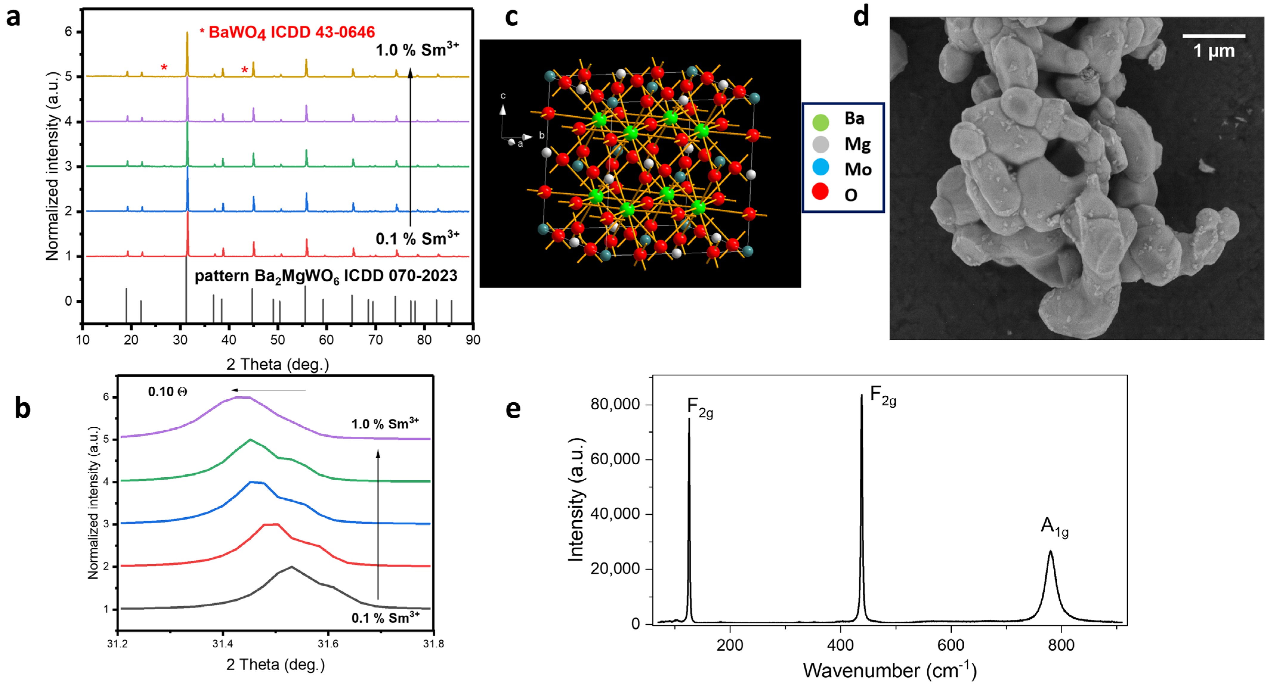
2.1. Structural Properties

Figure 1a shows X-ray diffractograms of molybdate materials synthesized using the co-precipitation method, along with the visualization of the structure. There is no standard pattern for the structure of BMM, but according to what was written in the previous publication [9], the structure of this material matches well with the ICDD 070-2023 pattern of Ba2MgWO6 (see Figure 1a). It was possible to obtain practically single-phase materials with a small amount of an additional phase of BaMoO4 ICDD 43-0646 (less than 1%), the presence of which does not in any way affect the physicochemical properties of the obtained materials. It can be seen that, as the Sm3+ concentration increases, the diffractograms shift towards lower theta angles (Figure 1b). This means that, as the amount of Sm3+ increases, the lattice constants increase because the smaller ion in the host (Mg2+, IR = 86 pm) is replaced by a larger one (Sm3+, IR = 109 pm). The obtained materials have a cubic structure, crystallizing in the Fm 3 ¯ m space group (Figure 1c), and are characterized by the following cell parameters: a = 8.1120 Å, unit cell volume V = 533.81 Å3 and Z = 4.
In the SEM image (Figure 1d), one can notice that the obtained material consists of crystallites of undefined shapes: oval with rounded edges. We determined the average crystallite sizes in the ImageJ 1.51j8 program, and they are 0.62 µm. The Supplementary Materials contains a histogram of the average crystal size, determined on the basis of 100 crystallites (Figure S1).
To further check the quality of the samples, Raman spectra were collected (see Figure 1e). The spectrum presents two narrow bands near 126 (F2g) and 438 cm−1 (F2g) as well as broader Raman peaks near 780 cm−1 (A1g), which is in good agreement with the previously reported experimental and theoretical data for molybdates and tungstates having double-perovskite architecture [21,22]. The cubic Fm 3 ¯ m structure exhibits only four optical phonons (A1g + Eg + 2F2g), but the Raman band corresponding to the Eg mode is usually too weak to be detected [22,23].

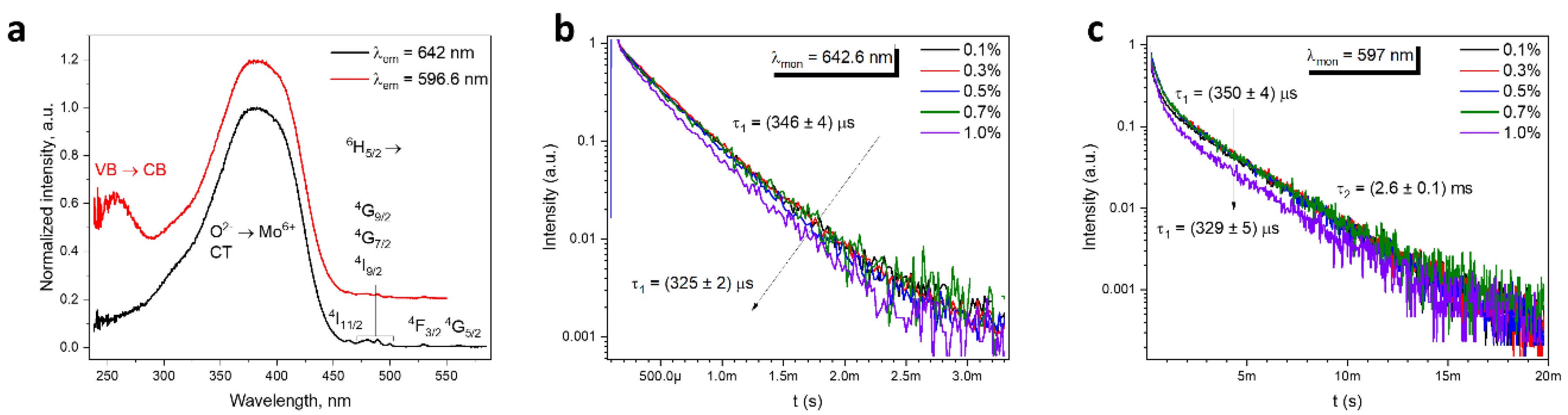
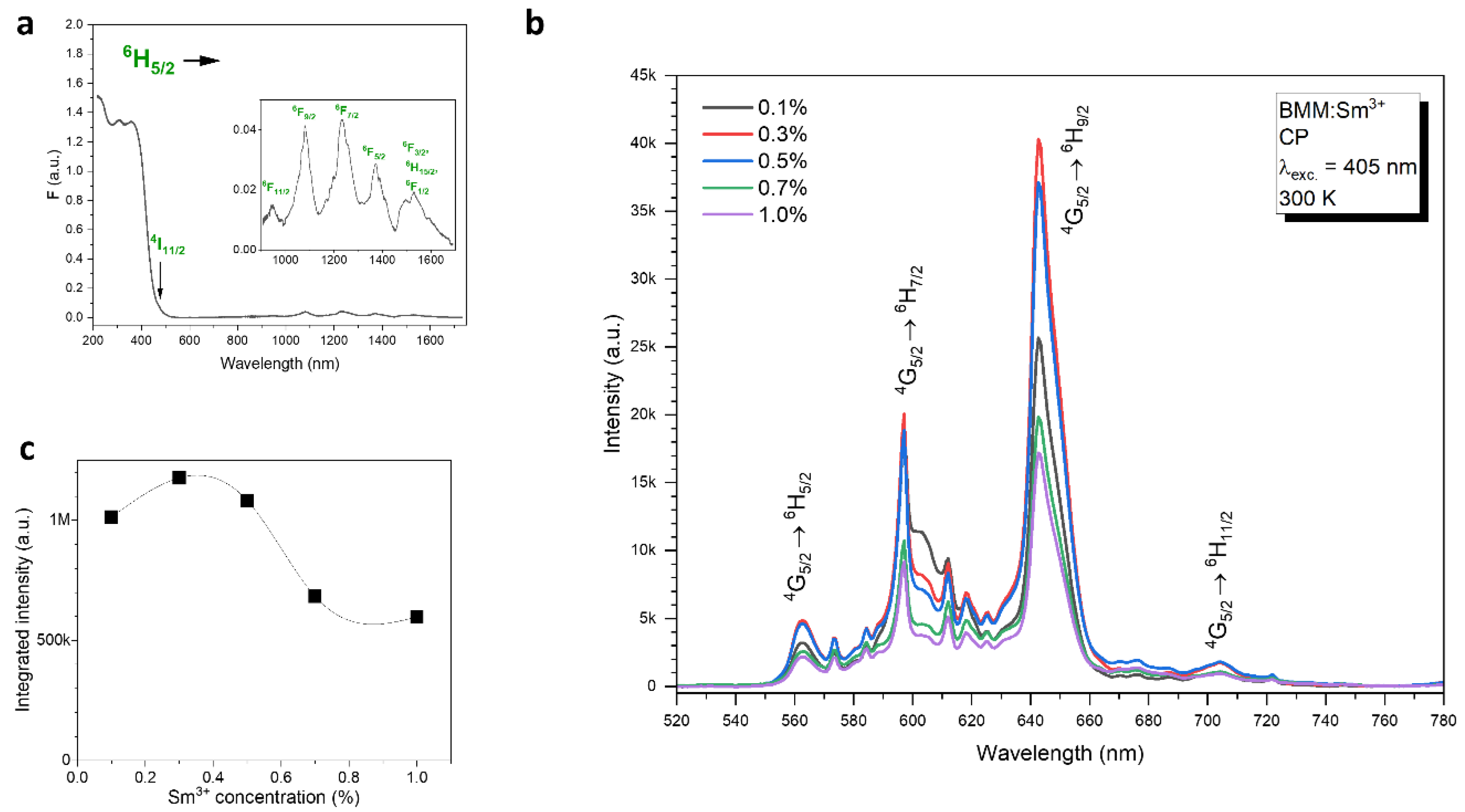
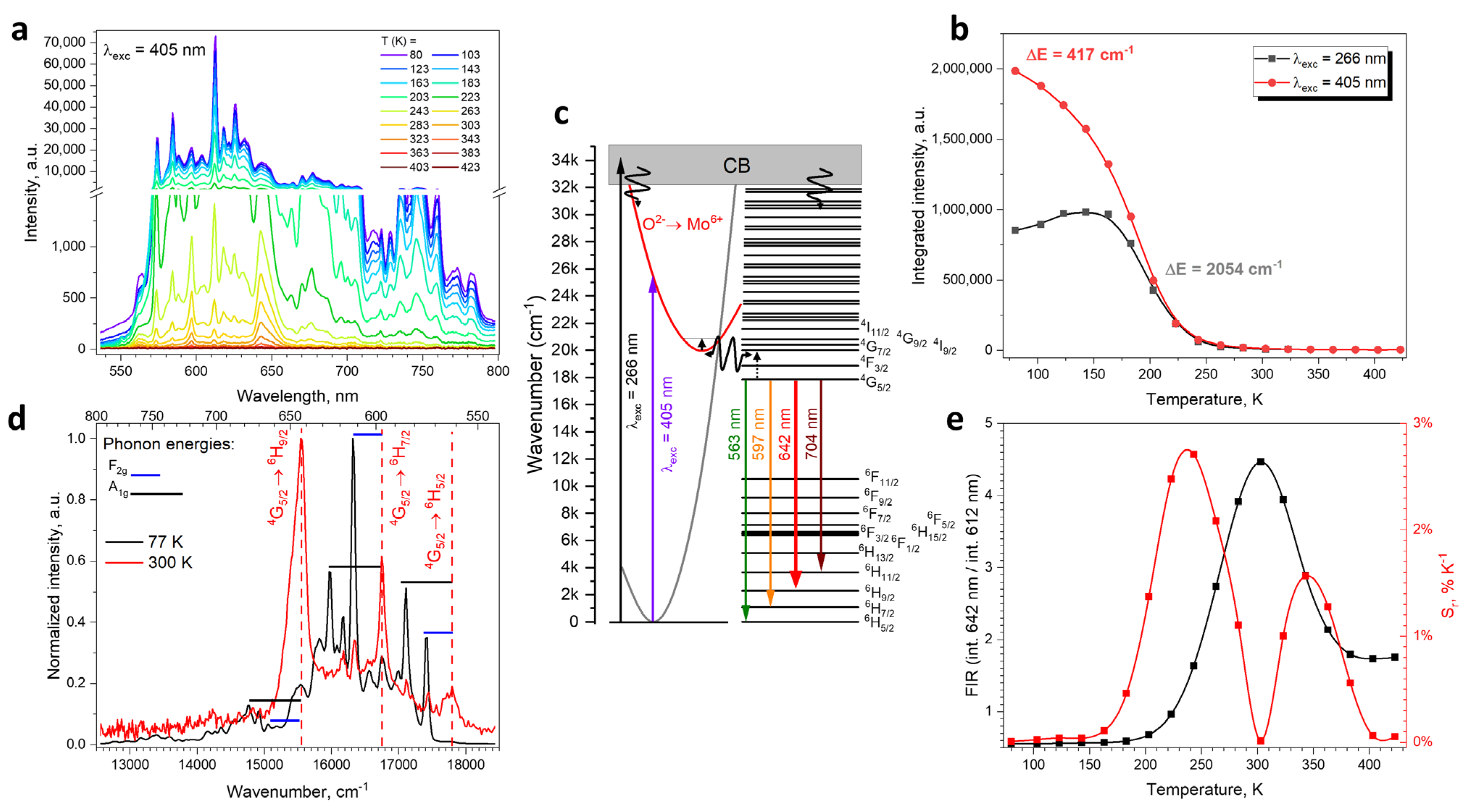
2.2. Spectroscopic Properties

The absorption spectra of BMM:Sm3+ exhibit absorption peaks in the near infrared, where the lower-lying levels of Sm3+ are located (Figure 2a inset). These absorption lines are primarily due to 6H5/26FJ (J = 1/2, …, 11/2) transitions.
The emission spectra of the BMM:Sm3+ (Figure 2b) exhibit four emission peaks resulting from the 4G5/26HJ (J = 5/2, …, 11/2) transitions, characteristic of Sm3+ along with smaller neighboring peaks assigned to the vibronic transitions of the mentioned main emission lines. The optimal concentration of Sm3+ is 0.3% (Figure 2c).
Although the XRD results (Figure 1b) suggest that the Sm3+ substitutes predominantly Mg2+ in the structure, one can identify the two-site emission of Sm3+ in the emission spectra of the sample with 0.1% Sm3+ by comparing the relative intensity of the maxima at 597 nm and 642.6 nm; the sample with 0.1% Sm3+ visibly exhibit stronger 597 nm emission as compared to other Sm3+ concentrations. This suggests that, in this sample, the Sm3+ ions are located in both Mg2+ and Ba2+ sites. For higher concentrations, as the lattice constant increases the Ba2+ site becomes less preferable for Sm3+ dopant, and as a result the emission becomes more uniform and seemingly one-site. The radio between the orange and red emission is known to indicate the symmetry of the Sm3+ ions site; however, it is usually inferred that the red emission at 642.6 nm resulting from the electric–dipole (ED) type 4G5/26H9/2 transition is dominant at low-symmetry sites [24], and orange emission at ~600 nm resulting from the mixed electric–dipole/magnetic–dipole (ED + MD) type 4G5/26H7/2 transition is dominant at high-symmetry octahedral sites [25]. This straightforward causation is, however, contested by the results of some highly-symmetrical double-perovskite materials such as Ba2ZnWO6:Sm3+, which exhibits dominant red emission [26] similar to BMM:Sm3+, as well as comparative studies between orthorhombic LiCaBO3:Sm3+ and cubic CsCaBO3:Sm3+ [27]. In the latter, the Sm3+ ions exhibit dominant red emission when located in octahedral sites of CsCaBO3 [28] and dominant orange emission when located in 7-coordinated sites in LiCaBO3 [29]. BMM:Sm3+ is to our best knowledge the most clear example of dominant red emission of Sm3+ resulting from the location of the dopant in octahedral sites of high-symmetry cubic structures.
The excitation spectrum of BMM:Sm3+ monitored at the maximum intensity at 642 nm (Figure 3a) consists of broad bands in the UV/blue spectral region 300–450 nm, which originates from the absorption of the O2− → Mo6+ charge transfer. The excitation spectrum monitored at the maximum of the emission of the Sm3+ at the second (Ba) site consists of an additional band with maximum at 260 nm, which is due to excitation into Ba states in the conduction band [9]. In the visible part of the excitation spectrum, there are weaker lines associated with the 4f-4f transitions labeled in the graph. Based on the complimentary data from the excitation, absorption, and emission spectra, the positions of the barycenter of Sm3+ energy levels were determined and listed in Table 1.
The decay curves have two components: a short one and a long one (Figure 3b,c), which confirm the two-site emission of Sm3+. The short component has a decay time around 350 μs (Figure 3b). Such short decay times for Sm3+ luminescence have been observed in materials where Sm3+ is located at octahedral sites: CaNb2O6 [30] and TiO2 [31]. The decay times become increasingly shorter when the dopant concentration increases, due to concentration quenching [32]. The long component is, as expected, more dominant in decay curves monitored at 597 nm (Figure 3c), and exhibit decay time 2.6 ± 0.2 ms, observed commonly for Sm3+-doped materials with a site coordination number other than 6, such as SrAl4O7 [33] and Sr2SiO4 [34].
The emission spectra of BMM:Sm3+ measured in the −193–150 °C range (Figure 4a) reveal that both the intensity and shape of the Sm3+ emission is strongly temperature-dependent. The thermal quenching of the emission intensity (Figure 4b) has two components when fitted with an Arrhenius equation (Figure S2) with corresponding activation energies of 417 cm−1 and 2054 cm−1. The former component with smaller activation energy is absent, when the sample is excited at 266 nm into the Ba states (which constitute the conduction band); therefore, it is concluded that it must be associated with the quenching of the molybdate group charge transfer state. This is depicted in Figure 4c as a crossing between the ground state and the excided state parabolas. Similarly, low activation energy of thermal quenching was observed for the pure BMM host and Eu3+–doped BMM [9]. The latter and larger activation energy can be matched with the thermalization of the 4G7/2 state, which may be involved in two-way energy transfer between the Sm3+ and MoO6 group, responsible for both Sm3+ emission and thermal quenching.
The shape of the emission spectra at a low temperature (77 K) differs significantly from the room-temperature emission spectra (Figure 4d). The most dominant lines at 77 K cannot be assigned directly to the 4G5/26HJ transitions, but are noticeably distant from the room-temperature emission lines by the same wavenumbers, which correspond to the phonon energies derived from the Raman spectrum (Figure 1e). This is uncommon for Sm3+ emission at low temperature, which usually features 4G5/26HJ transitions unaccompanied by their phonon-related components [35]. Apparently due to the high symmetry of the BMM structure, the 4G5/26HJ transitions are forbidden and must be coupled with phonons to occur. This causes the material to only exhibit anti-Stokes phonon-coupled transitions at low temperatures. When the temperature is higher, the lattice distortions allow for direct 6H5/26FJ transitions. This unique phenomenon, observed previously for only a handful of materials doped with transition metal ions, such as Cr3+ or Mn4+ [36,37], allow for very sensitive temperature sensing using the fluorescence intensity ration (FIR) between the phonon-coupled emission at 612 nm and regular 4G5/26H9/2 emission at 642 nm (Figure 4e). The relative sensitivity calculated [38] from this FIR has a maximum of 2.7% K−1 at −30 °C and another local maximum of 1.6% K−1 at 75 °C. Such a value is, to the best of our knowledge, one of the highest achieved for luminescent thermometry performed using only Sm3+ ions—comparable only to 3.83% K−1 obtained in Cs4PbBr6 borogermanate glass [39]—and significantly higher than, e.g., 0.5% K−1 for YAG [40] and 0.26% K−1 for Ba2Mg(PO4)2 [41]. Higher sensitivities were obtained only for materials exhibiting Sm-independent host emission, e.g., 7.08% K−1 in CaWO4 [42] or 10.14% K−1 for TiO2 nanoparticles [43]. The obtained result, resulting purely from the high symmetry of the BMM host, sets a promising precedent for temperature sensing using high-symmetry perovskite materials.
The emission spectra with dominant red emission results in CIE color coordinates located at (0.631, 0.370) (Figure 5a). Such color coordinates located towards the red part of the color coordinate scale, work efficiently in lowering the CCT and enhancing the CRI of the commonly used cool white LEDs. To demonstrate the impact of BMM:Sm emission on the luminescence of the wLED, its spectrum was added to the spectrum of the commonly used wLED with CRI equal 85 and CTT equal 4059 K. The result is a white emission with CRI of 91 and CCT of 2943 K constituting the warm white emission (Figure 5b).

3. Conclusions

Molybdates with a double-perovskite structure with the formula Ba2MgMoO6 doped with Sm3+ ions were successfully obtained for the first time using the co-precipitation method. The electric dipole (ED) 4G5/26H9/2 transition dominates; although, the Sm3+ ions are located in high symmetry, and the spectroscopic properties are characteristic for Sm3+ at octahedral sites with 6-fold coordination. Two-site Sm3+ emission was identified in the emission spectra of the 0.1% Sm3+ sample by comparing the relative intensity of the maxima at 597 nm and 642.6 nm. The obtained results suggest that in this sample Sm3+ ions are located in both Mg2+ and Ba2+ sites. At higher concentrations, the Ba2+ site is less preferable for Sm3+ doping, and as a result, the emission becomes more uniform and single-site. The aim of our work was to improve the properties of white-light-emitting diode lighting by supplying the missing red component and optimizing its correlated color temperature and color-rendering index. We can successfully conclude that the obtained BMM:Sm3+ materials are good candidates as a red phosphor, achieving a simulated CRI value of 91 and a CCT of 2943 K, giving a warm white emission. Temperature measurements brought us completely unexpected results. The relative sensitivity calculated from FIR is a maximum of 2.7% K−1 at −30 °C and another local maximum of 1.6% K−1 at 75 °C. This value is one of the highest achieved for luminescence thermometry performed only using Sm3+ ions. This is an extremely promising precedent for temperature sensing using perovskite materials with high symmetry that results from the high symmetry of the BMM host.

Supplementary Materials

The following supporting information can be downloaded at: https://www.mdpi.com/article/10.3390/ma17081897/s1, Figure S1: Histogram of the average crystal size; Figure S2: Fitting of an Arrhenius equation to temperature-dependent emission intensity of BMM: Sm3+ excited at 405 nm (left) and 266 nm (right).

Author Contributions

Conceptualization, N.M.-G.; Methodology, N.M.-G.; Formal analysis, N.M.-G. and B.B.; Investigation, N.M.-G.; Data curation, N.M.-G., B.B. and M.P.; Writing—original draft, N.M.-G. and B.B.; Writing—review & editing, N.M.-G. and B.B.; Supervision, P.J.D.; Project administration, N.M.-G. and P.J.D. All authors have read and agreed to the published version of the manuscript.

Funding

This research received no external funding.

Institutional Review Board Statement

Not applicable.

Informed Consent Statement

Not applicable.

Data Availability Statement

No new data were created or analyzed in this study. Data sharing is not applicable to this article.

Acknowledgments

The authors acknowledge support by a statutory activity subsidy from the Polish Ministry of Science and Higher Education for the Institute of Low Temperature and Structure Research Polish Academy of Sciences.

Conflicts of Interest

The authors declare no conflict of interest.

References

  1. Shkerin, S.N.; Uporov, S.A.; Yudin, V.N.; Solodovnikov, S.F.; Zolotova, E.S.; Tolkacheva, A.S. Paramagnetic properties of triple molybdates CsNa5M3(MoO4)6 (M = Ni, Co, Mn). Russ. J. Phys. Chem. A 2016, 90, 2292–2296. [Google Scholar] [CrossRef]
  2. Soltys, E.V.; Urazov, K.; Kharlamova, T.S.; Vodyankina, O.V. Redox and Catalytic Properties of Copper Molybdates with Various Composition. Kinet. Catal. 2018, 59, 58–69. [Google Scholar] [CrossRef]
  3. Soficha, D.; Tushinova, Y.L.; Shendrik, R.; Bazarov, B.G.; Dorzhieva, S.G.; Chimitova, O.D.; Bazarova, J.G. Optical spectroscopy of molybdates with composition Ln2Zr3(MoO4)9 (Ln: Eu, Tb). Opt. Mater. 2018, 81, 71–77. [Google Scholar] [CrossRef]
  4. Sofich, D.O.; Dorzhieva, S.G.; Chimitova, O.D.; Bazarov, B.G.; Tushinova, Y.L.; Bazarova, Z.G.; Shendrik, R.Y. Hypersensitive 5D0-7F2 Transition of Trivalent Europium in Double Molybdates. Bull. Russ. Acad. 2019, 83, 321–323. [Google Scholar] [CrossRef]
  5. Nagamani Naidu, B.; Koteswara Rao, K.; Murali Krishna, M.P.S. Sol-gel synthesis, crystal structure and luminescence properties of Eu3+ doped double perovskite molybdates NaCaBi1−xEuxMoO6 (x = 0–0.27). Mater. Res. Express. 2019, 6, 116210. [Google Scholar] [CrossRef]
  6. Takeda, T.; Ito, M.; Kikkawa, S. Preparation of magneto-resistive Sr2FeMoO6 through molybdic acid gelation. J. Alloys Compd. 2008, 449, 93–95. [Google Scholar] [CrossRef]
  7. Rath, M.K.; Lee, K.-T. Characterization of novel Ba2LnMoO6 (Ln = Pr and Nd) double perovskite as the anode material for hydrocarbon-fueled solid oxide fuel cells. J. Alloys Compd. 2018, 737, 152–159. [Google Scholar] [CrossRef]
  8. Chan, W.-L.; Liu, Z.; Lu, S.; Tanner, P.A.; Wong, K.-L. The reported anomalous emission intensity of the 5D07F4 transition of Eu3+ in a molybdate double perovskite. J. Mater. Chem. C 2015, 3, 860–963. [Google Scholar] [CrossRef]
  9. Bondzior, B.; Stefańska, D.; Vu, T.H.Q.; Dereń, P.J. Optimization of Eu3+-to-Host Emission Ratio in Double-Perovskite Molybdenites for Highly Sensitive Temperature Sensors. J. Phys. Chem. C 2022, 126, 13247–13255. [Google Scholar] [CrossRef]
  10. Krishnan, R.; Kroon, R.E.; Swart, H.C. Charge transfer characteristics and luminescence properties of Eu3+ activated Ba2YMoO6 and BaY2(MoO4)4 phosphors. Mater. Res. Bull. 2022, 145, 111554. [Google Scholar] [CrossRef]
  11. Zhang, S.; Hu, Y.; Chen, L.; Wang, X.; Ju, G.; Fan, Y. A novel Ba2MgMoO6:Eu3+ orange-red phosphor: Photoluminescence properties and mechanism of charge and energy transfer. J. Mater. Res. 2013, 28, 3130–3136. [Google Scholar] [CrossRef]
  12. Zhang, L.; Sun, B.; Shao, C.; Zhen, F.; Wei, S.; Bu, W.; Yao, Q.; Jiang, Z.; Chen, H. Preparation, band-structure and luminescence of double perovskite Ba2MgMoO6:Eu3+ orange-red phosphor for white LEDs. Ceram. Int. 2018, 44, 17305–17312. [Google Scholar] [CrossRef]
  13. Wang, Y.; Ke, Y.; Chen, S.; Luo, J.; Shu, S.; Gao, J.; Deng, B.; Yu, R. Luminescence investigation of red-emitting Sr2MgMoO6:Eu3+ phosphor for visualization of latent fingerprint. J. Colloid Interface Sci. 2021, 583, 89–99. [Google Scholar] [CrossRef] [PubMed]
  14. Cao, R.; Huang, T.; Nie, J.; Zhang, L.; Chen, Y.; Li, L.; Lan, B.; Wang, J. Energy transfer and tunable–color luminescence properties of a single-phase CaSrNb2O7:Sm3+; Bi3+. J. Mol. Struct. 2024, 1297, 136962. [Google Scholar] [CrossRef]
  15. Cao, R.; Wei, J.; Chen, T.; Lan, B.; Li, L.; Liu, R.; Luo, Z.; Wang, J. Synthesis, adjustable-color emission and energy transfer of Ba2MgSi2O7: Sm3+, Bi3+ phosphor. J. Mol. Struct. 2023, 1274, 134404. [Google Scholar] [CrossRef]
  16. Stefańska, D.; Bondzior, B.; Vu, T.H.Q.; Miniajluk-Gaweł, N.; Dereń, P.J. The Influence of Morphology and Eu3+ Concentration on Luminescence and Temperature Sensing Behavior of Ba2MgWO6 Double Perovskite as a Potential Optical Thermometer. J. Alloys Compd. 2020, 842, 155742. [Google Scholar] [CrossRef]
  17. Yan, L.; Xing, M.; Ma, Y.; Kang, L.; Fu, Y.; Pang, Q.; Xin, F.; Wang, H.; Luo, X. Promising lanthanide-doped double molybdates KYb(MoO4)2 phosphor for highly efficient upconversion luminescence and temperature sensing. Spectrochim. Acta A Mol. Biomol. Spectrosc. 2024, 308, 123751. [Google Scholar] [CrossRef] [PubMed]
  18. Chen, H.; Jin, Y.; Fang, F.; Lin, H.; Li, Y.; Feng, G.; Xiong, Y.; Meng, F.; Cao, L.; Ren, H. Up-conversion phosphor Na2MoO4: Er3+/Yb3+ for the optical temperature sensing and anticounterfeiting. J. Lumin. 2024, 265, 120244. [Google Scholar] [CrossRef]
  19. Tian, X.; Xu, S.; Wen, J.; Zhu, L.; Ji, C.; Huang, Z.; Wang, X.; Luo, F.; Liu, X.; Lu, Y.; et al. Thermochromic Pr3+ doped CaMoO4 phosphor with diverse thermal responses for temperature sensing. Ceram. Int. 2023, 49, 27126–27137. [Google Scholar] [CrossRef]
  20. Armaroli, N.; Bolink, H.J. Photoluminescent Materials and Electroluminescent Devices. In Topics in Current Chemistry Collections; Springer: Berlin/Heidelberg, Germany, 2017. [Google Scholar] [CrossRef]
  21. Ye, S.; Wang, C.-H.; Jing, X.-P. Photoluminescence and Raman Spectra of Double-Perovskite Sr2Ca(Mo/W)O6 with A- and B-Site Substitutions of Eu3+. J. Electrochem. Soc. 2008, 155, 148. [Google Scholar] [CrossRef]
  22. Pásztorová, J.; Bi, W.H.; Gaal, R.; Krämer, K.; Živković, I.; Rønnow, H.M. Structure, heat capacity and Raman spectra of Ba2MgWO6 single crystals grown in BaCl2-MgCl2 flux. J. Solid State Chem. 2023, 326, 124184. [Google Scholar] [CrossRef]
  23. Fujioka, Y.; Frantti, J.; Kakihana, M. Raman Scattering Studies of the Ba2MnWO6 and Sr2MnWO6 Double Perovskites. J. Phys. Chem. B 2006, 110, 777–783. [Google Scholar] [CrossRef]
  24. Görller-Walrand, C.; Binnemans, K. Chapter 167 Spectral Intensities of f-f Transitions. In Handbook on the Physics and Chemistry of Rare Earths; Elsevier: Amsterdam, The Netherlands, 1998; Volume 25, pp. 101–264. [Google Scholar]
  25. Shih, H.-R.; Chang, Y.-S. Structure and Photoluminescence Properties of Sm3+ Ion-Doped YInGe2O7 Phosphor. Materials 2017, 10, 779. [Google Scholar] [CrossRef]
  26. Chen, P.; Hu, W.; Yang, D.; Zhu, J.; Zhang, J.; Wu, Y. Ba2ZnWO6:Sm3+ as promising orange-red emitting phosphors: Photoluminescence properties and energy transfer process. Phys. B Condens. 2018, 530, 127–132. [Google Scholar] [CrossRef]
  27. Herrmann, A.; Friedrich, D.; Zscheckel, T.; Rüssel, C. Luminescence properties of Sm3+ doped alkali/earth alkali orthoborates of the type XZBO3 with X = Li, Na, Cs and Z = Ca, Sr, Ba. J. Lumin. 2019, 214, 116550. [Google Scholar] [CrossRef]
  28. Wen, M.; Wu, H.; Su, X.; Lu, J.; Yang, Z.; Wu, X.; Pan, S. ACaBO3 (A = Cs, Rb): Two new cubic borates with isolated BO3 groups. Dalton Trans. 2017, 46, 4968–4974. [Google Scholar] [CrossRef] [PubMed]
  29. Wu, L.; Chen, X.L.; Li, H.; He, M.; Dai, L.; Li, X.Z.; Xu, Y.P. Structure determination of a new compound LiCaBO3. J. Solid State Chem. 2004, 177, 1111–1116. [Google Scholar] [CrossRef]
  30. Li, K.; Liu, X.; Zhang, Y.; Li, X.; Lian, H.; Lin, J. Host-Sensitized Luminescence Properties in CaNb2O6:Ln3+ (Ln3+ = Eu3+/Tb3+/Dy3+/Sm3+) Phosphors with Abundant Colors. Inorg. Chem. 2015, 54, 323–333. [Google Scholar] [CrossRef]
  31. Luo, W.; Li, R.; Chen, X. Host-Sensitized Luminescence of Nd3+ and Sm3+ Ions Incorporated in Anatase Titania Nanocrystals. J. Phys. Chem. C 2009, 113, 8772–8777. [Google Scholar] [CrossRef]
  32. Timoshenko, A.D.; Matvienko, O.O.; Doroshenko, A.G.; Parkhomenko, S.V.; Vorona, I.O.; Kryzhanovska, O.S.; Safronova, N.A.; Vovk, O.O.; Tolmachev, A.V.; Baumer, V.N.; et al. Highly-doped YAG:Sm3+ transparent ceramics: Effect of Sm3+ ions concentration. Ceram. Int. 2023, 49, 7524–7533. [Google Scholar] [CrossRef]
  33. Puchalska, M.; Zych, E. The effect of charge compensation through alkali metal co-doping on the luminescence behaviour of SrAl4O7:Sm3+ phosphor. J. Lumin. 2018, 197, 211–218. [Google Scholar] [CrossRef]
  34. Gupta, S.K.; Pathak, N.; Thulasidas, S.K.; Natarajan, V. Local site symmetry of Sm3+ in sol–gel derived α′-Sr2SiO4: Probed by emission and fluorescence lifetime spectroscopy. J. Lumin. 2016, 169, 669–673. [Google Scholar] [CrossRef]
  35. Solarz, P.; Dominika-Dzik, G.; Lisiecki, R.; Ryba-Romanowski, W.; Beregi, E.; Foldvari, I.; Lengyel, K. Spectroscopic properties of Sm3+ impurity in YAl3(BO3)4 single crystal. Opt. Mater. 2010, 32, 1446–1450. [Google Scholar] [CrossRef]
  36. Li, R.; Li, P.; Wei, G.; Li, J.; Shi, Y.; Wang, Y.; He, S.; Yang, Y.; Ding, W.; Wang, Z. Ultra-sensitive low-temperature thermometer regulated by the crystal field strength. Ceram. Int. 2023, 49, 7913–7919. [Google Scholar] [CrossRef]
  37. Cai, P.; Wang, X.; Seo, H.J. Excitation power dependent optical temperature behaviors in Mn4+ doped oxyfluoride Na2WO2F4. Phys. Chem. Chem. Phys. 2018, 20, 2028–2035. [Google Scholar] [CrossRef]
  38. Brites, C.D.S.; Millán, A.; Carlos, L.D. Chapter 281—Lanthanides in Luminescent Thermometry. In Handbook on the Physics and Chemistry of Rare Earths; Elsevier: Amsterdam, The Netherlands, 2016; Volume 49, pp. 339–427. [Google Scholar]
  39. Yao, G.; Li, S.; Valiev, D.; Chen, M.; Stepanow, S.; Lu, Y.; Li, C.; Zhou, Y.; Su, Z.; Zeng, F. Luminescence behavior and temperature sensing properties of Sm3+-doped Cs4PbBr6 quantum dots encapsulated in borogermanate glass. J. Non-Cryst. Solids 2022, 582, 121462. [Google Scholar] [CrossRef]
  40. Zhu, K.; Zhou, H.; Qiu, J.; Wang, L.-G.; Ye, L. Optical temperature sensing characteristics of Sm3+ doped YAG single crystal fiber based on luminescence emission. J. Alloys Compd. 2022, 890, 161844. [Google Scholar] [CrossRef]
  41. Singn, R.; Bedyyal, A.K.; Manhas, M.; Durani, F.; Swart, H.C.; Kumar, V. Effect of the synthesis route on luminescence dynamics and thermographic properties of Sm3+ doped Ba2Mg(PO4)2 phosphor. J. Alloys Compd. 2024, 973, 172911. [Google Scholar] [CrossRef]
  42. Lan, Q.; Meng, Q. Temperature sensing materials based on the FIR of doped ions and the matrix in CaWO4:Sm3+ phosphors. Sens. Actuator A-Phys. 2023, 363, 114708. [Google Scholar] [CrossRef]
  43. Dramićanin, M.D.; Antić, Ž.; Ćulubrk, S.; Ahrenkiel, S.P.; Nedeljković, J.M. Self-referenced luminescence thermometry with Sm3+ doped TiO2 nanoparticles. Nanotechnology 2014, 25, 485501. [Google Scholar] [CrossRef]
Figure 1. (a) XRD results of the studied dopuble-perovskite BMM doped with Sm3+, (b) XRD lines shift. (c) Unit cell of BMM with the space group Fm 3 ¯ m (d) SEM image and (e) Raman spectra of the BMM: 1.0% Sm3+.
Materials 17 01897 g001
Figure 2. (a) Absorption spectra of the BMM: 1.0% Sm3+, (b) emission spectra, (c) integral emission intensity. The line in (c) serves only to guide the eye.
Materials 17 01897 g002
Figure 3. (a) Excitation spectra of the BMM:0.3% Sm3+ and (b,c) decay times of the BMM doped with Sm3+ monitored at 642.6 nm (b) and 597 nm (c).
Materials 17 01897 g003
Figure 4. (a) Temperature-dependent emission spectra of BMM:0.3% Sm3+, (b) thermal quenching curves, and corresponding activation energies of BMM:Sm3+ under 405 and 266 nm excitation, (c) energy level scheme of the luminescent centers in Sm3+-doped BMM, (d) 77 K and 300 K emission spectra of BMM:Sm3+, (e) FIR and relative sensitivity of temperature sensing.
Materials 17 01897 g004
Figure 5. (a) CIE diagram of the BMM:0.3% Sm3+, (b) normalized emission spectra of common wLED, BMM doped with Sm3+, and simulated luminescence of combined wLED and BMM:Sm3+.
Materials 17 01897 g005
Table 1. Barycenters of energy levels of Sm3+ in BMM host determined from the excitation (4I11/2, …, 4G5/2), absorption (6F11/2, …, 6F1/2), and emission spectra (6H13/2, …, 6H7/2).
LevelWavenumber
(cm−1)
4I11/221,575
4G9/220,812
4G7/220,429
4I9/220,008
4F3/218,879
4G5/217,857
6F11/210,520
6F9/29163
6F7/27999
6F5/27129
6F3/26634
6H15/26492
6F1/26380
6H13/25076
6H11/23663
6H9/22302
6H7/21095
6H5/20
Disclaimer/Publisher’s Note: The statements, opinions and data contained in all publications are solely those of the individual author(s) and contributor(s) and not of MDPI and/or the editor(s). MDPI and/or the editor(s) disclaim responsibility for any injury to people or property resulting from any ideas, methods, instructions or products referred to in the content.

