1. Introduction
In dentistry, one of the most significant challenges is the effective treatment of oral diseases, which are among the most prevalent noncommunicable diseases globally, affecting approximately 3.5 billion people each year [
1]. Although the quality of care for all oral disorders shows an increasing trend on a global scale, oral health conditions still rank among the top 10 of all diseases globally and have a negative impact on the global economy. The annual expenditure was estimated at USD 387 billion in direct costs and another USD 323 billion in indirect costs. In terms of indirect costs, severe periodontal disease generated USD 82 billion in expenses [
1,
2,
3]. The complexity of oral diseases, compounded by individual patient variability, the multifunctionality of the oral cavity, and its inherent mobility, complicates the development of drugs with optimal physicochemical properties that can meet therapeutic needs and enhance treatment efficacy [
3,
4].
The primary objectives of oral disease therapy are to protect and regenerate damaged tissues and mitigate inflammation, which can lead to the formation of pathological pockets within the oral cavity. Crucial considerations in drug design include ensuring safety, ease of administration, convenient application, and targeted release of the active substance at the site of action, thereby reducing dosing frequency [
4,
5].
The escalating rates of antimicrobial resistance (AMR) pose a significant global health concern, primarily driven by the overutilization of antifungal agents and antibiotics. This trend underscores the imperative for intensified research into novel substances exhibiting antimicrobial, antioxidant, and anti-inflammatory properties. The study of medicinal substances of natural origin, mainly processed medicinal plants, is of great interest to the scientific community. Moreover, medicinal plants are an essential source of bioactive compounds and antioxidant substances for the human body [
6]. Currently, local herbal therapy is successfully used, and according to the World Health Organization (WHO), over 80% of the global population prefers herbal remedies, highlighting these natural substances’ significant role in healthcare today [
7,
8]. Rhizomes of
Reynoutria japonica Houtt. (under the pharmacopoeial name
Polygoni cuspidati rhizoma et radix,
hu zhang in pinyin Chinese), used as a source of active substances, are plant medicinal raw materials whose use originated in traditional Chinese medicine. This herbal medicine was included in the European Pharmacopoeia in 2017.
R. japonica (common name: Japanese Knotweed) belongs to the
Polygonaceae family, and its natural habitat includes regions in East Asia [
9,
10,
11]. In Europe, this plant is considered an invasive species, posing a threat to native plant species due to its ability to produce substances that inhibit the growth of other plants [
11,
12]. The extract obtained from the rhizome of
R. japonica is rich in chemical substances from various groups, including stilbene derivatives like resveratrol and piceid—compounds known for their significant biological activities [
13,
14]. The high resveratrol content contributes to the anti-inflammatory and antipyretic effects of the extract. Additionally, resveratrol is known for its antioxidant properties, making it a valuable aid in combating various inflammatory conditions and infections [
11,
14]. Hence, this species may be a promising therapy in preventing and healing lesions in the oral cavity and treating periodontitis. Resveratrol is absorbed from food in the intestine through diffusion and carrier-mediated transport [
15]. In enterocytes, resveratrol undergoes biotransformation, leading to the formation of well-soluble, easily excretable, and biologically active derivatives [
16,
17,
18]. Interestingly, a specific metabolism of resveratrol was observed in cancer cells. They overexpress the cytochrome P450 enzyme CYP1B1, which catalyzes aromatic hydroxylation reactions. These cells hydroxylate resveratrol at positions 4 and/or 3’, producing three metabolites, one of which is piceatannol (3,5,3’,4’-tetrahydroxystilbene), a known anticancer and antileukemic agent, which is being considered as a potential anticancer drug [
19]. A major resveratrol glucosylated derivative—piceid—has antimicrobial effects against oral pathogens, which are important in preventing dental diseases. It has been shown to inhibit the biofilm formation of Fusobacterium nucleatum, a bacterium associated with periodontal diseases [
20]. Furthermore, piceid exhibits antimicrobial effects against both planktonic and biofilm forms of
Porphyromonas gingivalis, another key pathogen in periodontal disease [
20].
Our research team conducted extensive research to select the best extract from
R. japonica rhizomes to develop a polymer film for potential use in the treatment of periodontal and gingival diseases [
21,
22]. For the development of the film, we selected a 25% ethanolic extract which effectively stimulated human gingival fibroblasts to proliferate, migrate, and increase the synthesis of collagen III. This activity, according to the phytochemical analysis, may be related to the high contents of resveratrol and piceid and the relevant composition of procyanidins.
Modern carriers of active pharmaceutical substances, including fibers, mucoadhesive strips and films, gels, liposome systems, microparticles, nanoparticles, and nanofibers, play a vital role in periodontology. Film carriers can be applied to the surface of the oral mucosa in the area of the cheeks and gums but can also be applied directly to the gum pockets.
A considerable advantage of this type of pharmaceutical form is its suitability for cutting out smaller fragments of a specific size and shape tailored to the individual needs of the patient. Their flexibility allows them to be easily placed in the desired location, unlike other conventional pharmaceutical forms, such as mucoadhesive tablets. Adhesion to the oral mucosa makes polymer films preferred over oral solutions or oral gels, which are usually easily removed by saliva and therefore have a shorter residence time on the mucosa. The mucoadhesive properties do not allow the film to be easily swallowed or inhaled, which minimizes the risk of choking.
Recently, films based on a mixture of natural and synthetic polymers enriched with plant extracts have gained significant popularity in biomedical applications. Cellulose and its derivatives are widely used in pharmaceutical and periodontal formulations due to properties such as biocompatibility, biodegradability, nontoxicity, film-forming ability, the possibility of chemical modification, chemical stability, good moisture retention capacity, and flexibility in processing [
23,
24,
25]. Their mucoadhesive properties allow for prolonged contact time with the specific tissues, thus enhancing bioavailability and preventing too rapid degradation for drug delivery applications [
26]. At the nanoscale, cellulose displays distinct adhesion properties that play a critical role in its interactions with microbial surfaces. These nanoscale properties enable cellulose to effectively bind to bacterial cells, thereby modulating bacterial adherence and influencing biofilm formation dynamics [
27,
28].
However, the limitations of cellulose- and cellulose derivative-based carriers, such as insufficient mechanical strength and barrier properties, pose significant challenges. To mitigate these shortcomings, blending cellulose and its derivatives with other water-soluble polymers, such as polyvinyl alcohol (PVA), is a promising approach to improve polymer-based carrier thermomechanical and morphological properties, including enhanced tensile and modulus properties. PVA is a highly valuable polymer in dental applications due to its nontoxicity, high crystallinity, water solubility, biodegradability, flexibility, hydrophilicity, mucoadhesive properties, controlled tensile strength, and excellent film-forming ability [
29]. It does not exhibit mutagenic or clastogenic effects, ensuring that it does not cause genetic mutations or chromosomal damage. Additionally, PVA does not accumulate in the body, further supporting its safety profile [
30]. In our study, to ensure appropriate mucoadhesive properties [
31,
32,
33,
34] and to extend the release time of substances incorporated into the carrier, additional polyvinylpyrrolidone (PVP) was included in the composition of the polymeric film formulation. PVP aids in reducing the periodontal pocket depth and improving connective tissue attachment in deep periodontal pockets, thereby reducing the number of pathogenic microorganisms in the periodontium [
35,
36].
Considering that polymer films prepared with
R. japonica extract have not yet been investigated in dental applications, the optimal contents and proportions of these composite films to achieve better mechanical and bioactive properties remain unclear. This study aimed to develop the composition, preparation technology, and assessment of the physico-chemical properties of polymer films based on cellulose derivatives. The tested substances included hydroxypropylmethylcellulose (HPMC), methylcellulose (MC), sodium carboxymethylcellulose (NaCMC), pullulan, PVA, and PVP containing 25% ethanolic extract from rhizomes of
R. japonica as promising compositions for use on the oral mucosa in the treatment of periodontal and gum diseases [
37].
The researchers hypothesize that it will be possible to develop carriers with the required release profile through advanced cross-linking processing. This approach will serve as a disintegration-rate modeling technology to predict the release profile and facilitate the selection of therapies based on clinical needs. Null Hypothesis (H₀): The release of resveratrol and piceid from the polymeric films does not follow the multidimensional model. This hypothesis was tested by fitting the drug release data to the multidimensional equation.
3. Results and Discussion
3.1. Visual Assessment
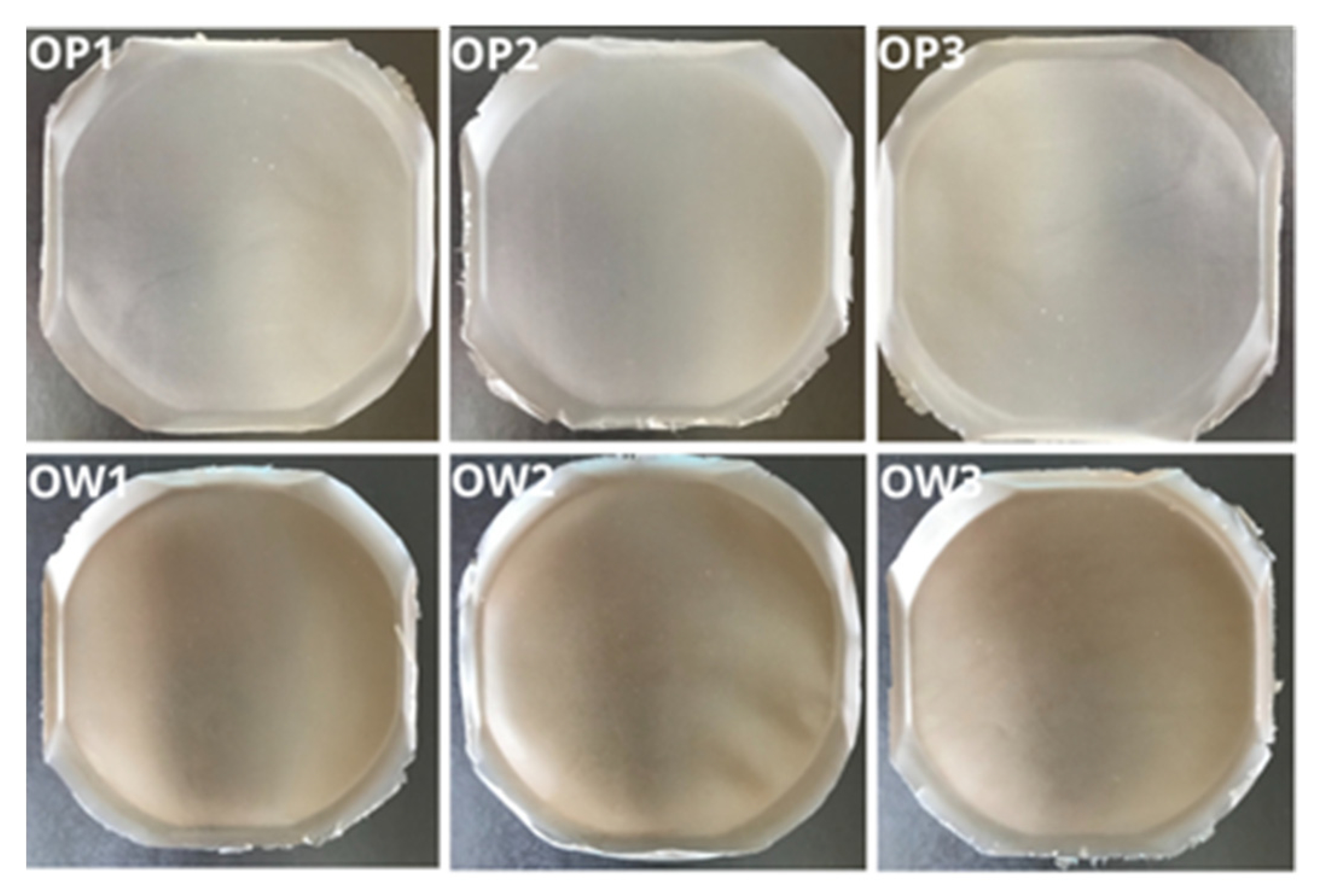
All developed polymer film formulations containing the extract from
R. japonica, namely, OW1, OW2, and OW3, demonstrated consistent morphological and structural properties (
Figure 4). No evidence of polymer aggregates, delaminations, or trapped air bubbles was observed within any of the formulations, indicating a high degree of uniformity. The films were notably flexible, nonsticky, easy to remove from molds, and exhibited a smooth surface. The inclusion of
R. japonica extract in the OW-series formulations imparted a slight brownish tint to the films. Apart from this color variation, no significant organoleptic differences were observed between the OW films and the placebo series (OP1, OP2, and OP3).
3.2. Thickness and Mass Measurements of Polymer Films
The evaluation of film thickness and mass is a critical parameter for determining the structural integrity and uniformity of polymeric film formulations. In this study, the thickness and mass of polymer films were measured to assess the reproducibility and uniformity of the manufacturing process across different formulations.
Table 3 presents the average thickness and mass values for fragments of each formulation, along with the calculated standard deviations (SDs).
The results of the measurements indicated that the OW1 formulation, containing the plant extract from R. japonica, and the placebo formulation OP1, both with the same polymer-based composition of film, exhibited the greatest average thickness. In contrast, the OW2 formulation and its placebo counterpart, OP2, demonstrated the lowest average thickness. These differences are a result of compositional differences. OW1 and OW3 were prepared using an aqueous solution of MC 400 cP at a 5% concentration (polymer ratio of 5:95), while OW2 and OP2 were formulated using an aqueous solution of MC A15C at a 3% concentration (polymer ratio of 3:97). The thickness of OW3 was similar to OW1, attributed to the use of a 5% aqueous HPMC solution (polymer ratio of 5:95). No significant differences in the average thickness were observed between the films containing R. japonica extract and their respective placebo formulations. Additionally, the calculated standard deviations for thickness across all formulations were lower than the precision threshold of the measurement device, indicating a high degree of uniformity in film thickness. Regarding mass measurements, the OW2 and OP2 formulations exhibited the lowest average mass, while OW1 and OP1 exhibited the highest. These results were consistent with the thickness measurements, as formulations with the lowest average thickness also had the lowest mass, and those with the greatest thickness had the highest mass. This correlation was due to the different concentrations of methylcellulose derivatives used in the formulations. The standard deviations for mass measurements across all formulations ranged from 2.00 to 2.45, indicating a comparable level of uniformity and reproducibility in the preparation method across all tested films.
3.3. Mechanical Bending Resistance Test of Polymer Films
The mechanical bending resistance of the polymer films was evaluated using a standardized method. All prepared formulations, including placebo films and those containing the plant extract from R. japonica, exhibited high resistance to bending. In all three repetitions for each formulation, the films withstood at least 300 bends without tearing or breaking. This demonstrates the optimal mechanical properties and high durability of the produced polymer films. The results indicate that the inclusion of the plant extract did not compromise the mechanical integrity of the films, suggesting that the formulations maintain their structural performance even with the addition of active components.
3.4. Mucoadhesion Strength Test
Mucoadhesion is a crucial parameter in evaluating polymeric carriers as dosage forms for application on the oral mucosa.
Table 4 presents the average mucoadhesion strength values, along with the standard deviations for the three placebo film formulations (OP1, OP2, and OP3) and three formulations containing
R. japonica extract (OW1, OW2, and OW3).
The results indicate that the highest average mucoadhesion strength values were obtained for the OP2 and OW2 formulations, while the lowest values were recorded for the OW3 and OP3 formulations. The OW2 and OP2 formulations contained MCA15C with a viscosity range of 1200–1800 mPa·s, whereas the OW3 and OP3 formulations contained HPMC with a viscosity range of 20–40 mPa·s. The higher viscosity of MCA15C contributed to the increased mucoadhesion strength of the films containing this polymer. Furthermore, all measurements were characterized by low standard deviation values, indicating the reproducibility of the applied film production method.
3.5. Wettability Test
Wettability is a critical parameter in evaluating the performance of polymer films, especially in applications such as oral mucosal delivery systems. The degree of wettability affects how a film behaves upon contact with biological fluids, such as saliva or gingival crevicular fluid, influencing both the rate of hydration and the subsequent dissolution of the active ingredients embedded within the film matrix. Films with lower contact angles are likely to absorb more fluid, potentially enhancing the release and diffusion of the active compounds at the site of application. The contact angle measurements, presented in
Table 5, indicate that all the polymer film formulations exhibit contact angles below 90°, signifying partial wettability. This suggests that the films possess a hydrophilic surface, as evidenced by the ability of water to spread to some degree across the surface. Notably, no statistically significant differences in contact angle values were observed between the films containing
R. japonica extract (OW series) and their placebo counterparts (OP series). The lowest contact angle values were recorded for the placebo formulation OP1 and its corresponding extract-containing film OW1, while the highest contact angles were associated with formulations OP3 and OW3. This trend in wettability can be directly attributed to the intrinsic hydrophilicity of the polymers used in the formulations. Specifically, the lower contact angles suggest that these films, particularly OP1 and OW1, may be more inclined to interact with and absorb moisture from their environment compared to the other formulations.
3.6. Effect of Syringe Filter Membrane on the Content of R. Japonica Extract Components
The results of the study shown in
Table 6 highlighted substantial variations in the measured contents of the key components of
R. japonica extract, namely, resveratrol and piceid, depending on the type of filter membrane employed in the filtration process. Notably, the use of a nylon filter resulted in a marked depletion of filtrate in both resveratrol and piceid. The resveratrol content was reduced to approximately 2.11% of the declared value, while piceid levels in the filtrate were below the detection limit of the analytical method employed. In contrast, the PVDF filter with a pore size of 0.45 µm led to an increase in resveratrol content, surpassing the declared content, with values exceeding 100% of the expected dose. This phenomenon reflects the retention or less efficient filtration of resveratrol through this membrane, possibly due to its molecular weight or solubility characteristics. Additionally, the PVDF filter with a 0.22 µm pore size exhibited a significant reduction in resveratrol content, down to approximately 20.87% of the declared dose, while the piceid content was moderately decreased to around 76.64%. The detailed HPLC-RI chromatograms of resveratrol and piceid released from the
R. japonica extract depending on the type of filter membrane employed in the filtration process are provided in the
Supplementary Materials.
These results suggest that membrane pore size and material composition play a pivotal role in the selective retention or loss of extract components during the filtration process. The observed differences underscore the necessity of carefully selecting the appropriate filter membrane when preparing plant extracts for analysis or formulation development, as filtration can substantially influence the integrity and composition of the extract. The use of inappropriate membranes may lead to the loss of key bioactive components, thereby affecting the reproducibility and reliability of subsequent analyses and applications of the extract.
3.7. Determination of Film Dissolution Time and pH Value of Extracts After Dissolution
Table 7 presents the average values obtained from three measurements of dissolution time for film fragments and the pH values of extracts after dissolution of placebo and film formulations containing
R. japonica extract.
The results of the study confirmed the extended dissolution time of the optimized polymeric films. Formulations OW1 and OW2 achieved complete dissolution within 24 h, whereas films from formulation OW3 were dissolved within 20 h. No significant differences were observed between the plant-extract-containing films from R. japonica and their placebo counterparts. The pH values of the extracts obtained after the dissolution of the film fragments ranged from 7.15 for formulation OW1 to 7.32 for formulation OW3. The obtained appropriate pH values of the extracts, close to a neutral value, in the planned application are beneficial for ensuring carrier tolerance and reducing the risk of irritation at the application site.
3.8. Fourier Transform Infrared Spectroscopy (FT-IR)
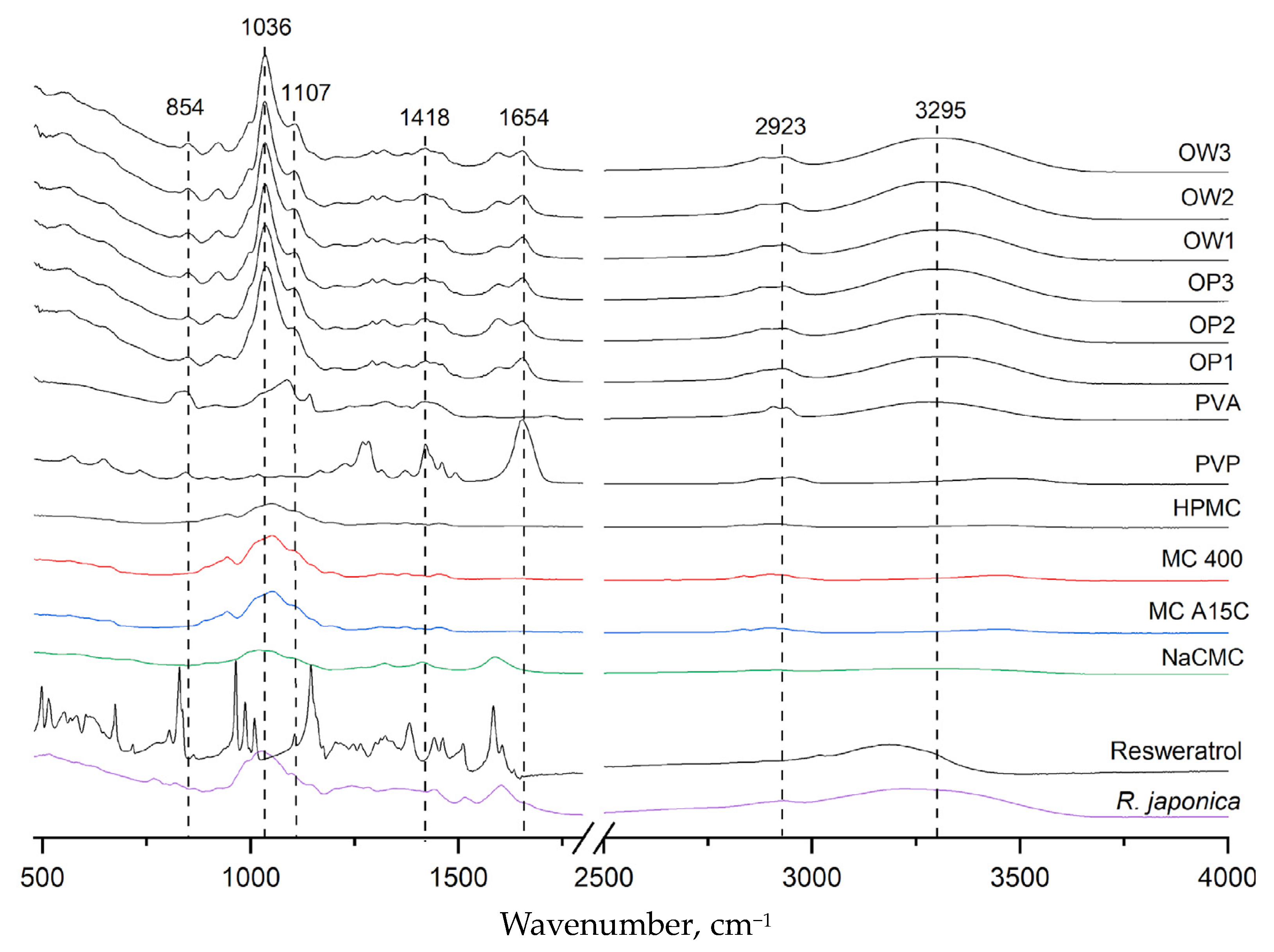
Figure 5 presents the infrared absorption spectra for all polymer components: polyvinyl alcohol (PVA), polyvinylpyrrolidone (PVP), methylcellulose (MC), hydroxypropyl methylcellulose (HPMC), sodium carboxymethylcellulose (NaCMC), powdered
Reynoutria japonica extract, pure resveratrol standard, placebo films (OP1, OP2, and OP3), and three film formulations containing
R. japonica extract (OW1, OW2, and OW3). The PVA spectrum exhibited a characteristic band in the range of 3000–3500 cm⁻
1, centered around 3295 cm⁻
1, corresponding to the stretching vibrations of hydroxyl (OH) groups involved in intra- and intermolecular hydrogen bonding. The PVP spectrum showed a peak at 1654 cm⁻
1, which is associated with the stretching vibrations of the carbonyl (C=O) bonds in the pyrrolidone ring. Additionally, a broad absorption band between 1050 and 1430 cm⁻
1 was observed, corresponding to the stretching vibrations of C-O bonds and the bending of -CH₂ and -CH groups. This broad band is prominent in the spectra of MC, NaCMC, and HPMC, indicating their structural similarity in this region. In the FT-IR spectra of the placebo formulations (OP1, OP2, and OP3) and the films containing
R. japonica extract (OW1, OW2, and OW3), no new absorption bands were observed beyond those characteristic of the formulation components. The 3295 cm⁻
1 band confirmed the presence of PVA, while the 1654 cm⁻
1 band corresponded to PVP. The broad band in the range of 1036–1418 cm⁻
1 was attributed to methylcellulose or its derivatives. The FT-IR results indicate a lack of interactions between the extract components and the polymers in the film.
3.9. In Vitro Release Profile of Resveratrol and Piceid from PVA/PVP/MCA15C/NaCMC Films with the R. Japonica Extract
To determine the amount of resveratrol and piceid released from the developed PVA/PVP/MCA15C/NaCMC/GLY formulations containing
R. japonica extract, the method described by Nawrot-Hadzik et al. [
33,
34] was employed. The detailed HPLC-RI chromatograms of resveratrol and piceid determined for the films and the
R. japonica extract are provided in the
Supplementary Materials. The results of the release study for the optimized OW2 film series are presented in
Figure 6 and
Figure 7, showing the percentage of resveratrol and piceid released at 1, 3, 5, 7, 12, and 24 test hours. The concentrations of these compounds were determined using calibration curves based on analytical-grade standards of pure resveratrol and piceid. The release profile of resveratrol from the OW2 polymeric film exhibited an initial rapid release, commonly referred to as the ‘burst effect’, with approximately 26.78% of the total dose released within the first 3 h. This was followed by a deceleration in the release rate until the fifth hour, after which a renewed increase was observed, reaching 44.33% by the seventh hour. This phase is characterized by a diffusion-controlled release mechanism, where the concentration gradient between the polymer matrix and the surrounding medium drives the migration of resveratrol molecules from the interior to the exterior of the matrix. The final stage of resveratrol release involves matrix erosion, during which the polymer gradually degrades or dissolves, resulting in the continuous release of the remaining resveratrol. This leads to a prolonged, gradual release phase lasting up to 24 h, culminating in the release of 51.26% of the total resveratrol dose. The piceid released from the OW2 polymeric film followed a similar pattern, with an initial rapid release of 25.63% within the first 3 h, followed by a noticeable deceleration until the fifth hour. After this period, the release rate increased again, reaching 43.29% by the seventh hour. In the subsequent phase, a significant slowdown in the release process was observed, with 46.63% of the total dose released by the 24th hour.
Three types of semi-empirical kinetic models, including the Korsmeyer–Peppas model (Equation (1)), a first-order model (Equation (2)), and a multidimensional model (Equation (3)), were used to explain the release mechanisms of resveratrol and piceid.
where
ft—the fraction of resveratrol or piceid released at time t;
ftmax—the maximum fraction of substance released during the process;
a—the kinetic constant of the Korsmeyer–Peppas equation that is dependent on the structural and geometric characteristics of the drug–polymer system;
n—the exponent defining the mechanism of the drug release;
k1–k5—kinetic constants.
The Korsmeyer–Peppas equation is frequently applied to describe drug release from polymeric systems, particularly when the release mechanism is not fully known or involves multiple processes. This model is most accurate when the percentage of drug released is below 60%, providing insight into both diffusion-controlled and anomalous transport mechanisms [
41,
42,
43,
44,
45]. Meanwhile, the first-order rate equation is useful for systems where the release rate is directly proportional to the concentration of the drug remaining in the formulation. This model effectively describes cases in which the release is concentration-dependent, making it suitable for water-soluble drugs or systems exhibiting first-order release [
44,
45].
Resveratrol and piceid release data for PVA/PVP/MCA 15C/NaCMC film layers, approximated with the Korsmeyer–Peppas, first-order kinetic, and multidimensional models, are presented in
Figure 6 and
Figure 7, and correlation coefficients (R
2) of the regression equation are collected in
Table 8 and
Table 9. The regression coefficient (R
2) values obtained from the first-order kinetic model are greater than those obtained from the Korsmeyer–Peppas model in the case of resveratrol and piceid released from OP2 film layers. The best fitting of the curves is presented by the multidimensional kinetic model.
3.10. Analysis of the Content of R. Japonica Extract in a Single Polymer Carrier
The content analysis of resveratrol and piceid in the
R. japonica extract was performed on three distinct fragments of the OW2 carrier (a, b, and c) based on the procedure presented in
Section 2.2.5, each fragment measuring 2.5 cm × 2.5 cm and sourced from different areas of the film. This specific formulation was chosen due to its superior performance in the release study, where it was demonstrated that it released the highest concentrations of bioactive compounds over time.
Table 10 provides a detailed account of the masses of the film fragments analyzed, the theoretical contents of resveratrol and piceid (calculated using established calibration curves for each substance), and the percentages of released compounds, along with the mean values and standard deviations.
The analysis revealed that the average resveratrol content in the dry OW2 film fragments was 36.31% of the theoretical dose. The standard deviation, ±6.16%, indicates significant variability in the measurements, pointing to potential procedural limitations. This suggests that improvements in the quantification method are necessary, particularly with respect to sample filtration post dissolution of the carrier.
Similarly, the measured piceid content was approximately 30.44% of the theoretical dose, with a standard deviation of ±6.55%. This considerable variability highlights the need for further refinement of the experimental protocols, including potential adjustments to improve accuracy and consistency in the analysis of both resveratrol and piceid contents.
The null hypothesis (H₀) stating that the release of resveratrol and piceid from the polymeric films does not follow the multidimensional model was rejected. This conclusion is supported by the obtained results, which demonstrate favorable mechanical properties, optimal mucoadhesion, and a sustained release profile for the active compounds, indicating the potential applicability of these films for the treatment of periodontal diseases.
3.11. Limitations of the Study
Although our studies offer valuable insights, it is important to acknowledge several limitations that warrant consideration. Firstly, although over 80 formulations were initially developed, only six (OP1–OP2 and OW1–OW3) were selected for detailed analysis. This selection may not have adequately captured the full spectrum of performance differences across all variants, such that potentially promising alternatives may have been overlooked. Moreover, the emphasis on these selected formulations may have led to the neglect of other critical factors, such as the stability of the films and their behavior under long-term storage conditions. Another limitation lies in the in vitro nature of the experiments. While these controlled conditions allow for precise analysis, they do not fully replicate the complexities of the human oral environment, where variables such as saliva composition, enzymatic activity, and oral microflora significantly influence the performance of polymer films. The absence of clinical studies further exacerbates this issue, restricting the ability to validate the therapeutic efficacy of the polymer films in the treatment of periodontal disease and hindering their application in practical healthcare settings. Lastly, the study does not address potential adverse effects or allergic reactions associated with the use of polymer films. These factors are critical for assessing the overall safety profile of the proposed treatment and represent a significant gap in the current research.
4. Conclusions
In this study, over 80 polymeric films incorporating cellulose derivatives, PVA, PVP, and pullulan were developed (series A to I) using the solvent-casting method. Ultimately, six formulations—the placebos OP1 (PVA/PVP/MC 400C P/NaCMC/GLY), OP2 (PVA/PVP/MCA 15C/NaCMC/GLY), and OP3 (PVA/PVP/HPMC/NaCMC/GLY) and OW1, OW2, and OW3 containing R. japonica extract—were selected. The created films exhibited uniform morphological and structural properties, optimal mechanical and mucoadhesive characteristics, and extended disintegration times, demonstrating suitability as potential active substance carriers for periodontal disease therapy.
The method for analyzing the components of R. japonica extract in the liquid obtained after the dissolution of the polymer films was also found to be important. The choice of filtration medium was crucial for obtaining accurate results regarding the content of resveratrol and piceid, two active ingredients in the extract. In a 24 h pharmaceutical availability test, the optimized formulations of films containing R. japonica extract achieved prolonged release of the active ingredients. This suggests that these film formulations may be effective in delivering these active ingredients over an extended period, potentially improving their therapeutic efficacy. Moreover, during the film release studies, it was observed that not only the main compound, resveratrol, was released from the R. japonica extract, but also piceid. This unexpected result emphasizes the need for further studies to better understand the release mechanism and potential interactions. Additionally, efforts should be directed toward optimizing the concentration of R. japonica extract and other cellulose derivatives to assess their impact on film properties and bioactivity, as well as exploring the specific biological mechanisms through which the active compounds in R. japonica contribute to periodontal healing. Further studies are necessary to assess the pharmaceutical usefulness of the developed formulations and their in vivo efficacy.


 ,
, 


