Article

Strongly Fluorescent Blue-Emitting La2O3: Bi3+ Phosphor for Latent Fingerprint Detection

by
Hanen Douiri
1,
Marwa Abid
1,
Lamia Rzouga Haddada
2,
Layla Brini
1,
Alessandra Toncelli
3,4,5,*,
Najoua Essoukri Ben Amara
6 and
Ramzi Maalej
1
1
Laboratory of Dielectric and Photonic Materials, Faculty of Sciences of Sfax, Sfax University, Sfax 3018, Tunisia
2
LATIS—Laboratory of Advanced Technology and Intelligent Systems, Higher Institute of Applied Sciences and Technology of Sousse, University of Sousse, Sousse 4011, Tunisia
3
Dipartimento di Fisica, Università di Pisa, Largo B. Pontecorvo 3, I-56127 Pisa, Italy
4
Istituto Nanoscienze CNR, Piazza San Silvestro 12, I-56127 Pisa, Italy
5
Istituto Nazionale di Fisica Nucleare-Sezione di Pisa, Largo B. Pontecorvo 3, I-56127 Pisa, Italy
6
LATIS—Laboratory of Advanced Technology and Intelligent Systems, National School of Engineers of Sousse, University of Sousse, Sousse 4023, Tunisia
*
Author to whom correspondence should be addressed.
Materials 2024, 17(17), 4217; https://doi.org/10.3390/ma17174217
Submission received: 23 July 2024 / Revised: 13 August 2024 / Accepted: 21 August 2024 / Published: 26 August 2024

Abstract

:
Blue-emitting bismuth-doped lanthanum oxide (La2O3: Bi3+) with various concentrations of Bi was synthesized using the sol–gel combustion method and used for visualization of latent fingerprints (LFPs). An X-ray diffraction (XRD) study revealed the hexagonal structure of the phosphors and total incorporation of the bismuth in the La2O3 matrix. Field Emission Scanning Electron Microscopy (FE-SEM) and Fourier Transform Infrared Spectroscopy (FTIR) were used to study the morphology and the relative vibrations of the synthesized samples. Photoluminescence (PL) studies showed strong blue emission around 460 nm due to the 3P11S0 transition. Clear bright-blue fingerprint images were obtained with the powder dusting method on various surfaces like aluminum, compact discs, glass, wood and marble. A first evaluation of these images indicated a clear visualization of all three levels of details and a very high contrast ranging from 0.41 on marble to 0.90 on aluminum. As a further step, we used an algorithm for extracting fingerprint minutiae with which we succeeded in detecting all three levels of fingerprint details and even the most difficult ones, like open and closed pores. According to these analyses, La2O3: Bi phosphor is demonstrated to be an effective blue fluorescent powder for excellent visualization of latent fingerprints.

1. Introduction

The detection and comparison of fingerprints is the most used biometric technique for personal identification, as they are an invaluable source of information for forensic scientists [1,2,3]. However, at crime scenes, fingerprints are not apparent to the naked eye. Furthermore, fingerprints left on smooth surfaces are especially difficult to locate. Only level 1 and level 2 ridge features are typically seen, although a small number of individuals are able to see level 3 ridge details or sweat pores [4,5,6]. Hence, they are referred to as latent fingerprints (LFPs) and require particular processes to make them sufficiently apparent [7], such as fuming, powder dusting and single-metal deposition methods [8]. For these reasons, it is critical to boost the effectiveness of the powder dusting method, which is the most popular and straightforward approach for visualizing fingerprints as compared to alternative methods like silver nitrate spraying and ninhydrin fuming [6,9]. Iodine, cyanoacrylate and multi-metallic deposition are examples of physical/chemical approaches that have been applied to enhance fingerprint detection [10,11].
In the last several decades, fluorescent materials have shown good potential in forensic applications, particularly in latent fingerprint detection for the development of fingerprints, due to their unique optical properties [12,13,14,15]. Indeed, by being excited at particular wavelengths, they can produce intense fluorescence. Hence, some of the most successful applications of fluorescent nanomaterials are biolabeling and bioimaging [16,17]. These properties have led researchers to suggest using fluorescent particles to create latent fingerprints [18,19], guaranteeing minimal toxicity, high developing selectivity, high developing sensitivity and high developing contrast [20]. Moreover, in order to minimize interaction with different possible background colored surfaces, it is important to develop fluorescent agents with various color tones [21].
Rare earth (RE) elements are thought to be important as promising activators for fluorescent materials at various wavelengths due to their electronic structures, since their luminescence features rely on the location of the lanthanide (Ln) dopants’ 4f energy levels in relation to the valence band (VB) and conduction band (CB) of the host [22,23]. Among all rare earth metals, lanthanum oxide (La2O3) is a basic rare earth oxide that has been extensively used in research due to its novel optical, catalytic, electronic, magnetic and mechanical properties [24,25,26,27,28]. La2O3 has also been investigated as a phosphor host for lanthanide ions [29], such as Yb3+, Tb3+ [30] and Eu3+ [31].
Bismuth (Bi) ions exhibit interesting optical properties, yet they have been less intensively studied compared to rare earth ions. Bismuth has gained a tremendous amount of attention, since it is a non-toxic post-transition metal with a large number of possible valence states, which, together with its tendency to form clusters, make it a versatile but complex dopant for possible luminescence applications, such as LED ones [32].
In this work, we report the successful preparation of non-toxic powder based on highly luminescent blue-emitting La2O3:Bi3+ phosphors for fingerprint detection through a facile sol–gel combustion method without the need for the intervention of a fuel. It includes inorganic polymerization reactions like hydrolysis, polycondensation, gelation, aging, drying and calcination. Hence, this method has been widely used for the synthesis of different hybrid and doped materials, especially nanomaterials [33,34,35]. Crystal structure and luminescence properties were studied as a function of the concentration of Bi3+ ions (0.2, 0.5 and 1 mol %). Furthermore, we applied the optimized La2O3:Bi3+ phosphors to identify fingerprints on a variety of surfaces. We selected a number of frequently used items from our everyday collection, including CDs, aluminum foil, wood, ceramics and glass, on which fingerprints are frequently left. The quality of the retrieved minutiae was finally analyzed using a fingerprint minutiae detection method. The analyses verified that, once a decent identification of their minutiae is established, the co-doped phosphors can be useful for the imaging of latent fingerprints.

2. Materials and Methods

2.1. Material Synthesis and Characterization

Powder samples of La2O3:Bi3+ with different concentrations were prepared by the citric acid sol–gel combustion synthesis technique. The starting materials (La (NO3)3·6H2O (99.999%), citric acid monohydrate (C6H8O7·H2O, 99.5%, analytical grade) and Bi (NO3)3·5H2O (99.999%)) were purchased from Sigma Aldrich (St. Louis, MO, USA). A quantity of 50 mL of distilled water was used to thoroughly dissolve 5 g of citric acid monohydrate and 5 g of lanthanum nitrate. After that, an appropriate amount of bismuth nitrate (Bi (NO3)3·5H2O) was added to the previously stated solution dropwise after being separately dissolved in 10 mL of distilled water and 0.5 mL of 70 % concentrated nitric acid. After three hours of intense stirring at 85 °C, this was placed in a 200 °C furnace and burned with a white flame for 2 h. Brown powder was the end outcome. In order to improve the crystallinity and to reduce the impurities of the synthesized phosphor, it was heat-treated in an oven at a temperature of 1200 °C, maintained for 2 h.
A phase composition analysis of the prepared compounds was performed using an X’Pert PRO MPD diffractometer (Malvern Panalytical, Malvern, UK) with a Cu Kα (λ = 1.5419Å). Scanning was in the 2θ range of 20–80°. Surface morphologies of all samples were examined using a field emission scanning electron microscope (TESCON-Mira III XMU) (Kohoutovice, Czech Republic) equipped with an energy dispersive spectrophotometer (EDS). The Fourier transform infrared (FT-IR) spectra of the compounds were recorded at room temperature on a Perkin Elmer Spectrum (Waltham, MA, USA) in the range of 500–4000 cm−1. The room-temperature photoluminescence (PL) spectra were recorded using a Shimadzu Spectrofluorophotometer (RF-6000) with a 150 W Xenon lamp (Duisburg, Germany). The CIE chromaticity coordinates were calculated using the emission spectra.

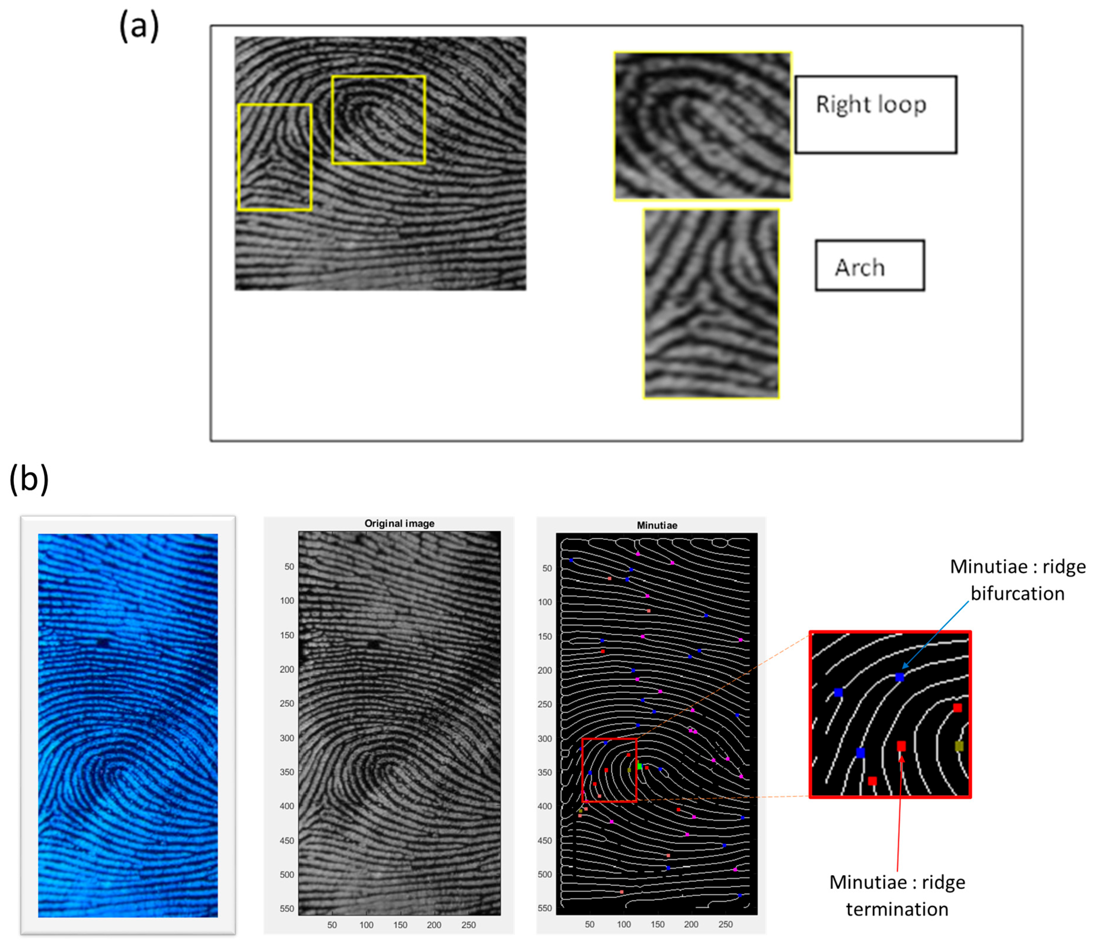
2.2. Latent Fingerprint Feature Extraction

Fingerprints hold significant importance in forensic science and are extensively studied due to their distinctive patterns and enduring characteristics [36,37,38]. The features of fingerprints are categorized into three levels:
-
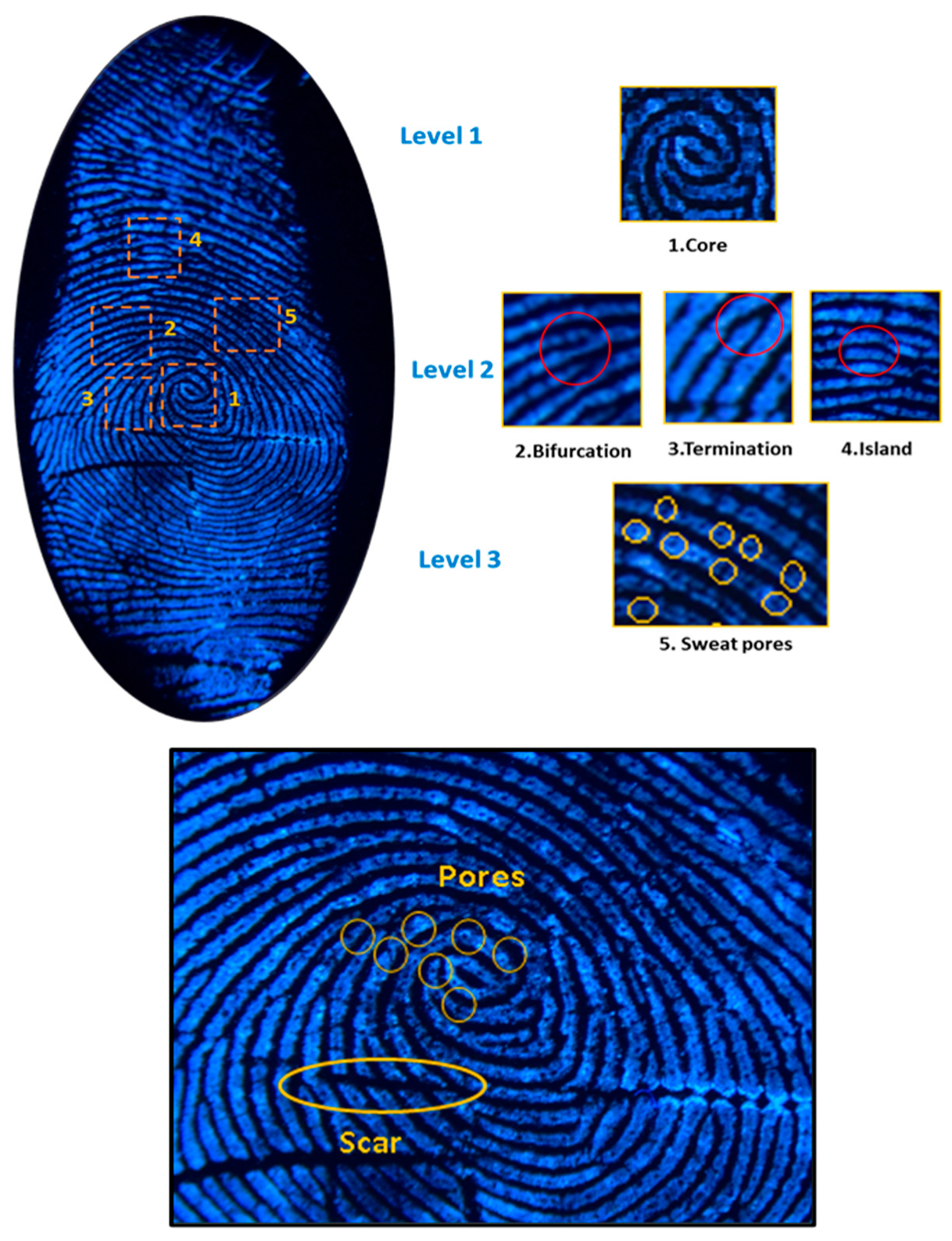
Level 1: This level consists of coarse features like arches, loops and whorls.
-
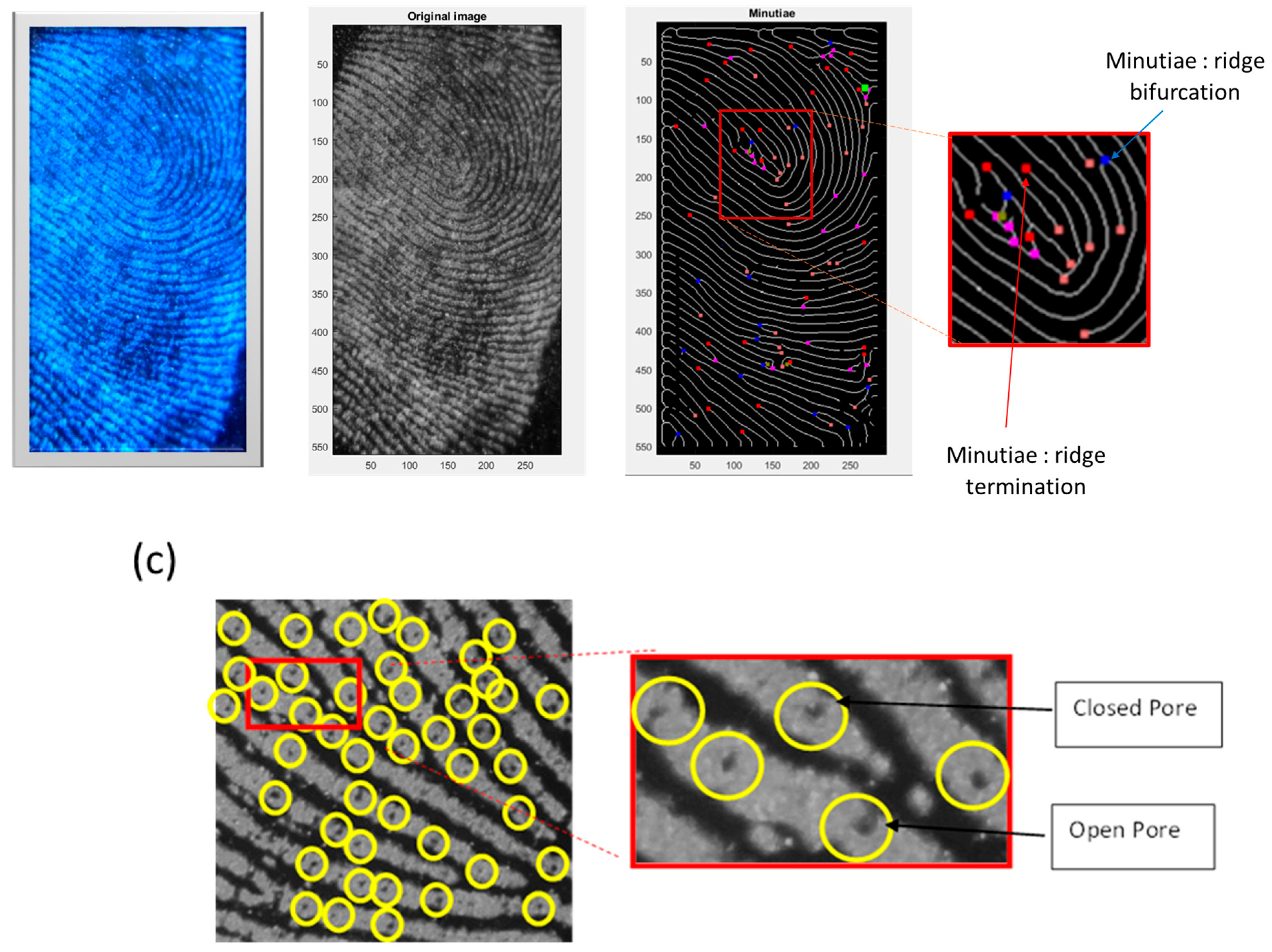
Level 2: This level involves identifying irregularities, known as minutiae, on capillary lines. These minutiae are then categorized into four types: terminations, bifurcations, islands and lakes.
-
Level 3: This level involves microscopic details, including pores.
These intricate fingerprint characteristics serve the purpose of identifying individual latent fingerprints, which are the impressions created when the grease and sweat on a finger transfer from the ridges to a contact surface upon touching it. Extensive research is conducted on fingerprints, owing to their distinctive patterns and enduring characteristics [37]. The visualization of latent fingerprints plays a crucial role in identifying both crime scenes and personal information. Thus, in order to demonstrate the satisfactory performance of the prepared La2O3: Bi phosphor as a fingerprint labeling agent, we chose to employ a fingerprint minutiae extraction algorithm to assess both the visualization and the quantity of fingerprint images. The algorithm in question, detailed in [36], involves preprocessing and minutiae extraction, as outlined below:
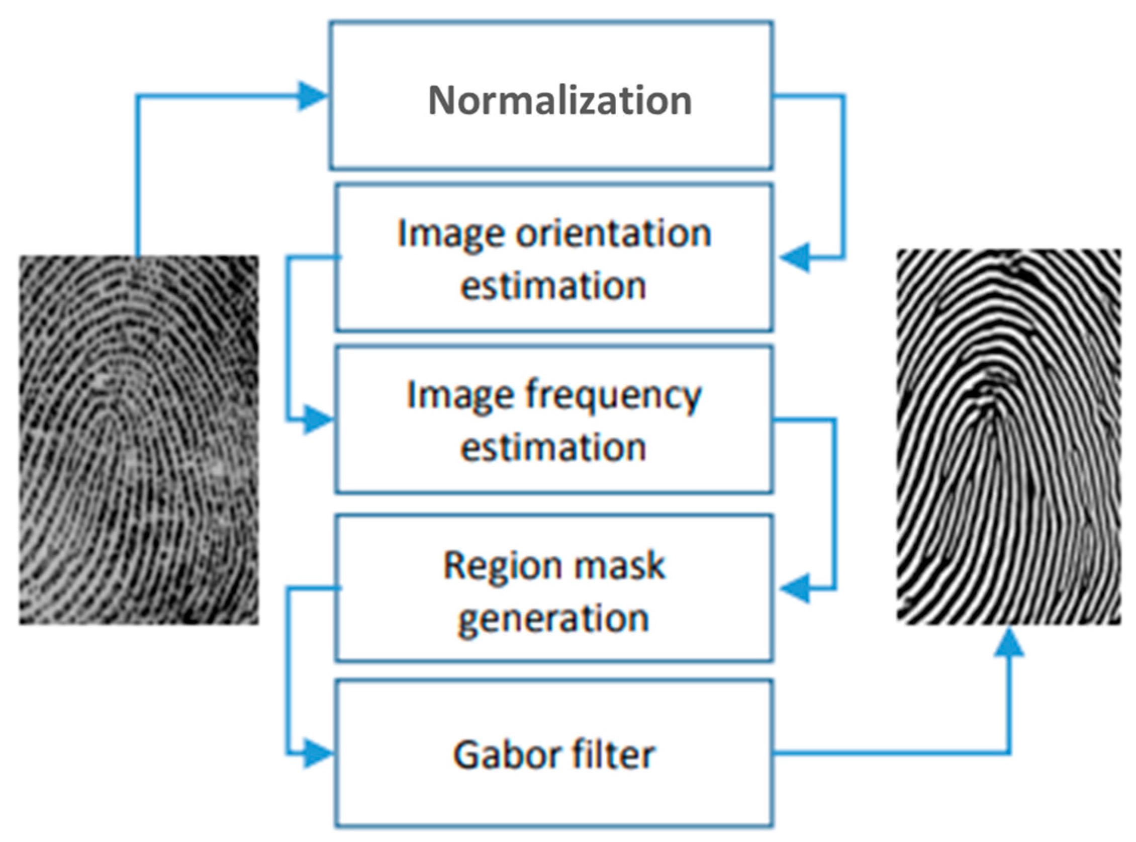
  • Preprocessing: This involves enhancing the image through a series of five main steps aimed at improving image quality for more effective feature extraction (Figure 1). The process begins with image normalization, followed by orienting the peaks of the fingerprint and estimating the frequency. Subsequently, a segmentation step is applied to identify the peak regions in a fingerprint image, creating a mask to delineate the region and normalizing image intensity values. Ultimately, oriented Gabor filters are utilized.
  • Feature extraction: The process of extracting minutiae features commences with an enhanced grayscale image. Following image binarization, a morphological thinning technique is applied to reduce ridge structures to a single pixel width, creating what is referred to as the skeleton. Subsequently, every pixel of the thinned binary image is analyzed to identify minutiae locations.

3. Results

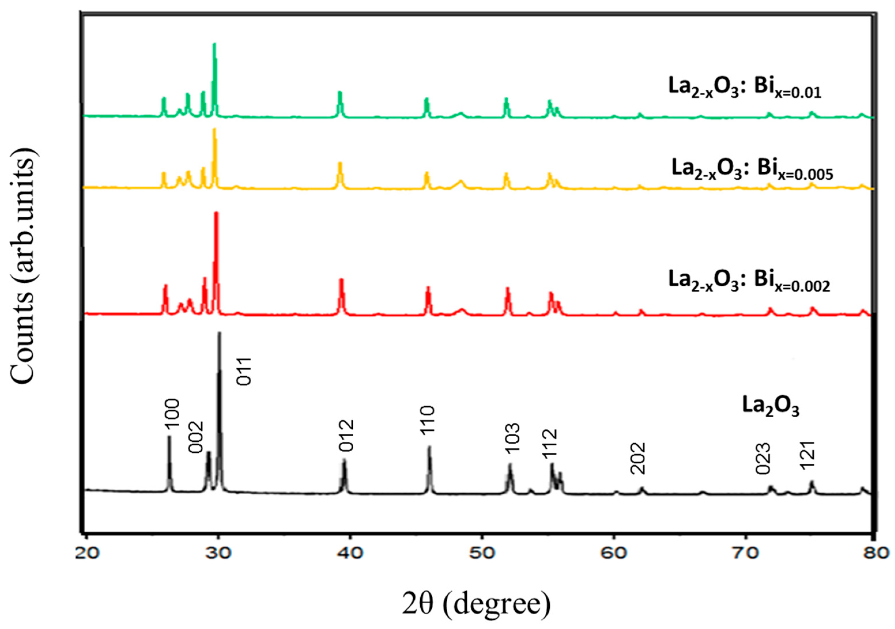
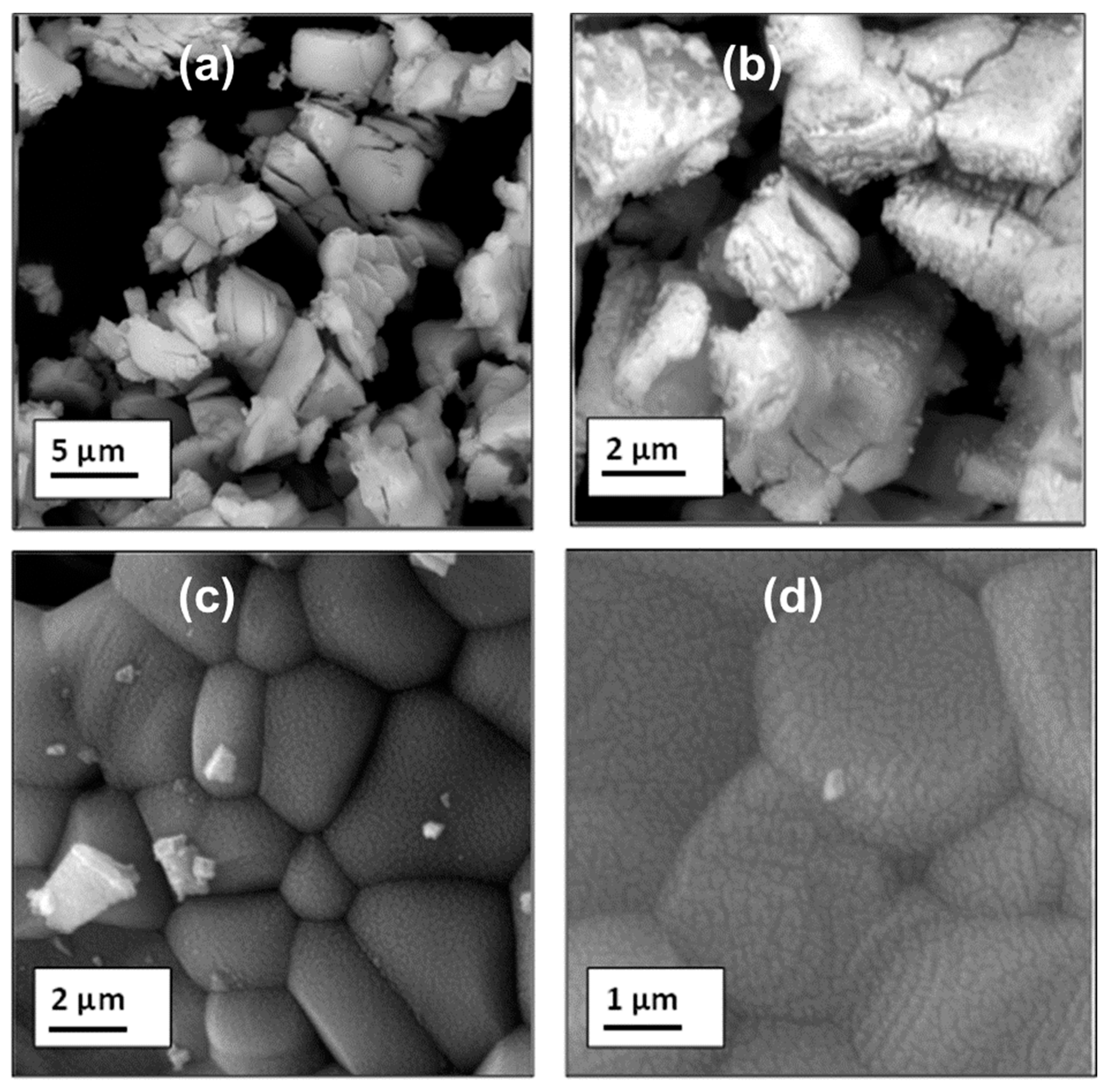
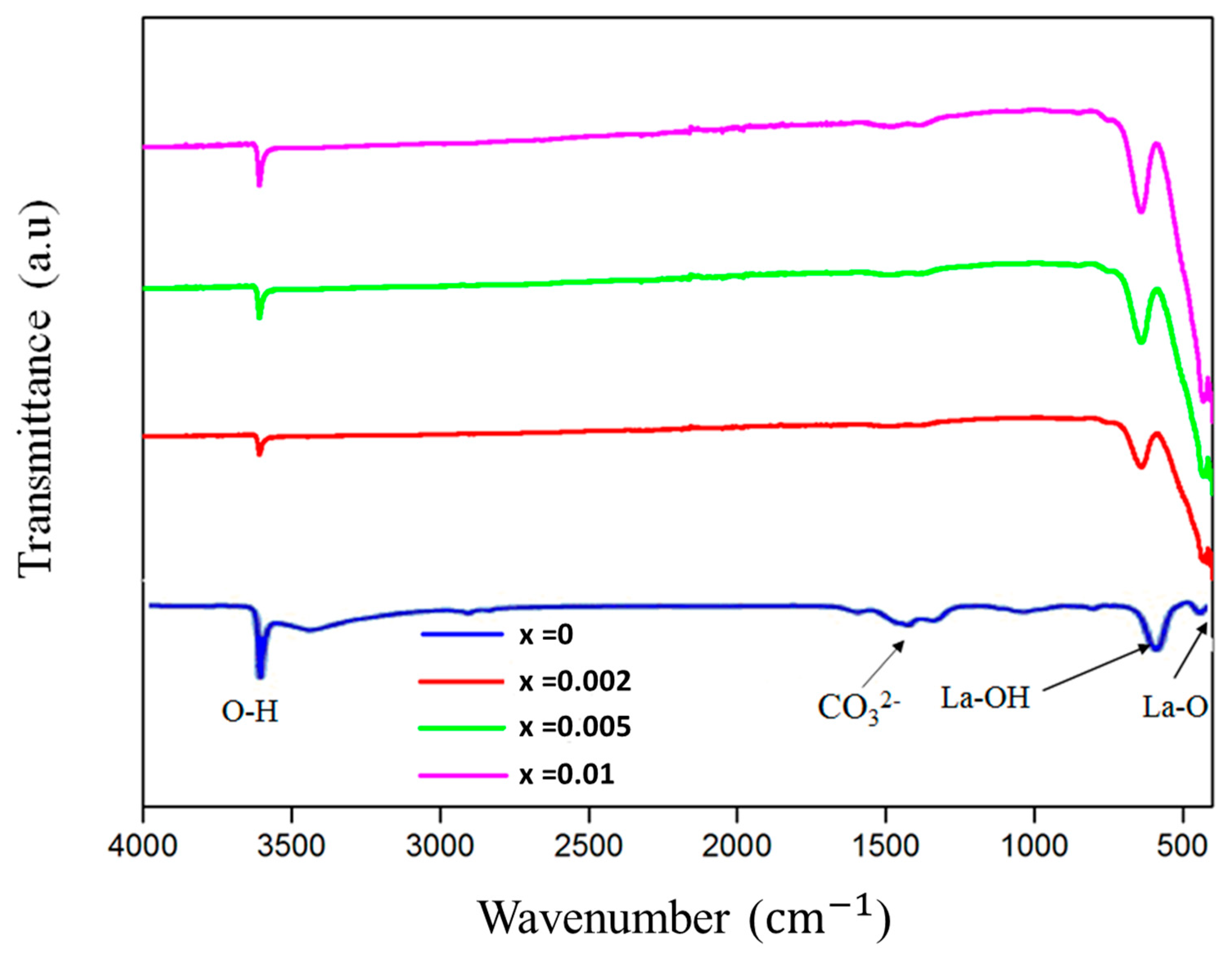
3.1. Structure and Morphology: XRD, SEM and EDX Studies

The XRD patterns of La2-xO3: Bix powder samples with x = 0.002, 0.005 and 0.01 are shown in Figure 2. On the basis of the Joint Committee on Powder Diffraction Standard (JCPDS) reference database, the diffraction peaks of the precursor are well indexed to hexagonal-phased La2O3 (JCPDS no. 05-0602). For synthesized samples, there is no variation in the location of diffraction peaks at different doping levels. However, additional diffraction angles of 27°, 27.7° and 48.5° located at 2θ = 27, 48 and 50 correlate with the (110), (101) and (211) and planes related to the presence of an La (OH)3 phase [39].
Figure 3 shows SEM images of the prepared La2O3: Bi3+ phosphors (x = 0.002 and 0.01). The particles of La2O3 have an irregular shape, and each particle contains many crystallites. However, some minor agglomerations can be observed, which is due to the fact that the Bi3+ ions influence the growi interface and change the chemical bonding.
The FTIR spectra in Figure 4 correspond to the samples prepared with different concentrations of bismuth (x = 0, 0.02, 0.05 and 0.1). The samples showed similar FTIR spectra with increasing peak intensities. Strong absorption and abrupt peaks at 3610 c m 1 could be attributed to stretching O–H vibrations brought on by moisture absorbed from the environment [40]. The appearance of several peaks in the range of 1000 1625   c m 1 may be attributed to the ν3 and ν1, ν2, and ν4 modes of the C O 3 2 , which confirms the presence of CO2 absorbed from the atmosphere [41]. The absorption peaks at about 645 c m 1 may be attributed to La–OH vibration, and the small band at around 430 c m 1 may be assigned to the characteristic metal oxide La–OH vibration [42].

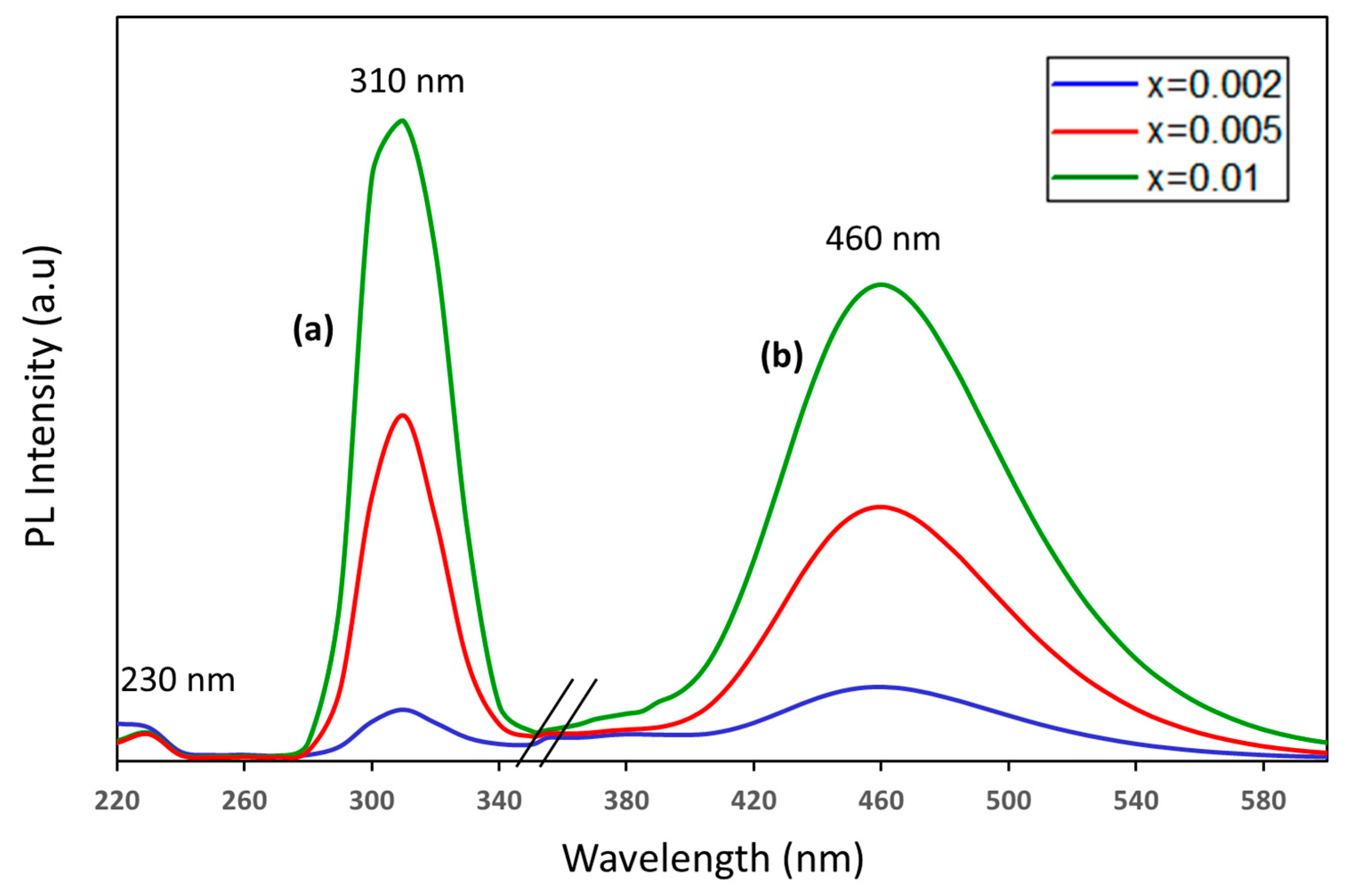
3.2. Photoluminescence (PL) Properties

Figure 5 shows the PL excitation and emission spectra of the La2O3 doped with different Bi concentrations. The excitation spectra of the blue emission (around 460 nm) consisted of two peaks at 230 nm and 310 nm corresponding to transitions from the 1S0 ground state to the 1P1 and 3P1 energy levels, respectively. On the other hand, broadband blue emission was observed at around 460 nm after 310 nm excitation. These Bi3+ ions’ radiative characteristics are attributed to parity-allowed transitions between the 6s2 ground state (1S0) and the 6s6p excited states, which are represented by the states 3PJ (J = 0–2) and 1P1. While the optical transition from 1S0 to 1P1 is spin-allowed, the optical transitions from 1S0 to 3P0 and 3P2 are totally spin-forbidden. The 3P1 level is permitted when the 1P1 and 3P1 levels are mixed via spin–orbit coupling [43].
Additionally, in Bi3+-doped materials, a lower energy luminescence is sometimes reported in addition to the characteristic broad band 3P0,11S0 luminescence. It might result from a charge transfer transition between metals (CB → Bi3+/Bi4+) as well as from Bi3+-Bi3+ pairs or clusters [44,45,46].
For the emission spectra, a single broad band of blue emission centered at 460 nm was detected and attributed to radiative transitions from the 3P1 excited state back to the 1S0 ground state. Moreover, the photoluminescence intensity increased with increasing Bi3+ ion concentration.

3.3. CIE Chromaticity

Color coordinates of the phosphors were calculated using the Commission Internationale de l’Eclairage (CIE) 1931 color chromaticity diagram. Figure 6 represents the CIE diagram of La2O3: x% Bi (x = 0.2, 0.5 and 1) phosphors. The CIE color coordinates of all the samples are almost equal and lie around (0.157, 0.172), which is very close to that of the ideal blue light.

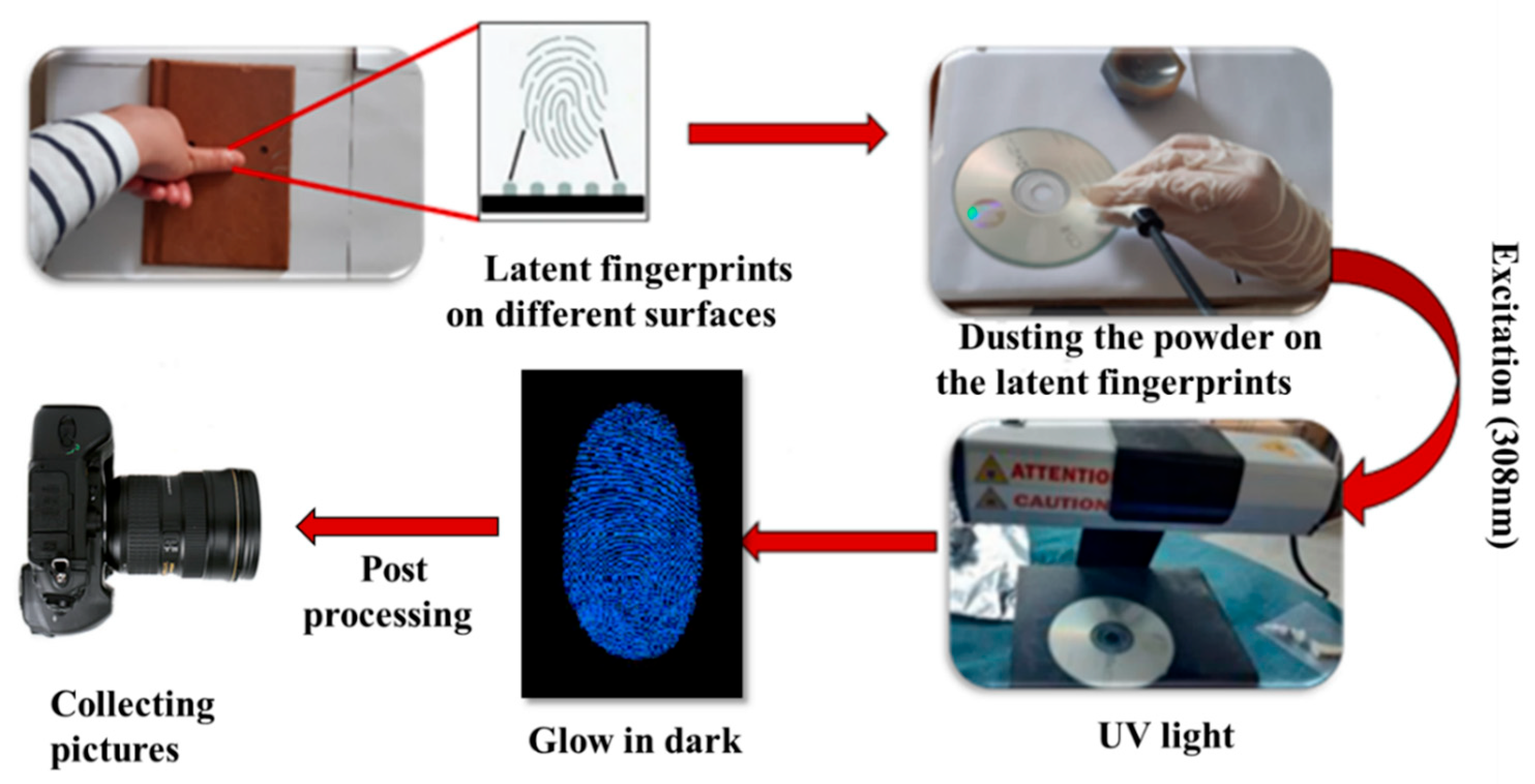
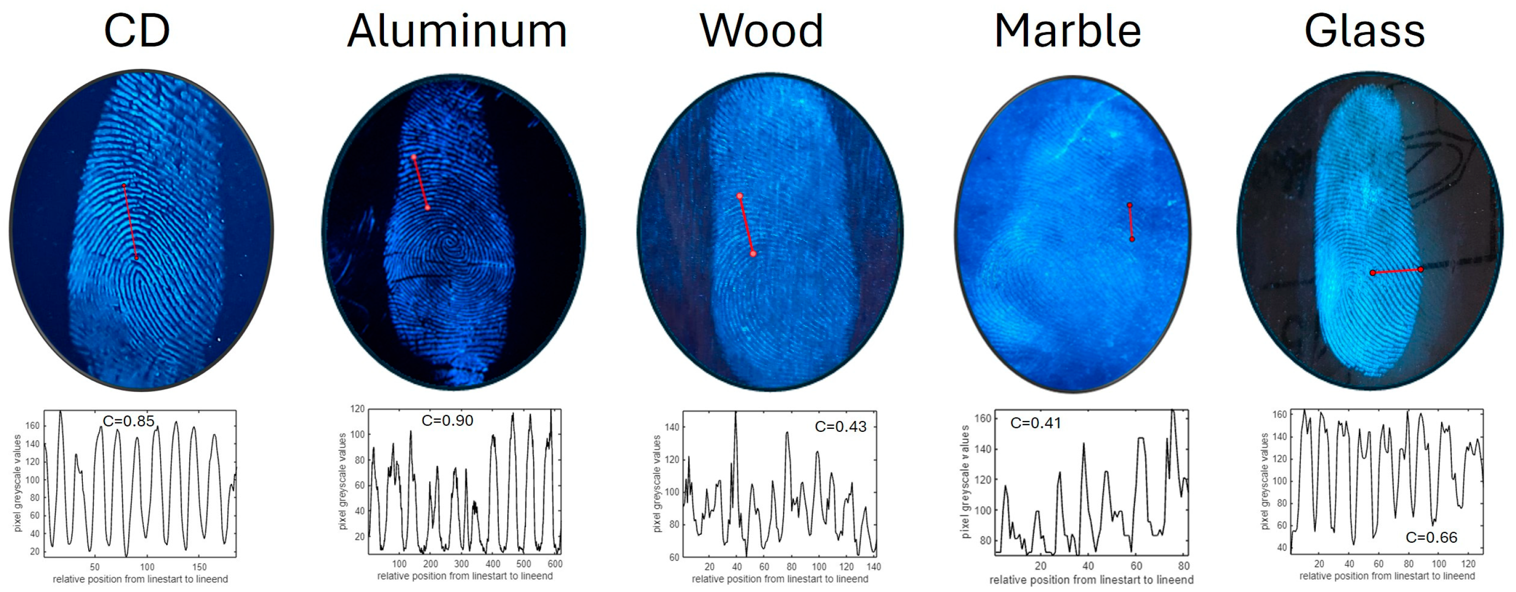
3.4. Latent Fingerprint Detection Using the Powder Dusting Method

Good visualization of the latent fingerprints (LFPs) on various surfaces was anticipated based on the photoluminescence results. Thus, to test our novel powders, both porous and non-porous surfaces (such as glass, plastic, aluminum foil and compact discs) were chosen. The donor’s hand was thoroughly cleaned and dried before the imprint was made. The donor’s fingerprints were then carefully wiped off their foreheads and their clean fingertips were pushed against a variety of room-temperature objects. After that, the LFPs were dyed with Bi-doped La2O3 using a soft feature brush and a smooth brushing technique. Finally, new fingerprints were developed and prepared for visualization. The production of fluorescent powder, which enables latent fingerprints to be seen under UV light, was the primary objective of this experiment. Figure 7 displays a schematic view of the development of the fingerprints.
Figure 8 shows the images of fresh fingerprints under UV lamp illumination (λ = 302 nm) developed by La2O3: 1% Bi3+ phosphors on CD, aluminum foil, wood, marble and glass surfaces, respectively. In general, the quality of the LFPs developed on different surfaces, based on visual assessment, was excellent, and information of levels 1–3 could be easily seen. In all cases, we extracted the intensity profile along a line after conversion of the RGB picture to grayscale. The intensity profile was then used to calculate the intensity fringe contrast as follows:
C = I M A X I m i n I M A X + I m i n
where I M A X and I m i n are the maximum and minimum intensities in the line profile, respectively. As expected, the lowest contrasts were obtained on difficult and porous surfaces, namely, wood and marble (0.43 and 0.41, respectively); the contrast on glass was 0.66; on CDs, it was 0.85; and the highest contrast of 0.90 was obtained on aluminum.
Figure 9 shows an enlarged fingerprint picture developed on aluminum foil under UV light (λ = 302 nm). All three levels of fingerprint ridge patterns are clearly visualized: the core (level 1); bifurcations, terminations and islands (level 2); and sweat pores (level 3).
Actually, general morphological details like orientation fields, ridge patterns and fingerprint ridge flows are level 1 details. Information regarding the pattern agreement of each fingerprint’s ridges is provided by level 2 details. Sweat pores, curvature and dots are examples of the dimensional fingerprint ridge features that are classified as level 3 details [47].

3.5. Automatic Minutiae Extraction

Macroscopic and microscopic details play a crucial role in personal identification. In order to demonstrate the feasibility of the La2O3: Bi phosphor as a fingerprint developing agent, we opted for the fingerprint minutiae extraction algorithm developed in [36] and performed assessments using the BioSecure, FVC DB1 and PolyU public databases [37]. The detected latent fingerprint images stained with La2O3: Bi on compact disc, aluminum, wood, marble and glass surfaces could be well visualized, and details of levels 1–3 were well identified. Figure 10 shows some examples of automatic identification of loops, arches, ridge terminations, and open and closed pores.

4. Conclusions

In summary, a facile sol–gel combustion method was employed to conveniently synthesize fluorescent La2O3: Bi3+ (0.2, 0.5 and 1 mol %) phosphors for latent fingerprint detection. According to a structural and spectroscopic analysis, the acquired samples had a good crystalline structure, indicating that the Bi3+ ions were effectively incorporated into the crystalline hexagonal structure of the La2O3 matrix. A photoluminescence study showed a strong broad band of blue fluorescence emission centered at 460 nm for excitation at 310 nm, corresponding to the radiative transition of the 3P1 excited state back to the 1S0 ground state. The CIE color coordinates were evaluated, and it was found that the photoluminescence emission from La2O3: Bi3+ phosphors lay in the blue region. By employing the powder dusting technique, when exposed to UV light, the blue emission from La2O3: Bi phosphors demonstrated significant promise for the viewing of latent fingerprints by generating latent fingerprint images with excellent contrast and recognition of all three levels of features on a variety of materials, including challenging ones like wood and marble. In parallel, an evaluation based on an automatic algorithm was performed by extracting fingerprint minutiae. We succeeded in detecting level 3 details corresponding to open and closed pores. The results of this work demonstrate the great potential of La2O3: Bi3+ phosphors for latent fingerprint feature extraction either in terms of visual evaluation or evaluation based on automatic recognition, even with a low Bi doping.

Author Contributions

Conceptualization, H.D., M.A. and R.M.; data curation, H.D., L.R.H. and A.T.; funding acquisition, R.M.; investigation, H.D., L.B. and A.T.; methodology, H.D., L.B., M.A. and L.R.H.; project administration, H.D.; resources R.M.; software and N.E.B.A.; supervision, A.T. and R.M.; validation, H.D., M.A. and L.R.H.; writing—original draft, H.D. and L.R.H.; writing—review and editing, A.T. and R.M. All authors have read and agreed to the published version of the manuscript.

Funding

This work was funded by the Tunisian Ministry of Higher Education and Scientific Research MHESR through the project PAQ Collabora T2-C3 “KIDAEM”.

Institutional Review Board Statement

Not applicable.

Informed Consent Statement

Informed consent was obtained from all subjects involved in the study.

Data Availability Statement

The raw data supporting the conclusions of this article will be made available by the authors on request.

Conflicts of Interest

The authors declare no conflicts of interest.

References

  1. Li, J.; Feng, J.; Kuo, C.-C.J. Deep convolutional neural network for latent fingerprint enhancement. Signal Process. Image Commun. 2018, 60, 52–63. [Google Scholar] [CrossRef]
  2. Cao, K.; Jain, A.K. Automated latent fingerprint recognition. IEEE Trans. Pattern Anal. Mach. Intell. 2018, 41, 788–800. [Google Scholar] [CrossRef] [PubMed]
  3. Wang, C.; Zhou, J.; Lulu, L.; Song, Q. Rapid Visualization of Latent Fingerprints with Color-Tunable Solid Fluorescent Carbon Dots. Part. Part. Syst. Charact. 2018, 35, 1700387. [Google Scholar] [CrossRef]
  4. Basavaraj, R.B.; Darshan, G.P.; Prasad, B.D.; Sharma, S.C.; Nagabhushana, H. Rapid visualization of latent fingerprints using novel CaSiO3:Sm3+ nanophosphors fabricated via ultrasound route. J. Rare Earths 2019, 37, 32–44. [Google Scholar] [CrossRef]
  5. Marappa, B.; Rudresha, M.S.; Basavaraj, R.B.; Darshan, G.P.; Daruka Prasad, B.; Sharma, S.C.; Sivakumari, S.; Amudha, P.; Nagabhushana, H. EGCG assisted Y2O3:Eu3+ nanopowders with 3D micro-architecture assemblies useful for latent figerprint recognition and anti-counterfeiting applications. Sens. Actuators B Chem. 2018, 264, 426–439. [Google Scholar] [CrossRef]
  6. Dhanalakshmi, M.; Basavaraj, R.B.; Darshan, G.P.; Sharma, S.C.; Nagabhushana, H. Pivotal role of fluxes in BaTiO3:Eu3+ nano probes for visualization of latent fingerprints on multifaceted substrates and anti-counterfeiting applications. Microchem. J. 2019, 145, 226–234. [Google Scholar] [CrossRef]
  7. Singla, N.; Kaur, M.; Sofat, S. Automated Latent Fingerprint Identification System: A Review. Forensic Sci. Int. 2020, 309, 110187. [Google Scholar] [CrossRef]
  8. Kolhatkar, G.; Parisien, C.; Ruediger, A.; Muehlethaler, C. Latent Fingermark Imaging by Single-Metal Deposition of Gold Nanoparticles and Surface Enhanced Raman Spectroscopy. Front. Chem. 2019, 7, 440–448. [Google Scholar] [CrossRef]
  9. Basavaraj, R.B.; Nagabhushana, H.; Darshan, G.P.; Daruka Prasad, B.; Rahul, M.; Sharma, S.C.; Sudaramani, R.; Archana, K.V. Red and green emitting CTAB assisted CdSiO3:Tb3+/Eu3+ nanopowders as fluorescent labeling agents used in forensic and display applications. Dye. Pigment. 2017, 147, 364–377. [Google Scholar] [CrossRef]
  10. Cantu, A.A. Silver physical developers for the visualization of latent prints on paper. Forensic Sci. Rev. 2001, 13, 29–64. [Google Scholar]
  11. Schnetz, B.; Margot, P. Technical note: Latent fingermarks, colloidal gold and multimetal deposition(MMD): Optimisation of the method. Forensic Sci. Int. 2001, 118, 21–28. [Google Scholar] [CrossRef] [PubMed]
  12. Brini, L.; Bennour, I.; Toncelli, A.; Maalej, R.; Abdelhedi, M. Eu-Doped Pyrochlore Crystal Nano-Powders as Fluorescent Solid for Fingerprint Visualization and for Anti-Counterfeiting Applications. Materials 2022, 15, 2423. [Google Scholar] [CrossRef]
  13. Chávez, D.; Garcia, C.R.; Oliva, J.; Diaz-Torres, L.A. A review of phosphorescent and fluorescent phosphors for fingerprint detection. Ceram. Int. 2021, 47, 10–41. [Google Scholar] [CrossRef]
  14. Ansari, A.A.; Aldajani, K.M.; AlHazaa, A.N.; Albrithen, H.A. Recent progress of fluorescent materials for fingermarks detection in forensic science and anti-counterfeiting. Coord. Chem. Rev. 2022, 462, 214523. [Google Scholar] [CrossRef]
  15. Lian, J.; Meng, F.; Wang, W.; Zhang, Z. Recent Trends in Fluorescent Organic Materials for Latent Fingerprint Imaging. Front. Chem. 2020, 8, 594864. [Google Scholar] [CrossRef]
  16. Wang, M.; Mi, C.C.; Wang, W.X.; Liu, C.H.; Wu, Y.F.; Xu, Z.R.; Mao, C.B.; Xu, S.K. Immunolabeling and NIR-excited fluorescent imaging of HeLa cells by using NaYF4: Yb, Er upconversion nanoparticles. ACS Nano 2009, 3, 1580–1586. [Google Scholar] [CrossRef]
  17. Ang, L.Y.; Lim, M.E.; Ong, L.C.; Zhang, Y. Applications of upconversion nanoparticles in imaging, detection and therapy. Nanomedicine 2011, 6, 1273–1288. [Google Scholar] [CrossRef]
  18. Wang, M. Latent fingermarks light up: Facile development of latent fingermarks using NIR-responsive upconversion fluorescent nanocrystals. RSC Adv. 2016, 6, 36264–36268. [Google Scholar] [CrossRef]
  19. Lundberg, P.; Lindh, E.M.; Tang, S.; Edman, L. Toward efficient and metal-free emissive devices: A solution-processed host-guest light-emitting electrochemical cell featuring thermally activated delayed fluorescence. ACS Appl. Mater. Interfaces 2017, 9, 28810–28816. [Google Scholar] [CrossRef]
  20. Wang, M.; Li, M.; Yu, A.; Zhu, Y.; Yang, M.; Mao, C. Fluorescent nanomaterials for the development of latent fingerprints in forensic sciences. Adv. Funct. Mater. 2017, 27, 1606243–1606259. [Google Scholar] [CrossRef]
  21. Brini, L.; Douiri, H.; Abid, M.; Toncelli, A.; Qasymeh, M.; Maalej, R.; Abdelhedi, M. Potential of Y2Sn2O7:Eu3+, Dy3+ Inoganic Nanophosphors in Latent Fingermark Detection. Crystals 2024, 14, 300. [Google Scholar] [CrossRef]
  22. Zhong, J.; Zhao, W.; Zhuo, Y.; Yan, C.; Wen, J.; Brgoch, J. Understanding the blue-emitting orthoborate phosphor NaBaBO3: Ce3+ through experiment and computation. J. Mater. Chem. C 2019, 7, 654–662. [Google Scholar] [CrossRef]
  23. Guss, P.; Foster, M.E.; Wong, B.M.; Doty, F.P.; Shah, K.; Squillante, M.R.; Shirwadkar, U.; Hawrami, R.; Tower, J.; Yuan, D. Results for aliovalent doping of CeBr3 with Ce2+. J. Appl. Phys. 2014, 115, 034908. [Google Scholar] [CrossRef]
  24. Wei, C.; Fan, J.; Gong, H. Structural, thermodynamic, and mechanical properties of bulk La and A-La2O3. J. Alloys Compd. 2015, 618, 615–622. [Google Scholar] [CrossRef]
  25. Pisecny, P.; Husekova, K.; Frohlich, K.; Harmatha, L.; Soltys, J.; Machajdik, D.; Espinos, J.P.; Jergel, M.; Jakabovic, J. Growth of lanthanum oxide films for application as a gate dielectric in CMOS technology. Mater. Sci. Semicon. Proc. 2004, 7, 231–236. [Google Scholar] [CrossRef]
  26. Mangla, O.; Srivastava, A.; Malhotra, Y.; Ostrikov, K. Lanthanum oxide nanostructured films synthesized using hot dense and extremely non-equilibrium plasma for nanoelectronic device applications. J. Mater. Sci. 2014, 49, 1594–1605. [Google Scholar] [CrossRef]
  27. Sasidharan, M.; Gunawardhana, N.; Inoue, M.; Yusa, S.-I.; Yoshio, M.; Nakashima, K. La2O3 hollow nanospheres for high performance lithium-ion rechargeable batteries. Chem. Commun. 2012, 48, 3200–3202. [Google Scholar] [CrossRef]
  28. Gangwar, B.P.; Palakollu, V.; Singh, A.; Kanvah, S.; Sharma, S. Combustion synthesized La2O3 and La (OH)3: Recyclable catalytic activity towards Knoevenagel and Hantzsch reactions. RSC Adv. 2014, 4, 55407–55416. [Google Scholar] [CrossRef]
  29. Xu, Z.; Bian, S.; Wang, J.; Liu, T.; Wang, L.; Gao, Y. Preparation and luminescence of La2O3:Ln3+ (Ln3+ = Eu3+, Tb3+, Dy3+, Sm3+, Er3+, Ho3+, Tm3+, Yb3+/Er3+, Yb3+/Ho3+) microspheres. RSC Adv. 2013, 3, 1410–1419. [Google Scholar] [CrossRef]
  30. Dey, R.; Pandey, A.; Rai, V.K. The Er3+- Yb3+ codoped La2O3 phosphor in fingerprint detection and optical heating. Spectrochim. Acta Part A Mol. Biomol. Spectrosc. 2014, 128, 508–513. [Google Scholar] [CrossRef]
  31. Pushpa, N.; Kokila, M.K.; Shivaramu, N.J. Luminescence properties of La2O3:Eu3+ nanophosphor prepared by sol–gel method. Nucl. Instrum. Methods Phys. Res. B 2016, 379, 69–72. [Google Scholar] [CrossRef]
  32. Reddy, P.V.S.S.S.N.; Babu, D.N.; Kishore, G.M.M.; Naveen, S.V.; Ramana, B.V.; Srinivas, B.; Naveen, B.V.; Rao, K.R.; Ch, S.K. Polyol derived Bi3+ and Er3+ co-doped lanthanum oxide nanophosphors for display & LED applications. Mater. Today Proc. 2019, 18, 2118–2122. [Google Scholar] [CrossRef]
  33. Catauro, M.; Ciprioti, S. Vecchio.Characterization of Hybrid Materials Prepared by Sol-Gel Method for Biomedical Implementations. A Critical Review. Materials 2021, 14, 1788. [Google Scholar] [CrossRef]
  34. Mura, S.; Ludmerczki, R.; Stagi, L.; Garroni, S.; Carbonaro, C.M.; Ricci, P.C.; Casula, M.F.; Malfatti, L.; Innocenzi, P. Integrating sol-gel and carbon dots chemistry for the fabrication of fluorescent hybrid organic-inorganic films. Sci. Rep. 2020, 10, 4770. [Google Scholar] [CrossRef] [PubMed]
  35. Parashar, M.; Shukla, V.K.; Singh, R. Metal oxides nanoparticles via sol–gel method: A review on synthesis, characterization and applications. J. Mater. Sci. Mater. Electron. 2020, 31, 3729–3749. [Google Scholar] [CrossRef]
  36. Haddada, L.R.; Amara, N.E.B. Score-Level Fusion of Fingerprint, Face and Iris based on Choquet Integral. In Proceedings of the 2019 5th International Conference on Nanotechnology for Instrumentation and Measurement (NanofIM), Sfax, Tunisia, 30–31 October 2019. [Google Scholar] [CrossRef]
  37. Gouiaa, M.; Bennour, I.; Haddada, L.R.; Toncelli, A.; Xu, J.; Mbarek, A.; Moscardini, A.; Amara, N.E.B.; Maalej, R. Spectroscopic characterization of Er, Yb: Y2Ti2O7 phosphor for latent fingerprint detection. Phys. B Condens. Matter 2020, 582, 412009. [Google Scholar] [CrossRef]
  38. Maalej, N.M.; Qurashi, A.; Bennour, I.; Haddada, L.R.; Shaikh, M.N.; Ilyas, M.; Amara, N.E.B.; Maalej, R.; Gondal, M.A. Green emitting rare earth Gd2O3:Tb3+ nanoparticles for rapid imaging of latent fingerprint. Methods Appl. Fluores 2021, 9, 025002. [Google Scholar] [CrossRef]
  39. Singh, A.; Palakollu, V.; Pandey, A.; Kanvah, S.; Sharma, S. Green synthesis of 1,4-benzodiazepines over La2O3 and La (OH)3 catalysts: Possibility of Langmuir-Hinshelwood adsorption. RSC Adv. 2016, 6, 103455–103462. [Google Scholar] [CrossRef]
  40. Mu, Q.; Wang, Y. Synthesis, characterization, shape-preserved transformation, and optical properties of La (OH)3, La2O2CO3, and La2O3 nanorods. J. Alloys Compd. 2011, 509, 396–401. [Google Scholar] [CrossRef]
  41. Méndez, M.; Cesteros, Y.; Marsal, L.F.; Giguère, A.; Drouin, D.; Salagre, P.; Formentıín, P.; Pallarès, J.; Aguiloó, M.; Dıaz, F.; et al. Effect of Thermal Annealing on the Kinetics of Rehydroxylation of Eu3+: La2O3 Nanocrystals. J. Inorg. Chem. 2012, 5, 16139–16146. [Google Scholar] [CrossRef]
  42. He, F.; Yang, P.; Wang, D.; Li, C.; Niu, N.; Gai, S.; Zhang, M. Preparation and up-conversion luminescence of hollow La2O3: Ln (Ln = Yb/Er, Yb/Ho) microspheres. Langmuir 2011, 27, 5616–5623. [Google Scholar] [CrossRef] [PubMed]
  43. Tabaza, W.A.I.; Swart, H.C.; Kroon, R.E. Optical properties of Bi and energy transfer from Bi to Tb in MgAl2O4 phosphor. J. Lumin. 2014, 148, 192–197. [Google Scholar] [CrossRef]
  44. Bautinaud, P. Revisiting the spectroscopy of the Bi3+ ion in oxide compounds. Inorg. Chem. 2013, 52, 6028–6038. [Google Scholar] [CrossRef]
  45. Krasnikov, A.; Tsiumra, V.; Vasylechko, L.; Zazubovich, S.; Zhydachevskyy, Y. Photoluminescence origin in Bi3+- doped YVO4, LuVO4, and GdVO4 orthovanadates. J. Lumin. 2019, 212, 52–60. [Google Scholar] [CrossRef]
  46. Srivastava, A.M.; Comanzo, H.A. The ultraviolet and visible luminescence of Bi3+ in orthorhombic perovskite. Opt. Mater. 2017, 63, 118–121. [Google Scholar] [CrossRef]
  47. Azman, A.R.; Mahat, N.A.; Wahab, R.A.; Ahmad, W.A.; Huri, M.A.M.; Hamzah, H.H. Relevant Visualization Technologies for Latent Fingerprints on Wet Objects and Its Challenges: A Review. Egypt. J. Forensic Sci. 2019, 9, 23. [Google Scholar] [CrossRef]
Figure 1. Description of the preprocessing phase.
Materials 17 04217 g001
Figure 2. La2O3 and La2-xO3: Bix powders (x = 0.002, 0.005 and 0.01).
Materials 17 04217 g002
Figure 3. SEM micrographs of prepared La2O3 Bi phosphors: (a,b) for x = 0.002; (c,d) for x = 0.01.
Materials 17 04217 g003
Figure 4. FTIR spectra of the La2O3 and La2-xO3: Bix powders.
Materials 17 04217 g004
Figure 5. (a) Excitation and (b) emission spectra of La2-xO3: Bix doped with different Bi concentrations.
Materials 17 04217 g005
Figure 6. CIE color chromaticity coordinates of La2O3: Bi3+ phosphors: A (x = 0.2), B (x = 0.5) and C (x = 1).
Materials 17 04217 g006
Figure 7. Illustration of the development of latent fingermarks using the 1%Bi: La2O3 phosphor dusting process.
Materials 17 04217 g007
Figure 8. Photographs (under 302 nm UV-illumination) of LFPs on different substrates developed by La2O3: Bi3+ fluorescent blue powder. For every picture, the line intensity profile along the red line and the best contrast obtained are shown at the bottom.
Materials 17 04217 g008
Figure 9. Latent fingerprint image visualization (under 302 nm UV illumination) displaying first-, second- and third-level details.
Materials 17 04217 g009
Figure 10. The latent fingerprint image features evidenced by the 1% Bi, La2O3 phosphors on aluminum foil under UV light based on an automatic algorithm ((a) level 1, (b) level 2, and (c) level 3).
Materials 17 04217 g010aMaterials 17 04217 g010b
Disclaimer/Publisher’s Note: The statements, opinions and data contained in all publications are solely those of the individual author(s) and contributor(s) and not of MDPI and/or the editor(s). MDPI and/or the editor(s) disclaim responsibility for any injury to people or property resulting from any ideas, methods, instructions or products referred to in the content.

