Article

Three-Dimensional Peri-Implant Tissue Changes in Immediately vs. Early Placed Tapered Implants Restored with Two Different Ceramic Materials—1 Year Results

1
Division of Fixed Prosthodontics and Biomaterials, University Clinic of Dental Medicine, University of Geneva, Rue Michel-Servet 1, 1211 Geneva, Switzerland
2
Department of Prosthodontics, College of Dentistry, Yonsei University, Seoul 03722, Republic of Korea
3
Clinic of Conservative and Preventive Dentistry, Division of Periodontology and Peri-Implant Diseases, Center of Dental Medicine, University of Zürich, 8032 Zürich, Switzerland
4
Clinic of General, Special Care and Geriatric Dentistry, Center of Dental Medicine, University of Zürich, 8032 Zürich, Switzerland
*
Authors to whom correspondence should be addressed.
Materials 2023, 16(16), 5636; https://doi.org/10.3390/ma16165636
Submission received: 16 June 2023 / Revised: 19 July 2023 / Accepted: 10 August 2023 / Published: 15 August 2023

Abstract

:
Background: A prospective multi-center randomized controlled clinical trial was performed to digitally analyze tissue volume changes in immediately and early placed implants with simultaneous bone augmentation restored with two different all-ceramic materials. Methods: A total of 60 patients received 60 bone-level tapered implants (BLT, Straumann AG) immediately (n = 30) or early placed, 8–10 weeks after tooth extraction, (n = 30). Implants were restored with all-ceramic single crowns fabricated out of zirconia (Lava Plus, 3M), or lithium disilicate (E.max CAD, Ivoclar Vivadent AG) bonded to titanium base abutments (Variobase for Cerec, Straumann AG). Impressions were taken at baseline (BL), 6 and 12 months, and STL data were used to define an area of interest (AOI) to analyze peri-implant volume changes and midfacial recessions. Results: For immediate placement, a mean volume loss of −5.56 mm3 (±5.83 mm3) was found at 6 months, and of −6.62 mm3 (±6.56 mm3) at 12 months. For early placement, a mean volume loss of −1.99 mm3 (±5.82 mm3) at 6 months, and of −3.7 mm3 (±5.62 mm3) at 12 months was found. The differences in volume loss at 12 months between the two implant placement protocols were significant (p = 0.005). In both groups, mean midfacial recessions of 0.48 mm (±0.52) occurred. Conclusions: A more pronounced peri-implant volume loss can be expected 12 months after immediate implant placement compared with early placement.

1. Introduction

Over many decades, dental implants have proven to be a successful and predictable treatment option for replacing missing teeth [1,2]. High long-term clinical survival and success rates have led to dental implants often being the treatment of choice for single and multiple tooth restorations [3,4,5]. Furthermore, evolved implant geometry, surface topographies, and electrochemical coatings have improved primary stability and rapid osseointegration [6,7,8,9]. As a result, newer treatment options, such as immediate implant placement protocols, have become available [10,11,12]. The currently available literature indicates similar treatment outcomes for immediately placed implants compared with early or delayed placed implants [11,13,14,15,16]. At the same time, however, different bone healing patterns may influence peri-implant tissue volume changes. Therefore, bone augmentation procedures are used to preserve or increase hard tissue volume, and thus further stabilizing the peri-implant soft tissue [16].
When evaluating the esthetic outcome of dental implant treatments in the esthetic zone, the peri-implant soft tissue has, among many other factors, been described as one key factor for esthetic success. Several well-established, two-dimensional methods to investigate esthetic outcomes and mucosal recessions around dental implants have been described in the literature [17,18,19,20,21]. The midfacial mucosal recession was reported to depend on several factors, such as implant position, implant type and diameter, gingival biotype, and surgical technique [22]. Newer and more complex three-dimensional protocols have allowed for a more comprehensive analysis of peri-implant soft tissue volume [23,24]. Additionally, 3D radiographical assessments of peri-implant bone augmentation procedures and their volume changes are available [25,26,27,28,29,30,31]. Nevertheless, most currently available 3D analysis methods only present an overall peri-implant volume gain or loss, whereas a more detailed analysis method would be desirable.
Other factors potentially influencing the quantity and quality of the peri-implant tissues are the type and material of implant-borne restorations [32,33]. The configuration and surface topography of the supra-structure and the presence or absence of a submucosal cement gap may influence the peri-implant tissues [34,35,36]. Clinical studies have shown that the smooth surface of polished zirconia implant abutments demonstrated excellent biological integration with the peri-implant soft tissue [37,38].
However, more data is needed regarding the three-dimensional peri-implant tissue volume changes depending on the implant placement protocol, tissue augmentation procedures, and restoration material. Therefore, one aim of this multi-center prospective randomized controlled clinical trial was to digitally analyze the tissue volume changes in immediately and early placed implants restored with two different all-ceramic restorative materials.

2. Materials and Methods

The present study was performed according to the Declaration of Helsinki on medical protocol and ethics, in compliance with ICH-GCP guidelines and ISO 14155, and the CONSORT guidelines for randomized trials. The study was registered in the US clinical trials register (NCT05079542) on 15 October 2021. The protocol was approved by the respective local ethical committees (CCER 15-117).
For this multi-center randomized controlled clinical trial (RCT), patients were recruited at the University of Geneva, Switzerland (Division of Fixed Prosthodontics and Biomaterials) and in four associated private practices from 2015 to 2021. Sixty patients received 60 bone-level tapered (BLT) implants (Straumann AG, Switzerland) either immediately placed (group 1; n = 30) or by early placement 8–10 weeks after tooth extraction (group 2; n = 30). Simultaneous bone augmentation procedures were performed using autologous bone chips, a bovine bone substitute (BioOss, Geistlich Pharma, Wolhusen, Switzerland), and a resorbable collagen membrane (BioGide, Geistlich Pharma, Wolhusen, Switzerland).
The implant-borne restorations were all-ceramic single crowns fabricated from two different materials: zirconia (Lava Plus, 3M Schweiz GmbH, Rüschlikon, Switzerland) and lithium disilicate (E.max CAD, Ivoclar Vivadent AG, Schaan, Liechtenstein) bonded to titanium base abutments (Variobase for Cerec, Institut Straumann AG, Basel, Switzerland). The detailed study protocol was published previously [39]. Alginate impressions of the peri-implant region were taken at the final crown insertion, which was this study’s baseline (BL), and at 6 and 12 months after BL. A laboratory technician prepared cast models and then scanned them with a laboratory desktop scanner (IScan D104, Imetric 4D Imaging Sàrl, Switzerland) to create STL files for the volumetric analysis.
Horizontal and vertical volume changes of the peri-implant region were analyzed between BL and the different follow-up time points. A specific area of interest (AOI) was defined and applied to all models for this analysis.

2.1. Definition of the Area of Interest (AOI)

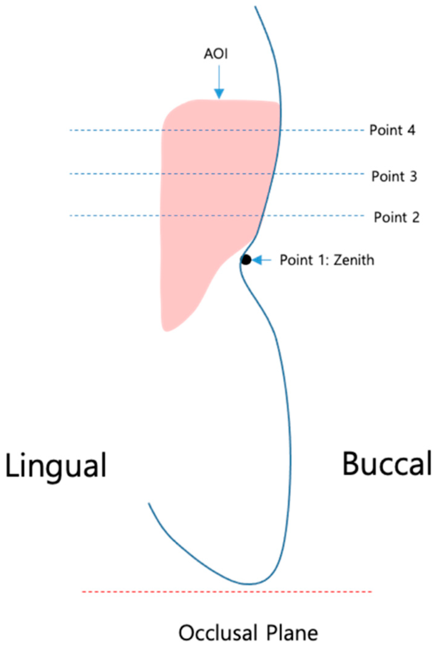
For the analysis of the peri-implant tissue volume changes, an AOI was defined and applied to all STL data in a standardized way (Figure 1).
The AOI was defined by tracing a line 0.5 mm below the mucosal margin, following the course of the gingival margin of the implant crown. For determining the mesial and distal borders of the AOI, vertical lines were drawn, going through the mesial and distal approximal contact points of the crown to the neighboring teeth and dividing the mesial and distal papilla into two halves each. The apical border of the AOI was defined by a horizontal line 3.5 mm below the zenith point of the crown.
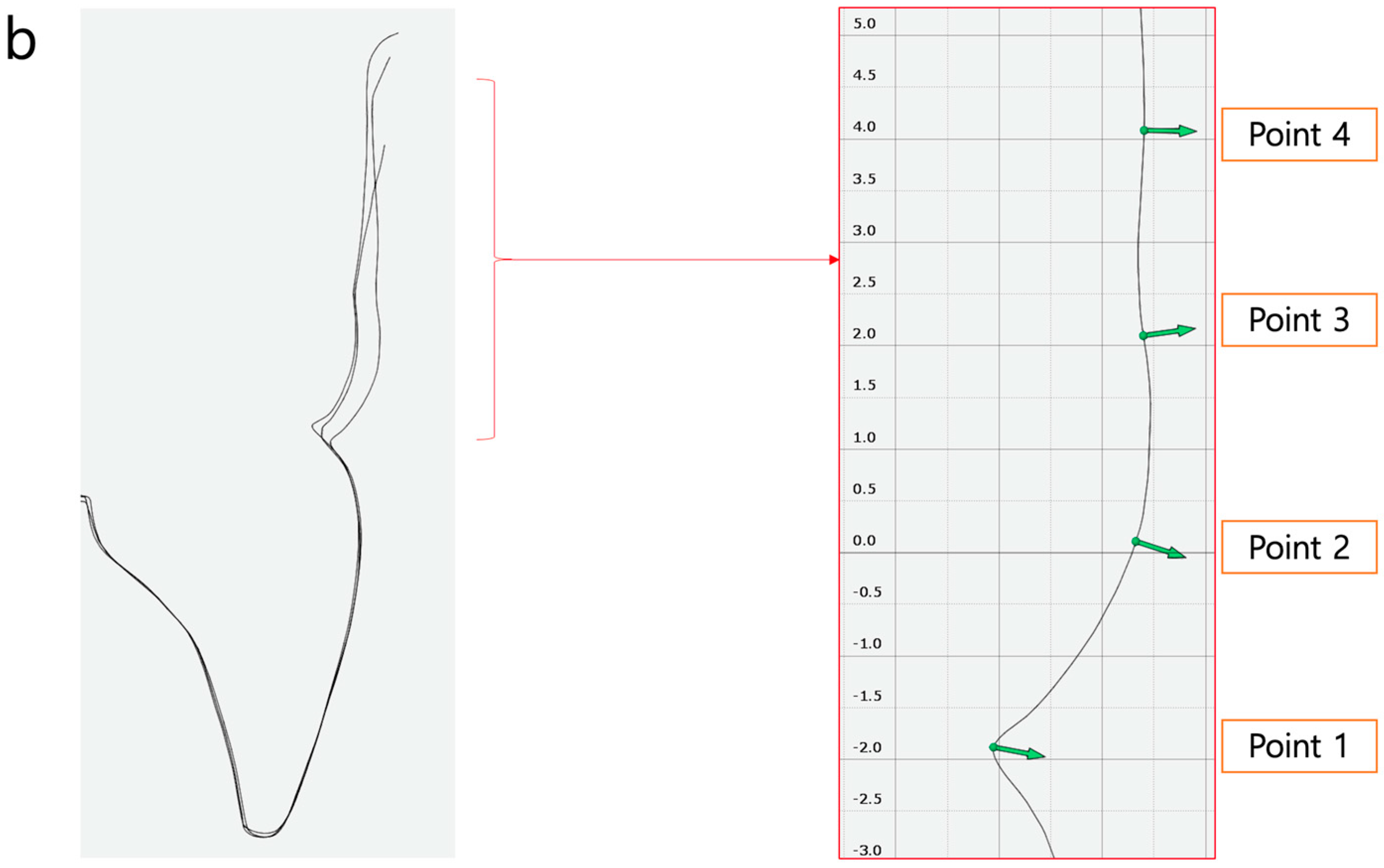
The midfacial mucosa height was defined as the distance between the incisal edge and point 1 = zenith. For the horizontal width measurements, three reference points were defined as reference points 2, 3, and 4, each 1 mm apart (Figure 2).

2.2. Definition of the Reference Points (Figure 2)

Point 1: reference point at zenith of the mucosal margin of the implant-borne crown.
Point 2: reference point 1 mm below zenith.
Point 3: reference point 2 mm below zenith.
Point 4: reference point 3 mm below zenith.

2.3. Volumetric Analysis

A previously developed method by Lee et al. was applied for this volumetric analysis of the peri-implant region using specialized software (GomInspect 2018, Gom, Braunschweig, Germany) [23]. This method allows detailed quantitative analysis of the volumetric changes at a specific site.

2.4. Analysis Procedure

  • Superimposition of STL data using best-fit algorithm focusing on the area of implant placement.
  • Alignment of the superimposed files with the x-, y-, and z-axes by each surface of the restoration of the implant.
  • Definition and marking of area of interest (AOI) in baseline STL and STL files at 6 and 12 months (Figure 3).
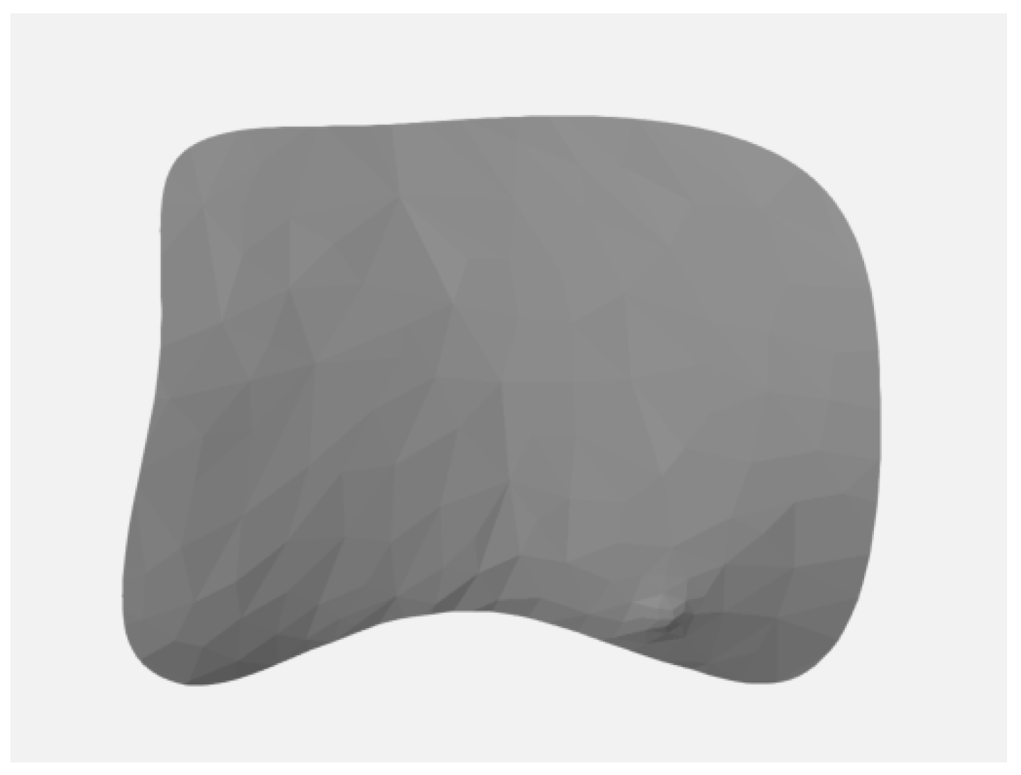
  • Separation of the AOI (Figure 4) from the original STL files.
  • Measurement of the AOI
  • Analysis by comparison of the AOI of each STL file: the separated AOI of the baseline visit was projected onto the AOI of the STL at 6-month and 12-month follow up to analyze any volume changes.
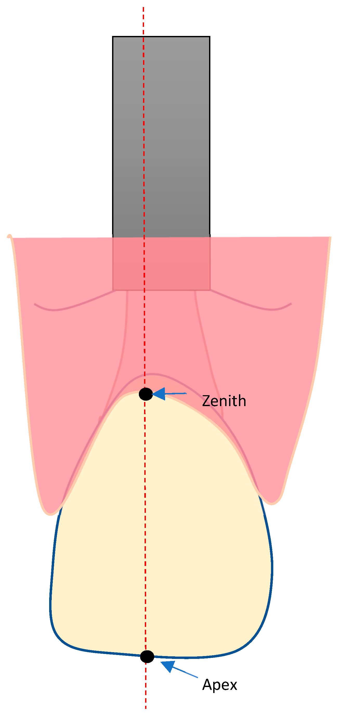
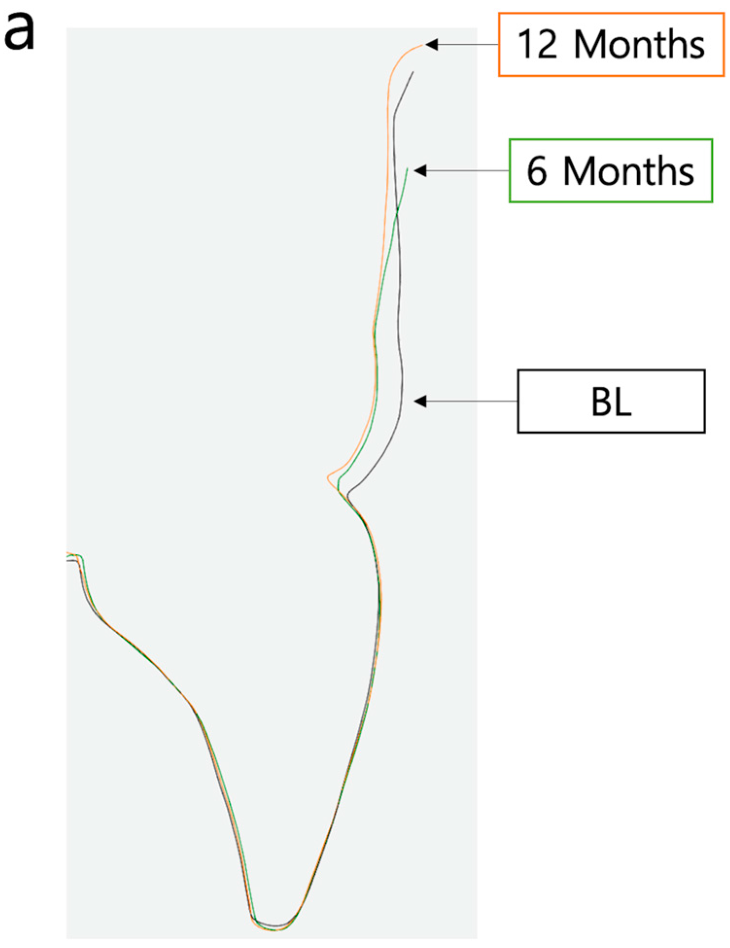
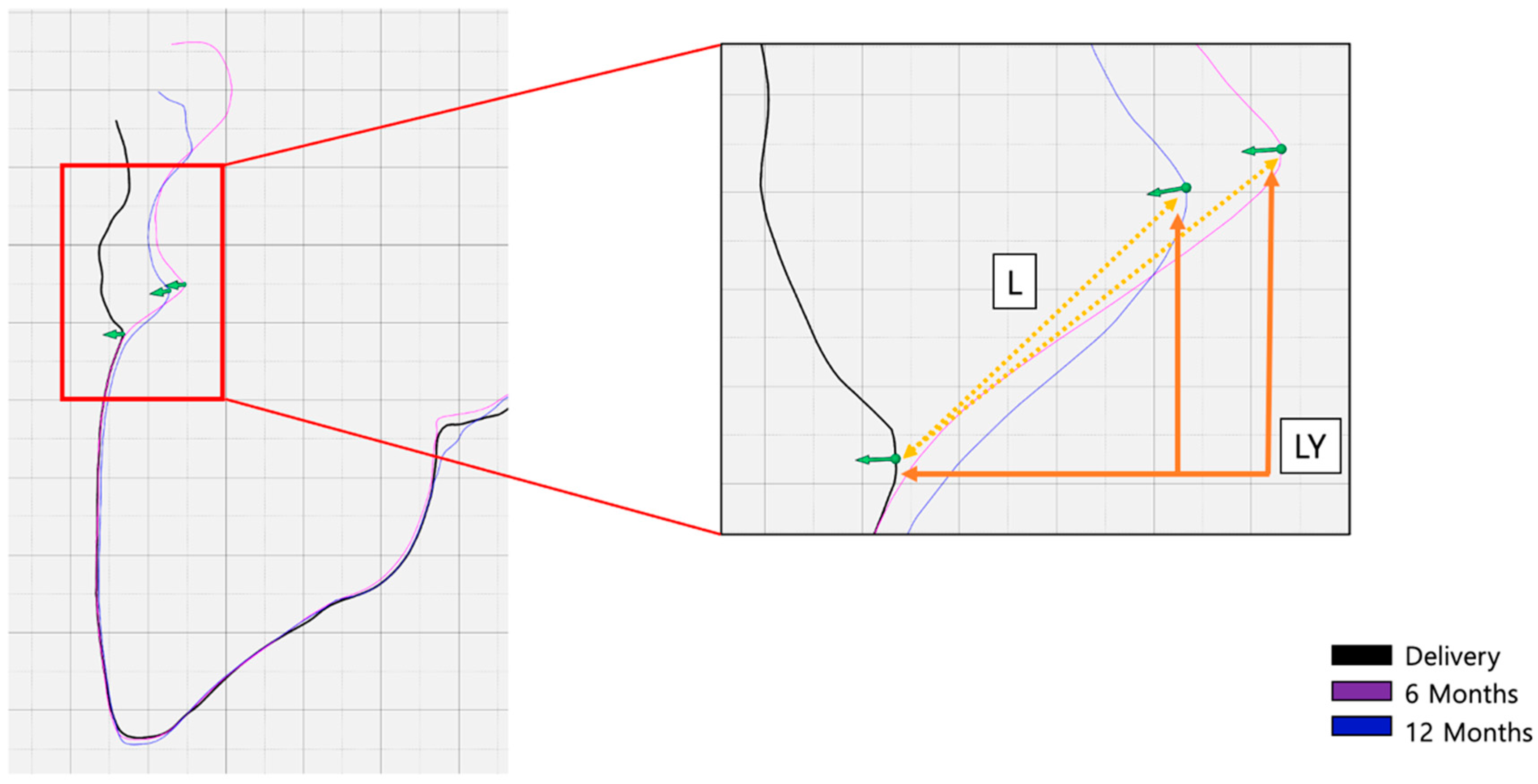
  • Vertical marginal height analysis using sectional analysis method. A vertical line was drawn from the zenith to the cusp/incisal edge of the tooth (Figure 5). Selection of reference points (points 1–4) on BL-STL was realized for analysis of horizontal changes in thickness at points 2, 3, and 4 and measurement of zenith changes (point 1) from BL to 6- and 12-month follow up (Figure 6 and Figure 7).
  • Analysis of volume changes from AOI of BL to AOI of 6 and 12 months, using the sectional analysis method described earlier [23].

2.5. Statistical Analysis

The measurements were analyzed using the statistical package SPSS Statistics 22 (IBM, Armonk, NY, USA). Normality was evaluated using the Shapiro–Wilk test, and a pairwise t-test was performed to compare the results of the two implant placement groups at 6 months and 12 months after baseline. The level of significance was at p < 0.05.

3. Results

Sixty patients, twenty-seven female (45%) and thirty-three male (55%), with a mean age of 56.6 ± 12.9 years, were enrolled in the study. All 60 subjects received one bone-level tapered (BLT) implant. Most frequently, premolars (48.4%), central incisors (21.6%), and lateral incisors (13.3%) of the maxilla were replaced, followed by maxillary canines (8.3%) and mandibular premolars (8.3%). For the present analysis, the baseline STL data of upper and lower jaw models of 56 patients were included.
One patient presented an early implant failure before implant loading; therefore, no STL model was available. Three other patients dropped out of the study during the follow-up visits as they could no longer be contacted, and their models were not included in the analysis. The models of 28 patients with 28 immediately placed implants and of 28 patients with 28 early placed implants were analyzed at BL. In the immediate placement group, 14 crowns were made of zirconia and 14 were made of lithium disilicate. In the early placement group, 15 were made of zirconia and 13 of lithium disilicate. Several scanned models could not be used for the analysis of the follow-up time points due to irregularities of the stone casts. Therefore, at the 6-month control, STL data of n = 51 patients and sites were available, whereas data of n = 47 patients and sites were available at the 12-month follow up.

3.1. Volume Changes

The overall volume changes of the AOI from BL to 6- and 12-month follow up were analyzed. The mean overall AOI volume change from baseline to 6-month follow up was −3.51 mm3 (±6.39 mm3) and −4.95 mm3 (±6.16 mm3) to 12-month follow up (Figure 8).
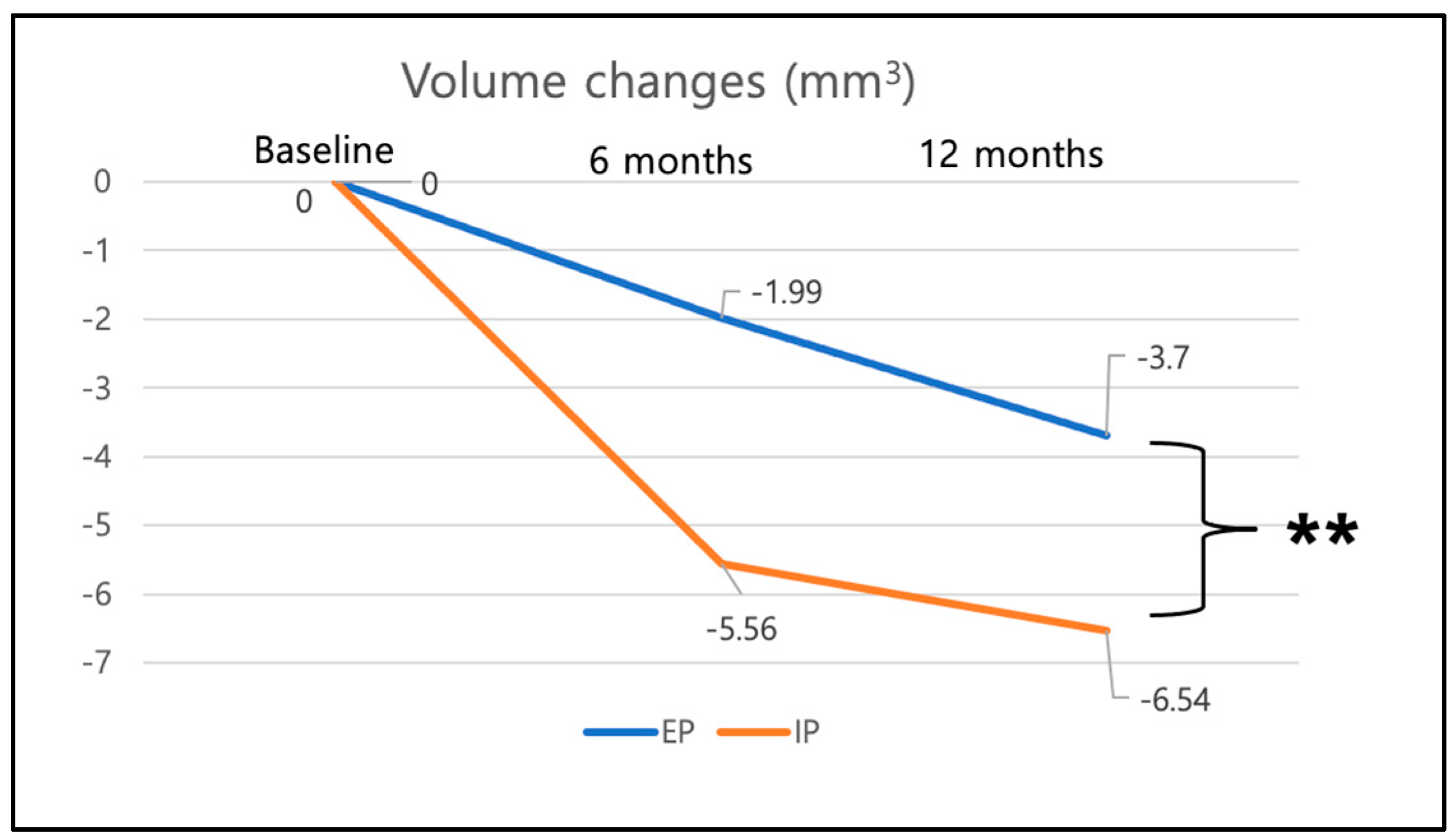
Furthermore, the AOI volume change from baseline to 6-month and 12-month follow up were analyzed between the groups immediate placement versus early placement (Figure 9). In the immediate placement group, a volume loss of −5.56 mm3 (±5.83 mm3) was found at 6 months and a mean volume loss of −6.62 mm3 (±6.56 mm3) at 12 months. For the early placement group, a mean volume loss of −1.99 mm3 (±5.82 mm3) at 6 months and of −3.7 mm3 (±5.62 mm3) at 12 months was found (Figure 9). Between baseline and 12 months, the immediate placement group (IP) showed significantly more overall volume loss in the AOI than the early placement group (EP) with p = 0.005.
No significant differences in volume changes were found between the two ceramic restorative materials (zirconia vs. lithium disilicate) with p = 0.972.

3.2. Overall Vertical Height Changes (Point 1)

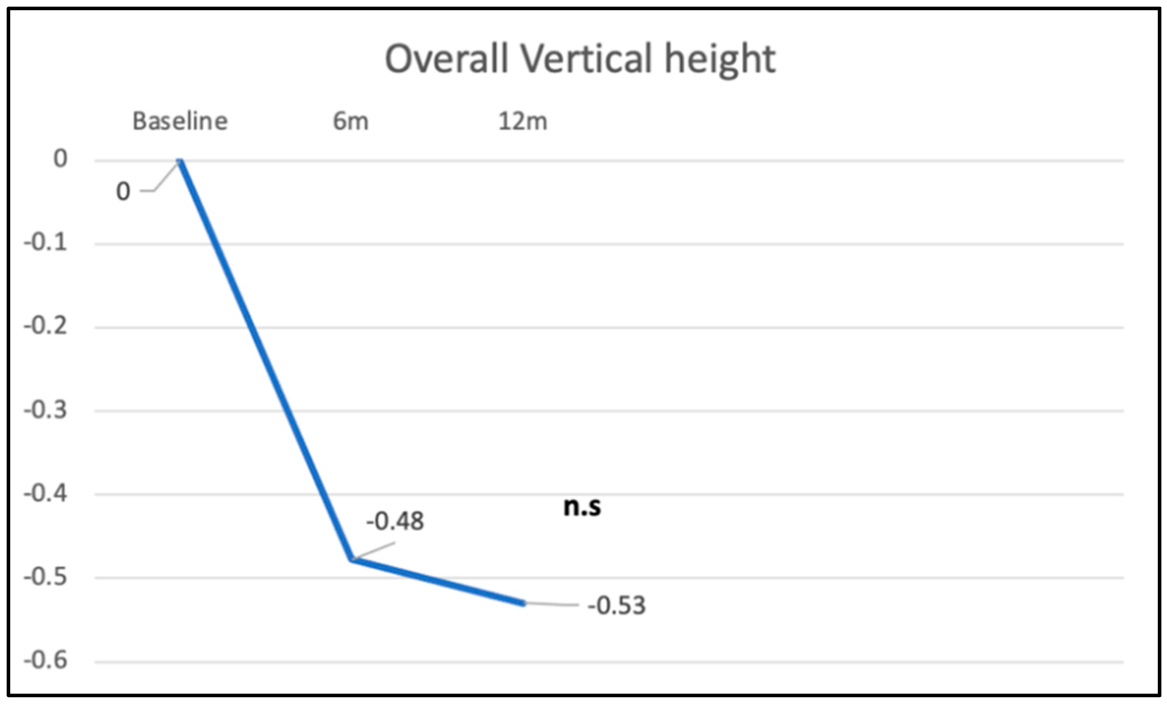
The mean vertical height of the zenith point (point 1) showed a trend to negatively change slightly (p = 0.692) from baseline to 6 months of −0.48 mm (±0.52), which corresponds to a recession of the mucosal margin of 0.48 mm. From the 6-month to the 12-month follow up, a non-significant yet slightly negative trend in the zenith position from −0.48 mm to −0.53 mm (±0.49 mm) of −0.05 mm was found, which implies a slight tendency for further recession (Figure 10).

3.3. Vertical Height of Zenith Point in Immediate vs. Early Placement and Zirconia vs. Lithium Disilicate

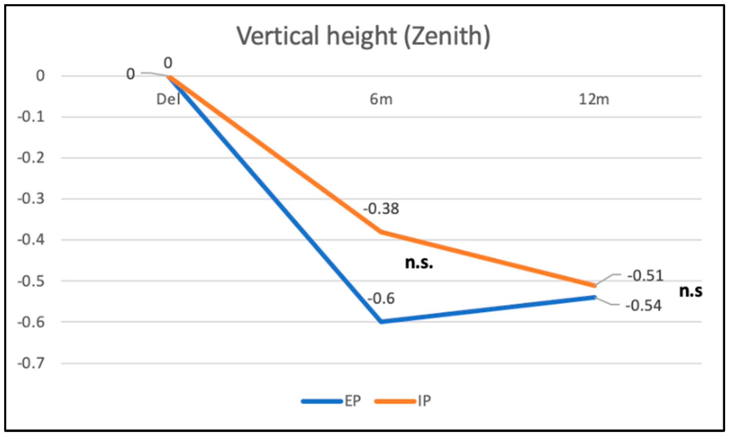
In the IP group, the vertical height of the zenith point (point 1) was reduced from baseline to 6 months by −0.38 mm (±0.32 mm), corresponding to a recession of the mucosal margin of 0.38 mm. From the 6-month to the 12-month follow up, a slightly negative change in the zenith position from −0.38 mm to −0.51 mm (±0.43 mm) of −0.13 mm could be found, representing a tendency for continuing recession (Figure 11). The changes in vertical height were at no time points statistically significant.
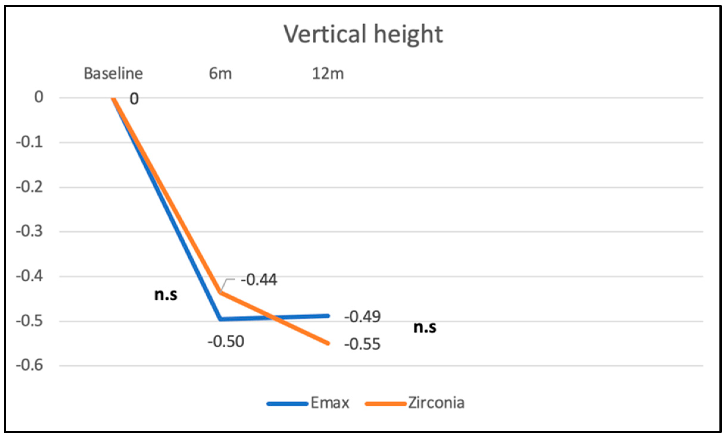
In the EP group, the vertical height of the zenith point (point 1) was reduced from baseline to 6 months by −0.60 mm (±0.51 mm), corresponding to a recession of the mucosal margin of 0.60 mm. From the 6-month to the 12-month follow up, a slightly positive change in the zenith position from −0.60 mm to −0.54 mm (±0.36 mm) of +0.06 mm could be found, which implies a slight decrease in recession (Figure 11). However, these changes in the zenith point position for the different follow-up time points did not reach statistical significance. The differences between the two groups were not significant (p = 0.5). Furthermore, no significant differences in vertical height changes between the two ceramic restorative materials (p = 0.253) could be observed (Figure 12).

3.4. Changes in Horizontal Width

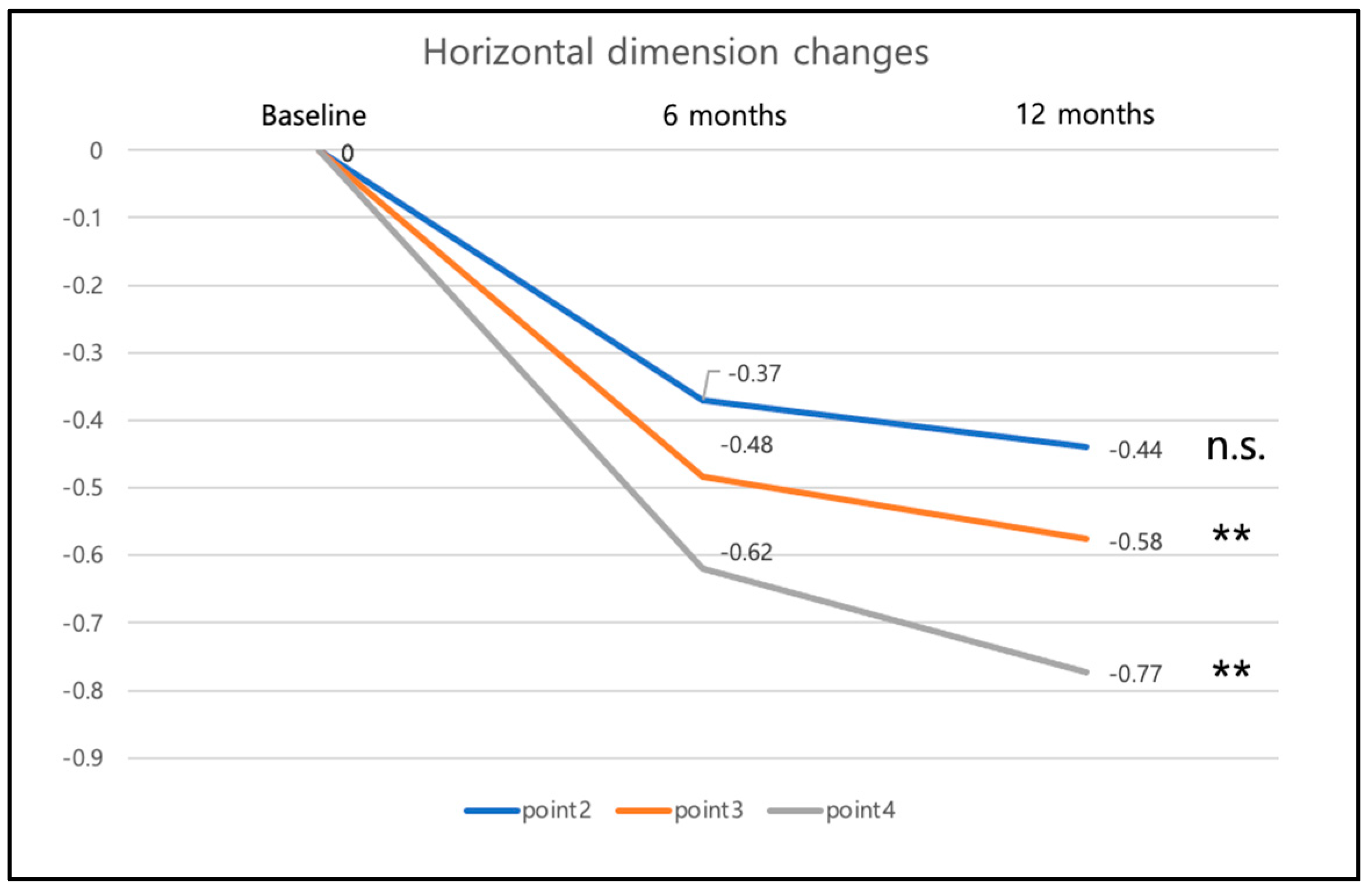
The changes in horizontal width were measured at each reference point 2–4 (Figure 13): point 2 was found 0.37 mm (±0.46 mm) less buccally at 6 months and 0.44 (±0.49) mm less buccally at 12 months. This implies a decrease in horizontal width over time. Between the 6-month and the 12-month visit, the horizontal width decrease was not statistically significant.
Similar results were observed for points 3 and 4: point 3 was found 0.48 mm (±0.54 mm) less buccally at 6 months and 0.58 mm (±0.57 mm) less buccally at 12 months. Between the 6-month and the 12-month visit, the horizontal decrease was significant with p = 0.005. Point 4 was found 0.62 mm (±0.63 mm) less buccally at 6 months and 0.77 mm (±0.59 mm) less buccally at 12 months. The horizontal decrease was significant between the 6-month and the 12-month visit with p = 0.007.
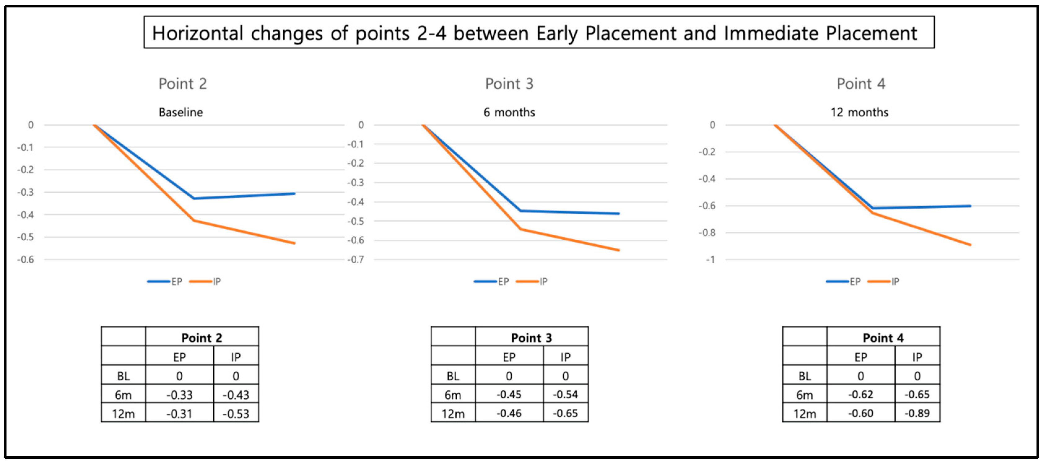
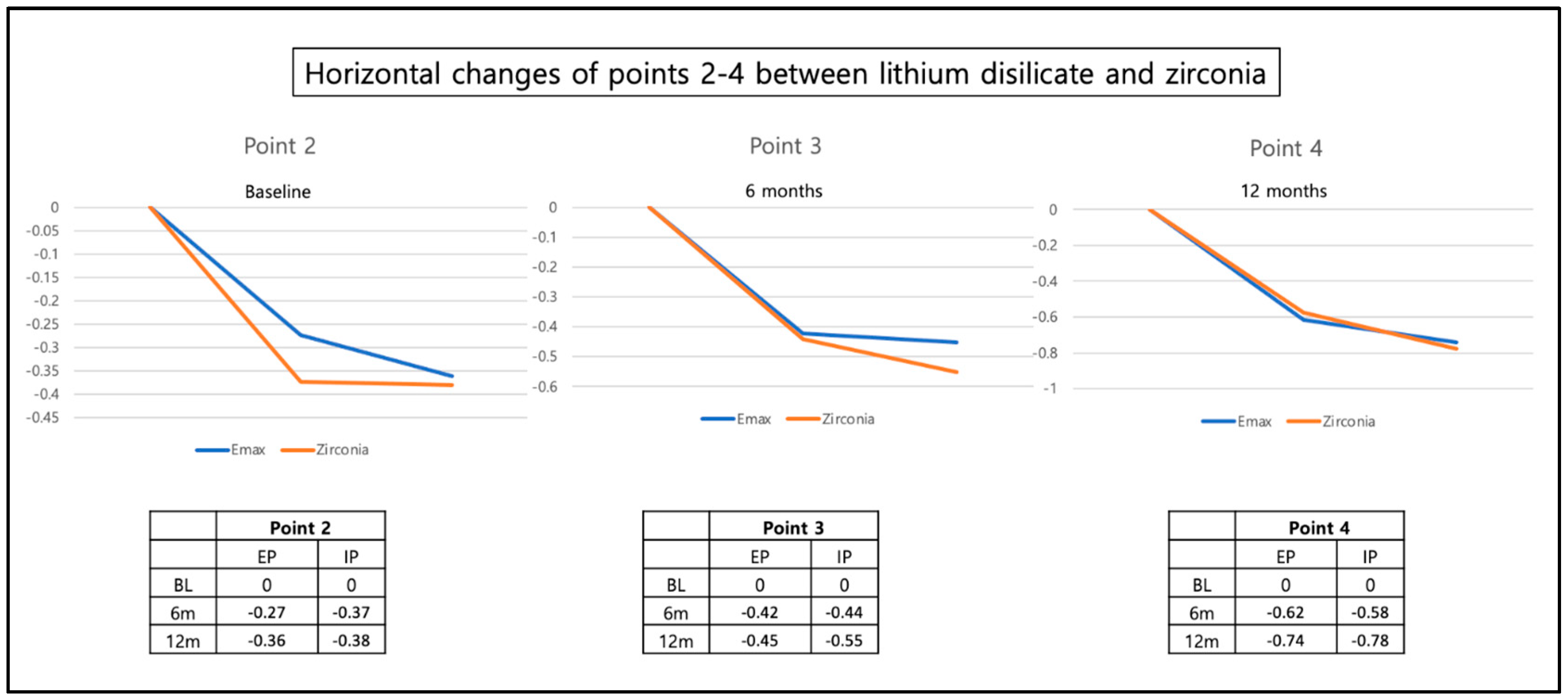
Neither the timepoint of the implant placement (immediate vs. early) nor the type of restorative material (zirconia vs. Lithium disilicate) had an influence (Figure 14 and Figure 15). The change in the horizontal position of reference points 2, 3, and 4 corresponds with a slight decrease in horizontal width in these regions.

4. Discussion

The present multi-center randomized controlled clinical trial demonstrated slight overall peri-implant volume loss following immediate and early implant insertion after one year of implant loading. A significant difference in peri-implant volume changes between the two placement groups was detected 12 months after loading. The early implant placement group showed less volume loss than the immediate implant placement group.
In the literature, the analysis of the stability of contour augmentation was often performed using radiographic CB-CT analysis after augmentation procedures [15,26,27,28]. Only a few studies analyzed the overall volume changes after implant insertion in three dimensions. Frequently, linear measurements were reported [40].
One study used a similar method as the present study [41]. The authors analyzed volume stability after implant insertion following an early implant placement protocol and soft tissue augmentation. Like the present study, alginate impressions of 15 patients were taken, and the resulting stone casts were scanned and transformed into STL data. The data were then superimposed, and volume changes were analyzed. The study group observed a volume decrease in the marginal area of the implant, whereas they found an increase in volume in the more apical part of their area of interest. Furthermore, they remarked on a high variability among their patient cohort. The present study’s data is in coherence with these findings of high inter-patient variability. In the mentioned study, the mean loss of facial prominence within the first year after crown insertion was −0.04 mm ± 0.31 mm. This distance corresponds to the change in volume over time and is the mean distance between all measured points in the bucco-oral direction. In the present study, the mean overall volume change for both groups was −5.1 mm3 after 12 months, corresponding to a mean bucco-oral distance of −0.3 mm at 12 months. No other studies were found that reported on three-dimensional volume changes (in mm3) similar to the present study after implant insertion and the bone augmentation procedure. Nevertheless, one study analyzing 3D radiographic data after immediate implant placement reported an almost complete loss of the buccal bone in one third of the 14 patients, which demonstrates a volume loss in the peri-implant area [27]. For this study, localized guided bone regeneration was performed during the immediate implant placement, likewise to the procedure performed in the present study.
The higher peri-implant volume loss in the immediate placement group compared with the early implant placement group can be explained by the expected bone remodeling and bone contour changes at post-extraction sites, especially within the first weeks and months after tooth extraction [40,42]. As volume changes were not analyzed before implant insertion, how much volume loss occurred in the early placement group after tooth extraction and before implant placement remains unknown. It can be assumed that a similar extent of volume changes occurred in both groups, but in the early placement group to an earlier time point, directly after tooth extraction and before implant placement.
Furthermore, midfacial recessions were observed in both groups after 6 and 12 months. In a study by Nimwegen et al., the presence of midfacial recessions after immediate implant placement and immediate insertion of the provisional restorations were compared between a study group receiving additional soft tissue augmentation (connective tissue graft) and a group not receiving soft tissue augmentation [43]. The results showed significantly higher midfacial recessions in the group without connective tissue grafting, with a mean difference of 0.68 mm between the study groups. In the control group with no connective tissue grafting, the mean midfacial recession amounted to 0.48 mm at 12 months in the latter study, which is equal to the present findings of a mean midfacial recession of 0.48 mm at the 12-month follow up [43]. Similar to the present study, another RCT comparing immediate and early implant placement with immediate insertion of provisional restorations in the anterior region found no statistically significant differences between the two groups [44]. Midfacial recessions amounted to 0.8 mm and 0.6 mm for the two groups, respectively, after two years of follow-up time.
One systematic review comparing midfacial recessions following immediate and early implant placement found notable deviations among the outcomes of the different included manuscripts [13]. The authors stated that immediate implant placement was associated with increased variability in the outcomes and a higher occurrence of midfacial recessions > 1 mm compared with the early implant placement protocol, where no recessions of >1 mm were reported. They concluded that immediate implant placement could increase the risk of midfacial mucosa recessions. The amount of detectable facial bone may influence the risk of midfacial mucosal recessions [13]. Furthermore, the bucco-oral position of the implant had an impact on the presence of midfacial recessions, with buccally placed implants significantly associated with recessions [13,45,46].
Overall, the presently reported extent of midfacial recessions after one year of 0.48 mm is in line with the expected average of midfacial recessions of 0.5 mm, for immediate and early implant placement, according to the comprehensive systematic review of Chen and co-workers [13].
In the present study, all implants were restored with a buccally micro-veneered all-ceramic crown made of lithium disilicate or zirconia and bonded to a titanium base abutment. This implies the submucosal contact of the peri-implant soft tissue with different restorative materials. After the 1-year observation period, no significant differences in peri-implant tissue volume changes were found when comparing the two ceramic materials. Several authors analyzed the short-term effects of various transmucosal restorative materials on peri-implant tissues, including the materials used in the present study, and found no significant differences regarding biological outcomes [47,48,49]. Similarly, several investigations demonstrated little to no long-term difference between metallic and ceramic implant abutments concerning the soft tissue inflammatory reaction [3,50,51,52]. Nevertheless, the evidence indicates that smoother material surfaces lead to less bacterial biofilm adhesion and, therefore, potentially improve peri-implant health [34]. However, more detailed long-term examinations of the peri-implant crevicular fluid analyzing inflammatory biomarkers such as interleukin or matrix-metalloproteinase of different restorative materials are necessary to provide clinical recommendations [53].
For the three-dimensional analysis performed in the present study, alginate impressions were made, and plaster models were cast, which were then scanned to obtain STL files. These digitized models were superimposed for digital volumetric analysis. An inherent shortcoming of this multi-step procedure is the risk of accumulated inaccuracies. A study using the same impression and data acquisition techniques to compare peri-implant volume changes after immediate implant placement with or without subsequent soft tissue grafting showed similar outcomes in inaccuracy as the present study [43]. For the present analysis, nine subjects had to be excluded from the analysis due to irregularities of the stone casts. Similarly, Nimwegen et al. excluded eight patients for the same reason [43]. This highlights the conventional impression techniques as a limitation of the chosen study design. A direct method of data acquisition using an intraoral scanner could reduce this risk and should, therefore, be preferred today.
The digital volumetric analysis method applied in the present study using the superimposition of STL data in specialized software has previously been described [23,54,55]. However, there is no consensus on a gold-standard technique for three-dimensional volume analysis in the currently available literature. This is shown by the heterogeneity of volumetric analysis methods used by other research groups. Other authors described the use of dental implant planning software not specialized in reporting volumetric outcomes [43,56].
Three-dimensional radiographic analysis stemming from cone-beam or micro-CT data is a widespread choice [57,58]. While excellent for the analysis of hard tissues such as teeth or bone, more evidence is needed to confirm the viability of this examination methodology for the precise inspection of the peri-implant soft tissues. In contrast, magnetic resonance imaging and subsequent volumetric analysis of soft tissues are standard in other fields of medicine, such as plastic and aesthetic surgery, but less common in dentistry [59].

5. Conclusions

This multi-center randomized controlled clinical trial showed that immediate and early placement protocols with simultaneous bone augmentation procedures lead to slight overall peri-implant tissue volume loss after one year. A significant difference in overall volume loss between the two time points of implant insertion was found. A slightly more pronounced peri-implant volume loss can be expected after immediate placement compared with early implant placement. Furthermore, slight midfacial mucosal recessions can be expected within the first year after implant placement.
Immediate placement protocols should therefore be avoided in esthetically demanding situations, such as anterior regions and high smile lines. It is essential to anticipate the post-extraction peri-implant volume and soft tissue changes and carefully choose between early and immediate placement protocols, depending on the patient’s clinical situation and esthetic demands.

Author Contributions

Conceptualization, M.S. (Malin Strasding), S.P.H. and I.S.; methodology, H.L.; software, H.L. and Y.J.; validation, H.L.; formal analysis, Y.J., H.L., M.S. (Minji Sun) and M.S. (Malin Strasding); investigation, M.S. (Malin Strasding) and S.P.H.; resources, I.S. and H.L.; data curation, M.S. (Malin Strasding), H.L. and Y.J.; writing—original draft preparation, M.S. (Malin Strasding) and L.M.; writing—review and editing, H.L., I.S., Y.J., S.P.H. and M.S. (Malin Strasding); visualization, Y.J., H.L., M.S. (Minji Sun) and M.S. (Malin Strasding); supervision, I.S. and H.L.; project administration, M.S. (Malin Strasding); funding acquisition, S.P.H. and I.S. All authors have read and agreed to the published version of the manuscript.

Funding

This research was funded by Straumann AG, grant number 15-117.

Institutional Review Board Statement

The study was conducted in accordance with the Declaration of Helsinki and approved by the Ethics Committee of the Commission Cantonale d’Ethique de la Recherche sur l’être humain (CCER 15-117, date of approval: 20 August 2015).

Informed Consent Statement

Informed consent was obtained from all subjects involved in the study.

Acknowledgments

The authors thank the dentists of the four external study centers: Léonard Brazzola, Robin Jaquet, Francesco Michelini, Jelena Gavric, and Philippe Rieder for their contribution to this multicentric study. Further, the authors thank the Master Dental Technician Vincent Fehmer and the pre-graduate student Yobel Solomon for the preparation and the scanning of the models. The authors thank Straumann AG for the financial and material support to the study.

Conflicts of Interest

The funders had no role in the design of the study; in the collection, analyses, or interpretation of data; in the writing of the manuscript; or in the decision to publish the results.

References

  1. Buser, D.; Janner, S.F.; Wittneben, J.G.; Bragger, U.; Ramseier, C.A.; Salvi, G.E. 10-year survival and success rates of 511 titanium implants with a sandblasted and acid-etched surface: A retrospective study in 303 partially edentulous patients. Clin. Implant Dent. Relat. Res. 2012, 14, 839–851. [Google Scholar] [CrossRef] [PubMed]
  2. Jimbo, R.; Albrektsson, T. Long-term clinical success of minimally and moderately rough oral implants: A review of 71 studies with 5 years or more of follow-up. Implant Dent. 2015, 24, 62–69. [Google Scholar] [CrossRef]
  3. Pjetursson, B.E.; Zarauz, C.; Strasding, M.; Sailer, I.; Zwahlen, M.; Zembic, A. A systematic review of the influence of the implant-abutment connection on the clinical outcomes of ceramic and metal implant abutments supporting fixed implant reconstructions. Clin. Oral Implant. Res. 2018, 29 (Suppl. S18), 160–183. [Google Scholar] [CrossRef] [Green Version]
  4. Pjetursson, B.E.; Valente, N.A.; Strasding, M.; Zwahlen, M.; Liu, S.; Sailer, I. A systematic review of the survival and complication rates of zirconia-ceramic and metal-ceramic single crowns. Clin. Oral Implant. Res. 2018, 29 (Suppl. S16), 199–214. [Google Scholar] [CrossRef] [PubMed] [Green Version]
  5. Sailer, I.; Strasding, M.; Valente, N.A.; Zwahlen, M.; Liu, S.; Pjetursson, B.E. A systematic review of the survival and complication rates of zirconia-ceramic and metal-ceramic multiple-unit fixed dental prostheses. Clin. Oral Implant. Res. 2018, 29 (Suppl. S16), 184–198. [Google Scholar] [CrossRef] [PubMed] [Green Version]
  6. Botticelli, D.; Berglundh, T.; Persson, L.G.; Lindhe, J. Bone regeneration at implants with turned or rough surfaces in self-c ontained defects. An experimental study in the dog. J. Clin. Periodontol. 2005, 32, 448–455. [Google Scholar] [CrossRef] [PubMed]
  7. Bornstein, M.M.; Schmid, B.; Belser, U.C.; Lussi, A.; Buser, D. Early loading of non-submerged titanium implants with a sandblasted and acid-etched surface. 5-year results of a prospective study in partially edentulous patients. Clin. Oral Implants Res. 2005, 16, 631–638. [Google Scholar] [CrossRef]
  8. Suzuki, S.; Kobayashi, H.; Ogawa, T. Implant stability change and osseointegration speed of immediately loaded photofunctionalized implants. Implant Dent. 2013, 22, 481–490. [Google Scholar] [CrossRef] [Green Version]
  9. Bosshardt, D.D.; Chappuis, V.; Buser, D. Osseointegration of titanium, titanium alloy and zirconia dental implants: Current knowledge and open questions. Periodontology 2000 2017, 73, 22–40. [Google Scholar] [CrossRef]
  10. Grunder, U.; Polizzi, G.; Goene, R.; Hatano, N.; Henry, P.; Jackson, W.J.; Kawamura, K.; Kohler, S.; Renouard, F.; Rosenberg, R.; et al. A 3-year prospective multicenter follow-up report on the immediate and delayed-immediate placement of implants. Int. J. Oral Maxillofac. Implant. 1999, 14, 210–216. [Google Scholar]
  11. Chen, S.T.; Wilson, T.G., Jr.; Hammerle, C.H. Immediate or early placement of implants following tooth extraction: Review of biologic basis, clinical procedures, and outcomes. Int. J. Oral Maxillofac. Implant. 2004, 19, 12–25. [Google Scholar]
  12. Lang, N.P.; Pun, L.; Lau, K.Y.; Li, K.Y.; Wong, M.C. A systematic review on survival and success rates of implants placed immediately into fresh extraction sockets after at least 1 year. Clin. Oral Implant. Res. 2012, 23 (Suppl. S5), 39–66. [Google Scholar] [CrossRef] [PubMed]
  13. Chen, S.T.; Buser, D. Esthetic outcomes following immediate and early implant placement in the anterior maxilla—A systematic review. Int. J. Oral Maxillofac. Implant. 2014, 29, 186–215. [Google Scholar] [CrossRef] [Green Version]
  14. Gallucci, G.O.; Hamilton, A.; Zhou, W.; Buser, D.; Chen, S. Implant placement and loading protocols in partially edentulous patients: A systematic review. Clin. Oral Implant. Res. 2018, 29 (Suppl. S16), 106–134. [Google Scholar] [CrossRef] [Green Version]
  15. Buser, D.; Chappuis, V.; Bornstein, M.M.; Wittneben, J.G.; Frei, M.; Belser, U.C. Long-term stability of contour augmentation with early implant placement following single tooth extraction in the esthetic zone: A prospective, cross-sectional study in 41 patients with a 5- to 9-year follow-up. J. Periodontol. 2013, 84, 1517–1527. [Google Scholar] [CrossRef] [PubMed]
  16. Buser, D.; Wittneben, J.; Bornstein, M.M.; Grutter, L.; Chappuis, V.; Belser, U.C. Stability of contour augmentation and esthetic outcomes of implant-supported single crowns in the esthetic zone: 3-year results of a prospective study with early implant placement postextraction. J. Periodontol. 2011, 82, 342–349. [Google Scholar] [CrossRef]
  17. Furhauser, R.; Florescu, D.; Benesch, T.; Haas, R.; Mailath, G.; Watzek, G. Evaluation of soft tissue around single-tooth implant crowns: The pink esthetic score. Clin. Oral Implant. Res. 2005, 16, 639–644. [Google Scholar] [CrossRef]
  18. Jemt, T. Restoring the gingival contour by means of provisional resin crowns after single-implant treatment. Int. J. Periodontics Restor. Dent. 1999, 19, 20–29. [Google Scholar]
  19. Meijer, H.J.; Stellingsma, K.; Meijndert, L.; Raghoebar, G.M. A new index for rating aesthetics of implant-supported single crowns and adjacent soft tissues—The Implant Crown Aesthetic Index. Clin. Oral Implant. Res. 2005, 16, 645–649. [Google Scholar] [CrossRef] [Green Version]
  20. Juodzbalys, G.; Wang, H.L. Esthetic index for anterior maxillary implant-supported restorations. J. Periodontol. 2010, 81, 34–42. [Google Scholar] [CrossRef]
  21. Hosseini, M.; Gotfredsen, K. A feasible, aesthetic quality evaluation of implant-supported single crowns: An analysis of validity and reliability. Clin. Oral Implant. Res. 2012, 23, 453–458. [Google Scholar] [CrossRef] [PubMed]
  22. Ross, S.B.; Pette, G.A.; Parker, W.B.; Hardigan, P. Gingival margin changes in maxillary anterior sites after single immediate implant placement and provisionalization: A 5-year retrospective study of 47 patients. Int. J. Oral Maxillofac. Implant. 2014, 29, 127–134. [Google Scholar] [CrossRef] [PubMed] [Green Version]
  23. Lee, H.; Fehmer, V.; Hicklin, S.; Noh, G.; Hong, S.J.; Sailer, I. Three-Dimensional Evaluation of Peri-implant Soft Tissue When Tapered Implants Are Placed: Pilot Study with Implants Placed Immediately or Early Following Tooth Extraction. Int. J. Oral Maxillofac. Implant. 2020, 35, 1037–1044. [Google Scholar] [CrossRef] [PubMed]
  24. Barone, A.; Toti, P.; Quaranta, A.; Alfonsi, F.; Cucchi, A.; Calvo-Guirado, J.L.; Negri, B.; Di Felice, R.; Covani, U. Volumetric analysis of remodelling pattern after ridge preservation comparing use of two types of xenografts. A multicentre randomized clinical trial. Clin. Oral Implant. Res. 2016, 27, e105–e115. [Google Scholar] [CrossRef] [PubMed]
  25. Buser, D.; Chappuis, V.; Kuchler, U.; Bornstein, M.M.; Wittneben, J.G.; Buser, R.; Cavusoglu, Y.; Belser, U.C. Long-term stability of early implant placement with contour augmentation. J. Dent. Res. 2013, 92, 176S–182S. [Google Scholar] [CrossRef]
  26. Abdelhamid, A.; Omran, M.; Bakhshalian, N.; Tarnow, D.; Zadeh, H.H. An open randomized controlled clinical trial to evaluate ridge preservation and repair using SocketKAP(™) and SocketKAGE(™): Part 2—Three-dimensional alveolar bone volumetric analysis of CBCT imaging. Clin. Oral Implant. Res. 2016, 27, 631–639. [Google Scholar] [CrossRef]
  27. Benic, G.I.; Mokti, M.; Chen, C.J.; Weber, H.P.; Hammerle, C.H.; Gallucci, G.O. Dimensions of buccal bone and mucosa at immediately placed implants after 7 years: A clinical and cone beam computed tomography study. Clin. Oral Implant. Res. 2012, 23, 560–566. [Google Scholar] [CrossRef]
  28. Omran, M.; Min, S.; Abdelhamid, A.; Liu, Y.; Zadeh, H.H. Alveolar ridge dimensional changes following ridge preservation procedure: Part-2—CBCT 3D analysis in non-human primate model. Clin. Oral Implant. Res. 2016, 27, 859–866. [Google Scholar] [CrossRef]
  29. Jung, R.E.; Benic, G.I.; Scherrer, D.; Hammerle, C.H. Cone beam computed tomography evaluation of regenerated buccal bone 5 years after simultaneous implant placement and guided bone regeneration procedures—A randomized, controlled clinical trial. Clin. Oral Implant. Res. 2015, 26, 28–34. [Google Scholar] [CrossRef]
  30. Botilde, G.; Colin, P.E.; Gonzalez-Martin, O.; Lecloux, G.; Rompen, E.; Lambert, F. Hard and soft tissue analysis of alveolar ridge preservation in esthetic zone using deproteinized bovine bone mineral and a saddle connective tissue graft: A long-term prospective case series. Clin. Implant Dent. Relat. Res. 2020, 22, 387–396. [Google Scholar] [CrossRef]
  31. Jung, R.E.; Brugger, L.V.; Bienz, S.P.; Husler, J.; Hammerle, C.H.F.; Zitzmann, N.U. Clinical and radiographical performance of implants placed with simultaneous guided bone regeneration using resorbable and nonresorbable membranes after 22–24 years, a prospective, controlled clinical trial. Clin. Oral Implant. Res. 2021, 32, 1455–1465. [Google Scholar] [CrossRef]
  32. Martin, W.C.; Pollini, A.; Morton, D. The influence of restorative procedures on esthetic outcomes in implant dentistry: A systematic review. Int. J. Oral Maxillofac. Implant. 2014, 29, 142–154. [Google Scholar] [CrossRef] [PubMed] [Green Version]
  33. Holst, S.; Blatz, M.B.; Hegenbarth, E.; Wichmann, M.; Eitner, S. Prosthodontic considerations for predictable single-implant esthetics in the anterior maxilla. J. Oral Maxillofac. Surg. 2005, 63, 89–96. [Google Scholar] [CrossRef] [PubMed]
  34. Abdalla, M.M.; Ali, I.A.A.; Khan, K.; Mattheos, N.; Murbay, S.; Matinlinna, J.P.; Neelakantan, P. The Influence of Surface Roughening and Polishing on Microbial Biofilm Development on Different Ceramic Materials. J. Prosthodont. 2021, 30, 447–453. [Google Scholar] [CrossRef] [PubMed]
  35. Corvino, E.; Pesce, P.; Mura, R.; Marcano, E.; Canullo, L. Influence of Modified Titanium Abutment Surface on Peri-implant Soft Tissue Behavior: A Systematic Review of In Vitro Studies. Int. J. Oral Maxillofac. Implant. 2020, 35, 503–519. [Google Scholar] [CrossRef] [PubMed]
  36. Wittneben, J.G.; Joda, T.; Weber, H.P.; Bragger, U. Screw retained vs. cement retained implant-supported fixed dental prosthesis. Periodontology 2000 2017, 73, 141–151. [Google Scholar] [CrossRef] [PubMed]
  37. Nothdurft, F.; Pospiech, P. Prefabricated zirconium dioxide implant abutments for single-tooth replacement in the posterior region: Evaluation of peri-implant tissues and superstructures after 12 months of function. Clin. Oral Implant. Res. 2010, 21, 857–865. [Google Scholar] [CrossRef]
  38. Ekfeldt, A.; Furst, B.; Carlsson, G.E. Zirconia abutments for single-tooth implant restorations: A retrospective and clinical follow-up study. Clin. Oral Implant. Res. 2011, 22, 1308–1314. [Google Scholar] [CrossRef]
  39. Strasding, M.; Hicklin, S.P.; Todorovic, A.; Fehmer, V.; Mojon, P.; Sailer, I. A multicenter randomized controlled clinical pilot study of buccally micro-veneered lithium-disilicate and zirconia crowns supported by titanium base abutments: 1-year outcomes. Clin. Oral Implant. Res. 2023, 34, 56–65. [Google Scholar] [CrossRef]
  40. Schropp, L.; Wenzel, A.; Kostopoulos, L.; Karring, T. Bone healing and soft tissue contour changes following single-tooth extraction: A clinical and radiographic 12-month prospective study. Int. J. Periodontics Restor. Dent. 2003, 23, 313–323. [Google Scholar]
  41. Schneider, D.; Grunder, U.; Ender, A.; Hammerle, C.H.; Jung, R.E. Volume gain and stability of peri-implant tissue following bone and soft tissue augmentation: 1-year results from a prospective cohort study. Clin. Oral Implant. Res. 2011, 22, 28–37. [Google Scholar] [CrossRef]
  42. Araujo, M.G.; Lindhe, J. Dimensional ridge alterations following tooth extraction. An experimental study in the dog. J. Clin. Periodontol. 2005, 32, 212–218. [Google Scholar] [CrossRef] [PubMed]
  43. van Nimwegen, W.G.; Raghoebar, G.M.; Zuiderveld, E.G.; Jung, R.E.; Meijer, H.J.A.; Muhlemann, S. Immediate placement and provisionalization of implants in the aesthetic zone with or without a connective tissue graft: A 1-year randomized controlled trial and volumetric study. Clin. Oral Implant. Res. 2018, 29, 671–678. [Google Scholar] [CrossRef] [PubMed] [Green Version]
  44. Palattella, P.; Torsello, F.; Cordaro, L. Two-year prospective clinical comparison of immediate replacement vs. immediate restoration of single tooth in the esthetic zone. Clin. Oral Implant. Res. 2008, 19, 1148–1153. [Google Scholar] [CrossRef] [PubMed]
  45. Evans, C.D.; Chen, S.T. Esthetic outcomes of immediate implant placements. Clin. Oral Implant. Res. 2008, 19, 73–80. [Google Scholar] [CrossRef] [PubMed]
  46. Chen, S.T.; Darby, I.B.; Reynolds, E.C. A prospective clinical study of non-submerged immediate implants: Clinical outcomes and esthetic results. Clin. Oral Implant. Res. 2007, 18, 552–562. [Google Scholar] [CrossRef] [PubMed]
  47. Enkling, N.; Marder, M.; Bayer, S.; Gotz, W.; Stoilov, M.; Kraus, D. Soft tissue response to different abutment materials: A controlled and randomized human study using an experimental model. Clin. Oral Implant. Res. 2022, 33, 667–679. [Google Scholar] [CrossRef] [PubMed]
  48. Dethier, F.; Bacevic, M.; Lecloux, G.; Seidel, L.; Rompen, E.; Lambert, F. The Effects of Abutment Materials on Peri-Implant Soft Tissue Integration: A Study in Minipigs. J. Prosthodont. 2022, 31, 585–592. [Google Scholar] [CrossRef]
  49. Mehl, C.; Gassling, V.; Schultz-Langerhans, S.; Acil, Y.; Bahr, T.; Wiltfang, J.; Kern, M. Influence of Four Different Abutment Materials and the Adhesive Joint of Two-Piece Abutments on Cervical Implant Bone and Soft Tissue. Int. J. Oral Maxillofac. Implant. 2016, 31, 1264–1272. [Google Scholar] [CrossRef] [Green Version]
  50. Zembic, A.; Bosch, A.; Jung, R.E.; Hammerle, C.H.; Sailer, I. Five-year results of a randomized controlled clinical trial comparing zirconia and titanium abutments supporting single-implant crowns in canine and posterior regions. Clin. Oral Implant. Res. 2013, 24, 384–390. [Google Scholar] [CrossRef]
  51. Sailer, I.; Philipp, A.; Zembic, A.; Pjetursson, B.E.; Hammerle, C.H.; Zwahlen, M. A systematic review of the performance of ceramic and metal implant abutments supporting fixed implant reconstructions. Clin Oral Implant. Res 2009, 20 (Suppl. S4), 4–31. [Google Scholar] [CrossRef] [PubMed] [Green Version]
  52. Linkevicius, T.; Vaitelis, J. The effect of zirconia or titanium as abutment material on soft peri-implant tissues: A systematic review and meta-analysis. Clin. Oral Implant. Res. 2015, 26 (Suppl. S11), 139–147. [Google Scholar] [CrossRef] [PubMed]
  53. Kuhn, K.; Rudolph, H.; Graf, M.; Moldan, M.; Zhou, S.; Udart, M.; Bohmler, A.; Luthardt, R.G. Interaction of titanium, zirconia and lithium disilicate with peri-implant soft tissue: Study protocol for a randomized controlled trial. Trials 2015, 16, 467. [Google Scholar] [CrossRef] [Green Version]
  54. Karasan, D.; Sailer, I.; Lee, H.; Demir, F.; Zarauz, C.; Akca, K. Occlusal adjustment of 3-unit tooth-supported fixed dental prostheses fabricated with complete-digital and -analog workflows: A crossover clinical trial. J. Dent. 2023, 128, 104365. [Google Scholar] [CrossRef] [PubMed]
  55. Marchand, L.; Sailer, I.; Lee, H.; Mojon, P.; Pitta, J. Digital wear analysis of different CAD/CAM fabricated monolithic ceramic implant-supported single crowns using two optical scanners. Int. J. Prosthodont. 2022, 35, 357–364. [Google Scholar] [CrossRef]
  56. Happe, A.; Debring, L.; Schmidt, A.; Fehmer, V.; Neugebauer, J. Immediate Implant Placement in Conjunction with Acellular Dermal Matrix or Connective Tissue Graft: A Randomized Controlled Clinical Volumetric Study. Int. J. Periodontics Restor. Dent. 2022, 42, 381–390. [Google Scholar] [CrossRef]
  57. Shanbhag, S.; Shanbhag, V.; Stavropoulos, A. Volume changes of maxillary sinus augmentations over time: A systematic review. Int. J. Oral Maxillofac. Implant. 2014, 29, 881–892. [Google Scholar] [CrossRef] [Green Version]
  58. Kuhl, S.; Brochhausen, C.; Gotz, H.; Filippi, A.; Payer, M.; d’Hoedt, B.; Kreisler, M. The influence of bone substitute materials on the bone volume after maxillary sinus augmentation: A microcomputerized tomography study. Clin. Oral Investig. 2013, 17, 543–551. [Google Scholar] [CrossRef]
  59. Kim, H.; Mun, G.H.; Wiraatmadja, E.S.; Lim, S.Y.; Pyon, J.K.; Oh, K.S.; Lee, J.E.; Nam, S.J.; Bang, S.I. Preoperative magnetic resonance imaging-based breast volumetry for immediate breast reconstruction. Aesthetic Plast. Surg. 2015, 39, 369–376. [Google Scholar] [CrossRef]
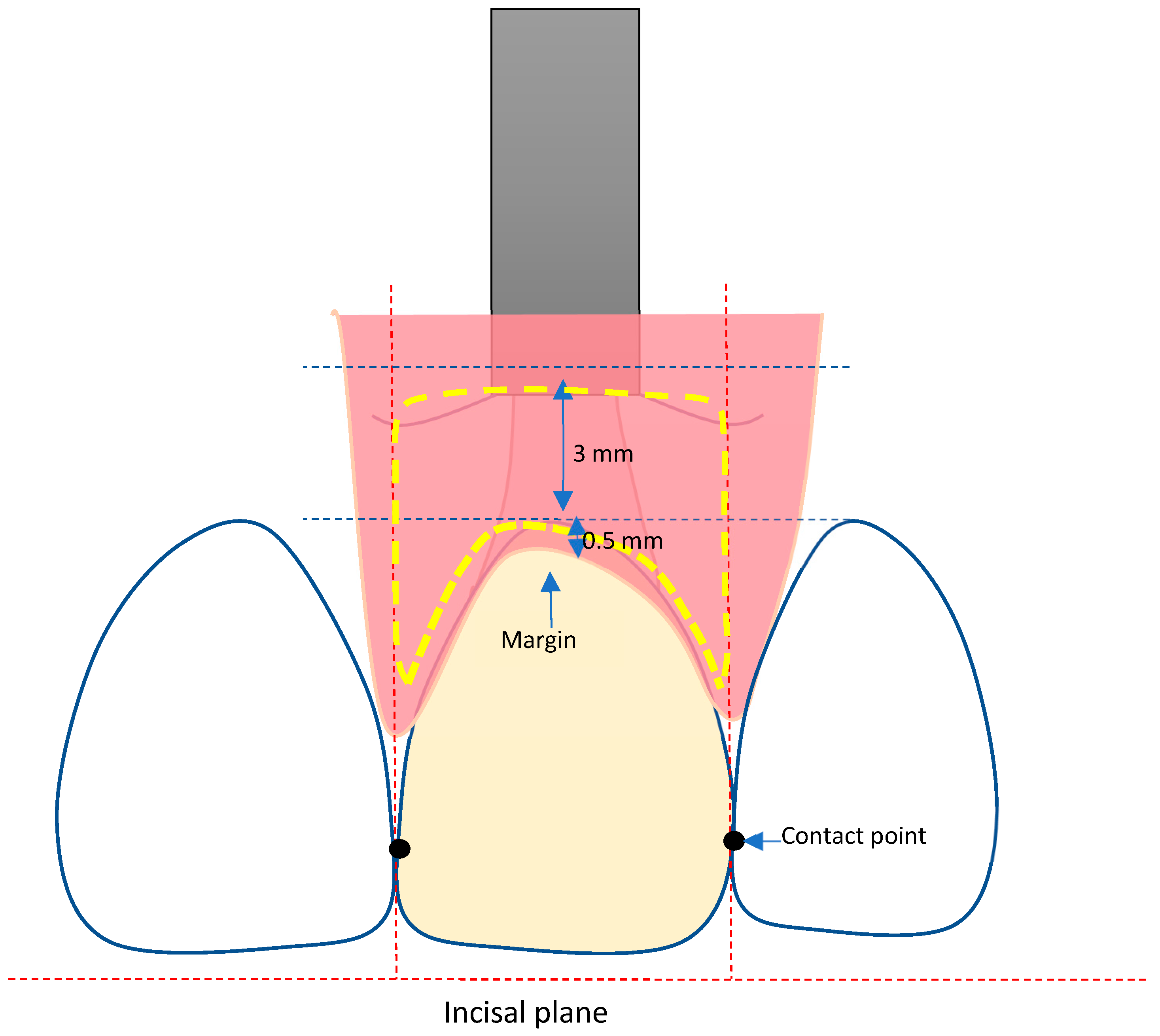
Figure 1. Definition of the AOI.
Materials 16 05636 g001
Figure 2. Defined position of points 1–4.
Materials 16 05636 g002
Figure 3. Selection of the AOI on 6-month and 12-month STL files.
Materials 16 05636 g003
Figure 4. Separation of the AOI.
Materials 16 05636 g004
Figure 5. Selection of the reference point (= zenith) for the vertical marginal height analysis.
Materials 16 05636 g005
Figure 6. (a) Superposition of STL scan data of the different time points (BL, 6 months, 12 months) for visualization of tissue volume changes during follow-up visits. (b) Reference points 1–4 for analysis of zenith changes and horizontal thickness changes from BL to 6- and 12-month follow up.
Materials 16 05636 g006aMaterials 16 05636 g006b
Figure 7. Analysis of zenith location changes (point 1) from BL to 6- and 12-month follow up.
Materials 16 05636 g007
Figure 8. Overall volume changes (immediate and early implant placement) from baseline to 12 months.
Materials 16 05636 g008
Figure 9. Volume changes between groups immediate vs. early implant placement at 6 months and 12 months. Significant difference at 12 months (p = 0.005) between the two groups. Statistical significance is marked as follows: ** = p < 0.01.
Materials 16 05636 g009
Figure 10. Overall vertical height changes (immediate and early implant placement) from baseline to 12 months. Differences were statistically not significant (n.s.).
Materials 16 05636 g010
Figure 11. Changes in vertical height (reference point 1) for the immediate and early placement group from BL to 6-month and 12-month follow up. Differences between the groups were statistically not significant (n.s.).
Materials 16 05636 g011
Figure 12. Changes in vertical height (reference point 1) for the lithium disilicate and the zirconia group from BL to 6-month and 12-month follow up. Differences between the groups were statistically not significant (n.s.).
Materials 16 05636 g012
Figure 13. Overall horizontal dimension changes of the reference points 2, 3, and 4 at BL, 6-month and 12-month follow-up. Differences between the groups were statistically not significant (n.s.). Statistical significance is marked as follows: ** = p < 0.01.
Materials 16 05636 g013
Figure 14. Horizontal dimension changes of the reference points 2, 3, and 4 at BL, 6-month and 12-month follow-up for immediate and early placement groups.
Materials 16 05636 g014
Figure 15. Horizontal dimension changes of the reference points 2, 3, and 4 at BL, 6-month and 12-month follow-up for Lithium disilicate and Zirconia restorations.
Materials 16 05636 g015
Disclaimer/Publisher’s Note: The statements, opinions and data contained in all publications are solely those of the individual author(s) and contributor(s) and not of MDPI and/or the editor(s). MDPI and/or the editor(s) disclaim responsibility for any injury to people or property resulting from any ideas, methods, instructions or products referred to in the content.