Review

Functionalization of TiO2 for Better Performance as Orthopedic Implants

School of Nanoscience and Technology, Beijing Institute of Nanoenergy and Nanosystems, University of Chinese Academy of Sciences, Beijing 100101, China
*
Authors to whom correspondence should be addressed.
These authors contributed equally to this work.
Materials 2022, 15(19), 6868; https://doi.org/10.3390/ma15196868
Submission received: 2 September 2022 / Revised: 23 September 2022 / Accepted: 27 September 2022 / Published: 3 October 2022
(This article belongs to the Special Issue Frontiers in Functional Materials for Bioelectronics and Biosensors)

Abstract

:
This review mainly focuses on the surface functionalization approaches of titanium dioxide (TiO2) to prevent bacterial infections and facilitate osteointegration simultaneously for titanium (Ti)-based orthopedic implants. Infection is one of the major causes of implant failure. Meanwhile, it is also critical for the bone-forming cells to integrate with the implant surface. TiO2 is the native oxide layer of Ti which has good biocompatibility as well as enriched physical, chemical, electronic, and photocatalytic properties. The formed nanostructures during fabrication and the enriched properties of TiO2 have enabled various functionalization methods to combat the micro-organisms and enhance the osteogenesis of Ti implants. This review encompasses the various modifications of TiO2 in aspects of topology, drug loading, and element incorporation, as well as the most recently developed electron transfer and electrical tuning approaches. Taken together, these approaches can endow Ti implants with better bactericidal and osteogenic abilities via the functionalization of TiO2.

1. Introduction

Titanium (Ti) and Ti alloy are currently the most widely used orthopedic materials. Their applications include micro-plates, micro-bone-screws, artificial bone joints, and fine surgical instruments. Ti-based materials have many excellent properties, including low density, high strength, corrosion resistance, good biocompatibility, magnetic compatibility, and no allergic reaction after implantation [1]. However, biomedical applications of Ti implants encounter a very important disadvantage; that is, Ti has no intrinsic antibacterial properties. Infections may occur after Ti implant surgery, resulting in implant failure, prolonged hospitalization, and increased cost to the health care system [2]. Microbial infections in the bones lead to a condition commonly known as osteomyelitis. A long-term and particular antibiotic medication is needed to cure it [3]. Production of implant material is a complex process [1] that requires safety standards in terms of biological and mechanical properties so that the health of patients may not be affected [4,5,6,7]. Ti and Ti alloys have advantages over Mg, Co, and stainless-steel alloys because Mg, as a degradable metal, cannot afford long-term service, Co has a high price, and stainless steel is not compatible with clinical magnetic resonance imaging (MRI) examination.
Titanium dioxide (TiO2) is the native oxide layer of Ti. Although TiO2 does not have intrinsic antibacterial properties, it brings many opportunities to improve the performance of Ti as a biomedical implant. In the process of oxide layer formation on the surface of Ti, various material treatment methods can be executed to obtain antibacterial properties. In addition, it is convenient to form TiO2 nanostructures that have better osteogenesis properties than Ti. In vivo, TiO2 may lead to better bone-implant contact by increasing cellular activity [8] and collagen type I expression [9]. Furthermore, TiO2 is one kind of semiconductor material with certain electronic and photocatalytic functions, which allows the development to find innovative methods to endow Ti implants with antibacterial properties and better osteogenesis ability.
In this review, we will discuss the various functionalization approaches of TiO2 to improve the performance of the Ti implant. The schematic diagram of this review is illustrated in Figure 1. The functionalization aims to endow the implant with antibacterial properties and enhance its ability to support osteogenesis. Apart from the well-established approaches such as topology, drug loading, and element incorporation, this review also discusses the most newly developed electron transfer and electrical tuning approaches. TiO2 is a material extensively involved in the electronic and photocatalytic fields for its ability to conduct electron separation and transfer. In recent years, it has been discovered that electron transfer between TiO2 and biological cells can also occur, and electrical tuning of TiO2 for the antibacterial properties is becoming possible. These novel approaches are worthy of more attention and research devotion.

2. Important Facts about Orthopedic Implant

2.1. Implant Failure

The success of bone surgical operations mainly depends on the quality of implantable biomaterials. Implant success is mainly halted by the infections caused by post-operative complications. Certain factors may lead to bacterial infections or even failure, including extensive damage to local tissues, improper fixation, smoking, diabetes, chemotherapy, irradiation, and inappropriate surgical techniques [10]. The implants may get an infection from surgery equipment, medical staff, room atmosphere, or bacteria in the patient’s blood. The outcome of these microbial infections sometimes becomes grave, leading to a second surgery, amputation, or even death [11]. Implant infections are mostly initiated by Staphylococcus epidermidis (S. epidermidis), Staphylococcus aureus (S. aureus), Pseudomonas aeruginosa (P. aeruginosa), and Enterobacteriaceae [12].
Implant failure may occur at early or late stage [13]. Lack of osseointegration may lead to early implant failure, whereas in late implant failures, osseointegration works well at the beginning but decreases later due to disease and biochemical overload [14]. Researchers have identified various reasons for implant failures, which include infectious and physical damage [15]. Implant failures can be minimized by maintaining hygienic measures, caring for physical damage, and regular review of implants.
Progressive bone loss occurs due to inflammatory lesions in the soft tissues associated with the implants [13] and peri-implant disease [16]. Poor hygienic measures, unmanaged diseases such as diabetes, and the use of corticosteroids in immune-compromised individuals may all lead to that situation [17,18]. Despite taking all necessary hygienic measures, bacterial infections may still occur. Studies have suggested that joint infections may take place in 1% of primary and 3–7% of multiple surgeries [19,20]. Patients with multiple surgeries have a higher risk of mortality and infection [19]. Implant infections and failures are a large economic burden on the health system. In the US, it costs more than $8.6 billion annually [19,21].

2.2. Fundamental Requirements of Orthopedic Implants

Bone is naturally composed of organic, inorganic, and collagen fibrils. The nano-hierarchical structures give shape and mechanical strength to bones [22]. The structures include small molecular amino acids forming tropocollagen helixes and nanoscale collagen fibers forming a microporous network of bones (Figure 2a). There is a crucial interaction between surface characteristics and the extracellular matrix for osteointegration [23]. Bone mesenchymal stem cells (BMSC) in the bone marrow are known to typically respond to metallic implants with the production of soft tissue rather than bone, which causes implants to fail [24,25]. Guiding stem cell differentiation to a desired specific line on the surface of the material is a key factor in the success of implants [26,27]. Osteoblasts are mature bone cells, whereas osteoprogenitor cells are pluripotent cells having the capacity to differentiate into different kinds of cells. Osteoblasts and osteoprogenitor cells are in direct contact with the implants.
For better outcomes, the hierarchical structures of the bone must be simulated by the implant with surface nanostructures to support bone tissue regeneration (Figure 2b,c). Apart from the surface nanostructures, other modifications, including nanoparticles, may help further. For example, bismuth oxide (Bi2O3) has features including electrochemical stability, high biocompatibility, and a medium band gap [28,29]. The contact of Bi2O3 nanoparticles and TiO2 nanocones resulted in a heterojunction that formed a built-in electric field and promoted the osteogenesis of BMSC on the basis of TiO2 nanostructures (Figure 2b) [30].
Figure 2. (a) The hierarchical structure of natural bone. Reprinted with permission from Ref. [31]. MDPI, 2022. (b) TiO2 nanocones with Bi2O3 quantum dots promotes osteogenesis. (i) Placing titanium implant in the bone defect; (ii) The electric field is built at the nanoscale interface of the implant. Reprinted with permission from Ref. [30]. John Wiley and Sons, 2021. (c) Osteogenesis pathways on TiO2 nanotubes in oxidative stress microenvironment brown arrow: enhancing the expression of a downstream gene; green arrow: inhibiting the expression of a downstream gene. Reprinted with permission from Ref. [32]. Elsevier, 2018.
Materials 15 06868 g002

3. Functionalization Approaches of TiO2 for Better Antibacterial and Osteogenesis Property

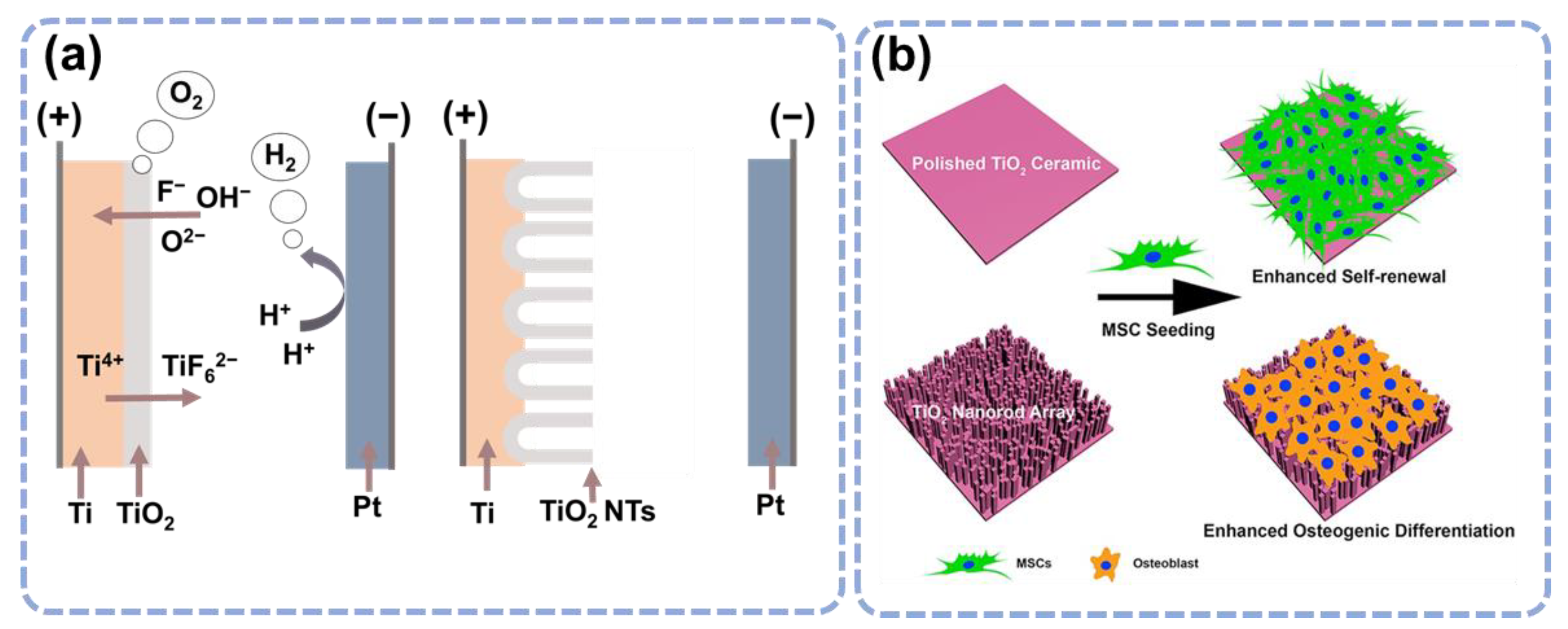
3.1. Topological Influence of the TiO2 Nanostructures

Topological modification is among the proposed methods to achieve surface functionalization. Studies have shown that surface nanostructure and topography may affect the migration, elongation, proliferation, and differentiation of stem cells [33,34,35]. In fact, cells and tissues in vivo will experience many topographic features ranging from nanoscale to microscale [36]. Thus, building a surface nanostructure on implants is an important research direction in the fields of artificial bones, joints, and dental implants [37,38,39]. The regulation of cell fate by surface topography is carried out by direct contact with adhering cells.
It has been widely accepted to form TiO2 nanotubes on Ti surfaces by doing anode oxidation (Figure 3a), and the annealing after anodization enhances the nanotube’s roughness and osseointegration capability [40,41]. Cell behavior is affected by the diameter of TiO2 nanotubes [40]. For instance, small nanotubes (30 nm in diameter) have been shown to promote BMSC adherence without significant differentiation, while larger nanotubes (70–100 nm in diameter) cause a dramatic lengthening of stem cells, which induces cytoskeletal stress and selective differentiation into osteoblast-like cells [42]. A diameter of 70 nm is the optimum size of TiO2 nanotubes for osteogenic differentiation of stem cells derived from human adiposity [43]. The diameters of TiO2 nanotubes are crucial for surface roughness and hydrophilicity. Several studies have shown that increasing diameter can increase antibacterial characteristics [44,45]. Ercan et al. found that nanotubes with a diameter of 80 nm had more antibacterial properties than the 30 nm diameter nanotubes against various strains of S. aureus due to higher hydrophobicity [46]. Other factors apart from the diameter, including the length, the gap between walls, and crystal forms, also influence the TiO2 nanotubes. Nano-engineered Ti prepared from hydrothermal etching has also been reported to be effective against gram-negative bacteria, E. coli [47].
TiO2 nanorod, another TiO2 nanostructure, also significantly influences the BMSC behavior [43]. The TiO2 nanorod array surface is very effective in regulating the differentiation of BMSC towards osteoblasts. In another study, TiO2 ceramics were synthesized and TiO2 nanorods were used to compare the BMSC cellular adhesion and self-renewal characteristics when commercial culture plates were used as the control group [48]. All samples demonstrated good biocompatibility from day 2 to day 8, suggesting that TiO2 ceramic promotes cell adhesion, renewal, and cellular morphology (Figure 3b).
Increasing the average surface roughness of the implant promotes osteointegration and is another topology-based surface modification [49]. The surface roughness enhances protein adsorption and osteoblastic functions [50]. The inorganic coating may include calcium phosphate/hydroxyapatite and certain peptides [51]. However, a thick layer of calcium phosphate coating has poor stability [52]. To address this issue, biomimetic strategies were devised, which have shown good versatility [49,53]. This coating has great osteoconductive potential in vivo [54].

3.2. Drug Loading and Release Based on the TiO2 Nanostructures

Antibiotics are very effective at killing bacteria, but antibiotics taken by oral or muscular injection have very low efficiency in treating infections in the bone. Localized drug release from the implant surface can solve the problem. TiO2 nanostructures such as nanotubes and nanopores are highly facilitated to do drug-loading [49,50]. TiO2 nanotubes are especially favored because of their larger surface area and one-end open feature [55]. The drug delivery of the nanotubes is significantly affected by the fabrication conditions. It is also found that drug release was promoted by increasing the dimensions (length, width, and diameter) of nanotubes [56]. Loading into the nanotubes with infection-reducing drugs, such as penicillin and streptomycin, largely improves the performance of titanium implants [57,58].
By increasing the dimensions of the nanotubes, drug release was promoted, but drug loss also increased during the rinsing process. To overcome this problem, periodic structures in the nanotubes are prevented, which demonstrated a significant improvement in the drug release control; the periodic structures largely reduced drug burst release from 77% to 50% and extended overall release from 4 days to more than 17 days [39].
The release control can also be improved by biodegradable layers (Figure 4a) [59]. Nanotubes can be coated with different layers of PLGA or CHI to improve drug release control and osteoblast adhesion [60,61]. Aw et al. enabled the release control of water-insoluble drugs by integrating TiO2 nanotubes with Pluronic F127 polymeric micelles and biopolymer chitosan coatings (Figure 4b). They reduced the drug release burst from 77% to 39% and extended the overall release from 9 days to more than 28 days [60]. These results suggest the great potential of a nanotube-based antibacterial system for sustained drug delivery to combat chronic infection and inflammation after surgery.

3.3. Element Incorporation

Apart from biotics, the antibacterial property can also be promoted by introducing antibacterial ions, such as silver (Ag), zinc (Zn), and magnesium (Mg) [62,63,64,65,66]. Jia et al. reported a method to incorporate Ag nanoparticles into TiO2 microporous coatings using polydopamine [62]. A sustained release of Ag+ ions for up to 28 days was observed, which endowed the Ti implant with long-term antibacterial ability. An additional trap-killing of the bacteria was enabled with these Ag nanoparticles (Figure 5a). Negatively charged bacteria were attracted toward the positively charged Ag nanoparticles and killed with more efficiency. More Ag doping to TiO2 for better antibacterial properties can be found in the literature [67,68,69].
Zn is an important trace element in the human body, and it has a pivotal role in DNA synthesis, enzymatic activities, biomineralization, hormonal activities, and antibacterial characteristics [70,71,72,73,74]. Zn doping in TiO2-based biomaterial has also been found to possess excellent antibacterial activities and better cell-material interactions [75,76]. The bacterial killing was due to the penetration of Zn2+ in the bacterial surface membranes [77].
Figure 5. (a) The incorporation of Ag nanoparticles in TiO2 microporous structures to conduct bacteria killing. a1: Releasing of Ag nanoparticles to kill the bacteria; a2: Killing of bacteria upon contact with Ag nanoparticles at the TiO2 microporous structures; a3: Killing of the bacteria by trapping them in the microporous structures. Reprinted with permission from Ref. [62]. Elsevier, 2016. (b) The incorporation of Mg into TiO2 nanotubes to achieve both antibacterial and osteogenesis purposes. Reprinted with permission from Ref. [78]. American Chemical Society, 2019.
Materials 15 06868 g005
Mg is a microelement in the body and contributes to numerous cellular functions including enzymatic reactions, proteins, and nucleic acid synthesis; it is also effective in reducing inflammation and bone loss [79,80]. The incorporation of Mg can inhibit bacterial infection and osteolysis. Yang Y et al. designed a surface with Mg incorporated into the TiO2 nanotubes [78]. The surface demonstrated remarkable antibacterial properties, enhanced cytocompatibility, and inhibited osteoclast genesis, both in vitro and in vivo. The nanostructures and alkaline microenvironment during degradation were responsible for the antimicrobial ability. The continuous release of Mg2+ suppressed the osteolysis via down-regulation of NF-κB/NFATc1 signaling (Figure 5b). Mg doping has multiple therapeutic effects; however, an alkaline environment may pose a serious challenge in clinical use. Controlled release of Mg is the possible solution but needs further exploration [81]. Many other studies support that Mg incorporation can enhance the antibacterial and osteogenesis property of the implants [81,82].

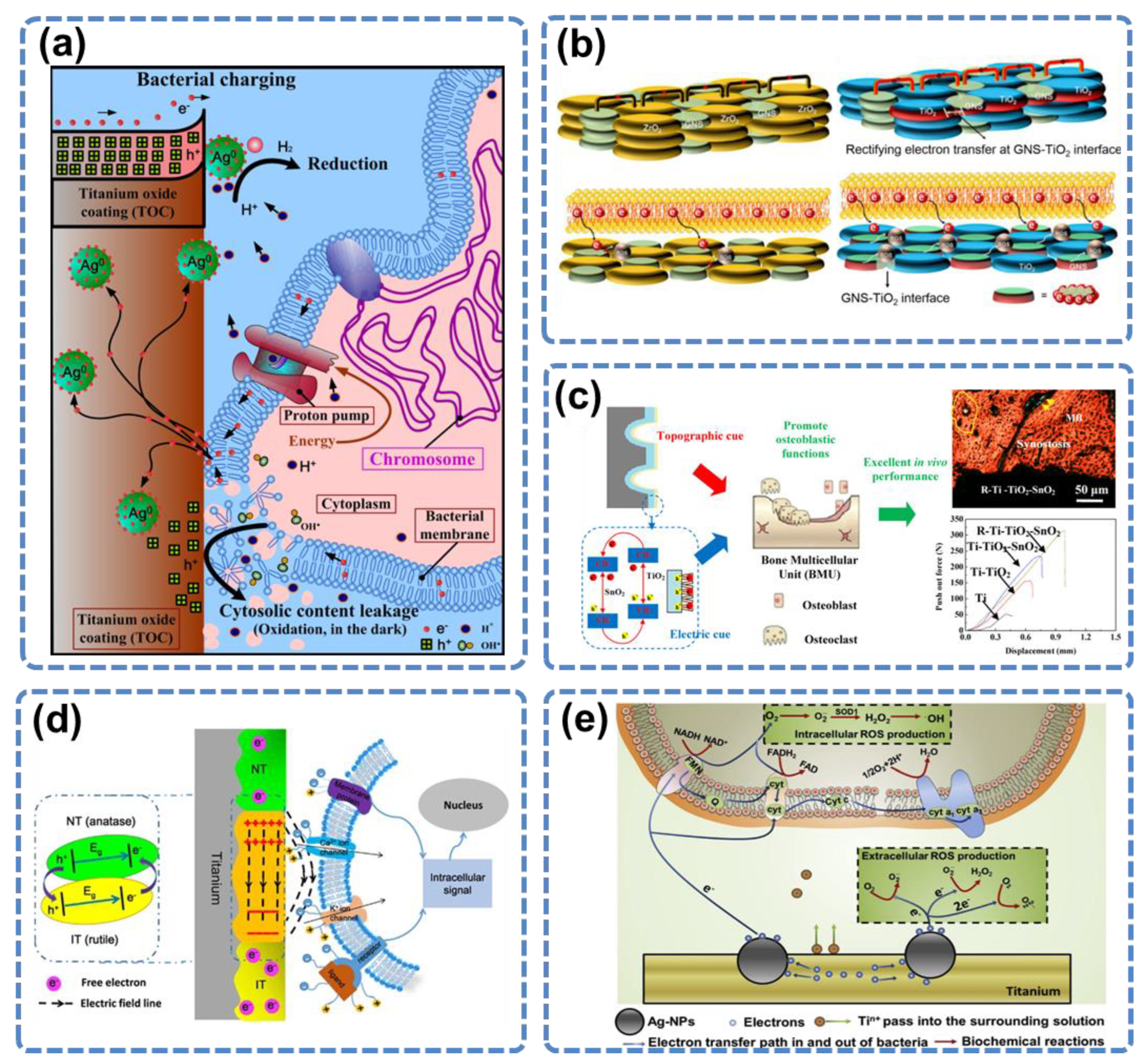
3.4. Electron Transfer

In recent years, an antibacterial theory based on the electron transfer between the material surface and the microbes has been proposed. Electron transfer is a common event in the photochemical modulation of materials, as well as a fundamental event for the energy generation of organisms [83]. A group of microbes can do extracellular electron transfer spontaneously by transferring the electron outside the cells to environmental minerals [84]. However, using the electron transfer approach to inhibit implant infection is a quite new topic [85].
Vecitis et al. found that the antibacterial properties of single-arm carbon nanotubes are closely related to their electronic state. With the same diameter and length, metallic carbon nanotubes can cause severe deformation and collapse of the bacterial cells, while those in a semi-conductive state have no antibacterial properties [86]. Faria et al. found that the composite structure of Ag nanoparticles and graphene lamellae has a strong bactericidal ability, but graphene lamellae itself does not, suggesting that the electronic interactions between the substrate and the modified materials have a dominant impact on the antibacterial property [87].
TiO2 also has complex interactions with the bacteria and osteoblasts via electron transfer. TiO2 is a semiconductor, and biological cells can also be regarded as semiconductors [88]. Once contacted, they form heterojunctions, which may involve electron transfer. Therefore, functionalization based on the electron transfer property also influences the performance of TiO2 as an orthopedic implant. Au and Ag nanoparticles or graphene sheets deposited on the TiO2 surface can endow TiO2 with antibacterial properties [88,89,90,91,92,93]. On the Ag@TiO2 surface, electrons were stored on the Ag nanoparticles, and induced valence-band hole (h+) accumulation, which caused cytosolic content leakage of the bacteria (Figure 6a) [89]. On the Au@TiO2 surface, electron transfer was due to the plasmon effect of Au nanoparticles, which captured the electrons in the respiratory chain on the living bacterial cell membrane and transferred them to the TiO2 substrate. Au@TiO2 formed the Schottky barrier, which prevented the return of electrons, causing continued electron loss in the bacteria until death [91,93]. Similarly, graphene coating resulted in a large increase in the electrical conductivity of TiO2 because of the combination of the unpaired π electrons of graphene and the Ti atoms [94]. The enhanced electron transfer from the bacterial cell membrane to the graphene-TiO2 interface leads to bacterial death (Figure 6b).
Electron transfer also works for osteogenesis. Zhou et al. fabricated a SnO2–TiO2 heterojunction and hierarchical structure on the surface of the Ti implant [95]. The electron transfer among the hierarchical Schottky barrier significantly improved the osteogenic function of the cells around the implant both in vitro and in vivo (Figure 6c). In another work, they constructed a layered double hydroxide (LDHs)–TiO2 heterojunction, which promoted the transfer of holes in materials to the physiological environment, enhancing the antibacterial effect of the implant [96]. Ning et al. generated a microscale electrostatic field (MEF) by doing patterned NT (rutile) and IT (anatase) surface modifications on Ti [97]. The electron transfer between NT and IT zones formed a sustained built-in MEF, which polarized the BMSC and activated the expression of osteogenic genes (Figure 6d). The MEF greatly promoted bone regeneration around the implant.
Apart from TiO2, the Ti surface can also make electron transfer-based interactions with the bacteria. In a study by Wang et al., Ag was implanted on the Ti surface using plasma technology, and this modification changed the Ti surface from non-antibacterial to antibacterial [93]. The bacteria-killing was not due to Ag+ ion release, but due to the micro galvanic reaction at the nano interface between Ag nanoparticles and Ti substrate. The reaction disturbed the process of electron transfer in the bacteria respiratory chain and produced a large number of reactive oxygen species (ROS) in the bacterial cells, resulting in their death (Figure 6e).
Figure 6. (a) Extracellular electron transfer induces bactericidal action of the Ag@TiO2 coating. Reprinted with permission from Ref. [89]. Elsevier, 2013. (b) The proposed mechanism explaining the antibacterial property of graphene nanosheets-TiO2 coatings is based on the electron transfer at the graphene-TiO2 interface. Reprinted with permission from Ref. [94]. Elsevier, 2020. (c) Illustration of the enhanced osteogenesis performance of titanium by an electric cue offered by the built-in electrical field of SnO2–TiO2. Topographic cue (red color arrow) and electric cue (blue color arrow) enhance the in vivo osteogenesis process (green color arrow). Reprinted with permission from Ref. [95]. American Chemical Society, 2018. (d) Demonstration of the mechanisms to generate MEF and the interactions between MEF and stem cells. Reprinted with permission from Ref. [97]. Springer Nature, 2016. (e) The electron transfer-based bacteria killing on the Ag@Ti surface. Reprinted with permission from Ref. [93]. Elsevier, 2017.
Materials 15 06868 g006

3.5. Electrical Functionalization

Based on the electron transfer mechanism of the above studies, researchers have further developed an innovative method to make the TiO2 surface obtain antibacterial properties through electrical tuning. In the beginning, it was found that an alternating current (AC) of about ±2 μA applied to the ZnO nanowires in a physiological solution could significantly improve the antibacterial property of ZnO after the current was removed (Figure 7a). The “sustained bacteria sterilization” was different from the “instant bacteria sterilization” because the latter was due to electroporation when AC was applied to the nanowires, but the former was due to surface functionalization by the electrical tuning [98]. After that, a 2 V low-voltage direct current (DC) power supply was used to conduct electrical treatment on the Ti plate with a TiO2 layer in the culture medium for 20 min. This DC tuning also changed the TiO2 surface from non-antibacterial to highly antibacterial [99]. After the electric tuning, TiO2 gained a strong ability to kill various bacteria and showed strong inhibition of biofilm formation. Meanwhile, the DC-tuned TiO2 surface had no negative effect on the osteoblast. The adhesion and proliferation of the cells were found to be as effective as those on the control TiO2 surface (Figure 7b).

4. Conclusions and Perspectives

In summary, Ti implants have a major concern regarding microbial infection, which may lead to implant failure. Various approaches have been carried out to deal with infections and promote osteointegration. TiO2 is not only the native oxide layer of the Ti biomedical implant but also a material widely studied in the photoelectronic and photocatalytic fields. Meanwhile, it is relatively easy to form nanostructures in the fabrication of TiO2 layers. Due to these properties, TiO2 has enabled various functionalization approaches to endow Ti with bactericidal and osteogenic abilities. In this review, we have discussed both the well-established and the newly-proposed approaches for TiO2 functionalization.
Topology, drug loading, and element incorporation are well-established approaches that have been developed for years. In some circumstances, the functionalization efforts may have conflicts. For the topological approaches, it is sometimes difficult to enhance the bactericidal and osteogenic properties at the same time because the topographies that can encourage BMSC and osteoblasts adhesion and proliferation will attract bacterial adhesion as well. In the ionic release approach, sometimes the released ions will harm the cells. However, innovations can still be made on the well-studied topics to overcome the problems they face. For example, nanoparticles can be used to help the TiO2 surface nanostructures, not from the aspect of topologies but by introducing a built-in electric field. Drugs can be incorporated into polymeric micelles, which may have more controllable behavior during the loading and releasing process. Metal ions such as Mg2+ and Zn2+ are found to have both bactericidal and osteo-enhancing properties; thus, incorporation of these elements into TiO2 via proper methods will be very helpful for Ti implants.
The electron transfer approaches are newly proposed in recent years. TiO2 is a material deeply involved in the electronic and photocatalytic fields for its ability to conduct electron separation and transfer. In the studies we discussed above, electron transfer between TiO2 and the biological cells also takes place and demonstrates advantages in balancing the two needs of the implant. Most of the electron transfer studies have declared that their approaches only have a killing effect on the bacteria but no adverse effect on the growth and differentiation of the osteoblasts. Based on electron transfer theory, the electrical tuning of TiO2 for anti-bacterial properties has also demonstrated success. However, the underlying mechanisms of the electron-based interactions of TiO2 with biological cells are far from explicit.
Most of the functionalization approaches are promising for clinical applications, as long as batch production can be realized and the production cost can be reduced. The electrical tuning method, however, needs extended evaluation because it involves in vivo electrical manipulation apart from material implantation. Whether the tuning parameters are safe for the body and their impacts on the surrounding biological systems are yet to be discovered. Of course, more animal experiments and clinical trials are needed for all approaches to translate them into human benefits.
In the future, it is welcome to conduct in-depth research on the bactericidal and osteogenesis mechanisms of the functionalization approaches to obtain a deeper understanding of the interactions among the implant surface, bacteria, and cells. This will enable researchers to design the functionalization methods more effectively and rationally and facilitate the clinical applications of Ti-based implants with more safety and convenience.

Author Contributions

Conceptualization, S.N. and E.W.; methodology, S.N and E.W.; software, S.N.; validation, S.N., E.W. and H.F.; formal analysis, S.N.; investigation, S.N. and E.W.; resources, S.N. and E.W; data curation, S.N.; writing—original draft preparation, S.N., E.W. and H.F.; writing—review and editing, S.N., H.F. and Z.L.; visualization, H.F.; supervision, Z.L.; project administration, Z.L. and H.F.; funding acquisition, Z.L. All authors have read and agreed to the published version of the manuscript.

Funding

This work was supported by the National Key R&D Program of China (2021YFB3200600), the Fundamental Research Funds for the Central Universities, the National Natural Science Foundation of China (81971770, T2125003, 61875015), Beijing Natural Science Foundation (JQ20038), and the Strategic Priority Research Program of the Chinese Academy of Sciences (XDA16021101).

Institutional Review Board Statement

Not applicable.

Informed Consent Statement

Not applicable.

Data Availability Statement

Not applicable.

Conflicts of Interest

The authors declare no conflict of interest.

References

  1. Baltatu, M.S.; Vizureanu, P.; Sandu, A.V.; Florido-Suarez, N.; Saceleanu, M.V.; Mirza-Rosca, J.C. New titanium alloys, promising materials for medical devices. Materials 2021, 14, 5934. [Google Scholar] [CrossRef] [PubMed]
  2. Filipović, U.; Dahmane, R.G.; Ghannouchi, S.; Zore, A.; Bohinc, K. Bacterial adhesion on orthopedic implants. Adv. Colloid Interface Sci. 2020, 283, 102228. [Google Scholar] [CrossRef]
  3. Losic, D.; Aw, M.S.; Santos, A.; Gulati, K.; Bariana, M. Titania nanotube arrays for local drug delivery: Recent advances and perspectives. Expert Opin. Drug Deliv. 2015, 12, 103–127. [Google Scholar] [CrossRef] [PubMed]
  4. Niinomi, M. Mechanical properties of biomedical titanium alloys. Mater. Sci. Eng. A 1998, 243, 231–236. [Google Scholar] [CrossRef]
  5. Elias, C.N.; Lima, J.H.C.; Valiev, R.; Meyers, M.A. Biomedical applications of titanium and its alloys. Jom 2008, 60, 46–49. [Google Scholar] [CrossRef]
  6. Sidambe, A.T. Biocompatibility of advanced manufactured titanium implants—A review. Materials 2014, 7, 8168–8188. [Google Scholar] [CrossRef] [PubMed] [Green Version]
  7. Park, Y.J.; Song, Y.H.; An, J.H.; Song, H.J.; Anusavice, K.J. Cytocompatibility of pure metals and experimental binary titanium alloys for implant materials. J. Dent. 2013, 41, 1251–1258. [Google Scholar] [CrossRef]
  8. von Wilmowsky, C.; Bauer, S.; Lutz, R.; Meisel, M.; Neukam, F.W.; Toyoshima, T.; Schmuki, P.; Nkenke, E.; Schlegel, K.A. In vivo evaluation of anodic TiO2 nanotubes: An experimental study in the pig. J. Biomed. Mater. Res. Part B Appl. Biomater. Off. J. Soc. Biomater. Jpn. Soc. Biomater. Aust. Soc. Biomater. Korean Soc. Biomater. 2009, 89, 165–171. [Google Scholar] [CrossRef]
  9. Zhao, G.; Schwartz, Z.; Wieland, M.; Rupp, F.; Geis-Gerstorfer, J.; Cochran, D.L.; Boyan, B.D. High surface energy enhances cell response to titanium substrate microstructure. J. Biomed. Mater. Res. Part A An Off. J. Soc. Biomater. Japanese Soc. Biomater. Aust. Soc. Biomater. Korean Soc. Biomater. 2005, 74, 49–58. [Google Scholar] [CrossRef]
  10. Chevalier, J.; Gremillard, L. Ceramics for medical applications: A picture for the next 20 years. J. Eur. Ceram. Soc. 2009, 29, 1245–1255. [Google Scholar] [CrossRef]
  11. Moriarty, T.F.; Schlegel, U.; Perren, S.; Richards, R.G. Infection in fracture fixation: Can we influence infection rates through implant design? J. Mater. Sci. Mater. Med. 2010, 21, 1031–1035. [Google Scholar] [CrossRef] [PubMed]
  12. Gittens, R.A.; Scheideler, L.; Rupp, F.; Hyzy, S.L.; Geis-Gerstorfer, J.; Schwartz, Z.; Boyan, B.D. A review on the wettability of dental implant surfaces II: Biological and clinical aspects. Acta Biomater. 2014, 10, 2907–2918. [Google Scholar] [CrossRef] [PubMed] [Green Version]
  13. Lindhe, J.; Meyle, J.; Group D of the European Workshop on Periodontology. Peri-implant diseases: Consensus report of the sixth European workshop on periodontology. J. Clin. Periodontol. 2008, 35, 282–285. [Google Scholar] [CrossRef] [PubMed] [Green Version]
  14. Chen, S.; Darby, I. Dental implants: Maintenance, care and treatment of peri-implant infection. Aust. Dent. J. 2003, 48, 212–220. [Google Scholar] [CrossRef] [PubMed] [Green Version]
  15. Periodontology, A.A. of Parameter on placement and management of the dental implant. J. Periodontol. 2000, 71, 870–872. [Google Scholar]
  16. Tonetti, M.S. Risk factors for osseodisintegration. Periodontol. 2000 1998, 17, 55–62. [Google Scholar] [CrossRef] [PubMed]
  17. Fujimoto, T.; Niimi, A.; Sawai, T.; Ueda, M. Effects of steroid-induced osteoporosis on osseointegration of titanium implants. Int. J. Oral Maxillofac. Implant. 1998, 13, 183–189. [Google Scholar]
  18. Farzad, P.; Andersson, L.; Nyberg, J. Dental implant treatment in diabetic patients. Implant Dent. 2002, 11, 262–267. [Google Scholar] [CrossRef]
  19. Halpern, M.; Kurtz, S.; Lau, E.; Mowat, F.; Ong, K. Projections of primary and revision hip and knee arthroplasty in the United States from 2005 to 2030. J. Bone Surg. 2007, 89, 780–785. [Google Scholar]
  20. Lentino, J.R. Prosthetic joint infections: Bane of orthopedists, challenge for infectious disease specialists. Clin. Infect. Dis. 2003, 36, 1157–1161. [Google Scholar] [CrossRef]
  21. Campoccia, D.; Montanaro, L.; Arciola, C.R. The significance of infection related to orthopedic devices and issues of antibiotic resistance. Biomaterials 2006, 27, 2331–2339. [Google Scholar] [CrossRef] [PubMed]
  22. Bosco, R.; Van Den Beucken, J.; Leeuwenburgh, S.; Jansen, J. Surface engineering for bone implants: A trend from passive to active surfaces. Coatings 2012, 2, 95–119. [Google Scholar] [CrossRef] [Green Version]
  23. Brammer, K.S.; Frandsen, C.J.; Jin, S. TiO2 nanotubes for bone regeneration. Trends Biotechnol. 2012, 30, 315–322. [Google Scholar] [CrossRef] [PubMed]
  24. Dalby, M.J.; Gadegaard, N.; Tare, R.; Andar, A.; Riehle, M.O.; Herzyk, P.; Wilkinson, C.D.W.; Oreffo, R.O.C. The control of human mesenchymal cell differentiation using nanoscale symmetry and disorder. Nat. Mater. 2007, 6, 997–1003. [Google Scholar] [CrossRef]
  25. Ratner, B.D.; Bryant, S.J. Biomaterials: Where we have been and where we are going. Annu. Rev. Biomed. Eng. 2004, 6, 41–75. [Google Scholar] [CrossRef]
  26. Lutolf, M.P.; Gilbert, P.M.; Blau, H.M. Designing materials to direct stem-cell fate. Nature 2009, 462, 433–441. [Google Scholar] [CrossRef] [Green Version]
  27. Chrzanowski, W.; Lee, J.H.; Kondyurin, A.; Lord, M.S.; Jang, J.; Kim, H.; Bilek, M.M.M. Nano-Bio-Chemical Braille for Cells: The Regulation of Stem Cell Responses using Bi-Functional Surfaces. Adv. Funct. Mater. 2015, 25, 193–205. [Google Scholar] [CrossRef]
  28. Huang, H.; He, L.; Zhou, W.; Qu, G.; Wang, J.; Yang, N.; Gao, J.; Chen, T.; Chu, P.K.; Yu, X.-F. Stable black phosphorus/Bi2O3 heterostructures for synergistic cancer radiotherapy. Biomaterials 2018, 171, 12–22. [Google Scholar] [CrossRef]
  29. Lu, H.; Hao, Q.; Chen, T.; Zhang, L.; Chen, D.; Ma, C.; Yao, W.; Zhu, Y. A high-performance Bi2O3/Bi2SiO5 pn heterojunction photocatalyst induced by phase transition of Bi2O3. Appl. Catal. B Environ. 2018, 237, 59–67. [Google Scholar] [CrossRef]
  30. Huang, X.; Xing, J.; Wang, Z.; Han, J.; Wang, R.; Li, C.; Xiao, C.; Lu, F.; Zhai, J.; Zhou, Z. 0D/1D Heterojunction Implant with Electro-Mechanobiological Coupling Cues Promotes Osteogenesis. Adv. Funct. Mater. 2021, 31, 2106249. [Google Scholar] [CrossRef]
  31. Fan, J.; Abedi-Dorcheh, K.; Vaziri, A.S.; Kazemi-Aghdam, F.; Rafieyan, S.; Sohrabinejad, M.; Ghorbani, M.; Adib, F.R.; Ghasemi, Z.; Klavins, K.; et al. A Review of Recent Advances in Natural Polymer-Based Scaffolds for Musculoskeletal Tissue Engineering. Polymers 2022, 14, 2097. [Google Scholar] [CrossRef] [PubMed]
  32. Yu, Y.; Shen, X.; Luo, Z.; Hu, Y.; Li, M.; Ma, P.; Ran, Q.; Dai, L.; He, Y.; Cai, K. Osteogenesis potential of different titania nanotubes in oxidative stress microenvironment. Biomaterials 2018, 167, 44–57. [Google Scholar] [CrossRef]
  33. Calabrese, G.; Franco, D.; Petralia, S.; Monforte, F.; Condorelli, G.G.; Squarzoni, S.; Traina, F.; Conoci, S. Dual-Functional Nano-Functionalized Titanium Scaffolds to Inhibit Bacterial Growth and Enhance Osteointegration. Nanomaterials 2021, 11, 2634. [Google Scholar] [CrossRef] [PubMed]
  34. Shao, Y.; Fu, J. Integrated micro/nanoengineered functional biomaterials for cell mechanics and mechanobiology: A materials perspective. Adv. Mater. 2014, 26, 1494–1533. [Google Scholar] [CrossRef] [PubMed] [Green Version]
  35. Kim, D.-H.; Provenzano, P.P.; Smith, C.L.; Levchenko, A. Matrix nanotopography as a regulator of cell function. J. Cell Biol. 2012, 197, 351–360. [Google Scholar] [CrossRef]
  36. Dvir, T.; Timko, B.P.; Kohane, D.S.; Langer, R. Nanotechnological strategies for engineering complex tissues. Nat. Nanotechnology 2011, 6, 13–22. [Google Scholar] [CrossRef]
  37. Dalby, M.J.; Gadegaard, N.; Oreffo, R.O.C. Harnessing nanotopography and integrin–matrix interactions to influence stem cell fate. Nat. Mater. 2014, 13, 558–569. [Google Scholar] [CrossRef]
  38. Yao, X.; Peng, R.; Ding, J. Cell–material interactions revealed via material techniques of surface patterning. Adv. Mater. 2013, 25, 5257–5286. [Google Scholar] [CrossRef]
  39. Li, Y.; Yang, Y.; Li, R.; Tang, X.; Guo, D.; Qing, Y.; Qin, Y. Enhanced antibacterial properties of orthopedic implants by titanium nanotube surface modification: A review of current techniques. Int. J. Nanomedicine 2019, 14, 7217–7236. [Google Scholar] [CrossRef] [Green Version]
  40. Zhao, L.; Liu, L.; Wu, Z.; Zhang, Y.; Chu, P.K. Effects of micropitted/nanotubular titania topographies on bone mesenchymal stem cell osteogenic differentiation. Biomaterials 2012, 33, 2629–2641. [Google Scholar] [CrossRef]
  41. Descamps, S.; Awitor, K.O.; Raspal, V.; Johnson, M.B.; Bokalawela, R.S.P.; Larson, P.R.; Doiron, C.F. Mechanical properties of nanotextured titanium orthopedic screws for clinical applications. J. Med. Devices 2013, 7, 021005. [Google Scholar] [CrossRef] [PubMed] [Green Version]
  42. Oh, S.; Brammer, K.S.; Li, Y.S.J.; Teng, D.; Engler, A.J.; Chien, S.; Jin, S. Stem cell fate dictated solely by altered nanotube dimension. Proc. Natl. Acad. Sci. USA 2009, 106, 2130–2135. [Google Scholar] [CrossRef] [Green Version]
  43. Lv, L.; Liu, Y.; Zhang, P.; Zhang, X.; Liu, J.; Chen, T.; Su, P.; Li, H.; Zhou, Y. The nanoscale geometry of TiO2 nanotubes influences the osteogenic differentiation of human adipose-derived stem cells by modulating H3K4 trimethylation. Biomaterials 2015, 39, 193–205. [Google Scholar] [CrossRef] [PubMed]
  44. Li, Q.; Cai, T.; Huang, Y.; Xia, X.; Cole, S.P.C.; Cai, Y. A review of the structure, preparation, and application of NLCs, PNPs, and PLNs. Nanomaterials 2017, 7, 122. [Google Scholar] [CrossRef] [PubMed]
  45. Peng, Z.; Ni, J.; Zheng, K.; Shen, Y.; Wang, X.; He, G.; Jin, S.; Tang, T. Dual effects and mechanism of TiO2 nanotube arrays in reducing bacterial colonization and enhancing C3H10T1/2 cell adhesion. Int. J. Nanomedicine 2013, 8, 3093. [Google Scholar] [PubMed] [Green Version]
  46. Ercan, B.; Taylor, E.; Alpaslan, E.; Webster, T.J. Diameter of titanium nanotubes influences anti-bacterial efficacy. Nanotechnology 2011, 22, 295102. [Google Scholar] [CrossRef] [PubMed]
  47. Puckett, S.D.; Taylor, E.; Raimondo, T.; Webster, T.J. The relationship between the nanostructure of titanium surfaces and bacterial attachment. Biomaterials 2010, 31, 706–713. [Google Scholar] [CrossRef]
  48. Qiu, J.; Li, J.; Wang, S.; Ma, B.; Zhang, S.; Guo, W.; Zhang, X.; Tang, W.; Sang, Y.; Liu, H. TiO2 nanorod array constructed nanotopography for regulation of mesenchymal stem cells fate and the realization of location-committed stem cell differentiation. Small 2016, 12, 1770–1778. [Google Scholar] [CrossRef]
  49. Buensuceso, C.S.; Woodside, D.; Huff, J.L.; Plopper, G.E.; O’Toole, T.E. The WD protein Rack1 mediates protein kinase C and integrin-dependent cell migration. J. Cell Sci. 2001, 114, 1691–1698. [Google Scholar] [CrossRef]
  50. Pegueroles, M.; Aparicio, C.; Bosio, M.; Engel, E.; Gil, F.J.; Planell, J.A.; Altankov, G. Spatial organization of osteoblast fibronectin matrix on titanium surfaces: Effects of roughness, chemical heterogeneity and surface energy. Acta Biomater. 2010, 6, 291–301. [Google Scholar] [CrossRef]
  51. Geesink, R.G.T.; de Groot, K.; KLEIN, C.P.A.T. Chemical implant fixation using hydroxyl-apatite coatings: The development of a human total hip prosthesis for chemical fixation to bone using hydroxyl-apatite coatings on titanium substrates. Clin. Orthop. Relat. Res. 1987, 225, 147–170. [Google Scholar] [CrossRef]
  52. Lee, J.J.; Rouhfar, L.; Beirne, O.R. Survival of hydroxyapatite-coated implants: A meta-analytic review. J. Oral Maxillofac. Surg. 2000, 58, 1372–1379. [Google Scholar] [CrossRef] [PubMed]
  53. Dalby, M.J.; Hart, A.; Yarwood, S.J. The effect of the RACK1 signalling protein on the regulation of cell adhesion and cell contact guidance on nanometric grooves. Biomaterials 2008, 29, 282–289. [Google Scholar] [CrossRef] [PubMed]
  54. Goodman, S.B.; Yao, Z.; Keeney, M.; Yang, F. The future of biologic coatings for orthopaedic implants. Biomaterials 2013, 34, 3174–3183. [Google Scholar] [CrossRef] [PubMed] [Green Version]
  55. Xiao, X.; Yang, L.; Guo, M.; Pan, C.; Cai, Q.; Yao, S. Biocompatibility and in vitro antineoplastic drug-loaded trial of titania nanotubes prepared by anodic oxidation of a pure titanium. Sci. China Ser. B Chem. 2009, 52, 2161–2165. [Google Scholar] [CrossRef]
  56. Yang, D.-J.; Kim, H.-G.; Cho, S.-J.; Choi, W.-Y. Thickness-conversion ratio from titanium to TiO2 nanotube fabricated by anodization method. Mater. Lett. 2008, 62, 775–779. [Google Scholar] [CrossRef]
  57. Kulkarni, M.; Mazare, A.; Gongadze, E.; Perutkova, Š.; Kralj-Iglič, V.; Milošev, I.; Schmuki, P.; Iglič, A.; Mozetič, M. Titanium nanostructures for biomedical applications. Nanotechnology 2015, 26, 62002. [Google Scholar] [CrossRef]
  58. Andersson, R.E.; Lukas, G.; Skullman, S.; Hugander, A. Local administration of antibiotics by gentamicin–collagen sponge does not improve wound healing or reduce recurrence rate after pilonidal excision with primary suture: A prospective randomized controlled trial. World J. Surg. 2010, 34, 3042–3046. [Google Scholar] [CrossRef] [PubMed]
  59. Kumeria, T.; Mon, H.; Aw, M.S.; Gulati, K.; Santos, A.; Griesser, H.J.; Losic, D. Advanced biopolymer-coated drug-releasing titania nanotubes (TNTs) implants with simultaneously enhanced osteoblast adhesion and antibacterial properties. Colloids Surf. B Biointerfaces 2015, 130, 255–263. [Google Scholar] [CrossRef] [Green Version]
  60. Aw, M.S.; Gulati, K.; Losic, D. Controlling drug release from titania nanotube arrays using polymer nanocarriers and biopolymer coating. J. Biomater. Nanobiotechnol. 2011, 2, 477. [Google Scholar] [CrossRef] [Green Version]
  61. Gulati, K.; Ramakrishnan, S.; Aw, M.S.; Atkins, G.J.; Findlay, D.M.; Losic, D. Biocompatible polymer coating of titania nanotube arrays for improved drug elution and osteoblast adhesion. Acta Biomater. 2012, 8, 449–456. [Google Scholar] [CrossRef] [PubMed]
  62. Jia, Z.; Xiu, P.; Li, M.; Xu, X.; Shi, Y.; Cheng, Y.; Wei, S.; Zheng, Y.; Xi, T.; Cai, H. Bioinspired anchoring AgNPs onto micro-nanoporous TiO2 orthopedic coatings: Trap-killing of bacteria, surface-regulated osteoblast functions and host responses. Biomaterials 2016, 75, 203–222. [Google Scholar] [CrossRef] [PubMed]
  63. Hwang, S.H.; Song, J.; Jung, Y.; Kweon, O.Y.; Song, H.; Jang, J. ElectrospunZnO/TiO2 composite nanofibers as a bactericidal agent. Chem. Commun. 2011, 47, 9164–9166. [Google Scholar] [CrossRef] [PubMed]
  64. Paladini, F.; Pollini, M.; Sannino, A.; Ambrosio, L. Metal-based antibacterial substrates for biomedical applications. Biomacromolecules 2015, 16, 1873–1885. [Google Scholar] [CrossRef]
  65. Chernousova, S.; Epple, M. Silver as antibacterial agent: Ion, nanoparticle, and metal. Angew. Chemie Int. Ed. 2013, 52, 1636–1653. [Google Scholar] [CrossRef] [PubMed]
  66. Campoccia, D.; Montanaro, L.; Arciola, C.R. A review of the biomaterials technologies for infection-resistant surfaces. Biomaterials 2013, 34, 8533–8554. [Google Scholar] [CrossRef]
  67. Zhang, Y.; Dong, C.; Yang, S.; Chiu, T.-W.; Wu, J.; Xiao, K.; Huang, Y.; Li, X. Enhanced silver loaded antibacterial titanium implant coating with novel hierarchical effect. J. Biomater. Appl. 2018, 32, 1289–1299. [Google Scholar] [CrossRef]
  68. Hou, X.; Mao, D.; Ma, H.; Ai, Y.; Zhao, X.; Deng, J.; Li, D.; Liao, B. Antibacterial ability of Ag–TiO2 nanotubes prepared by ion implantation and anodic oxidation. Mater. Lett. 2015, 161, 309–312. [Google Scholar] [CrossRef]
  69. Shanmuganathan, R.; MubarakAli, D.; Prabakar, D.; Muthukumar, H.; Thajuddin, N.; Kumar, S.S.; Pugazhendhi, A. An enhancement of antimicrobial efficacy of biogenic and ceftriaxone-conjugated silver nanoparticles: Green approach. Environ. Sci. Pollut. Res. 2018, 25, 10362–10370. [Google Scholar] [CrossRef]
  70. Tang, Y.; Chappell, H.F.; Dove, M.T.; Reeder, R.J.; Lee, Y.J. Zinc incorporation into hydroxylapatite. Biomaterials 2009, 30, 2864–2872. [Google Scholar] [CrossRef]
  71. Tas, A.C.; Bhaduri, S.B.; Jalota, S. Preparation of Zn-doped β-tricalcium phosphate (β-Ca3(PO4)2) bioceramics. Mater. Sci. Eng. C 2007, 27, 394–401. [Google Scholar]
  72. Lowe, N.M.; Fraser, W.D.; Jackson, M.J. Is there a potential therapeutic value of copper and zinc for osteoporosis? Proc. Nutr. Soc. 2002, 61, 181–185. [Google Scholar] [CrossRef] [PubMed]
  73. Storrie, H.; Stupp, S.I. Cellular response to zinc-containing organoapatite: An in vitro study of proliferation, alkaline phosphatase activity and biomineralization. Biomaterials 2005, 26, 5492–5499. [Google Scholar] [CrossRef] [PubMed]
  74. Applerot, G.; Lipovsky, A.; Dror, R.; Perkas, N.; Nitzan, Y.; Lubart, R.; Gedanken, A. Enhanced antibacterial activity of nanocrystalline ZnO due to increased ROS-mediated cell injury. Adv. Funct. Mater. 2009, 19, 842–852. [Google Scholar] [CrossRef]
  75. Liu, X.; Zhao, X.; Li, B.; Cao, C.; Dong, Y.; Ding, C.; Chu, P.K. UV-irradiation-induced bioactivity on TiO2 coatings with nanostructural surface. Acta Biomater. 2008, 4, 544–552. [Google Scholar] [CrossRef] [PubMed]
  76. de Assis, S.L.; Wolynec, S.; Costa, I. Corrosion characterization of titanium alloys by electrochemical techniques. Electrochim. Acta 2006, 51, 1815–1819. [Google Scholar] [CrossRef]
  77. Díez-Pascual, A.M.; Diez-Vicente, A.L. Development of nanocomposites reinforced with carboxylated poly (ether ether ketone) grafted to zinc oxide with superior antibacterial properties. ACS Appl. Mater. Interfaces 2014, 6, 3729–3741. [Google Scholar] [CrossRef] [Green Version]
  78. Yang, Y.; Liu, L.; Luo, H.; Zhang, D.; Lei, S.; Zhou, K. Dual-purpose magnesium-incorporated titanium nanotubes for combating bacterial infection and ameliorating osteolysis to realize better osseointegration. ACS Biomater. Sci. Eng. 2019, 5, 5368–5383. [Google Scholar] [CrossRef]
  79. Steinmetz, O.; Hoch, S.; Avniel-Polak, S.; Gavish, K.; Eli-Berchoer, L.; Wilensky, A.; Nussbaum, G. CX3CR1hi Monocyte/Macrophages Support Bacterial Survival and Experimental Infection–Driven Bone Resorption. J. Infect. Dis. 2016, 213, 1505–1515. [Google Scholar] [CrossRef] [Green Version]
  80. Wang, J.; Wu, X.; Duan, Y. Magnesium lithospermate B protects against lipopolysaccharide-induced bone loss by inhibiting RANKL/RANK pathway. Front. Pharmacol. 2018, 9, 64. [Google Scholar] [CrossRef] [Green Version]
  81. Chopra, D.; Gulati, K.; Ivanovski, S. Understanding and optimizing the antibacterial functions of anodized nano-engineered titanium implants. Acta Biomater. 2021, 127, 80–101. [Google Scholar] [CrossRef] [PubMed]
  82. Choe, H.J.; Kwon, S.-H.; Lee, J.-J. Tribological properties and thermal stability of TiAlCN coatings deposited by ICP-assisted sputtering. Surf. Coatings Technol. 2013, 228, 282–285. [Google Scholar] [CrossRef]
  83. Harris, H.W.; El-Naggar, M.Y.; Bretschger, O.; Ward, M.J.; Romine, M.F.; Obraztsova, A.Y.; Nealson, K.H. Electrokinesis is a microbial behavior that requires extracellular electron transport. Proc. Natl. Acad. Sci. USA 2010, 107, 326–331. [Google Scholar] [CrossRef] [PubMed] [Green Version]
  84. Shi, L.; Dong, H.; Reguera, G.; Beyenal, H.; Lu, A.; Liu, J.; Yu, H.-Q.; Fredrickson, J.K. Extracellular electron transfer mechanisms between microorganisms and minerals. Nat. Rev. Microbiol. 2016, 14, 651–662. [Google Scholar] [CrossRef] [PubMed]
  85. Wang, D.; Tan, J.; Zhu, H.; Mei, Y.; Liu, X. Biomedical Implants with Charge-Transfer Monitoring and Regulating Abilities. Adv. Sci. 2021, 8, 1–38. [Google Scholar] [CrossRef] [PubMed]
  86. Vecitis, C.D.; Zodrow, K.R.; Kang, S.; Elimelech, M. Electronic-structure-dependent bacterial cytotoxicity of single-walled carbon nanotubes. ACS Nano 2010, 4, 5471–5479. [Google Scholar] [CrossRef] [PubMed]
  87. de Faria, A.F.; Martinez, D.S.T.; Meira, S.M.M.; de Moraes, A.C.M.; Brandelli, A.; Souza Filho, A.G.; Alves, O.L. Anti-adhesion and antibacterial activity of silver nanoparticles supported on graphene oxide sheets. Colloids Surf. B Biointerfaces 2014, 113, 115–124. [Google Scholar] [CrossRef]
  88. Strahl, H.; Hamoen, L.W. Membrane potential is important for bacterial cell division. Proc. Natl. Acad. Sci. USA 2010, 107, 12281–12286. [Google Scholar] [CrossRef] [Green Version]
  89. Cao, H.; Qiao, Y.; Liu, X.; Lu, T.; Cui, T.; Meng, F.; Chu, P.K. Electron storage mediated dark antibacterial action of bound silver nanoparticles: Smaller is not always better. Acta Biomater. 2013, 9, 5100–5110. [Google Scholar] [CrossRef]
  90. Li, J.; Zhou, H.; Qian, S.; Liu, Z.; Feng, J.; Jin, P.; Liu, X. Plasmonic gold nanoparticles modified titania nanotubes for antibacterial application. Appl. Phys. Lett. 2014, 104, 261110. [Google Scholar] [CrossRef]
  91. Wang, G.; Feng, H.; Gao, A.; Hao, Q.; Jin, W.; Peng, X.; Li, W.; Wu, G.; Chu, P.K. Extracellular electron transfer from aerobic bacteria to Au-loaded TiO2 semiconductor without light: A new bacteria-killing mechanism other than localized surface plasmon resonance or microbial fuel cells. ACS Appl. Mater. Interfaces 2016, 8, 24509–24516. [Google Scholar] [CrossRef] [PubMed]
  92. Li, J.; Wang, G.; Zhu, H.; Zhang, M.; Zheng, X.; Di, Z.; Liu, X.; Wang, X. Antibacterial activity of large-area monolayer graphene film manipulated by charge transfer. Sci. Rep. 2014, 4, 1–8. [Google Scholar] [CrossRef] [Green Version]
  93. Wang, G.; Jin, W.; Qasim, A.M.; Gao, A.; Peng, X.; Li, W.; Feng, H.; Chu, P.K. Antibacterial effects of titanium embedded with silver nanoparticles based on electron-transfer-induced reactive oxygen species. Biomaterials 2017, 124, 25–34. [Google Scholar] [CrossRef] [PubMed]
  94. Yang, M.; Liu, H.; Qiu, C.; Iatsunskyi, I.; Coy, E.; Moya, S.; Wang, Z.; Wu, W.; Zhao, X.; Wang, G. Electron transfer correlated antibacterial activity of biocompatible graphene Nanosheets-TiO2 coatings. Carbon N. Y. 2020, 166, 350–360. [Google Scholar] [CrossRef]
  95. Zhou, R.; Han, Y.; Cao, J.; Li, M.; Jin, G.; Du, Y.; Luo, H.; Yang, Y.; Zhang, L.; Su, B. Enhanced osseointegration of hierarchically structured Ti implant with electrically bioactive SnO2–TiO2 bilayered surface. ACS Appl. Mater. Interfaces 2018, 10, 30191–30200. [Google Scholar] [CrossRef] [Green Version]
  96. Wang, D.; Li, Q.; Qiu, J.; Zhang, X.; Ge, N.; Liu, X. Corrosion motivated ROS generation helps endow titanium with broad-spectrum antibacterial abilities. Adv. Mater. Interfaces 2019, 6, 1900514. [Google Scholar] [CrossRef]
  97. Ning, C.; Yu, P.; Zhu, Y.; Yao, M.; Zhu, X.; Wang, X.; Lin, Z.; Li, W.; Wang, S.; Tan, G. Built-in microscale electrostatic fields induced by anatase–rutile-phase transition in selective areas promote osteogenesis. NPG Asia Mater. 2016, 8, e243. [Google Scholar] [CrossRef]
  98. Tian, J.; Feng, H.; Yan, L.; Yu, M.; Ouyang, H.; Li, H.; Jiang, W.; Jin, Y.; Zhu, G.; Li, Z. A self-powered sterilization system with both instant and sustainable anti-bacterial ability. Nano Energy 2017, 36, 241–249. [Google Scholar] [CrossRef]
  99. Wang, G.; Feng, H.; Hu, L.; Jin, W.; Hao, Q.; Gao, A.; Peng, X.; Li, W.; Wong, K.-Y.; Wang, H. An antibacterial platform based on capacitive carbon-doped TiO2 nanotubes after direct or alternating current charging. Nat. Commun. 2018, 9, 1–12. [Google Scholar]
Figure 1. The schematic illustration of functionalization approaches towards the desired orthopedic implant.
Materials 15 06868 g001
Figure 3. The topological nanostructures enhance osteogenic proliferation and differentiation. (a) The well-established fabrication process of TiO2 nanotubes. (b) The enhanced differentiation of BMSC on the nanorods. Reprinted with permission from Ref. [48]. John Wiley and Sons, 2016.
Materials 15 06868 g003
Figure 4. (a) Schematic illustration of the drug release control by TiO2 nanotubes. Reprinted with permission from Ref. [59]. Elsevier, 2015. (b) Polymeric micelles were used as nanocarriers and a chitosan polymer layer was coated on top of the nanotubes to control drug release. Reprinted with permission from Ref. [60]. Scientific Research, 2011.
Materials 15 06868 g004
Figure 7. Electrical functionalization to endow the surface with antibacterial properties. (a) The first study to discover ZnO nanowires has gained sustained bacteria killing ability after AC tuning, which is different from electroporation when AC is applied to the nanowires. Reprinted with permission from Ref. [98]. Elsevier, 2017. (b) The systematic study to verify the electrical functionalization of TiO2 nanotubes by DC to endow them with antibacterial properties. Reprinted with permission from Ref. [99]. Springer Nature, 2018.
Materials 15 06868 g007
Publisher’s Note: MDPI stays neutral with regard to jurisdictional claims in published maps and institutional affiliations.

