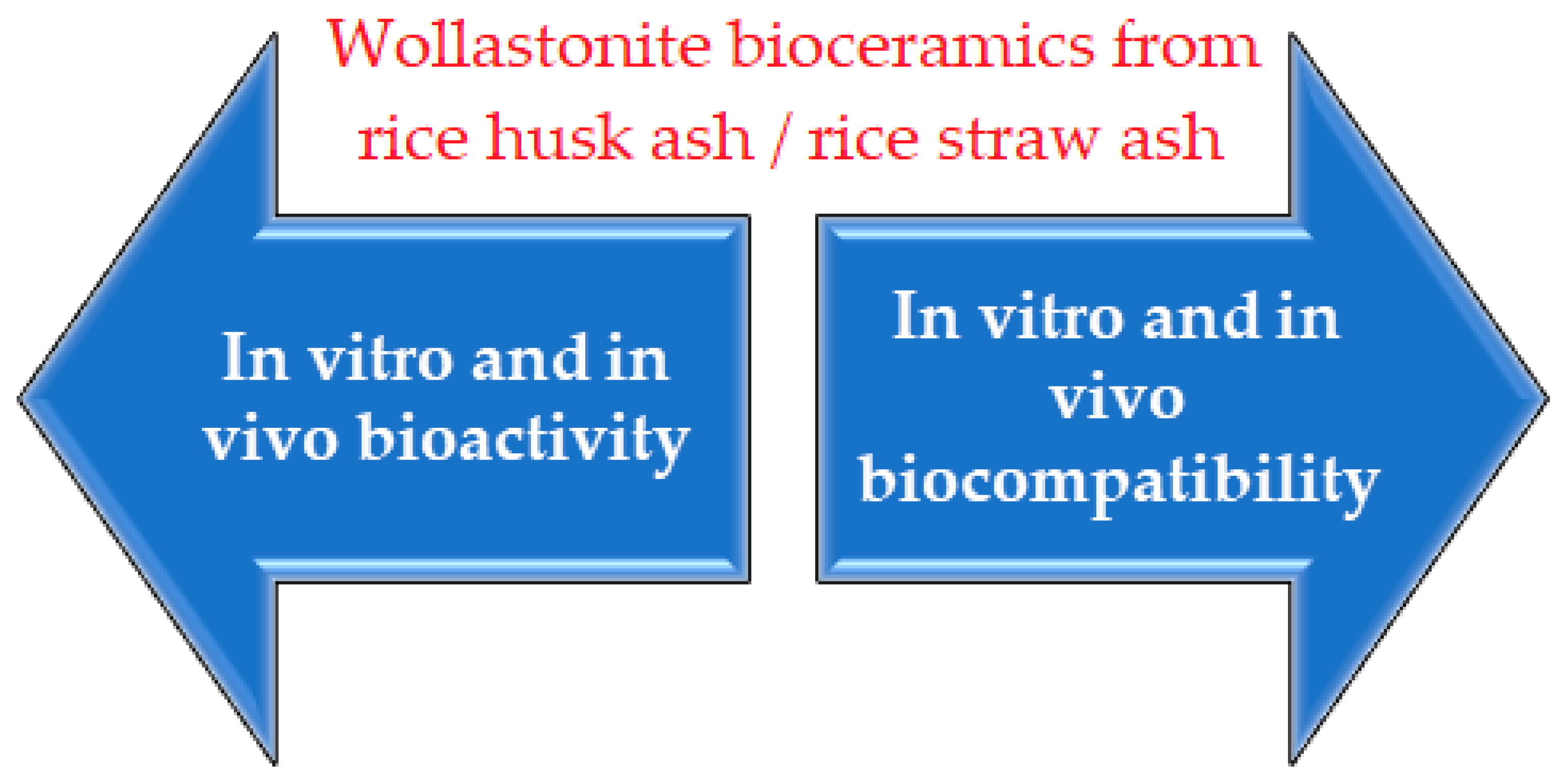
Bioactivity and Biocompatibility Properties of Sustainable Wollastonite Bioceramics from Rice Husk Ash/Rice Straw Ash: A Review


Abstract
:1. Introduction
2. Biomaterials
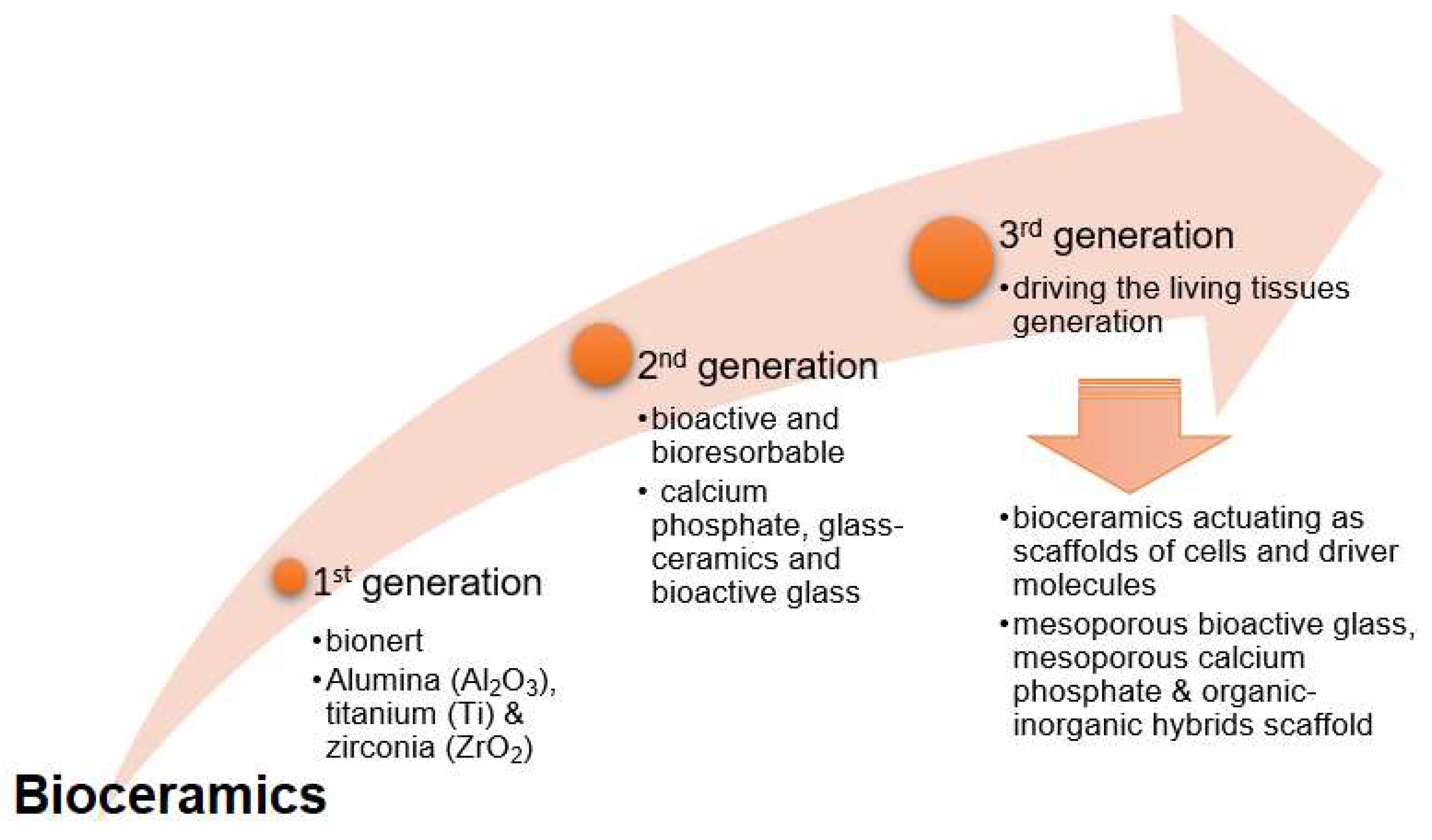
2.1. Bioceramics
- First generation: Inert bioceramics
- Second generation: Bioactive and bioresorbable bioceramics
- Third generation: Driving the living tissues generation
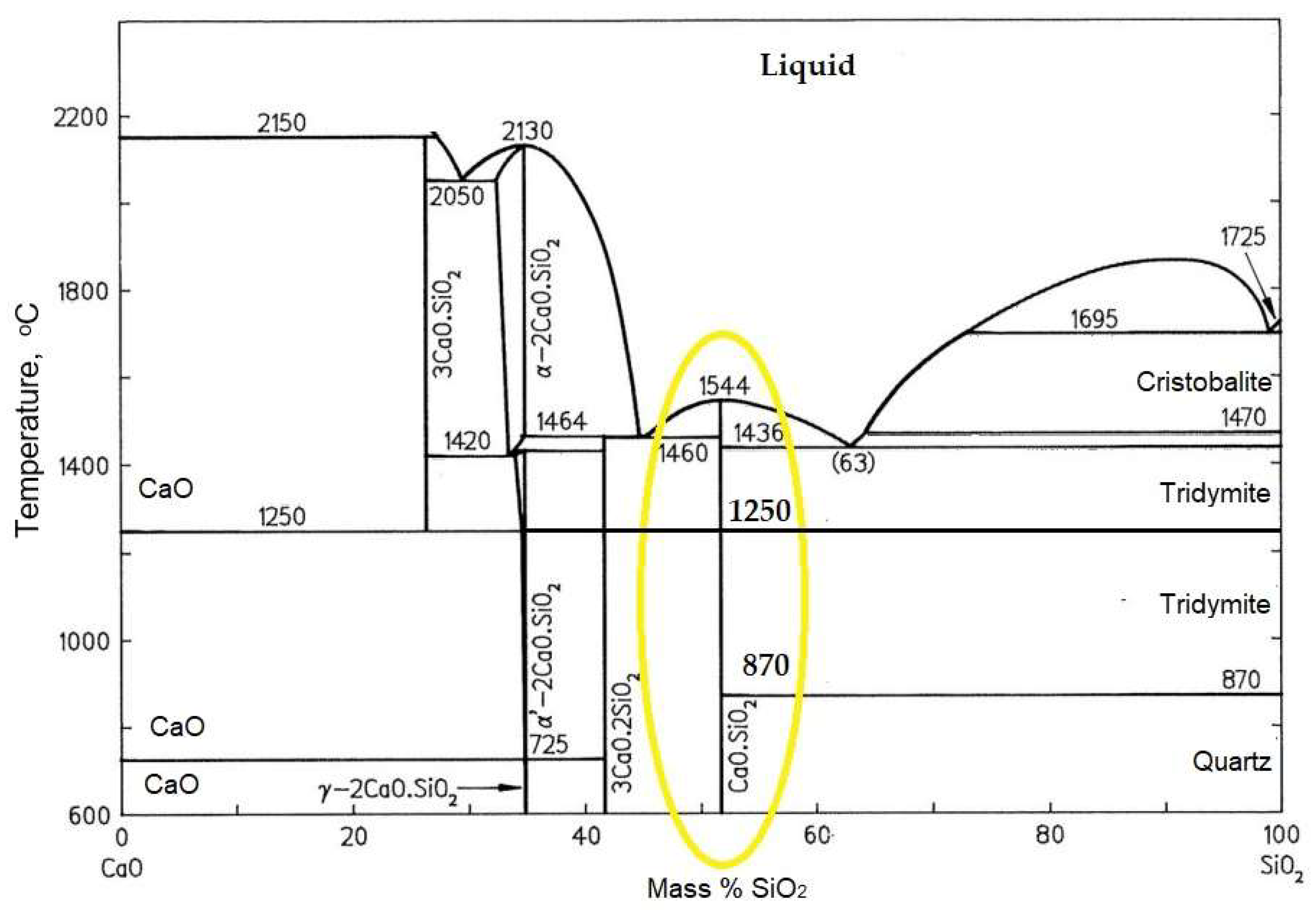
2.2. Wollastonite
2.2.1. Low-Temperature Wollastonite (β-CaSiO3)
2.2.2. High-Temperature Wollastonite or Pseudo-Wollastonite (α-CaSiO3)
2.2.3. Wollastonite in the Biomedical Field
3. Description of Agricultural Waste
3.1. Paddy
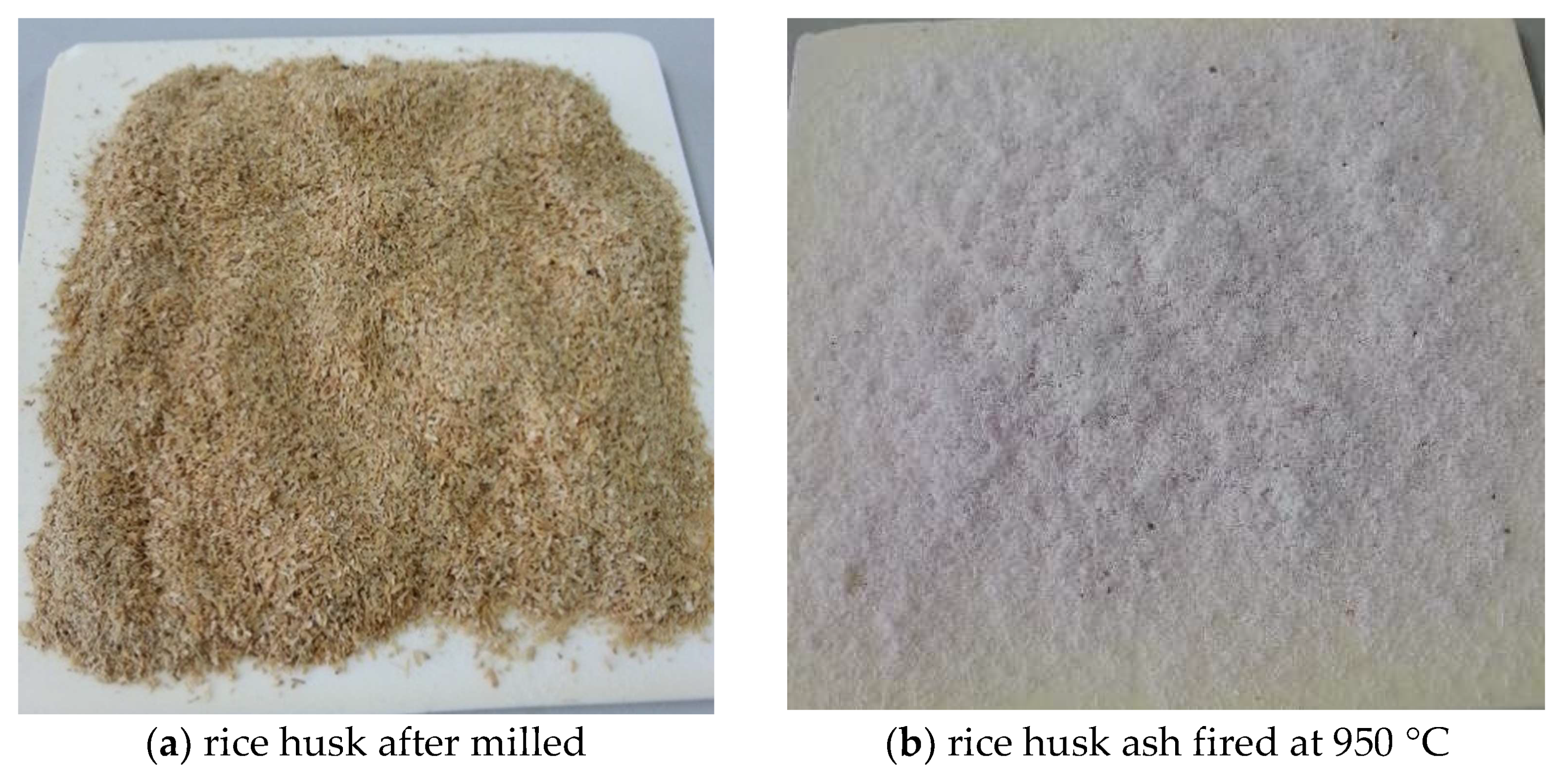
3.1.1. Rice Husk and Rice Husk Ash
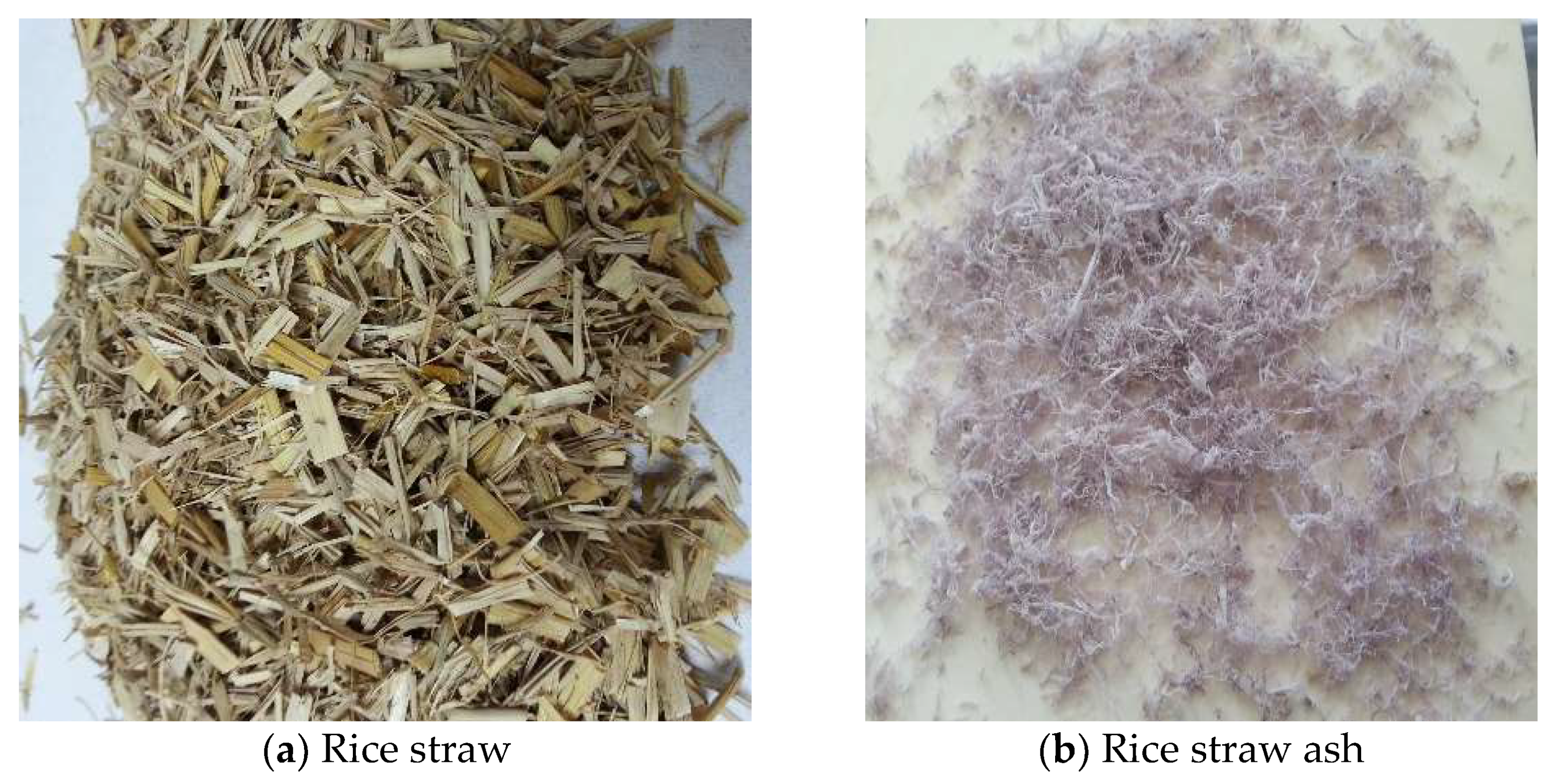
3.1.2. Rice Straw and Rice Straw Ash
4. Materials and Methods of Wollastonite Preparation
4.1. Materials
4.2. Method
4.2.1. Autoclaving
4.2.2. Solid-State Reaction
4.2.3. Melt-Quenching Technique
4.2.4. Sol–Gel
 where M is a metal atom, such as titanium, zirconia, aluminum, boron, etc.
4.2.5. Milling
4.3. Advantages and Limitations of the Processing Methods
5. Bioactivity and Biocompatibility Properties
5.1. Bioactivity Properties
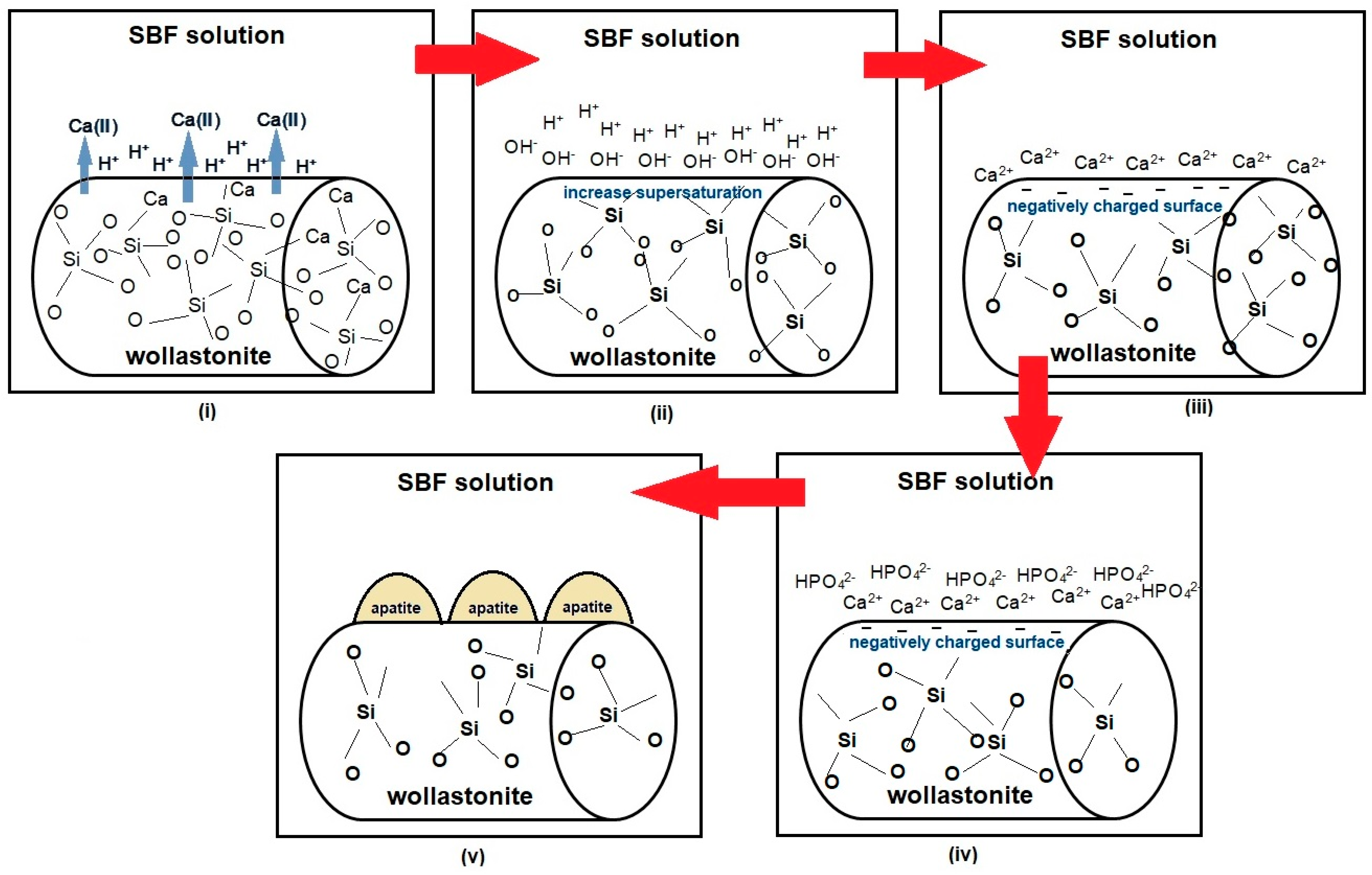
5.1.1. The Apatite Formation Mechanism for the CaO–SiO2 System In Vitro and In Vivo
- First step:
- Second step:
- Third step:
- Fourth step:
5.1.2. SBF
5.1.3. Bioactivity Studies
5.2. Biocompatibility Properties
5.2.1. In Vitro Biocompatibility
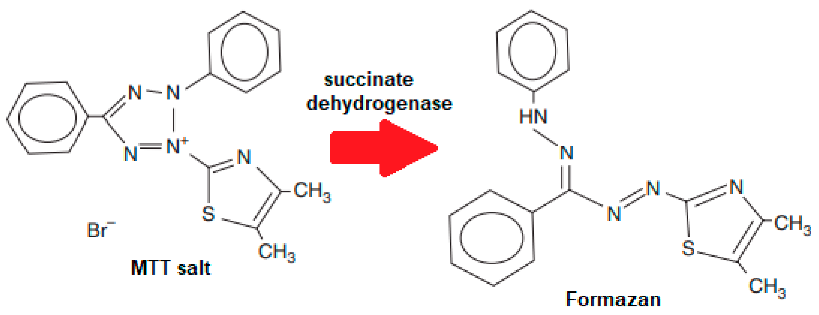
5.2.2. MTT Assay
5.2.3. In Vivo Biocompatibility
5.2.4. Biocompatibility Studies
6. General Overview and Future Perspectives
7. Conclusions
Author Contributions
Funding
Institutional Review Board Statement
Informed Consent Statement
Data Availability Statement
Conflicts of Interest
References
- Sadh, P.K.; Duhan, S.; Duhan, J.S. Agro-industrial wastes and their utilization using solid state fermentation: A review. Bioresour. Bioprocess. 2018, 5, 1. [Google Scholar] [CrossRef] [Green Version]
- Ayilara, M.S.; Olanrewaju, O.S.; Babalola, O.O.; Odeyemi, O. Waste Management through Composting: Challenges and Potentials. Sustainability 2020, 12, 4456. [Google Scholar] [CrossRef]
- Nevrlý, V.; Smejkalov, V. Strategic decisions leading to sustainable waste management: Separation, sorting and recycling possibilities. J. Clean. Prod. 2021, 278, 1–16. [Google Scholar]
- Agwa, I.S.; Omar, O.M.; Tayeh, B.A.; Abdelsalam, B.A. Effects of using rice straw and cotton stalk ashes on the properties of lightweight self-compacting concrete. Constr. Build. Mater. 2020, 235, 117541. [Google Scholar] [CrossRef]
- Zhang, W.; Lin, N.; Liu, D.; Xu, J.; Sha, J.; Yin, J.; Tan, X.; Yang, H.; Lu, H.; Lin, H. Direct carbonization of rice husk to prepare porous carbon for supercapacitor applications. Energy 2017, 128, 618–625. [Google Scholar] [CrossRef]
- Okahisa, Y.; Matsuoka, K.; Yamada, K.; Wataoka, I. Comparison of polyvinyl alcohol films reinforced with cellulose nano fibers derived from oil palm by impregnating and casting methods. Carbohydr. Polym. 2020, 250, 116907. [Google Scholar] [CrossRef] [PubMed]
- Chen, J.; Wang, X.; Zhang, B.; Yang, Y.; Song, Y.; Zhang, F.; Liu, B.; Zhou, Y.; Yi, Y.; Shan, Y.; et al. Integrating enzymatic hydrolysis into subcritical water pretreatment optimization for bio-ethanol production from wheat straw. Sci. Total Environ. 2021, 770, 145321. [Google Scholar] [CrossRef] [PubMed]
- Fang, Y.R.; Wu, Y.; Xie, G.H. Crop residue utilizations and potential for bioethanol production in China. Renew. Sustain. Energy Rev. 2019, 113, 109288. [Google Scholar] [CrossRef]
- Cantero-Tubilla, B.; Cantero, D.A.; Martinez, C.M.; Tester, J.W.; Walker, L.P.; Posmanik, R. Characterization of the solid products from hydrothermal liquefaction of waste feedstocks from food and agricultural industries. J. Supercrit. Fluids 2018, 133, 665–673. [Google Scholar] [CrossRef]
- Li, Q.; Ma, C.-L.; Zhang, P.-Q.; Li, Y.-Y.; Zhu, X.; He, Y.-C. Effective conversion of sugarcane bagasse to furfural by coconut shell activated carbon-based solid acid for enhancing whole-cell biosynthesis of furfurylamine. Ind. Crop. Prod. 2021, 160, 113169. [Google Scholar] [CrossRef]
- Guo, Y.; Tan, C.; Sun, J.; Li, W.; Zhang, J.; Zhao, C. Porous activated carbons derived from waste sugarcane bagasse for CO2 adsorption. Chem. Eng. J. 2020, 381, 122736. [Google Scholar] [CrossRef]
- Cha, L.; Torres-leo, C.; Martı, G.A. Recent advances on the microbiological and enzymatic processing for conversion of food wastes to valuable bioproducts. Curr. Opin. Food Sci. 2021, 38, 40–45. [Google Scholar]
- Basaglia, M.; Favaro, L.; Torri, C.; Casella, S. Is pyrolysis bio-oil prone to microbial conversion into added-value products? Renew. Energy 2021, 163, 783–791. [Google Scholar] [CrossRef]
- Dizaji, H.B.; Zeng, T.; Hartmann, I.; Enke, D.; Schliermann, T.; Lenz, V.; Bidabadi, M. Generation of High Quality Biogenic Silica by Combustion of Rice Husk and Rice Straw Combined with Pre- and Post-Treatment Strategies—A Review. Appl. Sci. 2019, 9, 1083. [Google Scholar] [CrossRef] [Green Version]
- Grimm, A.M.; Dorsch, L.Y.; Kloess, G.H.; Enke, D.; Roppertsz, A. Catalysing the combustion of rice husk and rice straw towards an energy optimized synthesis of metal modified biogenic silica. SSRN 2021, 1–31. [Google Scholar]
- Zareihassangheshlaghi, A.; Dizaji, H.B.; Zeng, T.; Huth, P.; Ruf, T.; Denecke, R.; Enke, D. Behavior of Metal Impurities on Surface and Bulk of Biogenic Silica from Rice Husk Combustion and the Impact on Ash-Melting Tendency. ACS Sustain. Chem. Eng. 2020, 8, 10369–10379. [Google Scholar] [CrossRef]
- Palakurthy, S.; Reddy, K.V.; Patel, S.; Azeem, P.A. A cost effective SiO2–CaO–Na2O bio-glass derived from bio-waste resources for biomedical applications. Prog. Biomater. 2020, 9, 239–248. [Google Scholar] [CrossRef] [PubMed]
- Srinath, P.; Azeem, P.A.; Reddy, K.V. Sol-gel synthesis of SiO2-CaO-Na2O bio-ceramics using- biowaste. AIP Conf. Proc. 2020, 2265, 030042. [Google Scholar]
- Alejandra, A.Q.S.; Diana, M.C.S. Synthesis and bioactivity evaluation of a Rice husk derived bioactive glass. Mater. Nanomed. Bioeng. 2019, 71, 302–307. [Google Scholar]
- Ismail, H.; Shamsudin, R.; Hamid, M.A.A.; Jalar, A. Synthesis and Characterization of Nano-Wollastonite from Rice Husk Ash and Limestone. Mater. Sci. Forum 2013, 756, 43–47. [Google Scholar] [CrossRef]
- Ismail, H.; Shamsudin, R.; Hamid, M.A.A. Characteristics of β-wollastonite derived from rice straw ash and limestone. J. Aust. Ceram. Soc. 2016, 52, 163–174. [Google Scholar]
- Choudhary, R.; Venkatraman, S.K.; Bulygina, I.; Senatov, F.; Kaloshkin, S.; Anisimova, N.; Kiselevskiy, M.; Knyazeva, M.; Kukui, D.; Walther, F.; et al. Biomineralization, dissolution and cellular studies of silicate bioceramics prepared from eggshell and rice husk. Mater. Sci. Eng. C 2021, 118, 111456. [Google Scholar] [CrossRef]
- Hossain, S.S.; Yadav, S.; Majumdar, S.; Krishnamurthy, S.; Pyare, R.; Roy, P. A comparative study of physico-mechanical, bioactivity and hemolysis properties of pseudo-wollastonite and wollastonite glass-ceramic synthesized from solid wastes. Ceram. Int. 2020, 46, 833–843. [Google Scholar] [CrossRef]
- Ridzwan, H.J.M.; Ismail, H.; Hamid, M.A.A.; Mohamad, H. A comparative study on physico-mechanical and bioactivity properties of β-wollastonite derived from rice husk ash and calcined limestone drying through freeze-dried and incubator technique. J. Aust. Ceram. Soc. 2021, 57, 755–766. [Google Scholar] [CrossRef]
- Punj, S.; Singh, K. Bioactive calcium silicate glass synthesized from sustainable biomass wastes. Biofuels Bioprod. Biorefin. 2020, 14, 1141–1151. [Google Scholar] [CrossRef]
- Pluta, K.; Florkiewicz, W.; Malina, D.; Rudnicka, K.; Michlewska, S.; Królczyk, J.; Sobczak-Kupiec, A. Measurement methods for the mechanical testing and biocompatibility assessment of polymer-ceramic connective tissue replacements. Measurement 2021, 171, 108733. [Google Scholar] [CrossRef]
- Teo, A.; Mishra, A.; Park, I.; Kim, Y.-J.; Park, W.-T.; Yoon, Y.-J. Polymeric Biomaterials for Medical Implants and Devices. ACS Biomater. Sci. Eng. 2016, 2, 454–472. [Google Scholar] [CrossRef]
- Saini, M.; Singh, Y.; Arora, P.; Arora, V.; Jain, K. Implant biomaterial: A comprehensive review. World J. Clin. Cases 2016, 3, 52–57. [Google Scholar] [CrossRef]
- Bose, S.; Bandyopadhyay, A. Introduction to Biomaterials; Elsevier BV: Amsterdam, The Netherlands, 2013; pp. 1–9. [Google Scholar]
- Ratner, B.D.; Hoffman, A.S.; Lemons, J.E.; Schoen, F.J. Biomaterials Science; Elsevier: Amsterdam, The Netherlands, 2004. [Google Scholar]
- Daculsi, G.; Bouler, J.-M.; LeGeros, R. Adaptive Crystal Formation in Normal and Pathological Calcifications in Synthetic Calcium Phosphate and Related Biomaterials. Adv. Virus Res. 1997, 172, 129–191. [Google Scholar] [CrossRef]
- Dorozhkin, S.V. Bioceramics of calcium orthophosphates. Biomaterials 2010, 31, 1465–1485. [Google Scholar] [CrossRef]
- Hench, L.L. An Introduction to Bioceramics; Imperial College Press: London, UK, 2013. [Google Scholar]
- Dorozhkin, S.V. Current State of Bioceramics Current State of Bioceramics. J. Ceram. Sci. Technol. 2018, 9, 353–370. [Google Scholar]
- Piconi, C.; Condo, S.G.; Kosmač, T. Advanced Ceramics for Dentistry; Elsevier: Amsterdam, The Netherlands, 2014. [Google Scholar]
- Nuevo-Ordóñez, Y.; Montes-Bayón, M.; Blanco-González, E.; Paz-Aparicio, J.; Raimundez, J.D.; Tejerina, J.M.; Peña, M.A.; Sanz-Medel, A. Titanium release in serum of patients with different bone fixation implants and its interaction with serum biomolecules at physiological levels. Anal. Bioanal. Chem. 2011, 401, 2747–2754. [Google Scholar] [CrossRef] [PubMed]
- Chevalier, J.; Gremillard, L. Zirconia as a Biomaterial. Compr. Biomater. 2011, 20, 95–108. [Google Scholar]
- Wu, C.; Ramaswamy, Y.; Kwik, D.; Zreiqat, H. The effect of strontium incorporation into CaSiO3 ceramics on their physical and biological properties. Biomaterials 2007, 28, 3171–3181. [Google Scholar] [CrossRef]
- Wang, C.; Lin, K.; Chang, J.; Sun, J. Osteogenesis and angiogenesis induced by porous β-CaSiO3/PDLGA composite scaffold via activation of AMPK/ERK1/2 and PI3K/Akt pathways. Biomaterials 2013, 34, 64–77. [Google Scholar] [CrossRef] [PubMed]
- Martinez, I.M.; Meseguer-Olmo, L.; Bernabeu-Esclapez, A.; Velasquez, P.A.; De Aza, P.N. In vitro behavior of α-tricalcium phosphate doped with dicalcium silicate in the system Ca2SiO4–Ca3(PO4)2. Mater. Charact. 2012, 63, 47–55. [Google Scholar] [CrossRef]
- Brannon-Peppas, L.; Ver, M. Polylactic and Polyglycolic Acids as Drug Delivery Carriers; Marcel Dekker: New York, NY, USA, 2000. [Google Scholar]
- Ren, D.; Feng, Q.; Bourrat, X. Effects of additives and templates on calcium carbonate mineralization in vitro. Micron 2011, 42, 228–245. [Google Scholar] [CrossRef] [Green Version]
- Sheikh, Z.; Najeeb, S.; Khurshid, Z.; Verma, V.; Rashid, H.; Glogauer, M. Biodegradable Materials for Bone Repair and Tissue Engineering Applications. Materials 2015, 8, 5744–5794. [Google Scholar] [CrossRef] [PubMed]
- Jeong, J.; Kim, J.H.; Shim, J.H.; Hwang, N.S.; Heo, C.Y. Bioactive calcium phosphate materials and applications in bone regeneration. Biomater. Res. 2019, 23, 1–11. [Google Scholar] [CrossRef] [Green Version]
- Kaya, S.; Cresswell, M.; Boccaccini, A.R. Mesoporous silica-based bioactive glasses for antibiotic-free antibacterial applications. Mater. Sci. Eng. C 2018, 83, 99–107. [Google Scholar] [CrossRef]
- Pourshahrestani, S.; Kadri, N.A.; Zeimaran, E.; Towler, M.R. Well-ordered mesoporous silica and bioactive glasses: Promise for improved hemostasis. Biomater. Sci. 2019, 7, 31–50. [Google Scholar] [CrossRef]
- Ramay, H.R.; Zhang, M. Biphasic calcium phosphate nanocomposite porous scaffolds for load-bearing bone tissue engineering. Biomater. 2004, 25, 5171–5180. [Google Scholar] [CrossRef] [PubMed]
- Sabudin, S.; Marzuke, M.A.; Hussin, Z. Effect of mechanical properties on porous calcium phosphate scaffold. Mater. Today Proc. 2019, 16, 1680–1685. [Google Scholar] [CrossRef]
- Abdraboh, A.S.; Abdel-Aal, A.A.; Ereiba, K.T. Preparation and Characterization of Inorganic Organic Hybrid Material Based on TEOS/MAPTMS for Biomedical Applications. Silicon 2020, 13, 613–622. [Google Scholar] [CrossRef]
- Cerqueira, A.; Romero-Gavilán, F.; García-Arnáez, I.; Martinez-Ramos, C.; Ozturan, S.; Iloro, I.; Azkargortae, M.; Elortzae, F.; Izquierdoa, R.; Gurruchagab, M.; et al. Bioactive zinc-doped sol-gel coating modulates protein adsorption patterns and in vitro cell responses. Mater. Sci. Eng. C 2021, 122, 111839. [Google Scholar] [CrossRef]
- Tegethoff, F.W. Calcium Carbonate: From the Cretaceous Period into the 21st Century; Springer: Basel, Switzerland, 2001. [Google Scholar]
- Mallick, K. Bone Substitute Biomaterials; Elsevier & Woodhead Publishing Limited: Cambridge, UK, 2014. [Google Scholar]
- Schmetterer, C.; Masset, P.J. Heat capacity of ccompounds in the CaO-SiO2 system—A Review. J. Phase Equilibr. Diffus. 2012, 33, 261–275. [Google Scholar] [CrossRef]
- Seryotkin, Y.; Sokol, E.V.; Kokh, S.N. Natural pseudowollastonite: Crystal structure, associated minerals, and geological context. Lithos 2012, 134–135, 75–90. [Google Scholar] [CrossRef]
- Höland, W.; Beall, G.H. Glass-Ceramic Technology; Wiley: Hoboken, NJ, USA, 2012. [Google Scholar]
- Pelan Pembangunan Pengurusan Sisa Makanan Bagi Sektor. 2016. Available online: https://jpspn.kpkt.gov.my/ (accessed on 4 January 2021).
- Ma’Ruf, A.; Pramudono, B.; Aryanti, N. Lignin isolation process from rice husk by alkaline hydrogen peroxide: Lignin and silica extracted. AIP Conf. Proc. 2017, 1823, 020013. [Google Scholar]
- Wang, S.; Zou, C.; Yang, H.; Lou, C.; Cheng, S.; Peng, C.; Wang, C.; Zou, H. Effects of cellulose, hemicellulose, and lignin on the combustion behaviours of biomass under various oxygen concentrations. Bioresour. Technol. 2021, 320, 124375. [Google Scholar] [CrossRef]
- Qu, T.; Guo, W.; Shen, L.; Xiao, J.; Zhao, K. Experimental Study of Biomass Pyrolysis Based on Three Major Components: Hemicellulose, Cellulose, and Lignin. Ind. Eng. Chem. Res. 2011, 50, 10424–10433. [Google Scholar] [CrossRef]
- Yuliansyah, A.T.; Hirajima, T.; Kumagai, S.; Sasaki, K. Production of Solid Biofuel from Agricultural Wastes of the Palm Oil Industry by Hydrothermal Treatment. Waste Biomass Valoriz. 2010, 1, 395–405. [Google Scholar] [CrossRef]
- Hong, L.S.; Ibrahim, D.; Omar, I.C. Oil palm frond for the production of bioethanol oil palm frond for the production of bioethanol. Int. J. Biochem. Biotechnol. 2012, 1, 7–11. [Google Scholar]
- El-Tayeb, T.S.; Abdelhafez, A.A.; Ali, S.H.; Ramadan, E.M. Effect of acid hydrolysis and fungal biotreatment on agro-industrial wastes for obtainment of free sugars for bioethanol production. Braz. J. Microbiol. 2012, 43, 1523–1535. [Google Scholar] [CrossRef] [PubMed] [Green Version]
- Bjerre, A.B.; Olesen, A.B.; Fernqvist, T.; Plöger, A.; Schmidt, A.S. Pretreatment of wheat straw using combined wet oxidation and alkaline hydrolysis resulting in convertible cellulose and hemicellulose. Biotechnol. Bioeng. 1996, 49, 568–577. [Google Scholar] [CrossRef]
- Food and Agriculture Data. Available online: http://www.fao.org/faostat/en (accessed on 6 April 2021).
- Juliano, B.O. Rice: Overview, 2nd ed.; Elsevier Ltd.: Amsterdam, The Netherlands, 2015. [Google Scholar]
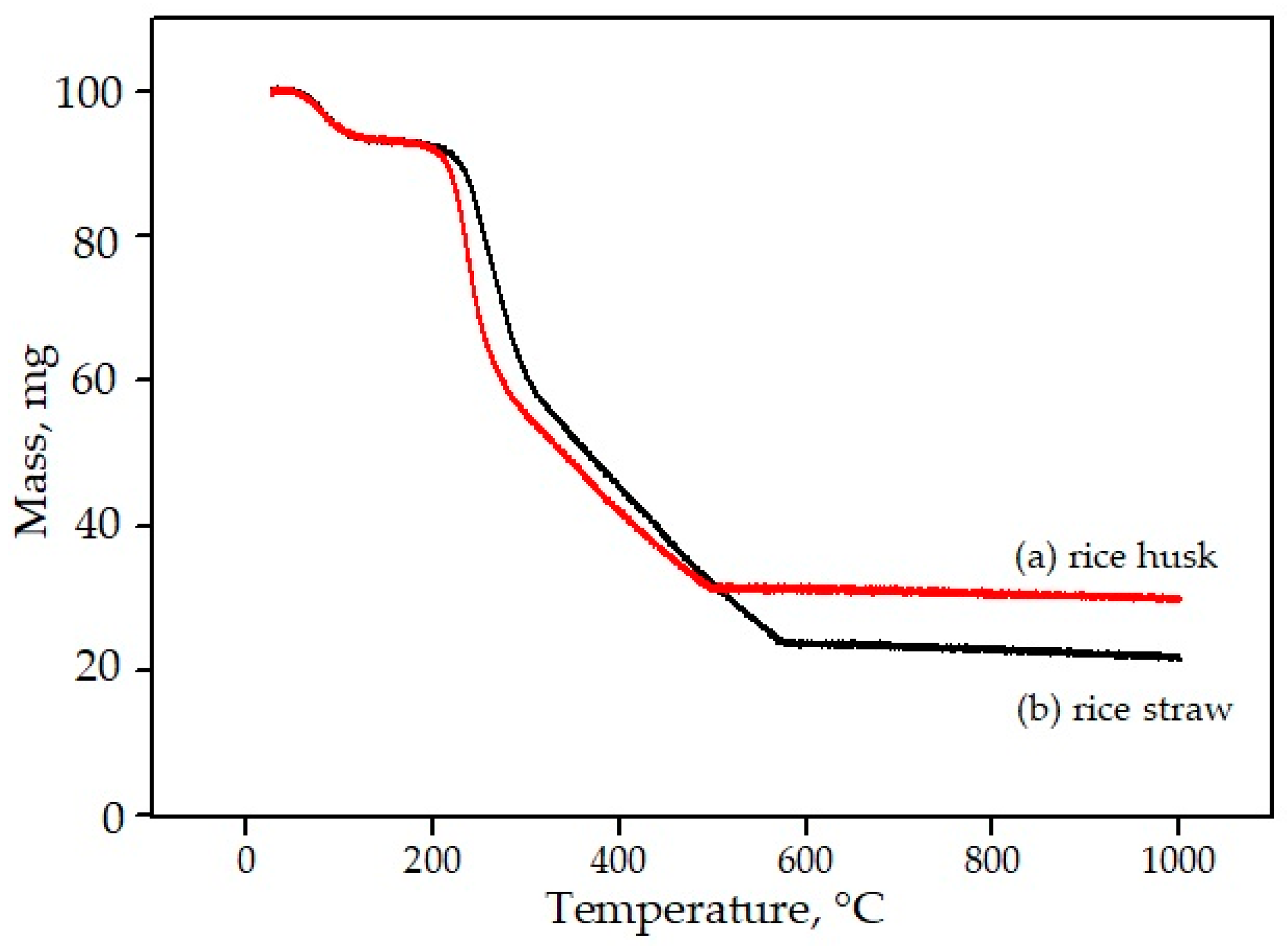
- Worasuwannarak, N.; Sonobe, T.; Tanthapanichakoon, W. Pyrolysis behaviors of rice straw, rice husk, and corncob by TG-MS technique. J. Anal. Appl. Pyrolysis 2007, 78, 265–271. [Google Scholar] [CrossRef]
- Ugheoke, B.I.; Mamat, O. A critical assessment and new research directions of rice husk silica processing methods and properties. Maejo Int. J. Sci. Technol. 2012, 6, 430–448. [Google Scholar]
- Alam, M.M.; Hossain, M.A.; Hossain, M.D.; Johir, M.A.H.; Hossen, J.; Rahman, M.S.; Zhou, J.L.; Hasan, A.T.M.K.; Karmakar, A.K.; Ahmed, M.B.; et al. The potentiality of rice husk-derived activated carbon: From synthesis to application. Processes 2020, 8, 203. [Google Scholar] [CrossRef] [Green Version]
- Teh, J.; Teoh, Y.; How, H.; Le, T.; Jason, Y.; Nguyen, H.; Loo, D. The Potential of Sustainable Biomass Producer Gas as a Waste-to-Energy Alternative in Malaysia. Sustainability 2021, 13, 3877. [Google Scholar] [CrossRef]
- Palakurthy, S.; Azeem, P.A.; Reddy, K.V.; Penugurti, V.; Manavathi, B. A comparative study on in vitro behavior of calcium silicate ceramics synthesized from biowaste resources. J. Am. Ceram. Soc. 2020, 103, 933–943. [Google Scholar] [CrossRef]
- Ismail, H.; Fadzil, N.F.M.; Shamsudin, R.; Hamid, M.A.A. Kesan Penambahan Magnesia dan Zirkonia terhadap Sifat Kebioaktifan dan Kekuatan Mampatan β-Wolastonit yang Disintesis daripada Kulit Telur. Sains Malays. 2019, 48, 1519–1527. [Google Scholar] [CrossRef]
- Ismail, H.; Shun, X.Y.; Shamsudin, R.; Hamid, M.A.A. Pengaruh Teknik Pengeringan yang Berbeza terhadap Ketumpatan, Keliangan dan Kebioaktifan β-Wolastonit daripada Batu Kapur Tempatan dan Jerami Padi. Sains Malays. 2019, 48, 165–172. [Google Scholar] [CrossRef]
- Palakurthy, S.; Samudrala, R.K. In vitro bioactivity and degradation behaviour of β-wollastonite derived from natural waste. Mater. Sci. Eng. C 2019, 98, 109–117. [Google Scholar] [CrossRef]
- Palakurthy, S.; Abdul, A.P.; Venugopal, R.K. In vitro evaluation of silver doped wollastonite synthesized from natural waste for biomedical applications. Ceram. Int. 2019, 45, 25044–25051. [Google Scholar] [CrossRef]
- Sultana, S.; Rahman, M.M.; Yeasmin, Z.; Ahmed, S.; Rony, F.K. Effect of the ratio of eggshell and rice husk as starting materials on the direct synthesis of bioactive wollastonite by solid state thermal method. J. Ceram. Process. Res. 2020, 21, 285–295. [Google Scholar]
- Azam, F.A.A.; Shamsudin, R.; Ng, M.H.; Ahmad, A.; Akbar, M.A.; Rashidbenam, Z. Silver-doped pseudowollastonite synthesized from rice husk ash: Antimicrobial evalution, bioactivity and cytotoxic effects on human mesenchymal stem cells. Ceram. Int. 2018, 44, 11381–11389. [Google Scholar]
- Shamsudin, R.; Azam, F.; Atiqah, A.; Hamid, M.A.A.; Ismail, H. Bioactivity and Cell Compatibility of β-Wollastonite Derived from Rice Husk Ash and Limestone. Materials 2017, 10, 1188. [Google Scholar] [CrossRef] [Green Version]
- Saravanan, S.; Vimalraj, S.; Vairamani, M.; Selvamurugan, N. Role of Mesoporous Wollastonite (Calcium Silicate) in Mesenchymal Stem Cell Proliferation and Osteoblast Differentiation: A Cellular and Molecular Study. J. Biomed. Nanotechnol. 2015, 11, 1124–1138. [Google Scholar] [CrossRef] [PubMed]
- Ismail, H.; Shamsudin, R.; Hamid, M.A.A.; Awang, R. Mekanisme Pembentukan Apatit pada Permukaan Sampel Β-Wolastonit yang Dihasilkan daripada Abu Sekam Padi. Sains Malays. 2016, 45, 1779–1785. [Google Scholar] [CrossRef]
- Alshatwi, A.A.; Athinarayanan, J.; Periasamy, V.S. Biocompatibility assessment of rice husk-derived biogenic silica nanoparticles for biomedical applications. Mater. Sci. Eng. C 2015, 47, 8–16. [Google Scholar] [CrossRef] [PubMed]
- Phuttawong, R.; Chantaramee, N.; Pookmanee, P.; Puntharod, R. Synthesis and Characterization of Calcium Silicate from Rice Husk Ash and Shell of Snail Pomacea canaliculata by Solid State Reaction. Adv. Mater. Res. 2015, 1103, 1–7. [Google Scholar] [CrossRef]
- Athinarayanan, J.; Periasamy, V.S.; Alhazmi, M.; Alatiah, K.A.; Alshatwi, A. Synthesis of biogenic silica nanoparticles from rice husks for biomedical applications. Ceram. Int. 2015, 41, 275–281. [Google Scholar] [CrossRef]
- Azeena, S.; Subhapradha, N.; Selvamurugan, N.; Narayanasamy, S.; Srinivasan, N.; Murugesan, R.; Chung, T.W.; Moorthi, A. Antibacterial activity of agricultural waste derived wollastonite doped with copper for bone tissue engineering. Mater. Sci. Eng. C 2017, 71, 1156–1165. [Google Scholar] [CrossRef] [PubMed]
- NaghiZadeh, F.; Sultana, N.; Kadir, M.R.A.; Shihabudin, T.M.T.M.; Hussain, R.; Kamarul, T. The Fabrication and Characterization of PCL/Rice Husk Derived Bioactive Glass-Ceramic Composite Scaffolds. J. Nanomater. 2014, 2014, 1–9. [Google Scholar] [CrossRef] [Green Version]
- Nayak, J.; Kumar, S.; Bera, J. Sol–gel synthesis of bioglass-ceramics using rice husk ash as a source for silica and its characterization. J. Non-Cryst. Solids 2010, 356, 1447–1451. [Google Scholar] [CrossRef]
- Nayak, J.; Bera, J. Effect of sintering temperature on mechanical behaviour and bioactivity of sol–gel synthesized bioglass-ceramics using rice husk ash as a silica source. Appl. Surf. Sci. 2010, 257, 458–462. [Google Scholar] [CrossRef]
- Byrappa, K.; Yoshimura, M. Handbook of Hydrothermal Technology; Elsevier BV: Amsterdam, The Netherlands, 2013. [Google Scholar]
- Ismail, H.; Yau, S.X.; Shamsudin, R.; Hamid, M.A.; Awang, R. Morphology and phase identification of bioactive freeze-dried β-CaSiO3. Key Eng. Mater. 2016, 694, 72–77. [Google Scholar] [CrossRef]
- Vallet-Regi, M. Bioceramics with Clinical Applications; Wiley: Hoboken, NJ, USA, 2014. [Google Scholar]
- Ramalingam, M.; Tiwari, A.; Ramakrishna, S.; Kobayashi, H. (Eds.) Integrated Biomaterials for Biomédical Technology; Wiley: Hoboken, NJ, USA, 2012. [Google Scholar]
- Nizami, M.; Iqbal, M.Z. Chemical kinetic aspects of solid state reaction producing wollastonite from rice husk silica and limestone. J. Mater. Sci. Technol. 2001, 17, 243–246. [Google Scholar]
- Nour, W.; Mostafa, A.; Ibrahim, D. Recycled wastes as precursor for synthesizing wollastonite. Ceram. Int. 2008, 34, 101–105. [Google Scholar] [CrossRef]
- Rashid, R.A.; Shamsudin, R.; Hamid, M.A.A.; Jalar, A. Low temperature production of wollastonite from limestone and silica sand through solid-state reaction. J. Asian Ceram. Soc. 2014, 2, 77–81. [Google Scholar] [CrossRef] [Green Version]
- Rashid, R.A.; Shamsudin, R.; Hamid, M.A.A.; Jalar, A. In-vitro bioactivity of wollastonite materials derived from limestone and silica sand. Ceram. Int. 2014, 40, 6847–6853. [Google Scholar] [CrossRef]
- Shukur, M.M.; Al-majeed, E.A.; Obied, M.M. Characteristic of wollastonite synthesized from local raw materials. Int. J. Eng. Technol. 2014, 4, 426–429. [Google Scholar]
- Kaur, G.; Pickrell, G.; Sriranganathan, N.; Kumar, V.; Homa, D. Review and the state of the art: Sol-gel and melt quenched bioactive glasses for tissue engineering. J. Biomed. Mater. Res. Part B Appl. Biomater. 2016, 104, 1248–1275. [Google Scholar] [CrossRef] [PubMed]
- Ibrahim, N.F.; Mohamad, H.; Noor, S.N.F.M.; Ahmad, N. Apatite formation on melt-derived bioactive glass powder based on SiO2-CaO-Na2O-P2O5 system. Ceram. Int. 2017, 43, 11676–11685. [Google Scholar] [CrossRef]
- Swe, T.T.; Shariff, K.A.; Mohamad, H.; Ishikawa, K.; Hayashi, K.; Abu Bakar, M.H. Behavioural response of cells and bacteria on single and multiple doped Sr and Ag S53P4 sol-gel bioglass. Ceram. Int. 2020, 46, 17881–17890. [Google Scholar] [CrossRef]
- Ibrahim, N.F.; Mohamad, H.; Noor, S.N.F.M. Characterization on melt-derived bioactive glass powder from SiO2-CaO-Na2O-P2O5 system. J. Non-Cryst. Solids 2017, 462, 23–31. [Google Scholar] [CrossRef]
- Hayashi, T.; Saito, H. Preparation of CaO-SiO2 glasses by the gel method. J. Mater. Sci. 1980, 15, 1971–1977. [Google Scholar] [CrossRef]
- Casa-Lillo, M.A.D.L.; Velásquez, P.; De Aza, P.N. Influence of thermal treatment on the “in vitro” bioactivity of wollastonite materials. J. Mater. Sci. Mater. Med. 2011, 22, 907–915. [Google Scholar] [CrossRef]
- Meiszterics, A.; Sinkó, K. Sol–gel derived calcium silicate ceramics. Colloids Surf. A Physicochem. Eng. Asp. 2008, 319, 143–148. [Google Scholar] [CrossRef]
- Saravanapavan, P.; Hench, L.L. Mesoporous calcium silicate glasses. I. Synthesis. J. Non-Cryst. Solids 2003, 318, 1–13. [Google Scholar] [CrossRef]
- Suryanarayana, C. Mechanical alloying and milling. Prog. Mater. Sci. 2001, 46, 1–184. [Google Scholar] [CrossRef]
- Ismail, H.; Shamsudin, R.; Hamid, M.A.A. Effect of autoclaving and sintering on the formation of β-wollastonite. Mater. Sci. Eng. C 2016, 58, 1077–1081. [Google Scholar] [CrossRef] [PubMed]
- Jamal, R.; Xu, F.; Shao, W.; Abdiryim, T. The study on the application of solid-state method for synthesizing the polyaniline/noble metal (Au or Pt) hybrid materials. Nanoscale Res. Lett. 2013, 8, 117. [Google Scholar] [CrossRef] [PubMed] [Green Version]
- Diba, M.; Boccaccini, A. Silver-Containing Bioactive Glasses for Tissue Engineering Applications; Woodhead Publishing Limited: Cambridge, UK, 2014. [Google Scholar]
- Predoi, D.; Iconaru, S.L.; Predoi, M.V.; Buton, N.; Motelica-Heino, M. Zinc Doped Hydroxyapatite Thin Films Prepared by Sol–Gel Spin Coating Procedure. Coatings 2019, 9, 156. [Google Scholar] [CrossRef] [Green Version]
- Du, Z.; Leng, H.; Guo, L.; Huang, Y.; Zheng, T.; Zhao, Z.; Liu, X.; Zhang, X.; Cai, Q.; Yang, X. Calcium silicate scaffolds promoting bone regeneration via the doping of Mg2+ or Mn2+ ion. Compos. Part B Eng. 2020, 190, 107937. [Google Scholar] [CrossRef]
- Ben-nissan, B. Advances in Calcium Phosphate Biomaterials; Springer: Berlin/Heidelberg, Germany, 2014. [Google Scholar]
- Eighmy, T.T.; Kinner, A.E.; Shaw, E.L.; Eusden, J.D.; Francis, C.A. Hydroxylapatite (Ca5(PO4)3OH) Characterization by XPS: An Environmentally Important Secondary Mineral. Surf. Sci. Spectra 1999, 6, 193–201. [Google Scholar] [CrossRef]
- Ptáček, P. Introduction to Apatites. In Apatites and their Synthetic Analogues—Synthesis, Structure, Properties and Applications; IntechOpen Limited: London, UK, 2016. [Google Scholar]
- Best, S.M.; Porter, A.E.; Thian, E.S.; Huang, J. Bioceramics: Past, present and for the future. J. Eur. Ceram. Soc. 2008, 28, 1319–1327. [Google Scholar] [CrossRef]
- Al-Khafaji, T.J.; Wong, F.; Fleming, P.S.; Karpukhina, N.; Hill, R. Novel fluoride and strontium-containing bioactive glasses for dental varnishes-design and bioactivity in Tris buffer solution. J. Non-Cryst. Solids 2019, 503–504, 120–130. [Google Scholar] [CrossRef]
- Rohanová, D.; Boccaccini, A.R.; Yunos, D.M.; Horkavcová, D.; Březovská, I.; Helebrant, A. TRIS buffer in simulated body fluid distorts the assessment of glass—Ceramic scaffold bioactivity. Acta Biomater. 2011, 7, 2623–2630. [Google Scholar] [CrossRef]
- Kokubo, T.; Takadama, H. How useful is SBF in predicting in vivo bone bioactivity? Biomaterials 2006, 27, 2907–2915. [Google Scholar] [CrossRef]
- Anderson, J.M. Biocompatibility and the Relationship to Standards: Meaning and Scope of Biomaterials Testing; Elsevier Ltd.: Amsterdam, The Netherlands, 2017. [Google Scholar]
- Williams, D. Concepts in Biocompatibility: New Biomaterials, New Paradigms and New Testing Regimes; Woodhead Publishing Limited: Cambridge, UK, 2012. [Google Scholar]
- Inayat-Hussain, S.; Rajab, N.F.; Siew, E.L. In Vitro Testing of Biomaterials Toxicity and Biocompatibility; Woodhead Publishing Limited: Cambridge, UK, 2008. [Google Scholar]
- Johnson, H.J.; Northup, S.J.; Seagraves, P.A.; Atallah, M.; Garvin, P.J.; Lin, L.; Darby, T.D. Biocompatibility test procedures for materials evaluation in vitro. II. Objective methods of toxicity assessment. J. Biomed. Mater. Res. 1985, 19, 489–508. [Google Scholar] [CrossRef]
- Mosmann, T. Rapid colorimetric assay for cellular growth and survival: Application to proliferation and cytotoxicity assays. J. Immunol. Methods 1983, 65, 55–63. [Google Scholar] [CrossRef]
- Lin, W.; Huang, Y.-W.; Zhou, X.-D.; Ma, Y. In vitro toxicity of silica nanoparticles in human lung cancer cells. Toxicol. Appl. Pharmacol. 2006, 217, 252–259. [Google Scholar] [CrossRef] [PubMed]


| Composition, wt.% | Bone | Hydroxyapatite | Enamel | Dentin |
|---|---|---|---|---|
| Calcium, Ca | 34.8 | 39.6 | 36.5 | 35.1 |
| Phosphorus, P | 15.2 | 18.5 | 17.7 | 16.9 |
| Ca/P molar ratio | 1.71 | 1.67 | 1.63 | 1.61 |
| Sodium, Na | 0.9 | - | 0.5 | 0.6 |
| Magnesium, Mg | 0.72 | - | 0.44 | 1.23 |
| Potassium, K | 0.03 | - | 0.08 | 0.05 |
| Carbonate, CO32− | 7.4 | - | 3.5 | 5.6 |
| Fluoride, F | 0.03 | - | 0.01 | 0.06 |
| Chloride, Cl | 0.13 | - | 0.30 | 0.10 |
| Pyrophosphate, P2O74− | 0.07 | 0.022 | 0.10 | |
| Total inorganic | 65 | 100 | 97 | 70 |
| Total organic | 25 | - | 1.5 | 20 |
| Water | 10 | - | 1.5 | 10 |
| Ignition products (800 °C) | HA + CaO | HA | Β-TCP + HA | Β-TCP + HA |
| Elastic modulus (GPa) | 0.34–13.8 | 10 | 80 | 15 |
| Tensile strength (MPa) | 150 | 100 | 10 | 100 |
| Description | Value |
|---|---|
| Color | White |
| Luster | Vitreous, Pearly |
| Molecular weight, gmol−1 | 116 |
| Specific gravity, gcm−3 | 2.86–3.09 |
| Refractive index | 1.63 |
| pH (10% slurry) | 9.9 |
| Solubility in water (g/100 cc) | 0.0095 |
| Density (g/cm3) | 2.70–3.00 |
| Hardness (Mohs) | 4.5–5 |
| Melting point (°C) | 1540 |
| Agricultural Waste | Chemical Composition (% w/w) | Ash (%) | References | ||
|---|---|---|---|---|---|
| Cellulose | Hemicellulose | Lignin | |||
| Rice husk | 32.7 | 31.7 | 18.8 | 16.3 | [14,57] |
| Rice straw | 41.9 | 25.6 | 0.8 | 16.5 | [58,59] |
| Palm oil trunk | 39.9 | 21.2 | 22.6 | 1.9 | [60] |
| Palm oil frond | 31.5 | 19.2 | 14.0 | 12.3 | [61] |
| Sugarcane bagasse | 30.2 | 56.7 | 13.4 | 1.9 | [62] |
| Corn stalks | 42.7 | 23.3 | 17.5 | 9.8 | [59] |
| Wheat straw | 32.8 | 38.0 | 8.9 | 1.4 | [63] |
| Soy stalks | 34.5 | 24.8 | 19.8 | 10.4 | [62] |
| Parameters | Husk | Straw |
|---|---|---|
| Origin | Rice milling | After harvesting |
| Quantity | 20–22% from rice weight | 2–8 ton/ha |
| Moisture content, (%) | 10 | 60 (wet weight) 10–12 (dry condition) |
| Density, (kgm−3) | 100–150 200–250 (on land) | 75 (loose straw) 100–180 (compact form) |
| Carbohydrate components, (%) (average) | Cellulose: 28–36 Hemicellulose: 12 Lignin 9–20 Gross protein: 1.9–3.0 | Cellulose: 24–34 Hemicellulose: 19–29 Lignin: 5–11 Gross protein:2.8–4.4 |
| Caloric content, (MJkg−1) | 14–16 (10% moisture content) | 14–16 (14% moisture content) |
| Ash, (%) | 13.2–21.0 | 10.4–21.8 |
| Silica, (mgg−1) | 18.8–22.3 | 11–15 |
| Calcium, (mgg−1) | 0.6–1.3 | 0.9–5.0 |
| Phosphorus, (mgg−1) | 0.3–0.7 | 0.61–0.65 |
| Raw Material | Method | References |
|---|---|---|
| Rice husk ash and limestone | Autoclaving | Ridzwan et al. [24] |
| Eggshell and rice husk ash | Solid-state reaction | Choudhary et al. [22] |
| Rice husk ash and eggshells | Solid-state reaction | Hossain et al. [23] |
| corn, sugarcane and eggshells | melt quench technique | Shivani and Kunjir [25] |
| Rice husk ash and eggshell | Sol–gel | Palakurthy et al. [70] |
| Rice husk ash and eggshell | Autoclaving | Ismail et al. [71] |
| Rice straw ash and limestone | Autoclaving | Ismail et al. [72] |
| Rice husk ash and eggshell | Sol–gel | Palakurty et al. [73] |
| Rice husk ash and eggshell | Sol–gel | Palakurthy et al. [74] |
| Rice husk ash and eggshells | Solid state | Sultana et al. [75] |
| Rice husk ash and limestone | Autoclaving | Farah ‘Atiqah et al. [76] |
| Rice husk ash and limestone | Autoclaving | Roslinda et al. [77] |
| Rice straw ash | Sol–gel | Saravanan et al. [78] |
| Rice husk ash and limestone | Autoclaving | Ismail et al. [79] |
| Rice husk | Autoclaving | Alshatwi et al. [80] |
| Rice husk ash and shell of a snail | Milling | Phuttawong et al. [81] |
| Rice husk | Autoclaving | Athinarayanan et al. [82] |
| Rice straw ash | Sol–gel | Azeena et al. [83] |
| Rice husk ash/PCL | Sol–gel | Naghizadeh et al. [84] |
| Rice husk ash | Sol–gel | Nayak et al. [85] |
| Rice husk ash | Sol–gel | Nayak et al. [86] |
| Methods | Advantages | Limitations | Ref. |
|---|---|---|---|
| Autoclaving | Very tough, able to bear high pressures and temperatures for long-term processing. | - | [105] |
| Solid-state | Easy control of operating conditions | The formation of toxic waste products; not suitable for mass production | [106] |
| Melt-quenching |
|
| [107] |
| Sol–gel |
|
| [90,108,109] |
| Milling | Non-mixed nanomaterial systems can be produced. | Contamination from milling jar | [90,104] |
| Ref | Bioactivity | Biocompatibility |
|---|---|---|
| Ridzwan et al. [24] | + | − |
| Choudhary et al. [22] | + | + |
| Hossain et al. [23] | + | + |
| Shivani and Kunjir [25] | + | − |
| Palakurthy et al. [70] | + | − |
| Ismail et al. [71] | + | − |
| Ismail et al. [72] | + | − |
| Palakurty et al. [73] | + | + |
| Palakurthy et al. [74] | + | + |
| Sultana et al. [75] | + | − |
| Farah ‘Atiqah et al. [76] | + | + |
| Roslinda et al. [77] | + | + |
| Saravanan et al. [78] | − | + |
| Ismail et al. [79] | + | − |
| Alshatwi et al. [80] | − | + |
| Phuttawong et al. [81] | − | − |
| Athinarayanan et al. [82] | − | + |
| Azeena et al. [83] | − | + |
| Naghizadeh et al. [84] | + | − |
| Nayak et al. [85] | + | − |
| Nayak et al. [86] | + | − |
| Ion Concentration | SBF (mM) | Human Blood Plasma (mM) |
|---|---|---|
| Na+ | 142.0 | 142.0 |
| K+ | 5.0 | 5.0 |
| Mg2+ | 1.5 | 1.5 |
| Ca2+ | 2.6 | 2.5 |
| Cl− | 148.8 | 103.0 |
| HPO42− | 1.0 | 1.0 |
| SO42− | 0.0 | 0.5 |
| pH | 7.4 | 7.2–7.4 |
| Order | Reagent | Amount (g) | Purity (%) ± 2.0 |
|---|---|---|---|
| 1 | NaCl | 8.035 | 99.0 |
| 2 | NaHCO3 | 0.355 | 99.0 |
| 3 | KCl | 0.225 | 99.0 |
| 4 | K2HPO4·3H2O | 0.231 | 99.0 |
| 5 | MgCl2·6H2O | 0.311 | 98.0 |
| 6 | 1.0 M HCl | 35.0 | - |
| 7 | CaCl2 | 0.292 | 95.0 |
| 8 | Na2SO4 | 0.072 | 99.0 |
| 9 | Tris | 6.118 | 99.0 |
| 10 | 1.0 M HCl | ±10.0 mL | - |
| Ref. | Bioactivity Study |
|---|---|
| Choudhary et al. [22] | The wollastonite scaffold’s XRD patterns showed that the HA process (JCPDS no. 09-0432) precipitated after 3 days of soaking. After 10 days, the apatite had fully covered the surface, indicating that the SBF was responsive. Increasing the soaking period caused apatite to appear as the main phase, with high peaks, and decreased the wollastonite peak. As a result, increasing the soaking time increased apatite deposition. |
| Hossain et al. [23] | After soaking in SBF, the peak of the α-wollastonite phases decreased with soaking time for both samples (α-wollastonite ceramic (WC) and α-wollastonite glass-ceramic (WGC)). This is due to the deposition of the hydroxyapatite (HA) process on the surface of WC and WGC. The characterization peak of HA at around 2θ = 32° confirms the formation of the HA process. As a result, the number of HA peaks increases as the soaking time increases from 7 to 28 days. By comparing the number of HA peaks between WGC and WC in the SBF solution, it was determined that WGC is more degradable than WC. As a result, the findings show that the WGC is more capable of forming HA than WC. |
| Ridzwan et al. [24] | After 7 days of soaking in SBF, hydroxyapatite (HA) was formed on the β-WI and β-WFD samples’ surfaces. At the end of the soaking period, amorphous calcium phosphate (ACP) and calcium-deficient hydroxyapatite (CDHA) were deposited on both samples’ surfaces. Because of single-phase HA formation after 21 days of soaking, β-WI was more bioactive than β-WFD. |
| Shivani and Kunjir [25] | The hydroxyapatite (HA) with ICDD no. 09-432 is observed when soaking in SBF for glasses derived from sugarcane leaf ash, corn husk ash, or eggshell powder. These peaks may be linked to amorphous HA. These peaks vanished later in the soaking phase, suggesting the formation of metastable HA in these glasses. |
| Palakurthy et al. [70] | The hydroxyapatite (HA) phase (JCPDS no: 090432) was detected in the XRD analysis, and an increasing amount of HA phase was observed with increasing soaking time. After 14 days of soaking, the wollastonite phase’s diffraction intensity almost completely disappeared, and was replaced by HA as the main phase of wollastonite derived from rice husk ash. These findings show that wollastonite ceramics made from RHA and eggshell have a faster rate of HA growth on their surface, related to their surface microstructure. |
| Ismail et al. [71] | All samples showed good bioactivity properties, with a thin layer of glass of amorphous calcium phosphate (ACP) on the sample surface. |
| Ismail et al. [72] | β-wollastonite samples dried in the incubator at body temperature were more bioactive than freeze-dried samples. Both sets of samples produced the same types of the calcium phosphate group on the surface—namely, amorphous calcium phosphate (ACP), and calcium-deficient hydroxyapatite (CDHA). |
| Palakurthy et al. [73] | The hydroxyapatite (HA) showed peaks characteristic of JCPDS no. 09-0432, suggesting that HA layer growth began immediately on the sample surface after soaking it in SBF solution. As the soaking time increased, the actual wollastonite phase’s diffraction intensity decreased dramatically, and was replaced by the HA phase. Because of the non-homogeneous distribution of HA on the sample’s surface, the wollastonite phase was still detectable after 21 days of soaking. |
| Palakurthy et al. [74] | The XRD pattern indicates the representative diffraction peaks of HA in all samples, consistent with JCPDS no. 09-0432. The diffraction strength of the initial wollastonite process was significantly reduced and replaced with HA. Furthermore, the peak intensities of HA increased with increasing exposure time in SBF solution from 7 to 21 days. It can also be shown that the intensity of these diffraction peaks increased from pure wollastonite (W) to silver-doped wollastonite (WAg), suggesting that WAg has more HA mineralization on its surface than W. |
| Sultana et al. [75] | As a result of immersion in SBF, the surface of the wollastonite samples come to be covered with newly formed apatite (HA) layers, and a continuous deposit of dense apatite took place over time. In SBF-treated wollastonite, coupled with wollastonite’s peaks, HA’s characteristic peak at 2θ position 31.79°, and this observation are in good agreement with the previous study. |
| Farah ′Atiqah et al. [76] | The pseudo-wollastonite peaks at 2θ angles 27, 33, 36.6, 45.7, and 62.9° were decreased from day 1 to day 7. The Ag peak, on the other hand, increased with increasing immersion time. After seven days of immersion, the presence of a significant peak means that the HA peak has begun to form. The HA peak was visible after 14 days of immersion, at a peak angle of 31–33°. |
| Roslinda et al. [77] | These findings suggest that the crystallinity of β-wollastonite decreased as the soaking time increased. The amorphous calcium phosphate (ACP) layer was discovered on day 3, and almost completely covered the wollastonite’s surface. An ACP structure is characterized by a broad peak centered between 30.0 and 35.0 degrees. The diminishing peak of β-wollastonite at 30.0 degrees verified this situation. The converted, unstable ACP structure began to form hydroxyapatite peaks (ICDD no. 72-1243), and no β-wollastonite or ACP structure peak was observed. The HA peak was discovered after just 21 days of soaking. The XRD pattern demonstrates that β-wollastonite transitions from a crystalline to an amorphous structure during the soaking phase. |
| Ismail et al. [79] | When β-wollastonite samples are immersed in SBF solution, apatite forms on their surfaces; after immersing β-wollastonite samples in SBF solution, two types of calcium phosphate groups were formed: amorphous calcium phosphate (ACP)—which is unstable—and calcium-deficient hydroxyapatite (CDHA). |
| Naghizadeh et al. [84] | The SEM micrographs confirmed that all silicate-based bioactive glass-ceramic (R-SBgC) scaffolds induced microsized apatite formation after SBF immersion. After 14 and 21 days, there was no substantial improvement in the pure polycaprolactone (PCL) scaffold. |
| Nayak et al. [85] | These crystalline phases are not present in specimens after soaking with SBF for three days. After 3 days of soaking, the specimens revealed the presence of an amorphous glassy phase and several crystalline calcium-phosphate-based phases. The specimens contained carbonated hydroxyapatite and hydrated calcium phosphate phases. The presence of these two phases increased from 14 to 21 days. |
| Nayak et al. [86] | In BGC900, these crystalline phases were almost non-existent. This is due to crystalline phase dissolution in SBF. The BGC900 included carbonated hydroxyapatite (Ca10(PO4)3(CO3)3(OH)2 and hydrated calcium phosphate (CaHPO4(H2O)2) phases. SBF incubation seems to have less impact on the crystalline peak of glass-ceramics sintered at higher temperatures. When glass-ceramics are sintered at higher temperatures, the crystalline phases can be less soluble in SBF. |
| Ref. | Biocompatibility Study |
|---|---|
| Choudhary et al. [22] | Hemolysis assay on the wollastonite was performed using the ASTM 756-00 and ISO 10 993–51,992 standards; both standards state that a sample is considered non-hemolytic if the hemolytic index range is less than 2%, slightly hemolytic if the range is between 2% and 5%, and hemolytic if the range is more than 5%. The results of the hemolysis assay show that, after 1 day of incubation, wollastonite was found to be hemocompatible at all concentrations (62.5, 125, and 250 g/mL). Nevertheless, after 24 h of incubation, a tendency toward increased RBC destruction was observed in the case of wollastonite. |
| Hossain et al. [23] | The author said that the hemolysis assay was carried out in accordance with ASTM F 756-00. Both waste-derived specimens had strong blood compatibility, with hemolysis indexes of less than 2% for 5 mg/mL concentrations. |
| Palakurthy et al. [73] | MTT assay of MG63 cells cultured with ceramic samples at different concentrations (50–1000 μg/mL) was used to examine cytocompatibility. A cell culture study showed that NCS (wollastonite from RHA and eggshells) ceramic particles are biocompatible and can proliferate in cells. |
| Palakurthy et al. [74] | The MTT assay was used to measure cytocompatibility, and the findings show that the synthesized wollastonite ceramic has no biological cell toxicity. |
| Farah ‘Atiqah et al. [76] | In this analysis, using 100% leachate with the medium leached for 1 and 3 days from each scaffold resulted in the death of all cells within 24 h of incubation using the PrestoBlue cell viability assay. A significant observation was that even though the cytotoxicity test showed a low growth rate, suggesting that the composite was toxic at the cellular level, there was a substantial increase in cell proliferation after 72 h. |
| Roslinda et al. [77] | β-wollastonite demonstrated biocompatibility with cells in the cell viability test. |
| Saravanan et al. [78] | The MTT findings demonstrated that the m-WS particles were nontoxic when revealed to mMSCs at different time points. |
| Alshatwi et al. [80] | The biocompatibility of the bSNPs was assessed using an MTT assay. For 24 and 48 h, the hLFCs were exposed to varying concentrations of bSNPs (25, 50, 100, 200, and 400 μg/mL). An in vitro model was used to conduct preliminary studies on the biocompatibility of rice-husk-derived, highly pure bSNPs with hLFCs. The cell viability results showed that the bSNPs did not affect the hLFCs. Lin et al. found that synthetic silica particles with diameters between 15 and 46 nm had important cytotoxic effects at low concentrations (50 μg/mL) [122]. The bSNPs permitted more than 85% cell viability in this study, even at concentrations as high as 400 g/mL, indicating that the bSNPs are biocompatible. |
| Athinarayanan et al. [82] | MTT assay was used to determine the biocompatibility of prepared bSNPs. According to the cell viability outcomes, bSNPs have great biocompatibility with hMScs. |
| Azeena et al. [83] | The particles’ cytocompatibility was investigated via MTT assay using murine mesenchymal stem cells at various concentrations and time intervals. The substance was found to be cyto-friendly up to a concentration of 0.05 mg/mL. |
Publisher’s Note: MDPI stays neutral with regard to jurisdictional claims in published maps and institutional affiliations. |
© 2021 by the authors. Licensee MDPI, Basel, Switzerland. This article is an open access article distributed under the terms and conditions of the Creative Commons Attribution (CC BY) license (https://creativecommons.org/licenses/by/4.0/).
Share and Cite
Ismail, H.; Mohamad, H. Bioactivity and Biocompatibility Properties of Sustainable Wollastonite Bioceramics from Rice Husk Ash/Rice Straw Ash: A Review. Materials 2021, 14, 5193. https://doi.org/10.3390/ma14185193
Ismail H, Mohamad H. Bioactivity and Biocompatibility Properties of Sustainable Wollastonite Bioceramics from Rice Husk Ash/Rice Straw Ash: A Review. Materials. 2021; 14(18):5193. https://doi.org/10.3390/ma14185193
Chicago/Turabian StyleIsmail, Hamisah, and Hasmaliza Mohamad. 2021. "Bioactivity and Biocompatibility Properties of Sustainable Wollastonite Bioceramics from Rice Husk Ash/Rice Straw Ash: A Review" Materials 14, no. 18: 5193. https://doi.org/10.3390/ma14185193
APA StyleIsmail, H., & Mohamad, H. (2021). Bioactivity and Biocompatibility Properties of Sustainable Wollastonite Bioceramics from Rice Husk Ash/Rice Straw Ash: A Review. Materials, 14(18), 5193. https://doi.org/10.3390/ma14185193