Article

Multi-Layer Technique (MLT) with Porcine Collagenated Cortical Bone Lamina for Bone Regeneration Procedures and Immediate Post-Extraction Implantation in the Esthetic Area: A Retrospective Case Series with a Mean Follow-Up of 5 Years

1
Private Practice, 80803 Munich, Germany and Charité Berlin, Department of Prosthodontics, Geriatric Dentistry and Craniomandibular Disorders, 14197 Berlin, Germany
2
Implaneo GmbH MVZ Dental Clinic, 81679 Munich, Germany and Charité Berlin, Department of Prosthodontics, Geriatric Dentistry and Craniomandibular Disorders, 14197 Berlin, Germany
3
Charité Berlin, Department of Prosthodontics, Geriatric Dentistry and Craniomandibular Disorders, 14197 Berlin, Germany
4
Department of Biomedical, Surgical and Dental Sciences, Università degli Studi di Milano, 20122 Milan, Italy
5
IRCCS Orthopedic Institute Galeazzi, Dental Clinic, 20161 Milan, Italy
6
Department of Periodontics and Oral Medicine, School of Dentistry, University of Michigan, Ann Arbor, MI 48109, USA
*
Author to whom correspondence should be addressed.
These authors share the first position.
These authors share the last position.
Materials 2021, 14(18), 5180; https://doi.org/10.3390/ma14185180
Submission received: 14 July 2021 / Revised: 24 August 2021 / Accepted: 7 September 2021 / Published: 9 September 2021

Abstract

:
Background: Augmentation of the edentulous atrophic anterior region is a challenging situation. The purpose of this article was to evaluate the effectiveness of a collagenated cortical bone lamina of porcine origin for horizontal ridge augmentation in patients with inadequate alveolar ridge width undergoing immediate post-extraction implantation in the anterior sites, and to report on implant survival rates/complications. Materials and methods: The cases were extracted electronically from a large database according to these specific inclusion criteria: patients with inadequate alveolar ridge width in the anterior maxilla or mandible, who underwent immediate post-extraction implant placement and simultaneous alveolar bone reconstruction using xenogeneic cortical bone lamina. An additional layer of palatal connective tissue graft was inserted between lamina and the vestibular mucosa, for improving soft tissue healing. A collagenated bone substitute was additionally placed in the gap between the lamina and implant surface in all patients. The main outcomes were implant survival and complications. Results: Forty-nine patients with 65 implants were included. Patients’ mean age at the time of implant surgery was 60.0 ± 13.6 years. The mean follow-up was 60.5 ± 26.6 months after implant placement. The implant survival was 100%. Four postoperative complications occurred in four patients. No specific factor was found to be associated with complication occurrence. Conclusion: The use of collagenated cortical bone lamina can be considered as a successful option for alveolar reconstruction in immediate post-extraction implant insertion procedures in anterior regions with inadequate alveolar ridge width.

1. Introduction

Reconstruction of atrophic jaws is still a challenging situation for surgeons [1,2], especially in the esthetic area where the treatment of anterior defects is essential to restore function and esthetics of the whole face [3,4]. There are various options described in literature for placing implants immediately after tooth extraction with successful results [2,3,4]. However, there is currently no established gold standard, nor a universal consensus, on which option is best. Disagreement still exists in the methods suitable to address all the problems associated with such clinical situations [1,2]. Decisions should always be related to several factors depending upon the hard and soft tissue conditions after tooth extraction which might affect the complexity of the procedure, and successful outcomes.
Adequate integrity and volume of the alveolar bone are fundamental to successful implant-supported rehabilitation in any clinical situation, and a diversity of grafting materials were proposed in literature to re-establish the lost functions and esthetics [5,6,7,8,9,10,11]. Among these options, xenogeneic bone substitutes are among the most popular methods due to many advantages such as osteoconductivity and avoidance of donor site morbidity [9,10,11]. Xenografts that could be either collagenated (fully resorbable) and non-collagenated (more similar to ceramic biomaterials with a very slow resorption rate) are valuable as bone substitutes because they show similar bone morphology with human bone, and can be used in conjunction with collagenated materials [12,13,14].
Collagenated cortical bone lamina of porcine origin is a recently developed material with biological and mechanical properties favorable to successfully achieving hard tissue augmentation [15]. The main purpose of this clinical case series report was to evaluate the prognosis of a novel clinical approach called the multi-layer technique (MLT), consisting of soft tissue graft, collagenated porcine cortical lamina, and a porcine particulated graft material in patients undergoing immediate implantation in the anterior post-extraction sites with missing socket buccal bone, and to report survival rates and complications, over a five-year follow-up period.

2. Materials and Methods

This multicentric retrospective case series study was carried out in private clinics and consisted of patients that had ridge augmentations with cortical bone lamina, simultaneous to immediate implant placement in postextraction sockets. All the patients were treated and followed between February 2012 and December 2020. The study was in compliance with the principles laid down in the Declaration of Helsinki on medical protocol and ethics. A signed informed consent form was obtained from all subjects for the medical and surgical procedure and for the use of data in the research. Institutional Review Board approval of the IRCCS Orthopedic Institute Galeazzi was obtained for retrospective studies on implant therapy, with number 2552377-L2058/RC 2019.

2.1. Patient Selection

Medical records were collected retrospectively from the clinics’ databases of patients who had bone augmentations with a multi-layer technique involving the use of cortical bone matrix (OsteoBiol® Lamina Soft, Tecnoss®, Giaveno, Italy), and received one or more dental implants for oral rehabilitation in the anterior maxilla.
The inclusion criteria for treatment were as follows:
  • Patients older than 18 years of age;
  • Patients without any general medical contraindications for grafting procedures;
  • Patients physically able to undergo surgical interventions: American Society of Anaesthesiologists ASA-1 or ASA-2; and
  • Patients with a missing buccal plate, in need of tooth extraction, in which the alveolar ridge was deficient in the buccolingual dimension (<5 mm), who required a lateral ridge augmentation for implant placement.
Smoking habits, controlled diabetes, osteoporosis, bruxism and minor systemic conditions were not considered as exclusion criteria.
The implants utilized in this study consisted of Nobel Active, Nobel Replace CC, Nobel Speedy Groovy, Nobel Parallel CC (Nobel Biocare, Kloten, Switzerland), Straumann BLX (Straumann, Andover, MA, USA), GM Helix (Straumann, Andover, MA, USA), 3i OsseoTap Certatin (Zimmer Biomet, Palm Beach Gardens, FL, USA). The implants were inserted immediately after extractions of teeth in the same surgical session with bone augmentation, with OsteoBiol© Lamina Soft (Tecnoss®, Giaveno, Italy) and bone substitute (OsteoBiol® mp3®, Tecnoss®, Giaveno, Italy). All the implants were placed according to the manufacturers’ surgical recommendations.
Prior to therapy, all patients were radiologically evaluated either with panoramic radiograph, periapical X-rays or CBCT. A detailed anamnesis of general health status was also performed by clinical intra/extra-oral examination. Patient preparation consisted of oral hygiene instruction several weeks before the surgical procedure, and in cases of poor oral health, a professional oral hygiene session was scheduled.

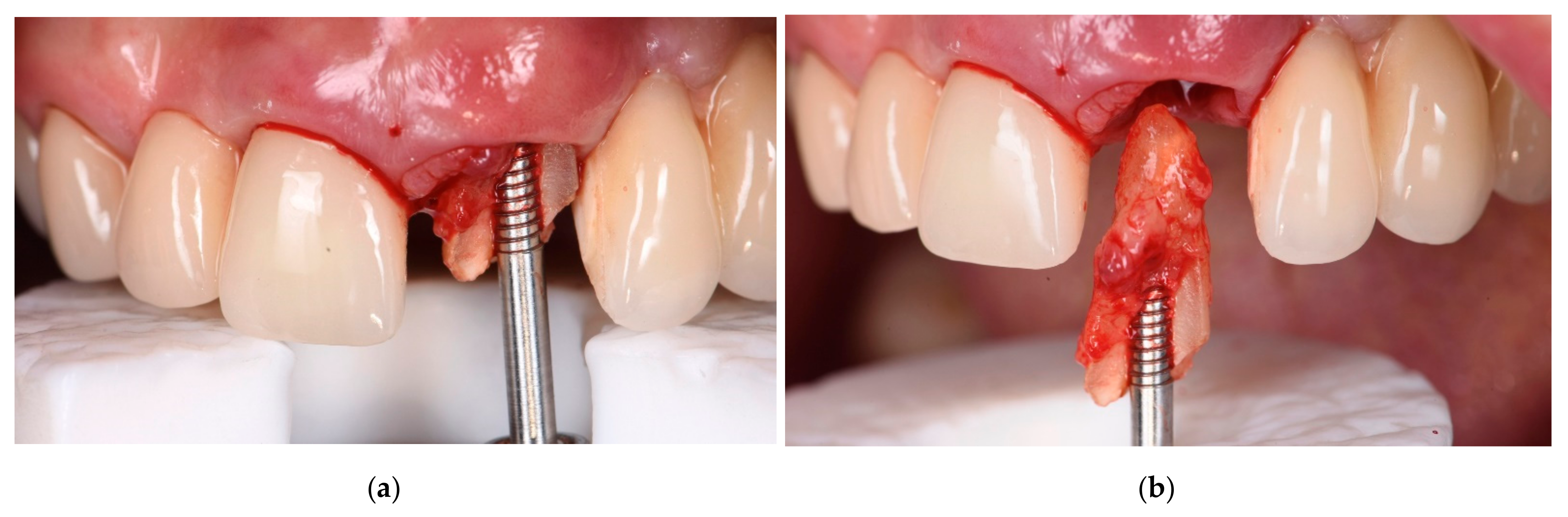
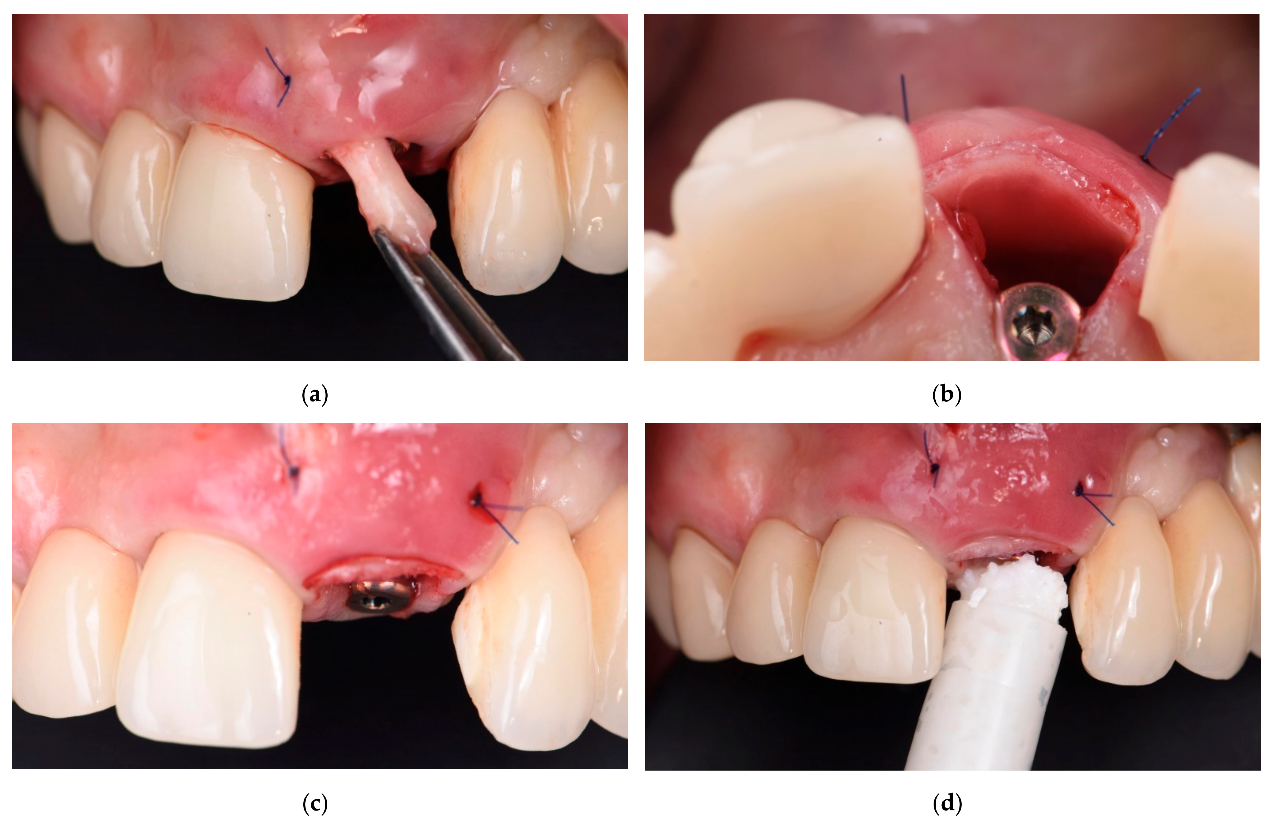
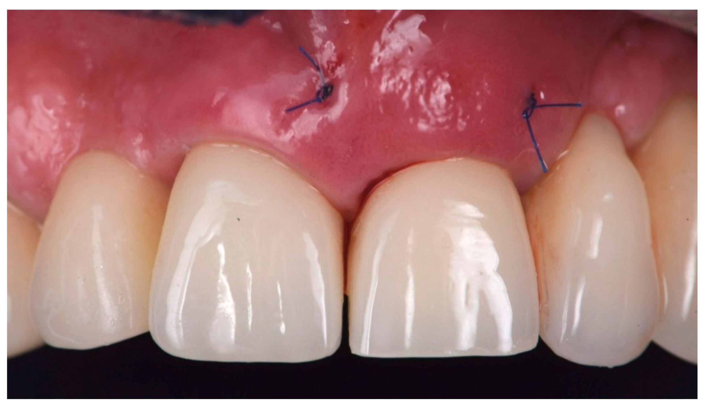
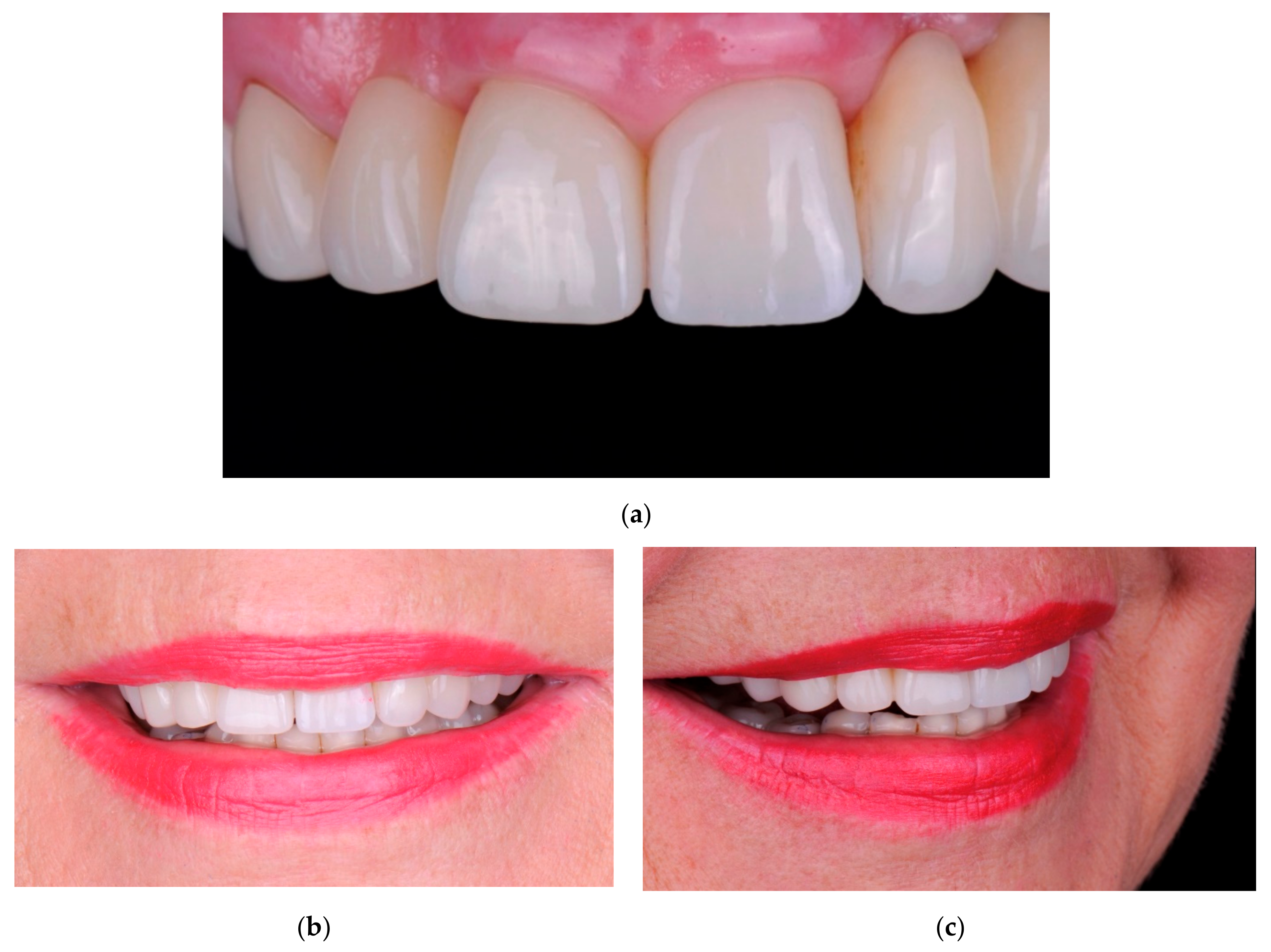
2.2. Surgical Technique

Starting from a day before surgery, prophylactic antibiotic was prescribed to each patient, with Augmentin (amoxicillin and clavulanate potassium) at a dosage of 1 g tablet every 12 h or Azithromycin 500 mg every 12 h in cases of allergy to penicillin. On the day of surgery, after rinsing with a 0.2% chlorhexidine-digluconate solution and application of local anesthesia (4% articaine with 1:100,000 adrenalin), an intrasulcular incision was made around the tooth with a #15c surgical scalpel. The site was additionally prepared with periotomes and microlevers for a minimally invasive dental extraction procedure, and the tooth was extracted with maximum attention. Following the extraction, for removal of the granulation tissue and the residual of the periodontal connective tissue, a careful curettage of the socket was performed.
At this point, if immediate implantation was planned, the implant site was prepared using drills and burs according to the instructions from the manufacturer. The implant was inserted with final insertion torque of 50 Ncm, with palatal anchorage. The next step was the augmentation of the buccal aspect of the tooth socket by a multi-layer technique. This technique consisted of the apposition of three distinct materials: a free soft tissue graft from the same patient, collagenated cortical bone lamina matrix (OsteoBiol® Lamina Soft, Tecnoss®, Giaveno, Italy), and collagenated porcine bone substitute (OsteoBiol® mp3©, Tecnoss®, Giaveno, Italy).
In brief, the surgery continued with soft tissue graft harvesting from the palatal site of the patient. The graft was de-epitalized, placed inside the extraction socket and fixed to the buccal mucosa by means of two horizontal mattress sutures. Next, the cortical lamina bone graft was inserted between the soft tissue graft and the implant. In this study, two models of soft lamina were used: 1/Code LS25FS OsteoBiol® Lamina Soft, Fine, Porcine, 25 × 25 × 0.5 mm (cortical flexible lamina); 2/Code LS10HS OsteoBiol® Curved soft Lamina, Porcine, 35 × 35 × 0.9 mm (curved cortical flexible lamina). The choice among them depended on the preferences of the surgeon in terms of handling and thickness, and according to the defect characteristics. Prior to use, the cortical bone matrix (OsteoBiol® Lamina Soft, Tecnoss®, Giaveno, Italy) was hydrated in sterile saline for 5/10 min. Once the Lamina Soft had reached the desired plasticity, it was cut, adapted and stabilized as a second layer over the soft tissue graft. The stabilization of the cortical lamina bone graft was achieved at the time of insertion, since the graft was modeled to adequately fit the defect dimensions.
Subsequently, a collagenated porcine bone substitute (OsteoBiol®, mp3©, Tecnoss®, Giaveno, Italy) was inserted and compacted between the lamina and the surface of the implant. Condensation of the particulated bone substitute was made in small increments, from an apical to a cervical direction, with caution to avoid graft dislocation. For this purpose, a small bone compactor (Schwert, Seitingen-Oberflacht, Germany) was used in the apical region and a larger diameter bone compactor was used in the more coronal region. Lastly, the provisional crown was installed over the implant with attachment screw (with 20 Ncm torque), allowing sealing of the gingival margin for protection of the multi-layered graft. In case of delayed loading, the provisional crown was attached to the adjacent teeth by using light-cured composite fillers.

2.3. Post-Operative Protocol

After surgery, patients were instructed to avoid any load on the surgical site and to perform topical application of 0.12% chlorhexidine gluconate (PerioGard®, Colgate-Palmolive, Hamburg, Germany) for seven days, twice a day, followed with 0.2% chlorhexidine di-gluconate mouth rinses three times daily for at least two weeks. In order to reduce swelling, ibuprofen 600 mg every 12 h for five days was prescribed. Oral antibiotic therapy was prescribed to all patients, consisting of amoxicillin and clavulanate potassium 1 g every 12 h for five days, or azithromycin 500 mg every 12 h for five days as an alternative in case of allergy to penicillin.

2.4. Follow-Up

The sutures were removed 8–10 days later and standard follow-up visits, including clinical examinations were scheduled on regular basis at 1 month, 3 months, 6 months, 12 months; and then, every 6 months for the following years. All the patients received their final/temporary prosthesis as planned, using the manufacturer’s components or cemented on customized abutments.

2.5. Outcomes

Implant survival was considered as the primary outcome of the study. The intra-surgical and post-surgical complications were assessed as outcomes. The clinical follow-up examinations were done by the same surgeon who had performed implant surgery. Implants were considered to be successful according to the following criteria:
  • implant that is still functional supporting a prosthetic restoration;
  • healthy peri-implant tissues;
  • absence of clinical mobility;
  • no radiographic evidence of peri-implant radiolucency; and
  • absence of any post-operative complications.
For each patient, the parameters listed in Table 1, Table 2, Table 3 and Table 4 were collected.

2.6. Definition of Peri-Implant Mucositis and Peri-Implantitis

Peri-implant mucositis: presence of bleeding on probing (BoP+) and/or inflammation.
Peri-implantitis: BoP+ and/or suppuration, increasing bone loss compared to the baseline (crown delivery).
Peri-implant tissues were considered as healthy when there was neither mucositis nor perimplantitis.

2.7. Statistical Analysis

Descriptive statistics of the data was done using mean values and standard deviation (SD) for quantitative variables normally distributed; in addition, 95% confidence intervals were estimated. Normality of distributions was evaluated through the D’Agostino and Pearson omnibus test. The effect of variables on complications was evaluated by using the Fisher’s exact test. The unit of analysis was the implant. p = 0.05 was considered as the significance threshold. Statistical analysis was performed using GraphPad Prism version 5.03 (GraphPad Software, Inc., La Jolla, CA, USA).

3. Results

A large personal database of patients was evaluated, and patients were extracted electronically according to the inclusion criteria strategy described in materials and methods.
The initial research in patient population data in the office management software since 2010 resulted in 28,507 patients. Soft lamina applications (757 patients) were extracted from these patients. There were 459 patients with “6311040 Lamina Oval” applications, plus 322 patients with “6311020 Lamina Dried” (Group A), and 1078 patients with implants and CTG (Group B).
The overlap of Group A and B resulted in 255 patients.
After applying exclusion criteria (1. All-on-implant cases; 2. All cases where the implants were in the posterior region; 3. Cases where the augmentation with the bone lamina or the soft tissue and the implantation were in different areas), 177 patients were excluded. Finally, only patients undergoing immediate implantation in postextraction sites were selected.
As a final result, a total of 49 patients and 65 implants with a mean follow-up of 60.5 ± 26.6 months after implant placement (mean 53.5 months after loading) were included in this study. The mean age of the patients at the time of implant surgery was 60.0 years (SD 13.6, range 31–93 years). Thirteen patients (18 implants) were smokers. Periapical infection was present in 53 teeth. Thirty-six teeth had previously undergone root canal treatment. Fifty-seven implants were placed in the anterior maxilla, and eight in the anterior mandible. In 34 patients (49 implants), all surgical procedures were done in the same surgical session, and no further surgery was required, while 15 patients (16 implants) underwent two surgical sessions. In eight patients (nine implants), the second session was necessary to perform an additional connective tissue graft, in order to optimize the vestibular contour. Immediate loading was performed in 40 cases, while in 25 implants the functional loading was applied after a mean period of 8.8 ± 4.5 months. Forty-one patients (57 implants) had screwed prosthesis, while eight (eight implants) had cemented prosthesis. Table 1 shows general characteristics of the patient population including age and smoking habits. In Table 2, characteristics of extracted teeth, implant insertion and surgical procedure are described. No implant was lost throughout the observation period, leading to an implant survival of 100%. Four post-operative complications occurred in four patients (listed on Table 3), while 45 patients had no complications. All complications were promptly addressed and had no consequences on the treatment effectiveness. Table 4 reports the synthetic results of the analysis of factors potentially affecting the incidence of complications. No factor among those examined was found to be related to post-surgical complications.
One complete clinical case, illustrating the steps of the multi-layer procedure for post-extraction site reconstruction, is documented in Figure 1, Figure 2, Figure 3, Figure 4 and Figure 5.

4. Discussion

Healing of bone defects, following the use of bone grafting materials, depends greatly on the interaction between the bone graft and the bone cells of the host [16,17]. The factors that are influencing the bone regeneration are the defect morphology, physico-chemical properties of the biomaterials and bone regenerative potential of the patients [16,17].
In a systematic review, Esposito et al. evaluated the horizontal and vertical bone augmentation techniques; as a conclusion, no protocol was found more efficient among the options [18]. Among the alternatives, combination of xenografts with autogenous bone and the use of titanium reinforced polytetrafluoroethylene (PTFE) membranes is one of the most applied, with successful results but also a high incidence of complications [19,20].
The bone lamina technique represents a peculiar solution for augmentation of edentulous alveolar ridges, with mechanical properties analogue to titanium membranes, but also with specific features, including full biocompatibility. The cortical lamina is a collagenated porcine bone graft barrier made of cortical bone of heterologous origin, which is produced with an exclusive process, and consists of three distinct products: Lamina Soft, Curved Soft Lamina, and Rigid Lamina. The exclusive process by the manufacturer avoids the ceramization of hydroxyapatite crystals, so that physiological resorption of the lamina barrier is accelerated. Following a process of superficial decalcification, the Lamina Soft achieves an elastic consistency, yet preserving the typical compactness of the bone tissue from which it originates. The borders of the product are soft, so as to avoid micro traumas to the neighboring tissues. It has properties such as gradual resorption, a rigid, moldable, and adjustable membrane that is actually made of bone, and it adapts well to local anatomy in edentulous mandible or maxilla. In fact, it can be shaped using sterile scissors, until the necessary size is reached, then it has to be hydrated for 5–10 min in sterile saline (0.9% NaCl) solution. Once it achieves the desired pliability, it can be shaped and applied to the defect site. It can be fixated with pins or osteosynthesis screws [13,15,21], or directly sutured (the Fine model) to the surrounding tissues, with a triangular section non-traumatic needle [22]. These features are particularly helpful when it is necessary to maintain the graft volume in aesthetic areas, as well as in horizontal augmentation [15,23,24] of two wall defects, and for covering antrostomy in lateral access sinus augmentation procedures [21,25,26]. Lamina can also be used in regeneration procedures with the risk of exposure. Curved Soft Lamina has a semi-rigid consistency, has a thickness of about 1.0 mm, and must be directly grafted without hydration, after being shaped to fit the defect morphology [27]; it can be particularly effective in association with bone substitutes for regeneration of ridges in which the buccal plate is compromised. Rigid Lamina has been documented for orbital floor and wall reconstruction [28]. The recently developed 0.7 mm thickness Rigid Lamina undergoes a process of superficial semi-decalcification (about 50% as compared to Lamina soft), therefore increasing its consistency, typical of the cortical bone tissue [29,30]. It may represent a viable alternative to autogenous cortical bone plates in the reconstruction of three-dimensional crestal defects with the shell technique [31].
Currently, publications on lamina are limited in number, however they show quite promising outcomes [13,27,32,33]. A multi-center study evaluated the efficacy of the lamina in protecting grafted defects over implants [13], while others tested the cortical lamina functioning as a membrane when treating horizontal defects, and both reported excellent clinical, biological and esthetic outcomes [32,33]. More recently, lamina was tested in the posterior atrophic mandibular bone in a clinical and histological study [27]. As a result, all of the implants were osteo-integrated, and after one year of loading there was no bone loss at the crestal level around the implants. Biopsies showed new formation of bone in the areas that were augmented with lamina.
The technique described in this study is a technical modification of a protocol that was originally described by da Rosa et al. in 2013 [34] for immediate dentoalveolar restoration of compromised sockets in a case report. The proposed treatment by da Rosa et al. [34,35] followed a protocol of immediate implantation, with a flapless surgery, and using corticocancellous bone graft harvested from the maxillary tuberosity to restore the bone defect [34,35]. The same team of authors additionally evaluated the esthetic outcomes and tissue stability of this protocol in a prospective study which included 18 patients with a mean follow up of 58 months, and reported promising outcomes [36]. In that prospective study, the patients had their teeth scheduled for extraction and immediate implantation. In brief, the technique by da Rosa et al. included the following steps: the tooth was extracted as atraumatically as possible, the socket was carefully curetted to remove any granulation tissue, dental implants were inserted in the cingulum axis of the extracted tooth, autogenous corticocancellous bone graft was harvested from maxillary tuberosity, the graft was shaped for adaptation to the defect site, the graft was carefully inserted into the defect between the implant and the mucosal tissue, particulate cancellous bone was inserted and packed to fill the spaces between the corticocancellous bone graft and the implant surface, and subsequently, a provisional crown restoration was placed onto the implant [34,35,36].
A similar protocol was also assessed by various authors with favorable results [37,38,39]. The chamber concept explained by Degidi et al. included immediate provisionalization of implants placed in fresh extraction sockets using a definitive abutment [37]. In that paper, ten patients were treated with immediate flapless extraction, implant placement, grafting with bone substitutes into the gap between the inner surface of the buccal wall and the implant surface, and the provisional crown was placed on the standard abutment [37]. De Molon et al., in 2015 [38], evaluated results of reconstruction of the alveolar buccal bone plate in compromised fresh socket after immediate implant placement followed by immediate provisionalization [38]. De Molon et al., in that case report in 2013, reported a hopeless maxillary left central incisor with loss of the buccal bone wall, which was treated with flapless extraction, immediate implant placement, and immediate reconstruction of the buccal bone plate using the tuberosity as the donor site (to obtain block bone and connective tissue grafts, as well as particulate bone). For this purpose, an autogenous block bone graft and connective tissue graft were harvested from the maxillary tuberosity. After reshaping to match the bone defect, the graft was placed in position with the connective portion facing the gingival mucosa, and the bone portion facing the implant. Subsequently, the particulate autogenous bone graft was placed to completely fill the remaining gap between the implant and the bone graft, followed by the placement of an immediate provisional restoration [38].
In a more recent report by Sheejith et al. in 2020, the “ice cream cone technique” was represented as a flapless bone regeneration technique [39]. In fact, that technique (“ice cream cone”) was originally invented by Tarnow to augment the socket with buccal dehiscence, but his technique was only recommended for simple dehiscence and not a wall defect [40]. In that technique, a collagen membrane in the form of an ice cream cone was used and bone filler material placed into it, to regenerate the buccal plate of a fresh socket without elevating a flap [39,40]. In brief, the tooth was extracted, collagen membrane was contoured into a modified V shape/ice cream cone shape and was placed into the socket, wide enough to extend laterally. The socket was filled with a bone graft with the top part of the membrane extended over the opening of the socket, and the membrane was then sutured to the palatal tissue. As a result, they reported no change in the mucogingival junction position, and prevention of invasion of soft tissue into the socket. However, as a disadvantage, there was a decrease in the depth of the buccal vestibule at the end of healing period, and difficulty in placing the membrane as it became softened when exposed to fluid [39].
The novelty of the multi-layer protocol presented in this paper is the three-layer method used for grafting: a free soft tissue flap that was obtained from the palatal site of the same patient, cortical bone lamina matrix (OsteoBiol® Soft Cortical Lamina, Tecnoss®, Giaveno, Italy), and the collagenated porcine bone substitute (OsteoBiol® mp3®, Tecnoss®, Giaveno, Italy). The cortical lamina used was easy to handle and shape for a perfect adaptation for the full coverage of the extraction defect, and volume maintenance. Utilization of soft tissue graft, together with cortical lamina and particulate bone substitute ensured the good esthetic results. Furthermore, the multi-layer technique has several advantages:
  • There is no donor site, and this reduces the invasiveness of the procedure;
  • it is more versatile, since it can be performed even in patients who still have upper wisdom teeth, compared to the original technique, which can be done only if the wisdom teeth have been extracted; and
  • the connective tissue graft could potentially make the soft tissue more stable in the long term.
The absence of radiological evaluation of marginal bone level change could be considered a limitation of this study. However, as the reconstruction procedure herein presented was associated with immediate implantation and mostly immediate loading, it is very challenging to identify a baseline peri-implant bone level for which to refer in subsequent radiographic assessments.
According to the results of the present study, lamina technique is a successful option to treat anterior defects, which represent a unique and challenging situation in the dental practice, especially in the maxilla. Bone lamina always integrated in a biologically correct way, serving as a semi-rigid barrier for the reconstruction of the resorbed alveolar ridge. Bone lamina contributed to the correct regeneration of the missing bone volumes, and as a result, patient esthetics, function and satisfaction was high in the study population. The post-operative complications were minor and few in number.

5. Conclusions

Based on the results of this study, the following conclusions can be drawn:
  • The present multi-layer technique using bone lamina can be considered as a successful and valuable option with mid-term follow-up up to five years for immediate postextraction implants in sockets with a missing buccal wall.
  • There are some limitations due to the retrospective nature of the study.
Future prospective studies, aiming at quantifying bone regeneration, bone level changes, and patient-related outcome measures, are needed to confirm the excellent clinical results herein observed.

Author Contributions

P.L.S., F.B., H.W., M.D.F., L.F. and T.T. conceived and designed the analysis. Data were collected by P.L.S., F.B., H.W. and T.T. All authors contributed equally in conceptualization, investigation, methodology and resources of the project. Data curation, software, validation, and formal analysis were done by P.L.S., F.G. and M.D.F. Original draft preparation was done by F.G., M.D.F. and T.T. All authors contributed in reviewing and editing the text. P.L.S., F.B., H.W., L.F. and T.T. contributed in visualization, supervision, project administration, and funding acquisition. All authors have read and agreed to the published version of the manuscript.

Funding

This research received no external funding.

Institutional Review Board Statement

Institutional Review Board approval of the IRCCS Orthopedic Institute Galeazzi was obtained for retrospective studies on implant therapy, with number 2552377-L2058/RC 2019.

Informed Consent Statement

Informed consent was obtained from all subjects involved in the study.

Data Availability Statement

Data can be made available if requested.

Conflicts of Interest

The authors declare no conflict of interest.

References

  1. Iyer, S.; Thankappan, K. Maxillary reconstruction: Current concepts and controversies. Indian J. Plast. Surg. 2014, 47, 8–19. [Google Scholar] [CrossRef]
  2. Masaki, C.; Nakamoto, T.; Mukaibo, T.; Kondo, Y.; Hosokawa, R. Strategies for alveolar ridge reconstruction and preservation for implant therapy. J. Prosthodont. Res. 2015, 59, 220–228. [Google Scholar] [CrossRef] [PubMed]
  3. Buser, D.; Chappuis, V.; Belser, U.C.; Chen, S. Implant placement post extraction in esthetic single tooth sites: When immediate, when early, when late? Periodontology 2000 2017, 73, 84–102. [Google Scholar] [CrossRef]
  4. Testori, T.; Weinstein, T.; Scutellà, F.; Wang, H.-L.; Zucchelli, G. Implant placement in the esthetic area: Criteria for positioning single and multiple implants. Periodontology 2000 2018, 77, 176–196. [Google Scholar] [CrossRef] [PubMed]
  5. Pilipchuk, S.P.; Plonka, A.B.; Monje, A.; Tau, A.D.; Lanisdro, A.; Kang, B.; Giannobile, W.V. Tissue Engineering for Bone Regeneration and Osseointegration in the Oral Cavity. Dent. Mater. 2015, 31, 317–338. [Google Scholar] [CrossRef] [Green Version]
  6. Sowmya, S.; Mony, U.; Jayachandran, P.; Reshma, S.; Kumar, R.A.; Arzate, H.; Nair, S.V.; Jayakumar, R. Tri-Layered Nanocomposite Hydrogel Scaffold for the Concurrent Regeneration of Cementum, Periodontal Ligament, and Alveolar Bone. Adv. Healthc. Mater. 2017, 6, 1601251. [Google Scholar] [CrossRef] [PubMed]
  7. Vieira, S.; Vial, S.; Reis, R.L.; Oliveira, J.M. Nanoparticles for bone tissue engineering. Biotechnol. Prog. 2017, 33, 590–611. [Google Scholar] [CrossRef] [Green Version]
  8. Goker, F.; Taschieri, S.; Giannì, A.B.; Grecchi, E.; Savadori, P.; Donati, G.; Del Fabbro, M. Nanotechnology Scaffolds for Alveolar Bone Regeneration. Materials 2020, 13, 201. [Google Scholar] [CrossRef] [Green Version]
  9. Barone, A.; Aldini, N.N.; Fini, M.; Giardino, R.; Calvo Guirado, J.L.; Covani, U. Xenograft versus extraction alone for ridge preservation after tooth removal: A clinical and histomorphometric study. J. Periodontol. 2008, 79, 1370–1377. [Google Scholar] [CrossRef]
  10. Barone, A.; Toti, P.; Quaranta, A.; Alfonsi, F.; Cucchi, A.; Calvo-Guirado, J.L.; Negri, B.; Di Felice, R.; Covani, U. Volumetric analysis of remodelling pattern after ridge preservation comparing use of two types of xenografts. A multicentre randomized clinical trial. Clin. Oral Implant. Res. 2016, 27, e105–e115. [Google Scholar] [CrossRef]
  11. Sanz, M.; Vignoletti, F. Key aspects on the use of bone substitutes for bone regeneration of edentulous ridges. Dent. Mater. 2015, 31, 640–647. [Google Scholar] [CrossRef] [PubMed]
  12. Nannmark, U.; Sennerby, L. The bone tissue responses to prehydrated and collagenated cortico-cancellous porcine bone grafts: A study in rabbit maxillary defects. Clin. Implant. Dent. Relat. Res. 2008, 10, 264–270. [Google Scholar] [CrossRef] [PubMed]
  13. Pagliani, L.; Andersson, P.; Lanza, M.; Nappo, A.; Verrocchi, D.; Volpe, S.; Sennerby, L. A collagenated porcine bone substitute for augmentation at Neoss implant sites: A prospective 1-year multicenter case series study with histology. Clin. Implant. Dent. Relat. Res. 2012, 14, 746–758. [Google Scholar] [CrossRef] [PubMed]
  14. Calvo Guirado, J.L.; Ramírez Fernández, M.P.; Negri, B.; Delgado Ruiz, R.A.; de-Val, J.E.M.S.; Gómez-Moreno, G. Experimental model of bone response to collagenized xenografts of porcine origin (OsteoBiol® mp3): A radiological and histomorphometric study. Clin. Implant. Dent. Relat. Res. 2013, 15, 143–151. [Google Scholar] [CrossRef] [PubMed]
  15. Wachtel, H.; Fickl, S.; Hinze, M.; Bolz, W.; Thalmair, T. The bone lamina technique: A novel approach for lateral ridge augmentation--a case series. Int. J. Periodontics Restor. Dent. 2013, 33, 491–497. [Google Scholar] [CrossRef] [PubMed] [Green Version]
  16. Götz, W.; Reichert, C.; Canullo, L.; Jäger, A.; Heinemann, F. Coupling of osteogenesis and angiogenesis in bone substitute healing—A brief overview. Ann. Anat. 2012, 194, 171–173. [Google Scholar] [CrossRef]
  17. García-Gareta, E.; Coathup, M.J.; Blunn, G.W. Osteoinduction of bone grafting materials for bone repair and regeneration. Bone 2015, 81, 112–121. [Google Scholar] [CrossRef]
  18. Esposito, M.; Grusovin, M.G.; Felice, P.; Karatzopulos, G.; Worthington, H.V.; Couithard, P. The efficacy of horizontal and vertical bone augmentation procedures for dental implants—A Cochrane systematic review. Eur. J. Oral Implantol. 2009, 2, 167–184. [Google Scholar]
  19. Fontana, F.; Maschera, E.; Rocchietta, I.; Simion, M. Clinical classification of complication in guided bone regeneration procedure by means of nonresorbable membrane. Int. J. Periodontics Restor. Dent. 2011, 31, 265–273. [Google Scholar]
  20. Gallo, P.; Diaz-Baez, D. Management of 80 complications in vertical and horizontal ridge augmentation with non-resorbable membranes (d-PTFE) A cross-sectional study. Int. J. Oral Maxillofac Implant. 2019, 34, 927–935. [Google Scholar] [CrossRef]
  21. Festa, V.M.; Addabbo, F.; Laino, L.; Femiano, F.; Rullo, R. Porcine-Derived Xenograft Combined with a Soft Cortical Membrane Versus Extraction alone for Implant Site Development: A Clinical Study in Humans. Clin. Implant. Dent. Relat. Res. 2013, 15, 707–713. [Google Scholar] [CrossRef]
  22. Rossi, R.; Foce, E. Reconstruction of a Horizontal and Vertical Bone defect using The Cortical Lamina Technique. Med. Res. Arch. 2019, 7, 1–11. [Google Scholar]
  23. Lopez, M.A.; Andreasi Bassi, M.; Confalone, L.; Carinci, F.; Ormianer, Z.; Lauritano, D. The Use of Resorbable Cortical Lamina and Micronized Collagenated Bone in the Regeneration of Atrophic Crestal Ridges: A Surgical Technique. Case Series. J. Biol. Regul. Homeost. Agents 2016, 30 (Suppl. S1), 81–85. [Google Scholar] [PubMed]
  24. Polis-Yanes, C.; Cadenas-Sebastián, C.; Gual-Vaqués, P.; Ayuso-Montero, R.; Marí-Roig, A.; López-López, J. Guided Bone Regeneration of an Atrophic Maxilla using Heterologous Cortical Lamina. Case Rep. Dent. 2019, 2019, 5216362. [Google Scholar] [CrossRef] [Green Version]
  25. Hinze, M.; Vrielinck, L.; Thalmair, T.; Wachtel, H.; Bolz, W. Zygomatic Implant Placement in Conjuction with Sinus Bone Grafting: The “Extended Sinus Elevation Technique”. A Case-Cohort Study. Int. J. Oral Maxillofac. Implant. 2013, 28, e376–e385. [Google Scholar] [CrossRef] [Green Version]
  26. Scarano, A.; Murmura, G.; Mastrangelo, F.; Lorusso, F.; Greco Lucchina, A.; Carinci, F. A Novel Technique to prevent Sinus Membrane Collapse during Maxillary Sinus Floor Augmentation without Bone Graft: Technical Note. J. Biol. Regul. Homeost. Agents 2018, 32, 1589–1592. [Google Scholar] [PubMed]
  27. Rossi, R.; Foce, E.; Scolavino, S. The Cortical Lamina Technique: A New Option for Alveolar Ridge Augmentation. Procedure, Protocol, and Case Report. J. Leban. Dent. Assoc. 2017, 52, 35–41. [Google Scholar]
  28. Rinna, C.; Reale, G.; Foresta, E.; Mustazza, M.C. Medial Orbital Wall Reconstruction with Swine Bone Cortex. J. Craniofac. Surg. 2009, 20, 881–884. [Google Scholar] [CrossRef]
  29. Di Carlo, R.; Zara, S.; Ventrella, A.; Siani, G.; Da Ros, T.; Iezzi, G.; Cataldi, A.; Fontana, A. Covalent Decoration of Cortical Membranes with Graphene Oxide as a Substrate for Dental Pulp Stem Cells. Nanomaterials 2019, 9, 604. [Google Scholar] [CrossRef] [PubMed] [Green Version]
  30. Caballé-Serrano, J.; Munar-Fraua, A.; Delgado, L.; Pérez, R.; Hernández-Alfaro, F. Physicochemical Characterization of Barrier Membranes for Bone Regeneration. J. Mech. Behav. Biomed. 2019, 97, 13–20. [Google Scholar] [CrossRef] [PubMed]
  31. Tunkel, J.; Würdinger, R.; de Stavola, L. Vertical 3D Bone Reconstruction with Simultaneous Implantation: A Case Series Report. Int. J. Periodontics Restor. Dent. 2018, 38, 413–421. [Google Scholar] [CrossRef] [Green Version]
  32. Wachtel, H.; Helf, C.; Thalmair, T. The Bone Lamina Technique: A Novel Approach to Bone Augmentation. In Bone Biomaterials and Beyond; Barone, A., Nannmark, U., Eds.; EDRA Lswr: Milano, Italy, 2014; pp. 93–106. [Google Scholar]
  33. Happe, A.; Slotte, C. Reconstruction of horizontal ridge defects. In Bone Biomaterials and Beyond; Barone, A., Nannmark, U., Eds.; EDRA Lswr: Milano, Italy, 2014; pp. 107–118. [Google Scholar]
  34. Da Rosa, J.C.; Rosa, A.C.; da Rosa, D.M.; Zardo, C.M. Immediate Dentoalveolar Restoration of compromised sockets: A novel technique. Eur. J. Esthet. Dent. 2013, 8, 432–443. [Google Scholar]
  35. Da Rosa, J.C.; Rosa, A.C.; Zardo, C.M.; da Rosa, D.M.; Adolfi, D.; Canullo, L.; Periera, L.A.V.D.; Fadanelli, M.A. Immediate Dentoalveolar Restoration: Immediately-Loaded Implants in Compromised Alveolar Sockets, 1st ed.; Quintessence Publishing Company, Ltd.: Batavia, IL, USA, 2014; pp. 178–185. [Google Scholar]
  36. Da Rosa, J.C.; Rosa, A.C.; Francischone, C.E.; Sotto-Maior, B.S. Esthetic outcomes and tissue stability of implant placement in compromised sockets following immediate dentoalveolar restoration: Results of a prospective case series at 58 months follow-up. Int. J. Periodontics Restor. Dent. 2014, 34, 199–208. [Google Scholar] [CrossRef] [PubMed] [Green Version]
  37. Degidi, M.; Daprile, G.; Nardi, D.; Piattelli, A. Immediate provisionalization of implants placed in fresh extraction sockets using a definitive abutment: The chamber concept. Int. J. Periodontics Restor. Dent. 2013, 33, 559–565. [Google Scholar] [CrossRef] [PubMed] [Green Version]
  38. De Molon, R.S.; de Avila, E.D.; de Barros-Filho, L.A.; Ricci, W.A.; Tetradis, S.; Cirelli, J.A.; de Barros, L.A.B. Reconstruction of the Alveolar Buccal Bone Plate in Compromised Fresh Socket after Immediate Implant Placement Followed by Immediate Provisionalization. J. Esthet. Restor. Dent. 2015, 27, 122–135. [Google Scholar] [CrossRef] [PubMed]
  39. Sheejith, M.; Viswanath, S.; Nazar, A. Ice cream cone technique: A flapless bone regeneration technique. J. Prosthet Implant. Dent. 2020, 4, 28–31. Available online: https://www.ipskerala.com/JPID-vol-4/journal-jpid/JPID-Vol-04-Issue-01-Article05.pdf (accessed on 10 July 2021).
  40. Tan-Chu, J.H.P.; Tuminelli, F.J.; Kurtz, K.S.; Tarnow, D.P. Analysis of buccolingual dimensional changes of the extraction socket using the “ice cream cone” flapless grafting technique. Int. J. Periodontics Restor. Dent. 2014, 34, 399–403. [Google Scholar] [CrossRef]
Figure 1. First clinical case. Pre-operative intra-oral view of a patient: (a) frontal view of the compromised tooth #21; (b) pre-operative CBCT radiographs.
Materials 14 05180 g001
Figure 2. (a,b). Intra-operative view of the atraumatic extraction procedure of tooth #21.
Materials 14 05180 g002
Figure 3. Intra-oral view of the patient showing the steps of the three-layer protocol after implant placement: (a) placement of the harvested free soft tissue graft; (b) Soft tissue graft was stabilized with two horizontal mattress sutures, and cortical lamina graft was inserted and put in contact with soft tissue graft. Note the gap between the cortical lamina graft and implant; (c) frontal view showing implant, soft tissue and lamina graft in place; (d) placement of the particulated bone graft (OsteoBiol® mp3®) into the gap between implant and lamina graft.
Materials 14 05180 g003
Figure 4. Intra-oral view of the patient, showing the provisional crown installed over the implant with attachment screw, allowing sealing of the gingival margin for protection of the multi-layered graft.
Materials 14 05180 g004
Figure 5. Images of the final case after 12 months follow-up. (a) Intra-oral view of the patient showing the final crown; (b,c) Extra-oral photographs of the patient.
Materials 14 05180 g005
Table 1. General anamnesis results of patient population including age and smoking habits.
Pat. No.Age at Surgery, YearsSmoker–n. Cigarettes/dayGeneral Anamnesis, Drugs Taken
151yes-10/dNon-contributory
278noBypass—Ramipril, Clopidogrel
372noBreast cancer, 2010—Votum, Toroxin 100
449yes-20/dNon-contributory
563yes-20/dFosamax (single shot)
657yes-10/dNon-contributory
746yesNon-contributory
872noNon-contributory
957noNon-contributory
1054noNon-contributory
1151noNon-contributory
1286noDiabetes
1355yes-10/dNon-contributory
1463noNon-contributory
1563yes-20/dDafiro HCT, Atorvastin
1670noNon-contributory
1731noNon-contributory
1846yes-5/dNickel intolerance
1975noPenicillin intolerance
2066noNon-contributory
2132noNon-contributory
2272noOsteoporosis, hypertension
2366noHypertension
2457noNon-contributory
2555noPenicillin intolerance
2669noPenicillin intolerance
2778noOlmetec plus, Metohexal, Carmen, Adenuric
2840noPenicillin intolerance
2977noTyronajod 75
3052noNon-contributory
3148noNickel intolerance
3270noMarcumar
3355noHypertension
3445noNon-contributory
35611 cigar/dNon-contributory
3647noDiabetes
3774noNon-contributory
3860noNon-contributory
3954noAspirin
4057noTyrox
4193noHeart valve replacement
4268yes-20/dPenicillin intolerance
4338noNon-contributory
4448yes-30/dNon-contributory
4572noCancer (Avastin), Oteoporoses, Hypertension (Bisopolol)
4672noHypertension (Losartan)
4753yes-10/dNon-contributory
4874noNon-contributory
4947yes-20/dNon-contributory
Table 2. Characteristics of extracted teeth, reconstruction procedures and implants placed.
Patient NumberNo. of SurgeriesNo. of Implants Implant SitePeriapical InfectionPrevious RCTImplant Type, Brand, ModelImplant Length, mmImplant Diameter, mmImmediate LoadingCTGCemented or Screwed Retention
12121yesnoNobelReplace CC133.5noyesscrewed
21111yesyesStraumann GM Helix133.75yesyesscrewed
31232yesnoNobelReplace CC133.5yesyesscrewed
42nonoNobelReplace CC133.5yesyesscrewed
41212yesyesNobelReplace CC163.5yesyesscrewed
22noyesNobelReplace CC163.5yesyesscrewed
52211noyesNobelActive154.3yesnoscrewed
22noyesNobelActive154.3yesnoscrewed
61112noyesNobel Speedy Groovy133.3noyesscrewed
73121yesnoStraumann GM Helix134yesyescemented
81112noyesNobelReplace CC133.5yesyesscrewed
91221yesyesStraumann GM Helix134yesyesscrewed
12nonoStraumann GM Helix134yesyesscrewed
101323yesnoNobelReplace CC163.5noyesscrewed
42yesnoNobelReplace CC133.5yesnoscrewed
32nonoNobelReplace CC134.3yesnoscrewed
111413yesyesStraumann GM Helix183.5yesyesscrewed
12yesnoStraumann GM Helix163.5yesyesscrewed
22yesnoStraumann GM Helix163.5yesyesscrewed
23yesnoStraumann GM Helix163.5yesyesscrewed
121211yesnoStraumann GM Helix163.5yesyesscrewed
13yesnoStraumann GM Helix11.54.3yesyesscrewed
131121yesnoStraumann GM Helix133.75yesyesscrewed
141121yesyesStraumann GM Helix133.75yesyesscrewed
151121yesnoNobelReplace CC134.3yesyesscrewed
161121yesyesNobelReplace CC134.3noyesscrewed
171211yesyesNobelReplace CC163.5yesyesscrewed
21nonoNobelReplace CC163.5yesyesscrewed
182211yesyesNobelReplace CC133.5nonoscrewed
22yesnoNobelReplace CC133.5nonoscrewed
192122yesyesNobelActive153.5noyesscrewed
201113yesyesNobelReplace CC163.5noyesscrewed
212112yesyesNobelReplace CC163.5noyesscrewed
221243noyesNobelReplace CC-PMC133.5noyesscrewed
41nonoNobelReplace CC-PMC133.5noyesscrewed
232111yesyesNobelReplace CC163.5yesyescemented
242111yesnoNobelActive133.5noyescemented
251143yesnoNobelReplace CC134.3yesnoscrewed
262213yesyesNobelReplace CC134.3nonocemented
23yesyesNobelReplace CC164.3nonoscrewed
271111yesyesNobleParallel CC153.75yesyescemented
282121yesyesNobelReplace CC11.53.5noyesscrewed
29 122yesnoGM Helix184.3noyesscrewed
302121yesyesNobleParallel CC163.5noyesscrewed
311121yesyesNobelReplace CC133.5yesyescemented
321121yesyesStraumann GM Helix163.75yesyesscrewed
332121yesyesNobelReplace CC11.53.5noyescemented
341123yesnoNobleParallel CC133.75noyesscrewed
351121yesnoNobelReplace CC133.5noyesscrewed
361112yesnoStraumann GM Helix133.5yesyesscrewed
371122yesyesStraumann GM Helix133.5yesyesscrewed
381111yesyesNobelActive153.5yesyescemented
391141yesyesNobelReplace CC163.5yesyesscrewed
401121yesyesStraumann GM Helix133.75noyesscrewed
411122yesyesNobelReplace CC134.3yesnoscrewed
422221yesyesStraumann GM Helix133.75yesyesscrewed
13yesyesStraumann GM Helix164nonoscrewed
432111yesyesNobelReplace CC133.5noyesscrewed
441111yesyesNobleParallel CC133.75yesyesscrewed
451121yesnoGM Helix163.75yesyesscrewed
461121yesyesNobleParallel CC133.75yesyesscrewed
472121yesnoNobleParallel CC133.75noyesscrewed
482111yesnoNeodent-Straumann CM Alvim163.5noyesscrewed
491212nonoStraumann BLX143.75yesnoscrewed
22yesnoStraumann BLX143.75yesnoscrewed
RCT = root canal treatment; CTG = connective tissue graft.
Table 3. List of postoperative complications occurred.
Patient No.Site No.Complication Type
511Peri-implantitis
1711A little piece of the lamina removed 18 months after surgery
3321Palatal cyst developed four years after surgery
4811Fracture of the provisional crown
Table 4. Synthetic table of potentially influencing factors and incidence of complications.
Potentially Influencing FactorN. of ImplantsN. of ComplicationsSignificance (p-Value)
Smoking statusSmoker1810.43
Non-smoker473
Periapical infectionYes5330.42
No121
Previous root canal treatmentYes3630.31
No291
Prosthetic connectionScrewed5730.35
Cemented81
LocationMaxilla5740.58
Mandible80
Loading modeImmediate4020.35
Delayed252
Publisher’s Note: MDPI stays neutral with regard to jurisdictional claims in published maps and institutional affiliations.

