Article

Lanthanum and Manganese Co-Doping Effects on Structural, Morphological, and Magnetic Properties of Sol-Gel Derived BiFeO3

1
Institute of Chemistry, Vilnius University, Naugarduko 24, LT-03225 Vilnius, Lithuania
2
Center of Physical Sciences and Technology, LT-02300 Vilnius, Lithuania
3
Institute of Chemical Physics, Faculty of Physics, Vilnius University, Sauletekio Ave. 3,LT-10257 Vilnius, Lithuania
4
Department of Bioelectrochemistry and Biospectroscopy, Institute of Biochemistry, Life Sciences Center, Vilnius University, LT-10257 Vilnius, Lithuania
*
Author to whom correspondence should be addressed.
Materials 2021, 14(17), 4844; https://doi.org/10.3390/ma14174844
Submission received: 26 July 2021 / Revised: 19 August 2021 / Accepted: 23 August 2021 / Published: 26 August 2021
(This article belongs to the Special Issue Sol-Gel Synthesis of Materials)

Abstract

:
In this work, lanthanum and manganese co-substitution effects on different properties of bismuth ferrite solid solutions Bi1-xLaxFe0.85Mn0.15O3 (x from 0 to 1) prepared by a sol-gel synthetic approach have been investigated. It was observed that the structural, morphological, and magnetic properties of obtained specimens are influenced by the amount of introduced La3+ ions. Surprisingly, only the compound with a composition of BiFe0.85Mn0.15O3 was not monophasic, and the presence of neighboring phases was determined from X-ray diffraction analysis and Mössbauer measurements. Structural transitions from orthorhombic to cubic and back to orthorhombic were also observed depending on the La3+ amount. Antiferromagnetic behaviour was observed for all of the samples, with the highest magnetisation values for Bi0.5La0.5Fe0.85Mn0.15O3. Additionally, structural attributes and morphological features were evaluated by Raman spectroscopy and scanning electron microscopy (SEM), respectively.

1. Introduction

Since the work of Spaldin in 2000, where the existence of only a few magnetic ferroelectrics was questioned [1], multiferroics became one of the most investigated topics in the past 20 years. In this type of material, two of the three primary ferroic (ferroelectricity, ferromagnetism, or ferroelasticity) properties coincide in one phase. The combination of magnetism and ferroelectricity are interesting from both theoretical and application point of views. From the practical side, these compounds could be applied in energy-efficient devices [2], photocatalysis [3], biomedicine [4], microwave phase shifter [5], etc. From the theoretical aspect, magnetism arises from partially filled d or f shells, while ferroelectricity is associated with the off-centring of different transition metal ions, such as Ti4+, where the d orbital is empty. Therefore, few mechanisms (lone pair, charge ordering, or “geometric”) explaining ferroelectricity are known [6].
Bismuth ferrite (BiFeO3) can be considered one of the most investigated perovskite-type multiferroic materials. This compound demonstrates magnetoelectric properties in both thin-film [7] and bulk [8] forms. Nanosized BiFeO3 structures, which display different morphologies, are also explored thoroughly since these nanostructures showed an increase in magnetisation values [9] and better photocatalytic activity [10]. The combination of both ferroelectric and (anti)ferromagnetic orders remains a difficult challenge for this compound due to the large leakage current, which arises from high oxygen vacancies concentration, weak magnetoelectric effect, and formation of impurities [11]. Another material, which can display both spontaneous polarisation and magnetisation with magnetoelectric effect, is lanthanum ferrite (LaFeO3) [12]. While phase-pure BiFeO3 is difficult to prepare, LaFeO3 can be synthesised by different types of techniques, including sol-gel combustion [13], solid-state [14], hydrothermal [15], co-precipitation, or high-energy ball milling [16].
Substitution of A-site, B-site, or both cations can lead to improvement in physical properties or formation of phase-pure BiFeO3. One of the examples can be La3+ intercalation to the BiFeO3 system, leading to an increase in magnetisation, conductivity, and improved ferroelectric properties [17,18,19]. Substituting Fe3+ ions with Mn3+ ions can lead to structural transition, a better magnetoelectric coupling effect, and enhancement of magnetisation [20,21,22]. Co-doping with both elements was performed to suppress the formation of oxygen vacancies and Fe2+ ions, which could improve the electrical properties [23]. Furthermore, it was shown that Bi1−xLaxFe1−yMnyO3/Ti3C2 hybrids have high catalytic activity towards Congo Red degradation [24]. While most of the studies regarding Bi1-xLaxFe1-yMnyO3 focus on intercalation of small amounts of La3+ up to 30% [25], we aimed to investigate the whole compositional range.
In this work, we prepared a series of Bi1-xLaxFe0.85Mn0.15O3 (with different x steps) solid solutions by an environmentally friendly, cost-effective, and simple sol-gel technique using only ethylene glycol as a complexing agent. Various characterisation techniques were used to evaluate structural, morphological, and magnetic properties regarding chemical composition.

2. Materials and Methods
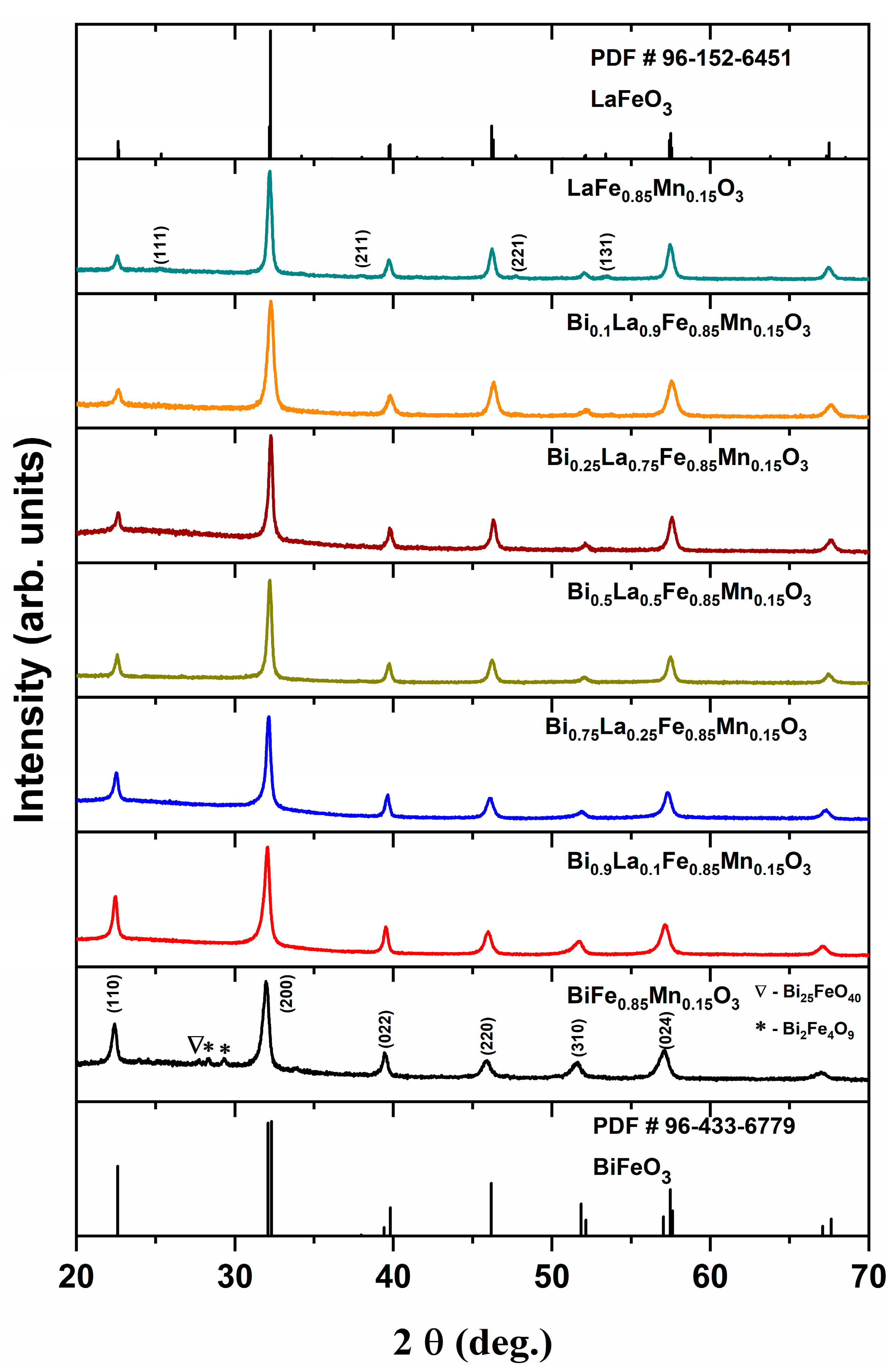
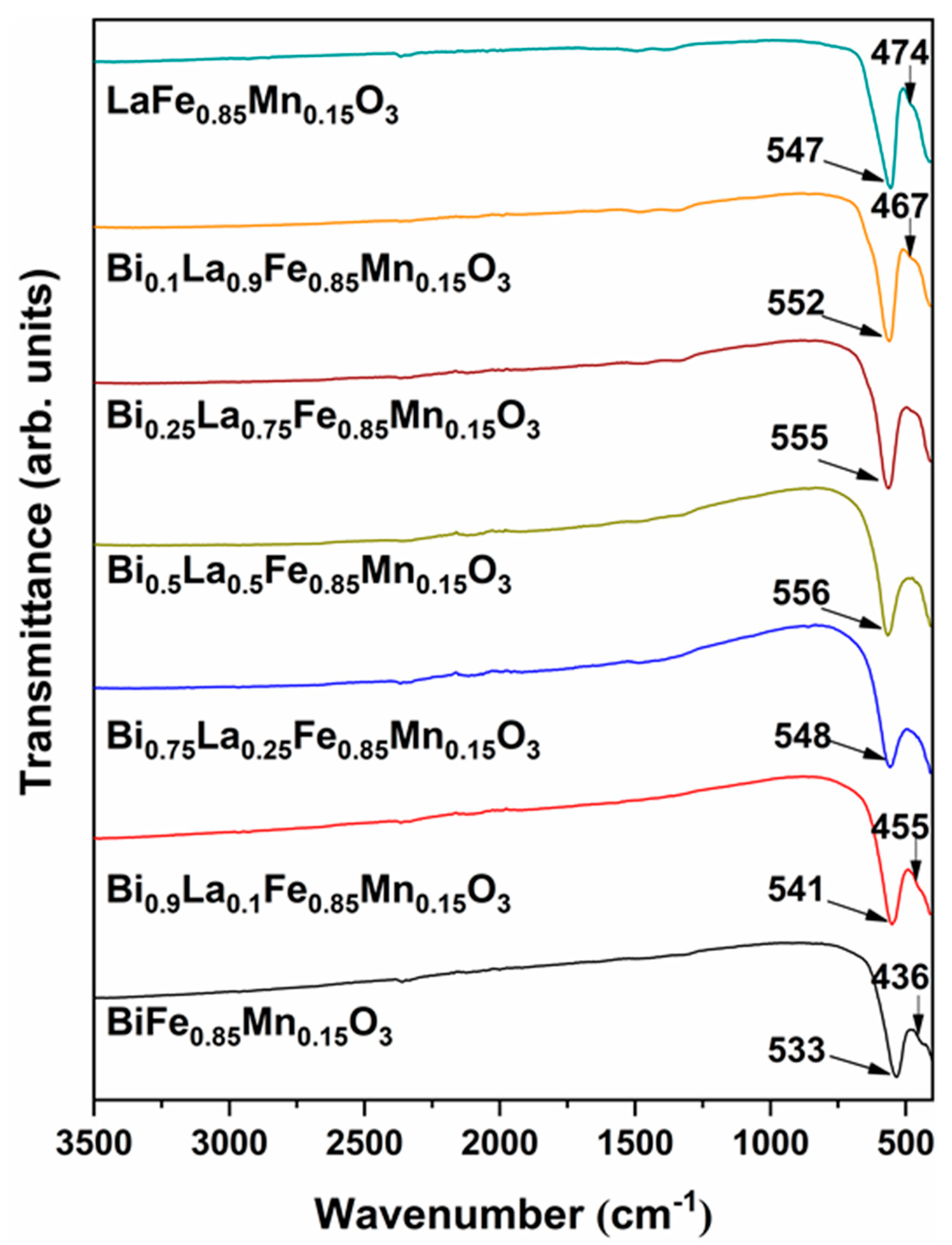
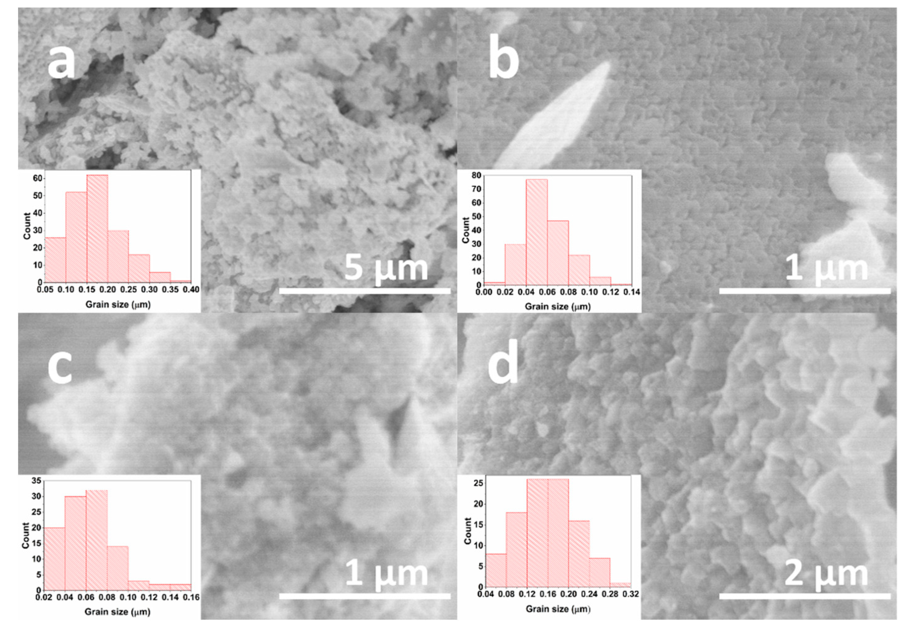
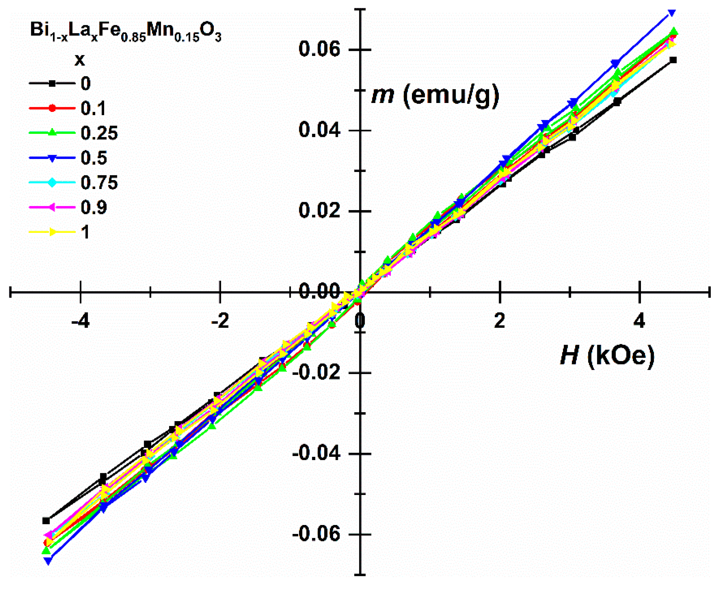
For the preparation of Bi1-xLaxFe0.85Mn0.15O3 solid solutions, bismuth (III) nitrate pentahydrate (Bi(NO3)3∙5H2O, Roth (Karlsruhe, Germany), 98%), lanthanum (III) nitrate hexahydrate (La(NO3)3∙6H2O, Alfa Aesar (Haverhill, MA, USA), 99.9%), iron (III) nitrate nonahydrate (Fe(NO3)3∙9H2O, Alfa Aesar, 99.9%), and manganese (II) nitrate tetrahydrate (Mn(NO3)2∙4H2O, Alfa Aesar, 99.9%) were used as starting materials. Firstly, required amounts of lanthanum, iron, and manganese nitrates were dissolved in 20 mL of distilled water. Before addition of bismuth nitrate, the pH of solution was adjusted to 1 by the addition of nitric acid (HNO3, 65%). After the dissolution of bismuth nitrate, the complexing agent, ethylene glycol (C2H6O2, Sigma-Aldrich (St. Louis, MO, USA), ≥99.5%), was added to the mixture. The molar ratio between total metal ions and ethylene glycol was 1:2. The obtained solution was homogenised under constant stirring at 90 °C for 1 h. Next, the temperature was increased to 120 °C for solvent evaporation and the formation of a gel. The resulting gel was dried in the oven at 120 °C for 6 h, ground in agate mortar, and annealed at 650 °C with a heating rate of 1 °C/min for 1.5 h in air.
Thermal decomposition of precursor gel was investigated by thermogravimetric and differential scanning calorimetric (TG/DTG-DSC) analysis using PerkinElmer STA 6000 Simultaneous Thermal Analyzer. About 5–10 mg of dried sample was heated from 30 °C to 900 °C at 10 °C/min heating rate in dry flowing air (20 mL/min). X-ray diffraction (XRD) analysis of obtained products was performed with a Rigaku Miniflex II diffractometer using a primary beam Cu Kα radiation (λ = 1.541838 Å). The 2θ angle of the diffractometer was set in the range from 20° to 70° while moving 10°/min. The obtained diffraction data were refined by the Rietveld method using the Fullprof suite. Crystallite size was calculated by applying Scherrer’s equation: D = K λ β c o s θ , with the following: K–shape factor (in our case, 0.89), λ–X-ray wavelength, β–full width at half maximum in radian, θ–Bragg diffraction angle. The β was measured for corundum standard in order to evaluate the instrumental broadening. Alpha FT-IR spectrometer (Bruker, Ettlingen, Germany) was used for FT-IR analysis of compounds. All spectra were recorded at ambient temperature in the range of 4000–400 cm−1. The morphology of samples was examined using a scanning electron microscope (SEM) (Hitachi SU-70, Tokyo, Japan). Raman spectra were acquired using LabRam HR800 (Horiba Jobin Yvon, Villeneuve d′Ascq, France) Raman spectrometer equipped with thermoelectrically cooled (−90 °C) CCD camera (DU920P-BR-DD), 600 lines/mm grating and microscope. Spectra were excited with a 532 nm beam from the CW diode-pumped solid-state (DPSS) laser (Cobolt Samba, Hübner Photonics, Stockholm, Sweden). The laser power at the sample was restricted to 0.2 mW to avoid laser-induced sample heating and photodegradation. The 50×/0.50 NA long working distance (LWD) objective was employed during the measurements. The overall integration time was 600 s. The position of the Raman bands on the wavenumber axis was calibrated by the Si Raman band at 520.7 cm−1. Parameters of the bands were determined by fitting the experimental spectra with Gaussian-Lorentzian shape components using GRAMS/A1 8.0 (Thermo Scientific, Waltham, MA, USA) software. Magnetometer consisting of the lock-in amplifier SR510 (Stanford Research Systems, Sunnyvale, CA, USA), the Gauss/teslameter FH-54 (Magnet Physics) and the laboratory magnet supplied by the power source SM 330-AR-22 (Delta Elektronika, Zierikzee, The Netherlands) was applied to record magnetisation dependences on an applied magnetic field. Mössbauer spectra were measured using 57Co(Rh) source and Mössbauer spectrometer (Wissenschaftliche Elektronik GmbH, Starnberg, Germany). Closed-cycle He cryostat (Advanced Research Systems, Macungie, PA, USA) was applied for low-temperature measurements. The doublets/singlets, sextets, and hyperfine field distributions were used to fit Mössbauer spectra applying WinNormos Site and Dist software. Isomer shift is given relative to α-Fe.

3. Results

Thermogravimetric analysis was employed to evaluate thermal decomposition behaviour and determine optimum annealing temperature, which is required for the formation of solid solutions. TG/DTG/DSC curves of precursor gel for Bi0.1La0.9Fe0.85Mn0.15O3 composition compound are presented in Figure 1. Three main degradation steps can be identified from the DTG curve. First, non-significant mass loss (about 3%) can be observed in the 60–150 °C temperature range; it can be ascribed to evaporation of adsorbed water. The most significant mass loss (about 46%) can be seen in the 160–500 °C temperature region. This step can be attributed to several processes, including the decomposition of metal nitrates or metal complexes with ethylene glycol and the residual organic part of the gel. Two small exothermic peaks, which are centred around 204 and 280 °C, are the result of a combustion reaction. Final mass loss (about 2%) can be seen in the 625–650 °C range. A similar degradation step previously was observed for La–Fe–O gel and was explained by the decomposition of residual organic compounds [26], ionic carbonates [27], or degradation of amorphous material [28]. From the TG curve, the total mass loss was determined to be 51%, and above 650 °C, the mass remained constant. For this reason, 650 °C was chosen as a final annealing temperature.
X-ray diffraction analysis was carried out to determine phase purity along with structural changes for the Bi1-xLaxFe0.85Mn0.15O3 solid solutions. The obtained results are demonstrated in Figure 2. Nearly all samples were identified as phase-pure with one exception—pristine BiFe0.85Mn0.15O3. It is known that Fe3+ substitution by the small amount of Mn3+ can lead to stabilisation of the perovskite phase while avoiding the formation of neighbouring phases [29]. On the other hand, the annealing temperature, which was used in this work, belongs to the range where BiFeO3 is considered to be metastable with respect to two Bi-rich Bi25FeO39 sillenite and Fe-rich Bi2Fe4O9 mullite phases [30]. Those two impurities phases were observed in our synthesised BiFe0.85Mn0.15O3 sample. By intercalating La3+ into the perovskite structure, few trends can be observed in the XRD profiles. The intensity of the peaks labelled (110), (022), and (310) decreases, while the intensity of the (220) peak increases for La-rich solid solutions. Since the ionic radius of the La3+ ion (ionic radius 1.16 Å in VIII-fold coordination) is slightly smaller than the Bi3+ ion (ionic radius 1.17 Å in VIII-fold coordination) [31], only a slight peak shift can be observed in the XRD patterns.
Rietveld refinement was carried out for all synthesised solid solution specimens. The splitting of the most intense (200) peak was not observed for any of our samples, which means that the perovskite structure deviates from the crystal symmetry of the BiFeO3 compound, which has a trigonal unit cell with rhombohedral or hexagonal axes. Fitting was performed with various structural models (R3c, Pbnm, Pm 3 ¯ m, R3c+Pbnm, R3c+Pm 3 ¯ m, etc.), previously completed by Kumar and Kar [25] for Bi1-xLaxFe1-xMnxO3 (x varying from 0 to 0.3) solid solutions. They observed that the structure of all co-doped samples consists of a mixture of two different phases: orthorhombic with Pbnm space group and rhombohedral with R3c space group. In our case, the BiFe0.85Mn0.15O3 sample has an orthorhombic unit cell with a Pbnm space group. B-site substitution with Mn3+ [32] or Ga3+ [33] ions can lead to identical structural transition. The compound with 10% La3+ has an identical structure to the BiFe0.85Mn0.15O3 sample, with a decrease in cell volume and slight variations in lattice parameters. Further increase in La3+ up to 75% resulted in a structural change to a cubic unit cell with Pm 3 ¯ m space group. Moreover, the appearance of new diffractions peaks labelled (111), (211), (221), and (131) for a sample containing 90% of La3+ confirms another structural transition from cubic to the orthorhombic unit cell with the Pbnm space group. Interestingly, in comparison with the solid solution with 90% of La3+ ions, the LaFe0.85Mn0.15O3 compound has an increase in unit cell parameters and cell volume. Analogous results were observed for Bi1-xLaxFeO3 solid solutions, where an increase in La3+ concentration resulted in an increase in cell parameters [34]. This was explained by the fact that Bi-containing compounds have a larger quantity of oxygen vacancies because of the lower strength of the Bi–O bond.
The most intense diffraction peak (200) was chosen for the calculation of crystallite size. The obtained results are presented in Table 1. Three distinctive regions can be excluded according to the structural transitions observed from XRD patterns. Firstly, the intercalation of 10% of La3+ leads to a small increase in crystallite size. After the structural change from orthorhombic to cubic unit cell, the crystallite size increased by 25% in comparison with the previous compound. The compound with 90% of La3+ showed a sudden decrease in crystallite size, which can be associated with the structural change. Furthermore, LaFe0.85Mn0.15O3 showed a similar crystallite size to the Bi0.25La0.75Fe0.85Mn0.15O3 solid solution.
The short-range structure of studied compounds was probed by Raman spectroscopy by using a 532 nm excitation wavelength. The technique is very sensitive to structural distortions and could provide additional structural information on unit cells as well as space-group type. Figure 3 compares the Raman spectra of BiFeO3 and BiFe0.85Mn0.15O3 samples. BiFe0.85Mn0.15O3 was chosen because this compound is known for possessing different structures with a variety of space groups. Pure BiFeO3 exhibits a characteristic pattern of bands related to a rhombohedrically distorted perovskite structure; the main bands are located at 78 cm−1 (symmetry E, band E-1), 141 cm−1 (A1-1), 173 cm−1 (A1-2), 221 cm−1 (A1-3), 270 cm−1 (E-3), 348 cm−1 (E-5), 366 cm−1 (E-6), 469 cm−1 (A1-5), and 527 cm−1 (E-8) [29,35,36,37,38]. The low-intensity band near 604 cm−1 might be associated with the E-9 mode or related with second-order vibrational transition [38,39,40]. The A1 symmetry bands located at 141, 173, and 221 cm−1 and one E symmetry mode located at 78 cm−1 are the most intense features in the spectrum of pure BiFeO3 compound.
Doping with Mn3+ ions results in drastic changes in the Raman spectrum (Figure 3). A strong and relatively broad band appears near 628 cm−1 along with the broad middle-intensity feature near 481 cm−1. In contrast, little changes are visible for lower frequency A1 symmetry bands located at 140, 170, and 220 cm−1. These low-frequency modes are mainly related to Bi−O stretching vibrations [41,42]. Thus, small spectra changes indicate little perturbation in the Bi−O bonding structure after the introduction of Mn3+ ions. The appearance of a strong band near 628 cm−1 indicates Mn3+ ions-induced perturbations in the Fe−O bonding range. Indeed, high-frequency E-8 and E-9 modes earlier were attributed as mainly related to Fe−O stretching bands [41,42]. These peaks are sensitive to the tilt of oxygen octahedra [40]. Intensification of a band at 628 cm−1 might be related to the transformation of short-range structure from rhombohedral (R3c space group) to orthorhombic (Pbnm space group) [43]. A similar band was observed for orthorhombic rare-earth manganites (RMnO3) (Pnma space group for oxygen octahedra) [44]. These observations confirm the results of the XRD analysis.
FT-IR spectroscopy analysis was carried out to further investigate structural changes in Bi1-xLaxFe0.85Mn0.15O3 solid solutions, and results are demonstrated in Figure 4. In general, the characteristic bands for orthoferrites and orthomanganites can be found in slightly different regions—600–250 cm−1 and 700–200 cm−1, respectively [45]. In our case, no absorption bands were observed in the 4000–600 cm−1 interval, which indicates the absence of residual organic species or carbonates in solid solutions. Our previous study [22] on BiFe1-xMnxO3 solid solutions revealed that the addition of Mn3+ could result in the most intense peak shift to higher wavenumbers. Samples with orthorhombic structure have two absorption bands: a strong and sharp band located at 533–552 cm−1 region as well as a weak and broad one at 436–474 cm−1. Only one peak at around 550 cm−1 can be observed for compounds with cubic structures. These bands can be ascribed to the stretching mode of the Fe–O bond in the FeO6 octahedra. With the increase of La3+ concentration, weaker absorption band shifts to higher wavenumbers, while a strong intensity peak only shifts to higher wavenumbers until the La3+ amount reaches 50%. Larger substitution results in a monotonic shift to lower wavenumbers. Furthermore, no stretching or bending vibrations belonging to Bi2O3 or La2O3 can be visible for all our samples, which indicate no presence of these impurity phases.
To investigate the influence of La3+ and Mn3+ co-doping on the morphology of mixed-metal ferrite powders, the SEM analysis was executed for solid solutions of four different compositions, and the results are demonstrated in Figure 5. The Bi0.9La0.1Fe0.85Mn0.15O3 sample (Figure 5a) consists of particles, which are necked to each other, forming some larger aggregates. Around 90% of particles for this compound are in the 50–250 nm range. Further increase in La3+ amount up to 25% resulted in a decrease in particle size (Figure 5b). For this sample, 95% of particles are in the 20–100 nm range. The Bi0.5La0.5Fe0.85Mn0.15O3 sample has analogous particle size distribution in comparison with a previous solid solution with 25% of La3+. With further increase in the concentration of La3+ ions, the particle size also increased, which can be seen in Figure 5d. For this Bi0.1La0.9Fe0.85Mn0.15O3 compound, nearly 92% of particles are in the 40–240 nm range. Interestingly, it was shown that an increase in Bi3+ content in BixLa1-xMnO3+δ solid solutions could lead to drastic particle growth [46]. On the other hand, our previous study revealed that Mn3+ could act as a particle growth inhibitor, limiting the grain size [22]. The intercalation of 15% Mn3+ ions limited the effect of particle growth, which could be related to the higher concentration of Bi3+ ions.
The linear dependence of magnetisation (Figure 6) is generally consistent with the antiferromagnetic order of spins in samples, but at least for BiFe0.85Mn0.15O3, the observed magnetisation is larger than that of bulk BiFeO3 [47,48]. It is noteworthy that in the case of nanocrystalline BiFeO3 and LaFeO3 (Table 1), weak ferromagnetism due to uncompensated magnetic moments on the surface of grains can cause an increase in magnetisation [49,50,51]. However, for BiFe1-xMnxO3 solutions, Mn influences magnetic order, which can cause the formation of additional paramagnetic compounds [22], which cause a decrease in uncompensated magnetisation.
Mössbauer spectra of Bi1-xLaxFe0.85Mn0.15O3 samples at room temperature (Figure 7a,b) showed broadened sextet lines, the increased width of which can be mostly related to the substitution of Fe by Mn. However, superparamagnetic relaxation of nanosized grains could also add to the line broadening here. Magnetically split part of spectra was fitted using hyperfine field distribution P(B) to account for line broadening. At low temperature (Figure 7b), however, the spectra could be fitted to two sextets. The line broadening caused the average hyperfine field <B> (Figure 7c) to be lower than the hyperfine field characteristic of BiFeO3 (49-50 T [48]) and LaFeO3 (51.3 T [51]). The parameters of Mössbauer spectra (Figure 7c) slightly depend on the composition of samples. A considerable amount of paramagnetic doublet (≈51% of the spectral area) appeared only for BiFe0.85Mn0.15O3. This doublet could be probably attributed to poor crystalline Bi2Fe4O9 as isomer shift δ = 0.31 ± 0.01 mm/s of the doublet is equal to the average isomer shift of two doublets characteristic of Bi2Fe4O9 [52].

4. Conclusions

An aqueous sol-gel method was successfully applied for the preparation of Bi1-xLaxFe0.85Mn0.15O3 (where 0 ≤ x ≤ 1) solid solution specimens. Nearly all compounds were phase-pure, with the exception of BiFe0.85Mn0.15O3, where the presence of Bi2Fe4O9 and Bi25FeO40 impurity phases was determined from X-ray diffraction analysis data and Mössbauer measurements. Depending on La3+ concentration, few structural transitions from orthorhombic to cubic and back to orthorhombic were observed in the series. Raman and FT-IR spectroscopies also provided additional structural information in good agreement with XRD analysis results. SEM analysis demonstrated that intercalation of Mn3+ ions limited the growth of the particles, and the largest particles were observed for Bi0.9La0.1Fe0.85Mn0.15O3. All samples demonstrated antiferromagnetic behaviour, with the highest magnetisation values observed for the Bi0.5La0.5Fe0.85Mn0.15O3 sample.

Author Contributions

Formal analysis, D.K., R.D. and A.Z.; Investigation, D.K., R.D., K.M., D.B., G.N. and M.T.; Resources, A.B. and A.K.; Data curation, D.K. and R.D.; Writing—Original Draft preparation, D.K. and R.D.; Writing—Review and Editing, D.K.; Visualization, D.K., R.D., K.M., D.B., G.N. and M.T.; Supervision, A.Z. and A.K. All authors have read and agreed to the published version of the manuscript.

Funding

This work was supported by a research grant BUNACOMP (No. S-MIP-19-9) from the Research Council of Lithuania.

Conflicts of Interest

The authors declare no conflict of interest.

References

  1. Hill, N.A. Why are there so few magnetic ferroelectrics? J. Phys. Chem. B 2000, 104, 6694–6709. [Google Scholar] [CrossRef]
  2. Manipatruni, S.; Nikonov, D.E.; Lin, C.C.; Gosavi, T.A.; Liu, H.; Prasad, B.; Huang, Y.L.; Bonturim, E.; Ramesh, R.; Young, I.A. Scalable energy-efficient magnetoelectric spin–orbit logic. Nature 2019, 565, 35–42. [Google Scholar] [CrossRef]
  3. Vanga, P.R.; Mangalaraja, R.V.; Ashok, M. Effect of (Nd, Ni) co-doped on the multiferroic and photocatalytic properties of BiFeO3. Mater. Res. Bull. 2015, 72, 299–305. [Google Scholar] [CrossRef]
  4. Guduru, R.; Liang, P.; Runowicz, C.; Nair, M.; Atluri, V.; Khizroev, S. Magneto-electric nanoparticles to enable field-controlled high-specificity drug delivery to eradicate ovarian cancer cells. Sci. Rep. 2013, 3, 1–8. [Google Scholar] [CrossRef]
  5. Ustinov, A.B.; Srinivasan, G.; Kalinikos, B.A. Ferrite-ferroelectric hybrid wave phase shifters. Appl. Phys. Lett. 2007, 90, 31913. [Google Scholar] [CrossRef]
  6. Khomskii, D. Trend: Classifying multiferroics: Mechanisms and effects. Physics 2009, 2, 20. [Google Scholar] [CrossRef] [Green Version]
  7. Wang, J.; Neaton, J.B.; Zheng, H.; Nagarajan, V.; Ogale, S.B.; Liu, B.; Viehland, D.; Vaithyanathan, V.; Schlom, D.G.; Waghmare, U. V Epitaxial BiFeO3 multiferroic thin film heterostructures. Science 2003, 299, 1719–1722. [Google Scholar] [CrossRef] [PubMed]
  8. Jia, D.C.; Xu, J.H.; Ke, H.; Wang, W.; Zhou, Y. Structure and multiferroic properties of BiFeO3 powders. J. Eur. Ceram. Soc. 2009, 29, 3099–3103. [Google Scholar] [CrossRef]
  9. Shokrollahi, H. Magnetic, electrical and structural characterization of BiFeO3 nanoparticles synthesized by co-precipitation. Powder Technol. 2013, 235, 953–958. [Google Scholar] [CrossRef]
  10. Gao, F.; Chen, X.Y.; Yin, K.B.; Dong, S.; Ren, Z.F.; Yuan, F.; Yu, T.; Zou, Z.G.; Liu, J. Visible-light photocatalytic properties of weak magnetic BiFeO3 nanoparticles. Adv. Mater. 2007, 19, 2889–2892. [Google Scholar] [CrossRef]
  11. Catalan, G.; Scott, J.F. Physics and applications of bismuth ferrite. Adv. Mater. 2009, 21, 2463–2485. [Google Scholar] [CrossRef]
  12. Acharya, S.; Mondal, J.; Ghosh, S.; Roy, S.K.; Chakrabarti, P.K. Multiferroic behavior of lanthanum orthoferrite (LaFeO3). Mater. Lett. 2010, 64, 415–418. [Google Scholar] [CrossRef]
  13. Theingi, M.; Tun, K.T.; Aung, N.N. Preparation, characterization and optical property of LaFeO3 nanoparticles via sol-gel combustion method. SciMedicine J. 2019, 1, 151–157. [Google Scholar] [CrossRef] [Green Version]
  14. Sazelee, N.A.; Idris, N.H.; Din, M.F.M.; Yahya, M.S.; Ali, N.A.; Ismail, M. LaFeO3 synthesised by solid-state method for enhanced sorption properties of MgH2. Results Phys. 2020, 16, 102844. [Google Scholar] [CrossRef]
  15. Zheng, W.; Liu, R.; Peng, D.; Meng, G. Hydrothermal synthesis of LaFeO3 under carbonate-containing medium. Mater. Lett. 2000, 43, 19–22. [Google Scholar] [CrossRef]
  16. Thuy, N.T.; Minh, D. Le Size effect on the structural and magnetic properties of nanosized perovskite LaFeO3 prepared by different methods. Adv. Mater. Sci. Eng. 2012, 2012. [Google Scholar] [CrossRef] [Green Version]
  17. Lee, Y.H.; Wu, J.M.; Lai, C.H. Influence of La doping in multiferroic properties of BiFeO3 thin films. Appl. Phys. Lett. 2006, 88, 42903. [Google Scholar] [CrossRef]
  18. Pandit, P.; Satapathy, S.; Gupta, P.K. Effect of La substitution on conductivity and dielectric properties of Bi1− xLaxFeO3 ceramics: An impedance spectroscopy analysis. Phys. B Condens. Matter. 2011, 406, 2669–2677. [Google Scholar] [CrossRef]
  19. Yan, X.; Chen, J.; Qi, Y.; Cheng, J.; Meng, Z. Hydrothermal synthesis and characterization of multiferroic Bi1−xLaxFeO3 crystallites. J. Eur. Ceram. Soc. 2010, 30, 265–269. [Google Scholar] [CrossRef]
  20. Li, Y.; Zhou, S.D.; Zhu, L.; Wang, Y.G. Structural transition and its effect on magnetoelectric coupling in the BiFe1− xMnxO3 ceramics prepared by sol–gel method. J. Magn. Magn. Mater. 2018, 465, 784–788. [Google Scholar] [CrossRef]
  21. Pálová, L.; Chandra, P.; Rabe, K.M. Magnetostructural Effect in the Multiferroic BiFeO3− BiMnO3 Checkerboard from First Principles. Phys. Rev. Lett. 2010, 104, 37202. [Google Scholar] [CrossRef] [PubMed] [Green Version]
  22. Diliautas, R.; Beganskiene, A.; Karoblis, D.; Mazeika, K.; Baltrunas, D.; Zarkov, A.; Raudonis, R.; Kareiva, A. Reinspection of formation of BiFe1-xMnxO3 solid solutions via low temperature sol-gel synthesis route. Solid State Sci. 2021, 111, 106458. [Google Scholar] [CrossRef]
  23. Kolte, J.; Daryapurkar, A.S.; Agarwal, M.; Gulwade, D.D.; Gopalan, P. Effect of substrate temperature on the structural and electrical properties of La and Mn co-doped BiFeO3 thin films. Thin Solid Films 2016, 619, 308–316. [Google Scholar] [CrossRef]
  24. Iqbal, M.A.; Ali, S.I.; Amin, F.; Tariq, A.; Iqbal, M.Z.; Rizwan, S. La-and Mn-codoped Bismuth Ferrite/Ti3C2 MXene composites for efficient photocatalytic degradation of Congo Red dye. ACS Omega 2019, 4, 8661–8668. [Google Scholar] [CrossRef] [Green Version]
  25. Kumar, P.; Kar, M. Effect of structural transition on magnetic and dielectric properties of La and Mn co-substituted BiFeO3 ceramics. Mater. Chem. Phys. 2014, 148, 968–977. [Google Scholar] [CrossRef] [Green Version]
  26. Chung, S.H.; Chiu, K.C.; Jean, J.H. Preparation and electrical properties of LaFeO3 compacts using chemically synthesized powders. Jpn. J. Appl. Phys. 2008, 47, 8498. [Google Scholar] [CrossRef]
  27. Köferstein, R.; Ebbinghaus, S.G. Synthesis and characterization of nano-LaFeO3 powders by a soft-chemistry method and corresponding ceramics. Solid State Ionics 2013, 231, 43–48. [Google Scholar] [CrossRef] [Green Version]
  28. Liu, T.; Xu, Y. Synthesis of nanocrystalline LaFeO3 powders via glucose sol–gel route. Mater. Chem. Phys. 2011, 129, 1047–1050. [Google Scholar] [CrossRef]
  29. Awasthi, R.R.; Asokan, K.; Das, B. Structural, dielectric and magnetic domains properties of Mn-doped BiFeO3 materials. Int. J. Appl. Ceram. Technol. 2020, 17, 1410–1421. [Google Scholar] [CrossRef]
  30. Selbach, S.M.; Einarsrud, M.A.; Grande, T. On the thermodynamic stability of BiFeO3. Chem. Mater. 2009, 21, 169–173. [Google Scholar] [CrossRef]
  31. Shannon, R.D. Revised effective ionic radii and systematic studies of interatomic distances in halides and chalcogenides. Acta Crystallogr. Sect. A 1976, 32, 751–767. [Google Scholar] [CrossRef]
  32. Chauhan, S.; Kumar, M.; Chhoker, S.; Katyal, S.C.; Singh, H.; Jewariya, M.; Yadav, K.L. Multiferroic, magnetoelectric and optical properties of Mn doped BiFeO3 nanoparticles. Solid State Commun. 2012, 152, 525–529. [Google Scholar] [CrossRef]
  33. Gholam, T.; Ablat, A.; Mamat, M.; Wu, R.; Aimidula, A.; Bake, M.A.; Zheng, L.; Wang, J.; Qian, H.; Wu, R. An experimental study of the local electronic structure of B-site gallium doped bismuth ferrite powders. Phys. Lett. A 2017, 381, 2367–2373. [Google Scholar] [CrossRef]
  34. Abdel-Khalek, E.K.; Ibrahim, I.; Salama, T.M.; Elseman, A.M.; Mohamed, M.M. Structural, optical, dielectric and magnetic properties of Bi1− xLaxFeO3 nanoparticles. J. Magn. Magn. Mater. 2018, 465, 309–315. [Google Scholar] [CrossRef]
  35. Tang, P.; Kuang, D.; Yang, S.; Zhang, Y. Hydrothermal synthesis and the structural, morphologic and magnetic characteristics of Mn doped bismuth ferrite crystallites. J. Mater. Sci. Mater. Electron. 2016, 27, 2594–2600. [Google Scholar] [CrossRef]
  36. Karpinsky, D.V.; Pakalniškis, A.; Niaura, G.; Zhaludkevich, D.V.; Zhaludkevich, A.L.; Latushka, S.I.; Silibin, M.; Serdechnova, M.; Garamus, V.M.; Lukowiak, A. Evolution of the crystal structure and magnetic properties of Sm-doped BiFeO3 ceramics across the phase boundary region. Ceram. Int. 2021, 47, 5399–5406. [Google Scholar] [CrossRef]
  37. Ahlawat, A.; Satapathy, S.; Sathe, V.G.; Choudhary, R.J.; Singh, M.K.; Kumar, R.; Sharma, T.K.; Gupta, P.K. Modification in structure of La and Nd co-doped epitaxial BiFeO3 thin films probed by micro Raman spectroscopy. J. Raman Spectrosc. 2015, 46, 636–643. [Google Scholar] [CrossRef]
  38. Yang, Y.; Sun, J.Y.; Zhu, K.; Liu, Y.L.; Wan, L. Structure properties of BiFeO3 films studied by micro-Raman scattering. J. Appl. Phys. 2008, 103, 93532. [Google Scholar] [CrossRef]
  39. Hlinka, J.; Pokorny, J.; Karimi, S.; Reaney, I.M. Angular dispersion of oblique phonon modes in BiFeO3 from micro-Raman scattering. Phys. Rev. B 2011, 83, 20101. [Google Scholar] [CrossRef]
  40. Sharma, P.; Satapathy, S.; Varshney, D.; Gupta, P.K. Effect of sintering temperature on structure and multiferroic properties of Bi0. 825Sm0. 175FeO3 ceramics. Mater. Chem. Phys. 2015, 162, 469–476. [Google Scholar] [CrossRef]
  41. Thang, D.V.; Hung, N.M.; Khang, N.C.; Oanh, L.T.M. Structural and multiferroic properties of (Sm, Mn) co-doped BiFeO3 materials. AIMS Mater. Sci. 2020, 7, 160–169. [Google Scholar] [CrossRef]
  42. Yuan, G.L.; Or, S.W.; Chan, H.L.W. Raman scattering spectra and ferroelectric properties of Bi1−xNdxFeO3 (x= 0–0.2) multiferroic ceramics. J. Appl. Phys. 2007, 101, 64101. [Google Scholar] [CrossRef]
  43. Pandey, R.; Pradhan, L.K.; Kumar, P.; Kar, M. Double crystal symmetries and magnetic orderings in co-substituted (Y and Mn) bismuth ferrite. Ceram. Int. 2018, 44, 18609–18616. [Google Scholar] [CrossRef]
  44. Martín-Carrón, L.; De Andres, A.; Martínez-Lope, M.J.; Casais, M.T.; Alonso, J.A. Raman phonons as a probe of disorder, fluctuations, and local structure in doped and undoped orthorhombic and rhombohedral manganites. Phys. Rev. B 2002, 66, 174303. [Google Scholar] [CrossRef] [Green Version]
  45. Rao, G.S.; Rao, C.N.R.; Ferraro, J.R. Infrared and Electronic Spectra of Rare Earth Perovskites. Ortho- Chromites, -Manganites and—Ferrites. Appl. Spectrosc. 1970, 24, 436–444. [Google Scholar] [CrossRef]
  46. Karoblis, D.; Mazeika, K.; Baltrunas, D.; Lukowiak, A.; Strek, W.; Zarkov, A.; Kareiva, A. Novel synthetic approach to the preparation of single-phase BixLa1−xMnO3+δ solid solutions. J. Sol-Gel Sci. Technol. 2020, 93, 650–656. [Google Scholar] [CrossRef]
  47. Lebeugle, D.; Colson, D.; Forget, A.; Viret, M.; Bonville, P.; Marucco, J.-F.; Fusil, S. Room-temperature coexistence of large electric polarization and magnetic order in BiFeO3 single crystals. Phys. Rev. B 2007, 76, 24116. [Google Scholar] [CrossRef] [Green Version]
  48. Karoblis, D.; Griesiute, D.; Mazeika, K.; Baltrunas, D.; Karpinsky, D.V.; Lukowiak, A.; Gluchowski, P.; Raudonis, R.; Katelnikovas, A.; Zarkov, A. A Facile Synthesis and Characterization of Highly Crystalline Submicro-Sized BiFeO3. Materials 2020, 13, 3035. [Google Scholar] [CrossRef]
  49. Huang, F.; Wang, Z.; Lu, X.; Zhang, J.; Min, K.; Lin, W.; Ti, R.; Xu, T.; He, J.; Yue, C. Peculiar magnetism of BiFeO3 nanoparticles with size approaching the period of the spiral spin structure. Sci. Rep. 2013, 3, 2907. [Google Scholar] [CrossRef] [PubMed] [Green Version]
  50. Lee, W.Y.; Yun, H.J.; Yoon, J.W. Characterization and magnetic properties of LaFeO3 nanofibers synthesized by electrospinning. J. Alloys Compd. 2014, 583, 320–324. [Google Scholar] [CrossRef]
  51. Sendil Kumar, A.; Manivel Raja, M.; Bhatnagar, A.K. Surface driven effects on magnetic properties of antiferromagnetic LaFeO3 nanocrystalline ferrite. J. Appl. Phys. 2014, 116, 113912. [Google Scholar] [CrossRef]
  52. Papaefthymiou, G.C.; Viescas, A.J.; Le Breton, J.M.; Chiron, H.; Juraszek, J.; Park, T.J.; Wong, S.S. Magnetic and Mössbauer characterization of the magnetic properties of single-crystalline sub-micron sized Bi2Fe4O9 cubes. Curr. Appl. Phys. 2015, 15, 417–422. [Google Scholar] [CrossRef] [Green Version]
Figure 1. TG/DTG/DSC curves of (0.1)Bi-(0.9)La-(0.85)Fe-(0.15)Mn gel.
Materials 14 04844 g001
Figure 2. XRD patterns of Bi1-xLaxFe0.85Mn0.15O3 samples.
Materials 14 04844 g002
Figure 3. Raman spectra of polycrystalline BiFeO3 and BiFe0.85Mn0.15O3 samples in the frequency region of 60−800 cm−1. The excitation wavelength is 532 nm (0.2 mW).
Materials 14 04844 g003
Figure 4. FT-IR spectra of Bi1-xLaxFe0.85Mn0.15O3 samples.
Materials 14 04844 g004
Figure 5. SEM images and grain size distribution of Bi0.9La0.1Fe0.85Mn0.15O3. (a), Bi0.75La0.25Fe0.85Mn0.15O3 (b), Bi0.5La0.5Fe0.85Mn0.15O3 (c) and Bi0.1La0.9Fe0.85Mn0.15O3 (d) solid solutions.
Materials 14 04844 g005
Figure 6. Hysteresis loops of Bi1-xLaxFe0.85Mn0.15O3 samples.
Materials 14 04844 g006
Figure 7. Mössbauer spectra of Bi1-xLaxFe0.85Mn0.15O3 samples at 296 K (a) and 10 K (b) and dependence of parameters: isomer shift δ and quadrupole shift 2ε, and average hyperfine field <B> of magnetically split part at 296 K on x (c).
Materials 14 04844 g007
Table 1. Crystallite size for Bi1-xLaxFe0.85Mn0.15O3 solid solutions.
FormulaD, nm
BiFe0.85Mn0.15O319.18
Bi0.9La0.1Fe0.85Mn0.15O320.39
Bi0.75La0.25Fe0.85Mn0.15O325.18
Bi0.5La0.5Fe0.85Mn0.15O328.72
Bi0.25La0.75Fe0.85Mn0.15O329.39
Bi0.1La0.9Fe0.85Mn0.15O319.60
LaFe0.85Mn0.15O329.53
Publisher’s Note: MDPI stays neutral with regard to jurisdictional claims in published maps and institutional affiliations.

