
SEM Evaluation of Tooth Surface after a Composite Filling Removal Using Er:YAG Laser, Drills with and without Curettes, and Optional EDTA or NaOCl Conditioning


 ,
,  ,
,  , ,
, ,  and
and 
Abstract
:1. Introduction
2. Materials and Methods
2.1. Mechanical and Mechanochemical Preparation of Dentin
2.2. Scanning Electron Microscopy
2.3. Semi-Quantitative Evaluation of Test Samples
2.4. Statistical Analysis
3. Results
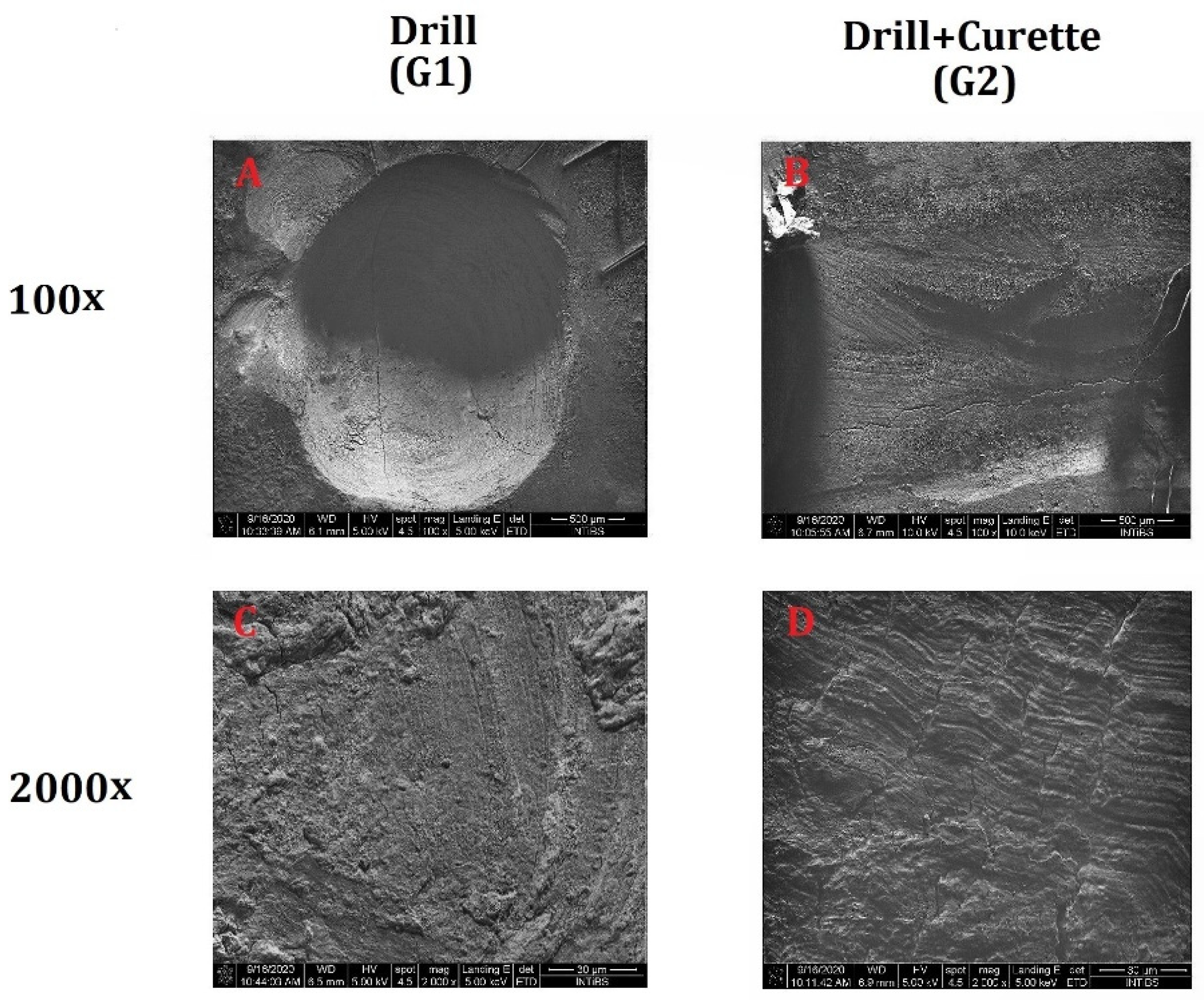
3.1. Effects of the Drill and Curette on Dentin Structure
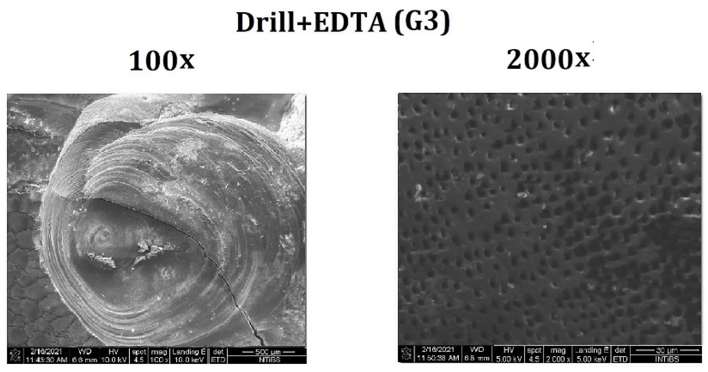
3.2. EDTA Application on the Dentin Surface
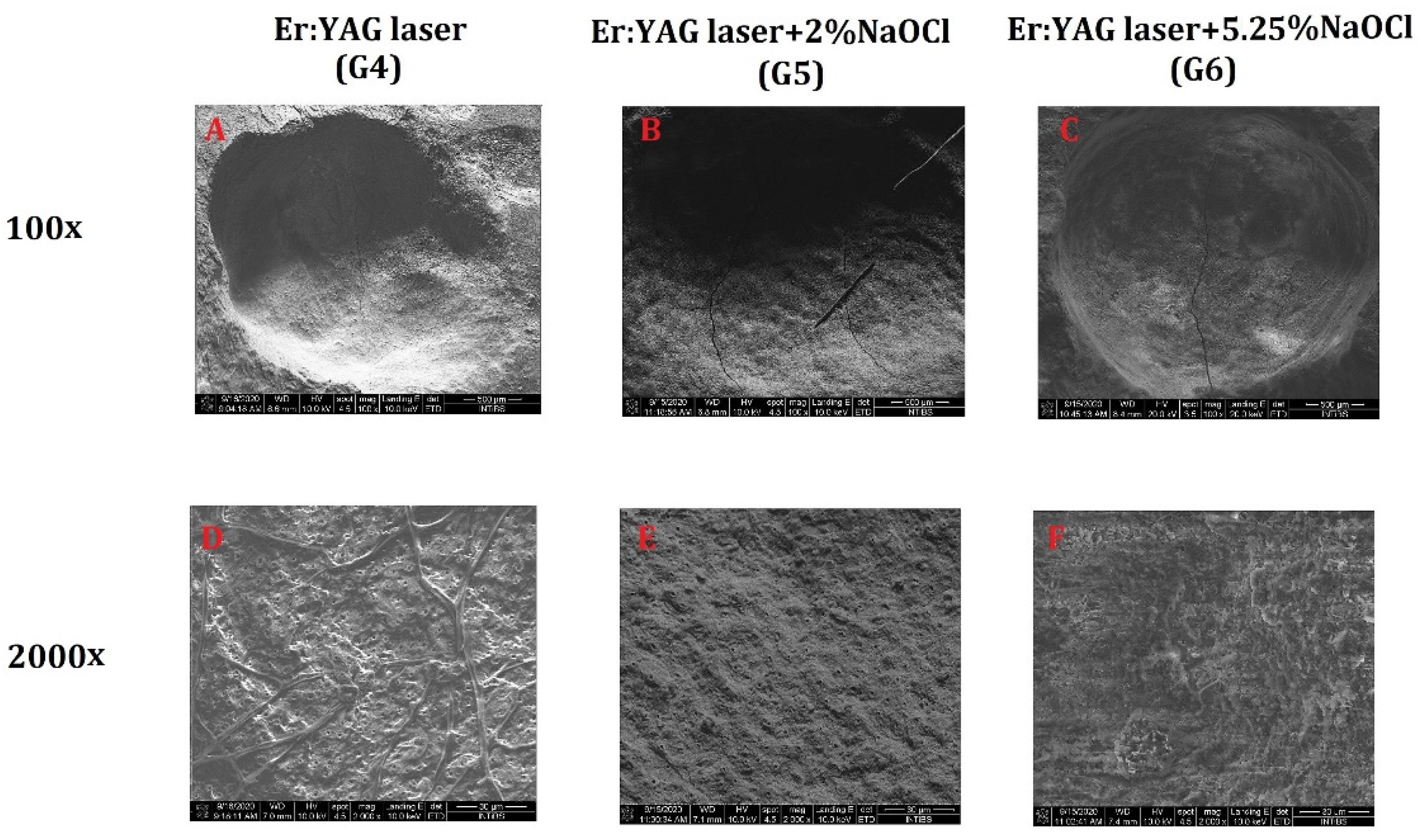
3.3. Er:YAG Laser Applicaion on the Dentin Surface
3.4. Semi-Quantitative Evaluation
4. Discussion
5. Conclusions
Author Contributions
Funding
Institutional Review Board Statement
Informed Consent Statement
Data Availability Statement
Conflicts of Interest
References
- Zakrzewski, W.; Dobrzynski, M.; Kuropka, P.; Matys, J.; Malecka, M.; Kiryk, J.; Rybak, Z.; Dominiak, M.; Grzech-Lesniak, K.; Wiglusz, K.; et al. Removal of Composite Restoration from the Root Surface in the Cervical Region Using Er: YAG Laser and Drill—In Vitro Study. Materials 2020, 13, 3027. [Google Scholar] [CrossRef]
- Nammour, S.; El Mobadder, M.; Maalouf, E.; Namour, M.; Namour, A.; Rey, G.; Matamba, P.; Matys, J.; Zeinoun, T.; Grzech-Leśniak, K. Clinical Evaluation of Diode (980 nm) Laser-Assisted Nonsurgical Periodontal Pocket Therapy: A Randomized Comparative Clinical Trial and Bacteriological Study. Photobiomodulation Photomed. Laser Surg. 2021, 39, 10–22. [Google Scholar] [CrossRef]
- Rosema, N.A.M.; Adam, R.; Grender, J.M.; Van der Sluijs, E.; Supranoto, S.C.; Van der Weijden, G.A. Gingival abrasion and recession in manual and oscillating-rotating power brush users. Int. J. Dent. Hyg. 2014, 12, 257–266. [Google Scholar] [CrossRef] [Green Version]
- McCracken, G.I.; Heasman, L.; Stacey, F.; Swan, M.; Steen, N.; De Jager, M.; Heasman, P.A. The impact of powered and manual toothbrushing on incipient gingival recession. J. Clin. Periodontol. 2009, 36, 950–957. [Google Scholar] [CrossRef]
- Zucchelli, G.; Mounssif, I. Periodontal plastic surgery. Periodontol. 2000 2015, 68, 333–368. [Google Scholar] [CrossRef]
- Chrysanthakopoulos, N.A. Gingival recession: Prevalence and risk indicators among young greek adults. J. Clin. Exp. Dent. 2014, 6, e243–e249. [Google Scholar] [CrossRef] [PubMed]
- Tugnait, A.; Clerehugh, V. Gingival recession—Its significance and management. J. Dent. 2001, 29, 381–394. [Google Scholar] [CrossRef]
- Tonetti, M.S.; D’Aiuto, F.; Nibali, L. Treatment of periodontitis and endothelial function. Jpn. J. Chest Dis. 2008, 67, 353. [Google Scholar]
- Lasho, D.J.; O’Leary, T.J.; Kafrawy, A.H. A Scanning Electron Microscope Study of the Effects of Various Agents on Instrumented Periodontally Involved Root Surfaces. J. Periodontol. 1983, 54, 210–220. [Google Scholar] [CrossRef]
- Aleo, J.J.; De Renzis, F.A.; Farber, P.A.; Varboncoeur, A.P. The Presence and Biologic Activity of Cementum-Bound Endotoxin. J. Periodontol. 1974, 45, 672–675. [Google Scholar] [CrossRef] [PubMed]
- Kassab, M.; Cohen, R.E. The effect of root modification and biomodification on periodontal therapy. Compend. Contin. Educ. Dent. 2003, 24, 31–34, 36. [Google Scholar]
- Matys, J.; Hadzik, J.; Dominiak, M. Schneiderian Membrane Perforation Rate and Increase in Bone Temperature during Maxillary Sinus Floor Elevation by Means of Er:YAG Laser–An Animal Study in Pigs. Implant Dent. 2017, 26, 238–244. [Google Scholar] [CrossRef]
- Matys, J.; Dominiak, M. Assessment of pain when uncovering implants with Er:YAG laser or scalpel for second stage surgery. Adv. Clin. Exp. Med. 2016, 25, 1179–1184. [Google Scholar] [PubMed] [Green Version]
- Matys, J.; Świder, K.; Flieger, R. Laser instant implant impression method: A case presentation. Dent. Med. Probl. 2017, 54, 101–106. [Google Scholar] [CrossRef] [Green Version]
- Matys, J.; Grzech-Leśniak, K. Dental Aerosol as a Hazard Risk for Dental Workers. Materials 2020, 13, 5109. [Google Scholar] [CrossRef] [PubMed]
- Grzech-Leśniak, K.; Matys, J. The Effect of Er:YAG Lasers on the Reduction of Aerosol Formation for Dental Workers. Materials 2021, 14, 2857. [Google Scholar] [CrossRef]
- Kiryk, J.; Matys, J.; Nikodem, A.; Burzyńska, K.; Grzech-Leśniak, K.; Dominiak, M.; Dobrzyński, M. The effect of er:Yag laser on a shear bond strength value of orthodontic brackets to enamel—A preliminary study. Materials 2021, 14, 2093. [Google Scholar] [CrossRef]
- Hibst, R.; Keller, U. Experimental studies of the application of the Er:YAG laser on dental hard substances: I. Measurement of the ablation rate. Lasers Surg. Med. 1989, 9, 338–344. [Google Scholar] [CrossRef]
- Ceballos, L.; Osorio, R.; Toledano, M.; Marshall, G.W. Microleakage of composite restorations after acid or Er-YAG laser cavity treatments. Dent. Mater. 2001, 17, 340–346. [Google Scholar] [CrossRef]
- Glockner, K.; Rumpler, J.; Ebeleseder, K.; Städtler, P. Intrapulpal temperature during preparation with the Er:YAG laser compared to the conventional burr: An in vitro study. J. Clin. Laser Med. Surg. 1998, 16, 153–157. [Google Scholar] [CrossRef]
- Grzech-Leśniak, K.; Bencharit, S.; Skrjanc, L.; Kanduti, D.; Matys, J.; Deeb, J.G. Utilization of Er:YAG Laser in Retrieving and Reusing of Lithium Disilicate and Zirconia Monolithic Crowns in Natural Teeth: An In Vitro Study. Appl. Sci. 2020, 10, 4357. [Google Scholar] [CrossRef]
- Grzech-Leśniak, K.; Matys, J.; Zmuda-Stawowiak, D.; Mroczka, K.; Dominiak, M.; Brugnera, A.; Gruber, R.; Romanos, G.E.G.E.; Sculean, A. Er:YAG Laser for Metal and Ceramic Bracket Debonding: An In Vitro Study on Intrapulpal Temperature, SEM, and EDS Analysis. Photomed. Laser Surg. 2018, 36, 595–600. [Google Scholar] [CrossRef] [PubMed]
- DenBesten, P.K.; White, J.M.; Pelino, J.E.P.; Furnish, G.; Silveira, A.; Parkins, F.M. The safety and effectiveness of an Er:YAG laser for caries removal and cavity preparation in children. Med. Laser Appl. 2001, 16, 215–222. [Google Scholar] [CrossRef]
- Attin, T. Methods for assessment of dental erosion. Monogr. Oral Sci. 2006, 20, 152–172. [Google Scholar]
- Joshi, M. Techniques to Evaluate Dental Erosion: A Systematic Review of Literature. J. Clin. Diagn. Res. 2016, 10, ZE01. [Google Scholar] [CrossRef]
- Esteves-Oliveira, M.; Zezell, D.M.; Apel, C.; Turbino, M.L.; Aranha, A.C.C.; Eduardo, C.D.P.; Gutknecht, N. Bond strength of self-etching primer to bur cut, Er,Cr:YSGG, and Er:YAG lased dental surfaces. Photomed. Laser Surg. 2007, 25, 373–380. [Google Scholar] [CrossRef] [PubMed]
- Kocherova, I.; Bryja, A.; Błochowiak, K.; Kaczmarek, M.; Stefańska, K.; Matys, J.; Grzech-Leśniak, K.; Dominiak, M.; Mozdziak, P.; Kempisty, B.; et al. Photobiomodulation with Red and Near-Infrared Light Improves Viability and Modulates Expression of Mesenchymal and Apoptotic-Related Markers in Human Gingival Fibroblasts. Materials 2021, 14, 3427. [Google Scholar] [CrossRef]
- Dompe, C.; Moncrieff, L.; Matys, J.; Grzech-Leśniak, K.; Kocherova, I.; Bryja, A.; Bruska, M.; Dominiak, M.; Mozdziak, P.; Skiba, T.H.I.; et al. Photobiomodulation—Underlying Mechanism and Clinical Applications. J. Clin. Med. 2020, 9, 1724. [Google Scholar] [CrossRef] [PubMed]
- Polson, A.M.; Frederick, G.T.; Ladenheim, S.; Hanes, P.J. The Production of a Root Surface Smear Layer by Instrumentation and its Removal by Citric Acid. J. Periodontol. 1984, 55, 443–446. [Google Scholar] [CrossRef]
- Brännström, M.; Johnson, G. Effects of various conditioners and cleaning agents on prepared dentin surfaces: A scanning electron microscopic investigation. J. Prosthet. Dent. 1974, 31, 422–430. [Google Scholar] [CrossRef]
- Leidal, T.I.; Eriksen, H.M. A scanning electron microscopic study of the effect of various cleansing agents on cavity walls in vitro. Eur. J. Oral Sci. 1979, 87, 443–449. [Google Scholar] [CrossRef]
- Eick, J.D.; Wilko, R.A.; Anderson, C.H.; Sorensen, S.E. Scanning Electron Microscopy of Cut Tooth Surfaces and Identification of Debris by Use of the Electron Microprobe. J. Dent. Res. 1970, 49, 1359–1368. [Google Scholar] [CrossRef]
- Demiryürek, E.Ö.; Külünk, Ş.; Saraç, D.; Yüksel, G.; Bulucu, B. Effect of different surface treatments on the push-out bond strength of fiber post to root canal dentin. Oral Surgery Oral Med. Oral Pathol. Oral Radiol. Endodontol. 2009, 108, e74–e80. [Google Scholar] [CrossRef]
- Guerisoli, D.M.Z.; Marchesan, M.A.; Walmsley, A.D.; Lumley, P.J.; Pecora, J.D. Evaluation of smear layer removal by EDTAC and sodium hypochlorite with ultrasonic agitation. Int. Endod. J. 2002, 35, 418–421. [Google Scholar] [CrossRef] [PubMed]
- Torabinejad, M.; Cho, Y.; Khademi, A.A.; Bakland, L.K.; Shabahang, S. The effect of various concentrations of sodium hypochlorite on the ability of MTAD to remove the smear layer. J. Endod. 2003, 29, 233–239. [Google Scholar] [CrossRef] [PubMed]
- Lo Giudice, G.; Lizio, A.; Lo Giudice, R.; Centofanti, A.; Rizzo, G.; Runci, M.; Alibrandi, A.; Cicciù, M. The effect of different cleaning protocols on post space: A SEM study. Int. J. Dent. 2016, 2016, 1907124. [Google Scholar] [CrossRef] [PubMed] [Green Version]
- Violich, D.R.; Chandler, N.P. The smear layer in endodontics—A review. Int. Endod. J. 2010, 43, 2–15. [Google Scholar] [CrossRef] [PubMed]
- Mohammadi, Z.; Shalavi, S.; Yaripour, S.; Kinoshita, J.I.; Manabe, A.; Kobayashi, M.; Giardino, L.; Palazzi, F.; Sharifi, F.; Jafarzadeh, H. Smear layer removing ability of root canal irrigation solutions: A review. J. Contemp. Dent. Pract. 2019, 20, 395–402. [Google Scholar] [CrossRef]
- Keller, U.; Hibst, R.; Geurtsen, W.; Schilke, R.; Heidemann, D.; Klaiber, B.; Raab, W.H.M. Erbium:YAG laser application in caries therapy. Evaluation of patient perception and acceptance. J. Dent. 1998, 26, 649–656. [Google Scholar] [CrossRef]
- Kuhn, K.; Rudolph, H.; Luthardt, R.G.; Stock, K.; Diebolder, R.; Hibst, R. Er:YAG laser activation of sodium hypochlorite for root canal soft tissue dissolution. Lasers Surg. Med. 2013, 45, 339–344. [Google Scholar] [CrossRef]
- Matys, J.; Dominiak, M.; Flieger, R. Energy and power density: A key factor in lasers studies. J. Clin. Diagnostic Res. 2015, 9, ZL01. [Google Scholar] [CrossRef] [PubMed]
- Dumitriu, D.; Dobre, T. Effects of temperature and hypochlorite concentration on the rate of collagen dissolution. J. Endod. 2015, 41, 903–906. [Google Scholar] [CrossRef]
- Reshma Raj, V.; Varma, R.; Sureshkumar, J.; Kumaran, P.; Xavier, A.; Madhavan, M. Comparison of cytotoxicity and smear layer removal efficacy of triphala (an Indian ayurvedic herbal formulation) and 5.25% sodium hypochlorite as root canal irrigants: An in vitro study. J. Indian Soc. Pedod. Prev. Dent. 2020, 38, 343–349. [Google Scholar] [PubMed]
- Ballal, V.; Rao, S.; Al-Haj Husain, N.; Özcan, M. Evaluation of Smear Layer Removal Using Different Irrigation Methods In Root Canals. Eur. J. Prosthodont. Restor. Dent. 2019, 27, 97–102. [Google Scholar]
- Cardoso, L.R.; Baldasso, F.E.R.; Delai, D.; Montagner, F.; Kopper, P.M.P. Effect of EDTA, sodium, and calcium hypochlorite on the inorganic component of root canal dentin: A SEM analysis. Microsc. Res. Tech. 2019, 82, 128–133. [Google Scholar] [CrossRef]
- Deeb, J.G.; Smith, J.; Belvin, B.R.; Grzech-Leśniak, K.; Lewis, J. Er:YAG laser irradiation reduces microbial viability when used in combination with irrigation with sodium hypochlorite, chlorhexidine, and hydrogen peroxide. Microorganisms 2019, 7, 612. [Google Scholar] [CrossRef] [Green Version]
- Scholz, K.J.; Bittner, A.; Cieplik, F.; Hiller, K.-A.; Schmalz, G.; Buchalla, W.; Federlin, M. Micromorphology of the Adhesive Interface of Self-Adhesive Resin Cements to Enamel and Dentin. Materials 2021, 14, 492. [Google Scholar] [CrossRef] [PubMed]

| Group | Drill G1 | Drill + Curette (G2) | Drill + EDTA (G3) | Er:YAG Laser (G4) | Er:YAG Laser + 2% NaOCl (G5) | Er:YAG Laser + 5.25% NaOCl (G6) |
|---|---|---|---|---|---|---|
| Quality of preparation (0–2) | 1 | 1 | 1 | 2 | 2 | 2 |
| Exposure of dentinal tubules (0–1) | 0 | 0 | 1 | 1 | 1 | 1 |
| Absence of smear layer (0–1) | 0 | 0 | 1 | 1 | 1 | 1 |
| Repeatability of results obtained (Similar sample image) (0–2) | 0 | 0 | 1 | 2 | 2 | 2 |
| Scoring | 1 a,b,c | 1 a,b,c | 4 d | 6 a | 6 b | 6 c |
| p Value | G4 vs. G2, G1 p < 0.05 G5 vs. G2, G1 p < 0.05 G6 vs. G2, G1 p < 0.05 | |||||
Publisher’s Note: MDPI stays neutral with regard to jurisdictional claims in published maps and institutional affiliations. |
© 2021 by the authors. Licensee MDPI, Basel, Switzerland. This article is an open access article distributed under the terms and conditions of the Creative Commons Attribution (CC BY) license (https://creativecommons.org/licenses/by/4.0/).
Share and Cite
Kiryk, J.; Matys, J.; Grzech-Leśniak, K.; Dominiak, M.; Małecka, M.; Kuropka, P.; Wiglusz, R.J.; Dobrzyński, M. SEM Evaluation of Tooth Surface after a Composite Filling Removal Using Er:YAG Laser, Drills with and without Curettes, and Optional EDTA or NaOCl Conditioning. Materials 2021, 14, 4469. https://doi.org/10.3390/ma14164469
Kiryk J, Matys J, Grzech-Leśniak K, Dominiak M, Małecka M, Kuropka P, Wiglusz RJ, Dobrzyński M. SEM Evaluation of Tooth Surface after a Composite Filling Removal Using Er:YAG Laser, Drills with and without Curettes, and Optional EDTA or NaOCl Conditioning. Materials. 2021; 14(16):4469. https://doi.org/10.3390/ma14164469
Chicago/Turabian StyleKiryk, Jan, Jacek Matys, Kinga Grzech-Leśniak, Marzena Dominiak, Małgorzata Małecka, Piotr Kuropka, Rafał J. Wiglusz, and Maciej Dobrzyński. 2021. "SEM Evaluation of Tooth Surface after a Composite Filling Removal Using Er:YAG Laser, Drills with and without Curettes, and Optional EDTA or NaOCl Conditioning" Materials 14, no. 16: 4469. https://doi.org/10.3390/ma14164469
APA StyleKiryk, J., Matys, J., Grzech-Leśniak, K., Dominiak, M., Małecka, M., Kuropka, P., Wiglusz, R. J., & Dobrzyński, M. (2021). SEM Evaluation of Tooth Surface after a Composite Filling Removal Using Er:YAG Laser, Drills with and without Curettes, and Optional EDTA or NaOCl Conditioning. Materials, 14(16), 4469. https://doi.org/10.3390/ma14164469