
Long-GRIN-Lens Microendoscopy Enabled by Wavefront Shaping for a Biomedical Microdevice: An Analytical Investigation


Abstract
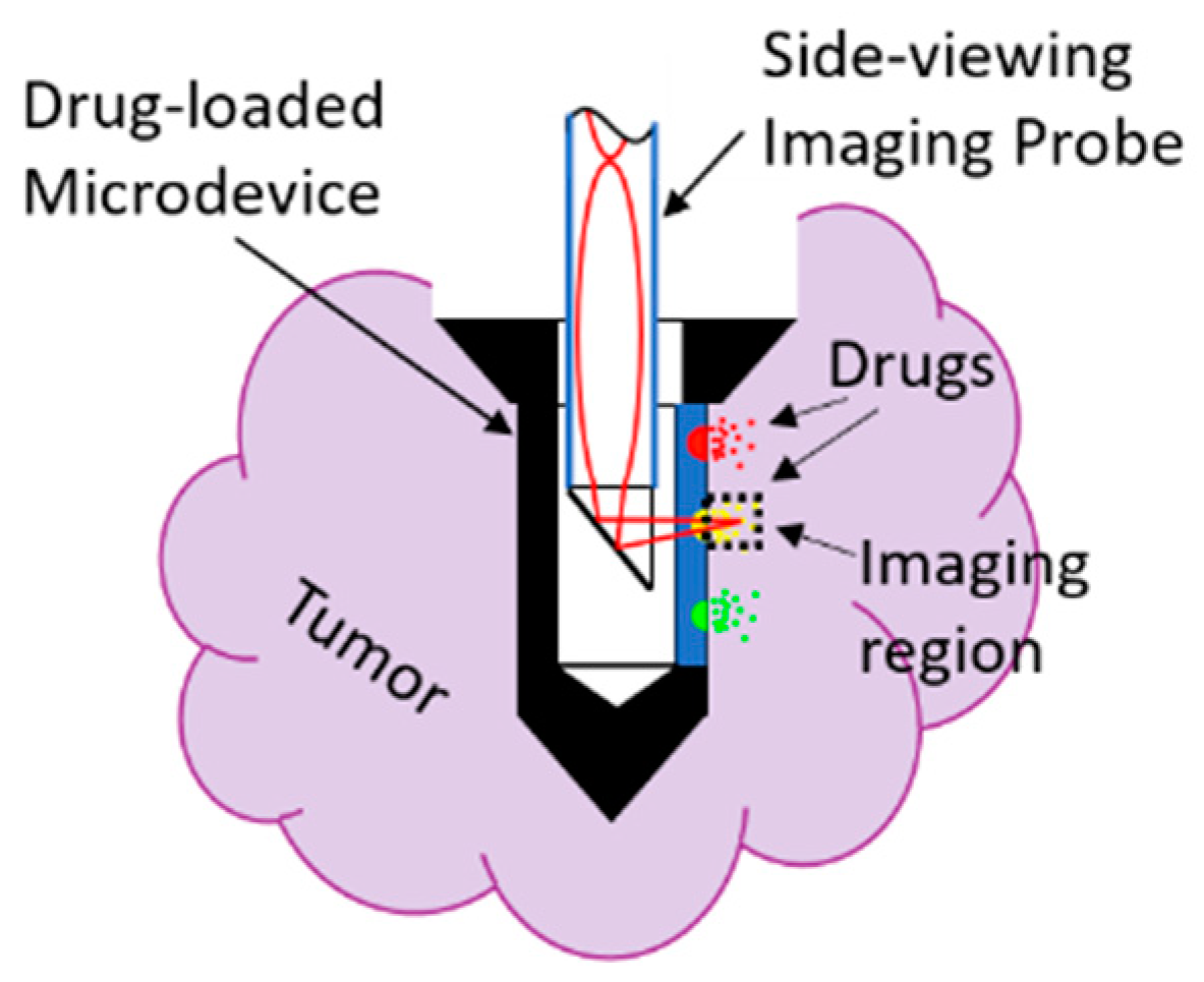
1. Introduction
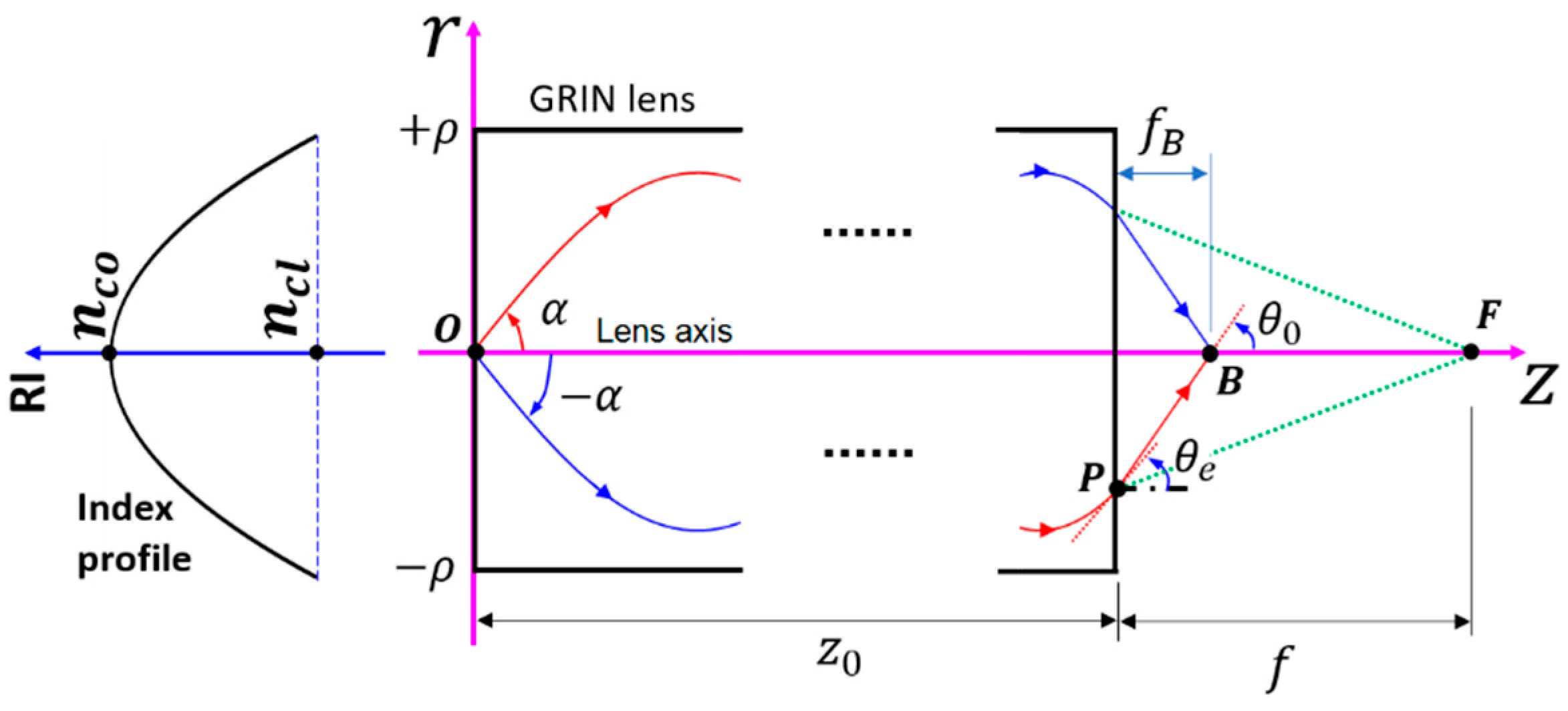
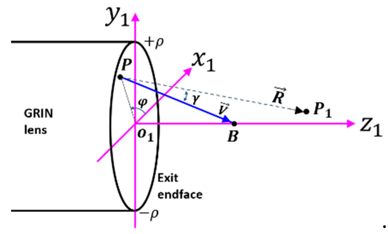
2. Theory
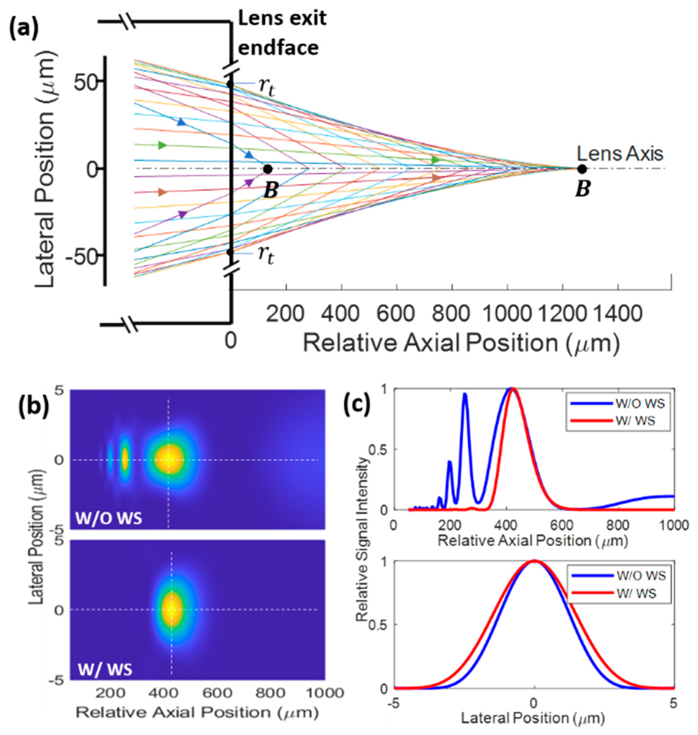
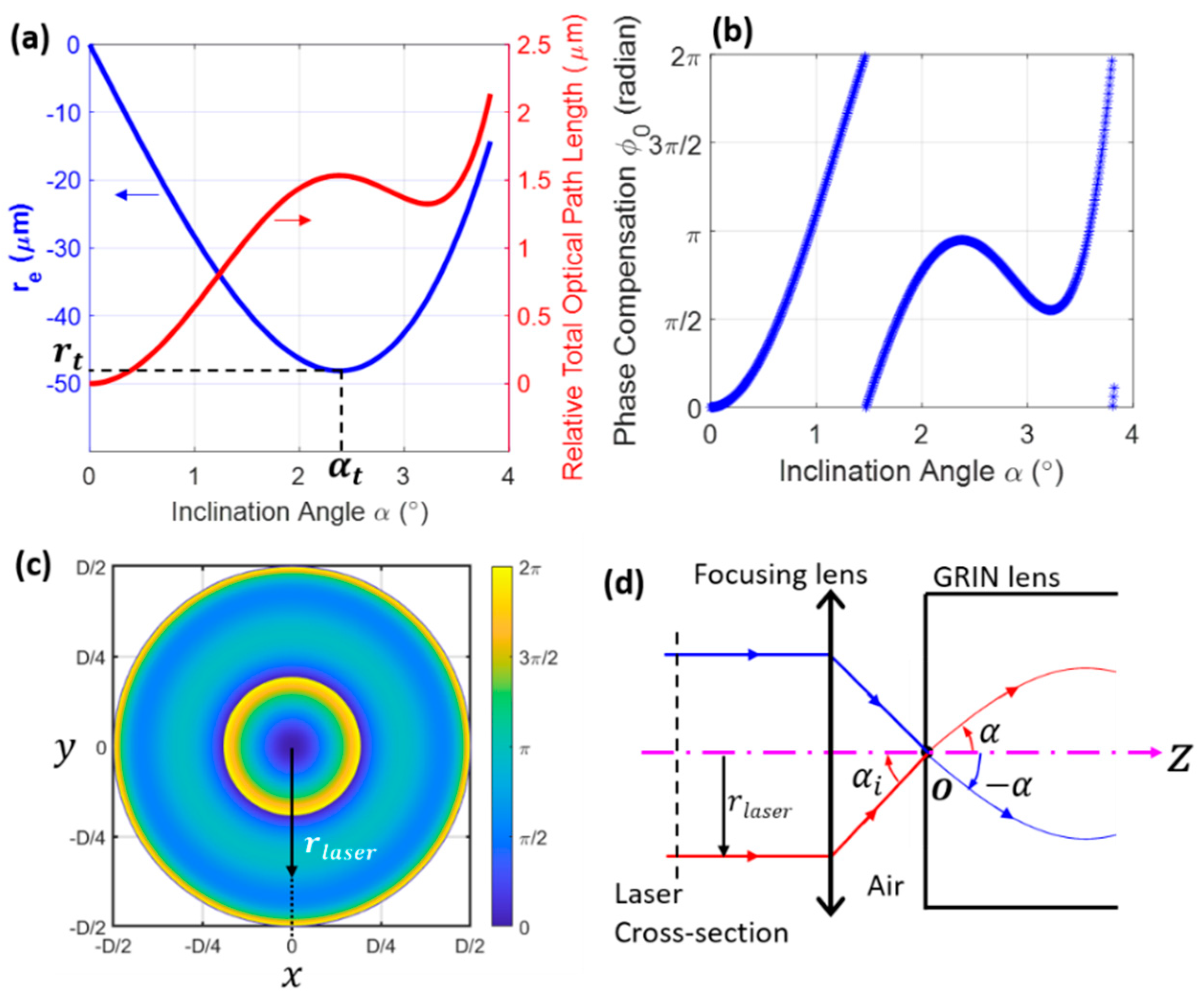
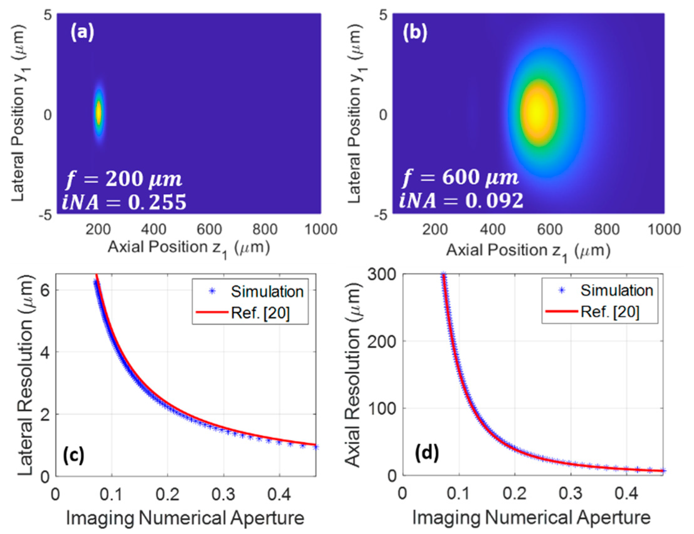
3. Results and Discussion
4. Conclusions
Author Contributions
Funding
Institutional Review Board Statement
Informed Consent Statement
Data Availability Statement
Conflicts of Interest
References
- Jonas, O.H.; Landry, H.M.; Fuller, J.E.; Santini, J.T.S., Jr.; Baselga, J.; Tepper, R.I.; Cima, M.J.; Langer, R. An implantable microdevice to perform high-throughput in vivo drug sensitivity testing in tumors. Sci. Transl. Med. 2015, 7, 284ra57. [Google Scholar] [CrossRef] [PubMed]
- Jonas, O.; Calligaris, D.; Methuku, K.R.; Poe, M.; Francois, J.P.; Tranghese, F.; Changelian, A.; Sieghart, W.; Ernst, M.; Krummel, D.P.; et al. First In Vivo Testing of Compounds Targeting Group 3 Medulloblastomas Using an Implantable Microdevice as a New Paradigm for Drug Development. J. Biomed. Nanotechnol. 2016, 12, 1297–1302. [Google Scholar] [CrossRef] [PubMed]
- Davidson, S.M.; Jonas, O.; Keibler, M.A.; Hou, H.W.; Luengo, A.; Mayers, J.R.; Wyckoff, J.; Del Rosario, A.M.; Whitman, M.; Chin, C.; et al. Direct evidence for cancer-cell-autonomous extracellular protein catabolism in pancreatic tumors. Nat. Med. 2017, 23, 235–241. [Google Scholar] [CrossRef] [PubMed]
- Bhagavatula, S.; Thompson, D.; Ahn, S.; Upadhyaya, K.; Lammers, A.; Deans, K.; Dominas, C.; Ferland, B.; Valvo, V.; Liu, G.; et al. A Miniaturized Platform for Multiplexed Drug Response Imaging in Live Tumors. Cancers 2021, 13, 653. [Google Scholar] [CrossRef]
- Lee, W.M.S.; Yun, S.H. Adaptive aberration correction of GRIN lenses for confocal endomicroscopy. Opt. Lett. 2011, 36, 4608–4610. [Google Scholar] [CrossRef]
- Kim, P.; Puoris’Haag, M.; Co^té, D.; Lin, C.P.; Yun, S.-H. In vivo confocal and multiphoton microendoscopy. J. Biomed. Opt. 2008, 13, 010501. [Google Scholar] [CrossRef] [PubMed]
- Kim, J.K.; Lee, W.M.S.; Kim, P.; Choi, M.; Jung, K.; Kim, S.; Yun, S.H. Fabrication and operation of GRIN probes for in vivo fluorescence cellular imaging of internal organs in small animals. Nat. Protoc. 2012, 7, 1456–1469. [Google Scholar] [CrossRef]
- Jung, J.C.; Schnitzer, M.J. Multiphoton endoscopy. Opt. Lett. 2003, 28, 902–904. [Google Scholar] [CrossRef]
- Dilipkumar, A.; Al-Shemmary, A.; Kreiß, L.; Cvecek, K.; Carlé, B.; Knieling, F.; Menezes, J.G.; Thoma, O.; Schmidt, M.; Neurath, M.F.; et al. Label-Free Multiphoton Endomicroscopy for Minimally Invasive In Vivo Imaging. Adv. Sci. 2019, 6, 1801735. [Google Scholar] [CrossRef]
- Wang, T.; Li, Q.; Xiao, P.; Ahn, J.; Kim, Y.E.; Park, Y.; Kim, M.; Song, M.; Chung, E.; Chung, W.K.; et al. Gradient index lens based combined two-photon microscopy and optical coherence tomography. Opt. Express 2014, 22, 12962–12970. [Google Scholar] [CrossRef] [PubMed]
- Huland, D.M.; Brown, C.M.; Howard, S.S.; Ouzounov, D.G.; Pavlova, I.; Wang, K.; Rivera, D.R.; Webb, W.W.; Xu, C. In vivo imaging of unstained tissues using long gradient index lens multiphoton endoscopic systems. Biomed. Opt. Express 2012, 3, 1077–1085. [Google Scholar] [CrossRef]
- Flaes, D.E.B.; Stopka, J.; Turtaev, S.; De Boer, J.F.; Tyc, T.; Čižmár, T. Robustness of Light-Transport Processes to Bending Deformations in Graded-Index Multimode Waveguides. Phys. Rev. Lett. 2018, 120, 233901. [Google Scholar] [CrossRef] [PubMed]
- Savelyev, D.A.; Ustinov, A.V.; Khonina, S.N.; Kazanskiy, N.L. Layered lens with a linear dependence of the refractive index change. Optical Technol. Telecommun. 2015 2016, 9807, 98070. [Google Scholar] [CrossRef]
- Snyder, A.W.; Love, J.D. Optical Waveguide Theory; Chapman and Hall: New York, NY, USA, 1983. [Google Scholar]
- Resisi, S.; Viernik, Y.; Popoff, S.M.; Bromberg, Y. Wavefront shaping in multimode fibers by transmission matrix engineering. APL Photon- 2020, 5, 036103. [Google Scholar] [CrossRef]
- Begel, L.; Galstian, T. Dynamic compensation of gradient index rod lens aberrations by using liquid crystals. Appl. Opt. 2018, 57, 7618–7621. [Google Scholar] [CrossRef]
- Karsakov, A.V.; Khonina, S.N. Investigation of focusing of the fundamental linearly polarized mode using microrelief on the end of an optical fiber. In Optical Technologies for Telecommunications 2013; SPIE—International Society for Optics and Photonics: Bellingham, WA, USA, 2014; Volume 9156, p. 91560A. [Google Scholar]
- Williams, H.E.; Freppon, D.J.; Kuebler, S.M.; Rumpf, R.C.; Melino, M.A. Fabrication of three-dimensional micro-photonic structures on the tip of optical fibers using SU-8. Opt. Express 2011, 19, 22910–22922. [Google Scholar] [CrossRef]
- Gu, M.; Gu, P.M. Advanced Optical Imaging Theory; Springer Science and Business Media LLC: Berlin, Germany, 2000; Volume 75. [Google Scholar]
- Zipfel, W.; Williams, R.M.; Webb, W.W. Nonlinear magic: Multiphoton microscopy in the biosciences. Nat. Biotechnol. 2003, 21, 1369–1377. [Google Scholar] [CrossRef] [PubMed]
- Sivankutty, S.; Andresen, E.R.; Cossart, R.; Bouwmans, G.; Monneret, S.; Rigneault, H. Ultra-thin rigid endoscope: Two-photon imaging through a graded-index multi-mode fiber. Opt. Express 2016, 24, 825–841. [Google Scholar] [CrossRef]
- Caravaca-Aguirre, A.M.; Piestun, R. Single multimode fiber endoscope. Opt. Express 2017, 25, 1656–1665. [Google Scholar] [CrossRef] [PubMed]
- Ploschner, M.; Tyc, T.; Čižmár, T. Seeing through chaos in multimode fibres. Nat. Photon- 2015, 9, 529–535. [Google Scholar] [CrossRef]
- Papadopoulos, I.N.; Farahi, S.; Moser, C.; Psaltis, D.; Papadopoulos, I.N.; Farahi, S.; Moser, C.; Psaltis, D. High-resolution, lensless endoscope based on digital scanning through a multimode optical fiber. Biomed. Opt. Express 2013, 4, 260–270. [Google Scholar] [CrossRef] [PubMed]
- Borhani, N.; Kakkava, E.; Moser, C.; Psaltis, D. Learning to see through multimode fibers. Optica 2018, 5, 960–966. [Google Scholar] [CrossRef]
- Gordon, G.S.D.; Gataric, M.; Ramos, A.G.C.P.; Mouthaan, R.; Williams, C.; Yoon, J.; Wilkinson, T.D.; Bohndiek, S.E. Characterizing Optical Fiber Transmission Matrices Using Metasurface Reflector Stacks for Lensless Imaging without Distal Access. Phys. Rev. X 2019, 9, 041050. [Google Scholar] [CrossRef]


Publisher’s Note: MDPI stays neutral with regard to jurisdictional claims in published maps and institutional affiliations. |
© 2021 by the authors. Licensee MDPI, Basel, Switzerland. This article is an open access article distributed under the terms and conditions of the Creative Commons Attribution (CC BY) license (https://creativecommons.org/licenses/by/4.0/).
Share and Cite
Liu, G.; Kang, J.W.; Jonas, O. Long-GRIN-Lens Microendoscopy Enabled by Wavefront Shaping for a Biomedical Microdevice: An Analytical Investigation. Materials 2021, 14, 3392. https://doi.org/10.3390/ma14123392
Liu G, Kang JW, Jonas O. Long-GRIN-Lens Microendoscopy Enabled by Wavefront Shaping for a Biomedical Microdevice: An Analytical Investigation. Materials. 2021; 14(12):3392. https://doi.org/10.3390/ma14123392
Chicago/Turabian StyleLiu, Guigen, Jeon Woong Kang, and Oliver Jonas. 2021. "Long-GRIN-Lens Microendoscopy Enabled by Wavefront Shaping for a Biomedical Microdevice: An Analytical Investigation" Materials 14, no. 12: 3392. https://doi.org/10.3390/ma14123392
APA StyleLiu, G., Kang, J. W., & Jonas, O. (2021). Long-GRIN-Lens Microendoscopy Enabled by Wavefront Shaping for a Biomedical Microdevice: An Analytical Investigation. Materials, 14(12), 3392. https://doi.org/10.3390/ma14123392