
Revealing Inflammatory Indications Induced by Titanium Alloy Wear Debris in Periprosthetic Tissue by Label-Free Correlative High-Resolution Ion, Electron and Optical Microspectroscopy

 , ,
, ,  , , and
Abstract

1. Introduction
2. Materials and Methods
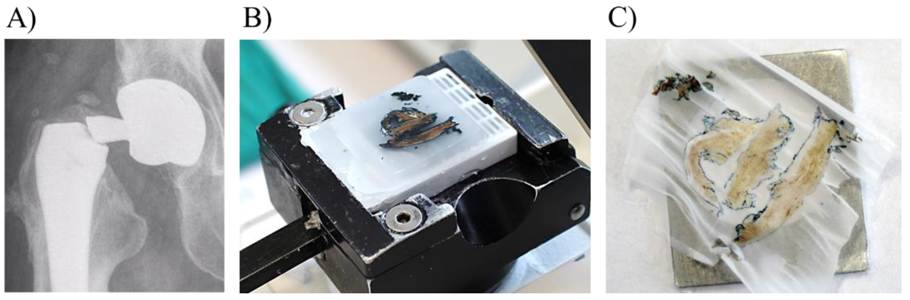
2.1. Preparation of Tissue Samples
2.2. Hybrid Fluorescence Confocal Laser Scanning Microscopy (CLSM) and NIR Reflectance Confocal Microscopy (RCM)
2.3. Fluorescence Lifetime (FLIM) and Fluorescence Hyperspectral Imaging (fHSI)
2.4. Helium Ion Microscopy (HIM)
2.5. Scanning Electron Microscopy with Energy-Dispersive X-ray Spectroscopy (SEM-EDS)
2.6. Particle Induced X-ray Emission (Micro-PIXE)
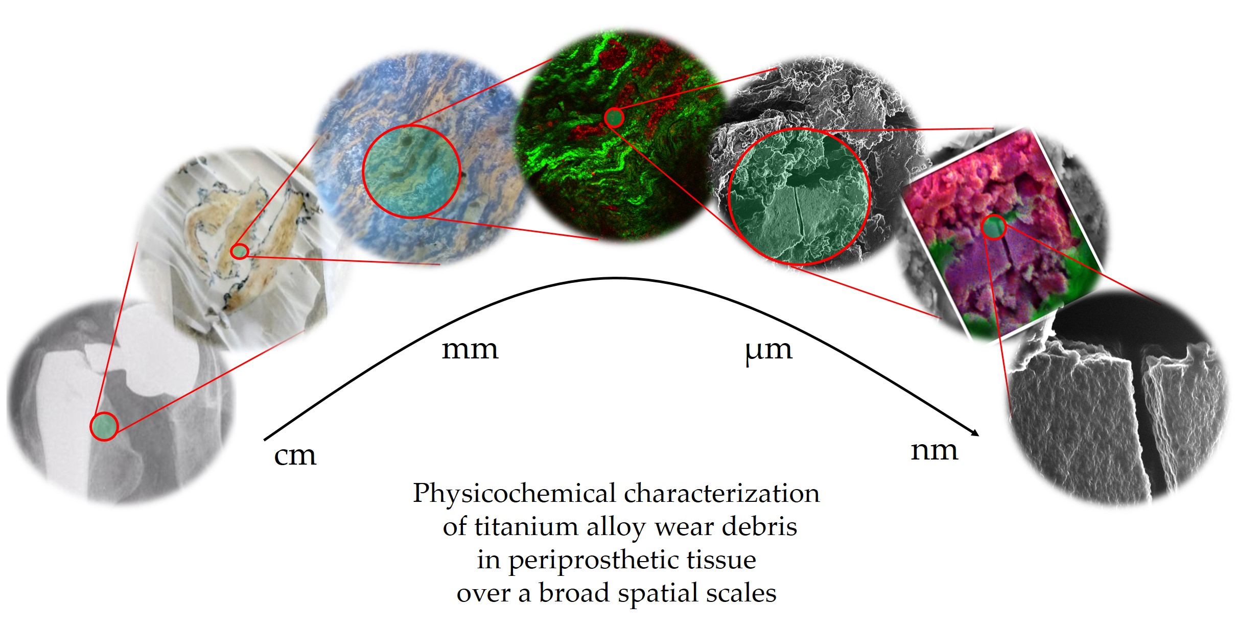
3. Results and Discussion
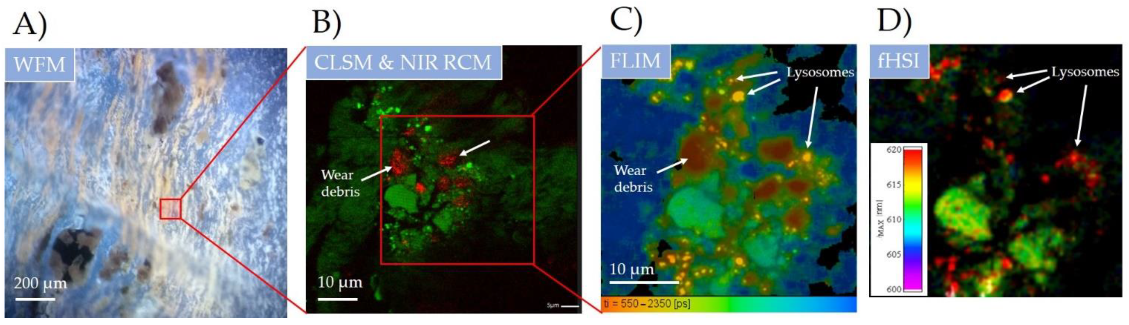
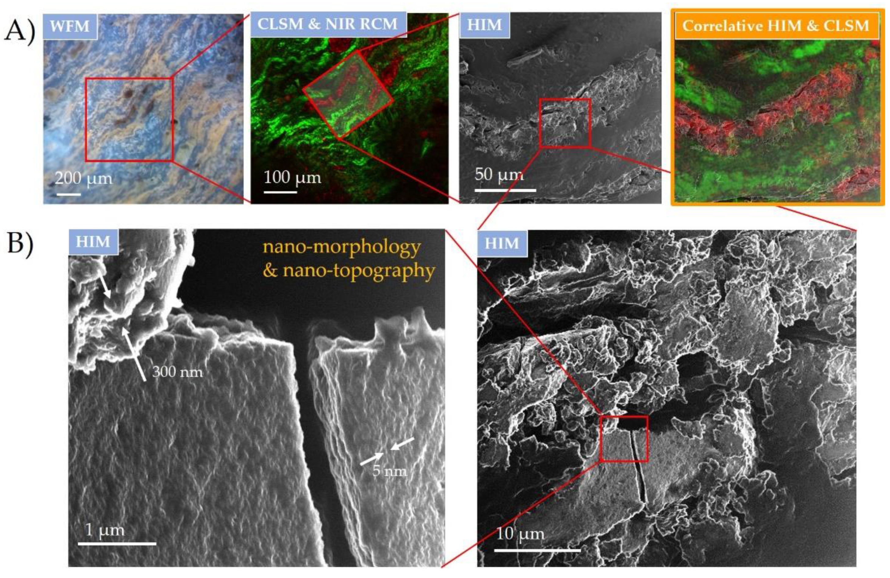
3.1. Wear Debris Cause Oxidative Stress in the Nearby Biological Tissue Detected by Label-Free Multimodal Optical Microspectroscopy
3.2. Wear-Debris Nanotopography Detected by HIM Reveals Significant Fretting and Corrosion That Can Affect Biological Tissue
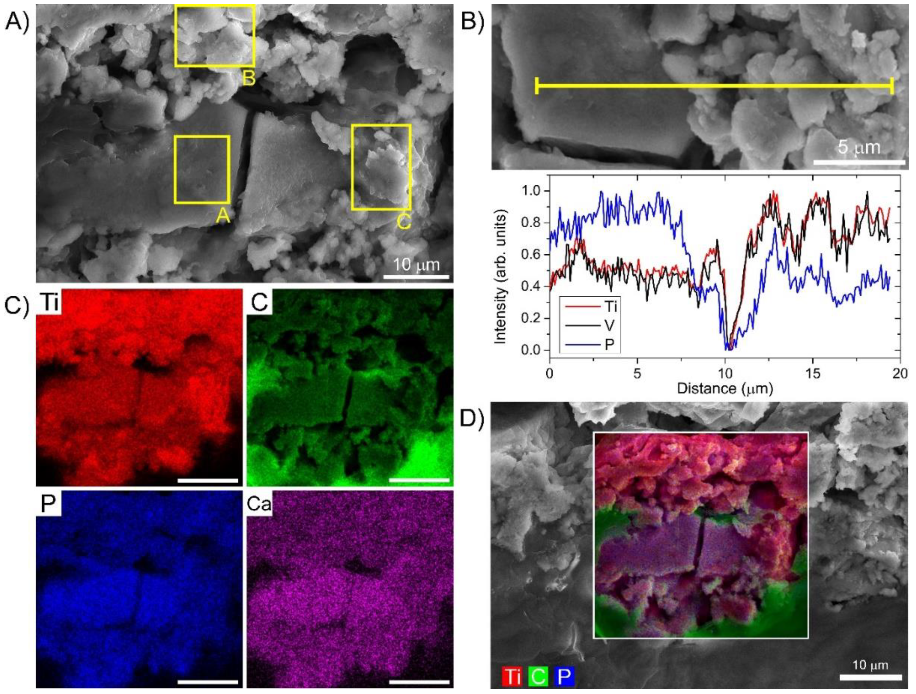
3.3. Preferential Hydroxyapatite Layer Formation and Selective Metal Leaching Detected by High-Resolution SEM-EDS on the Same Wear Debris
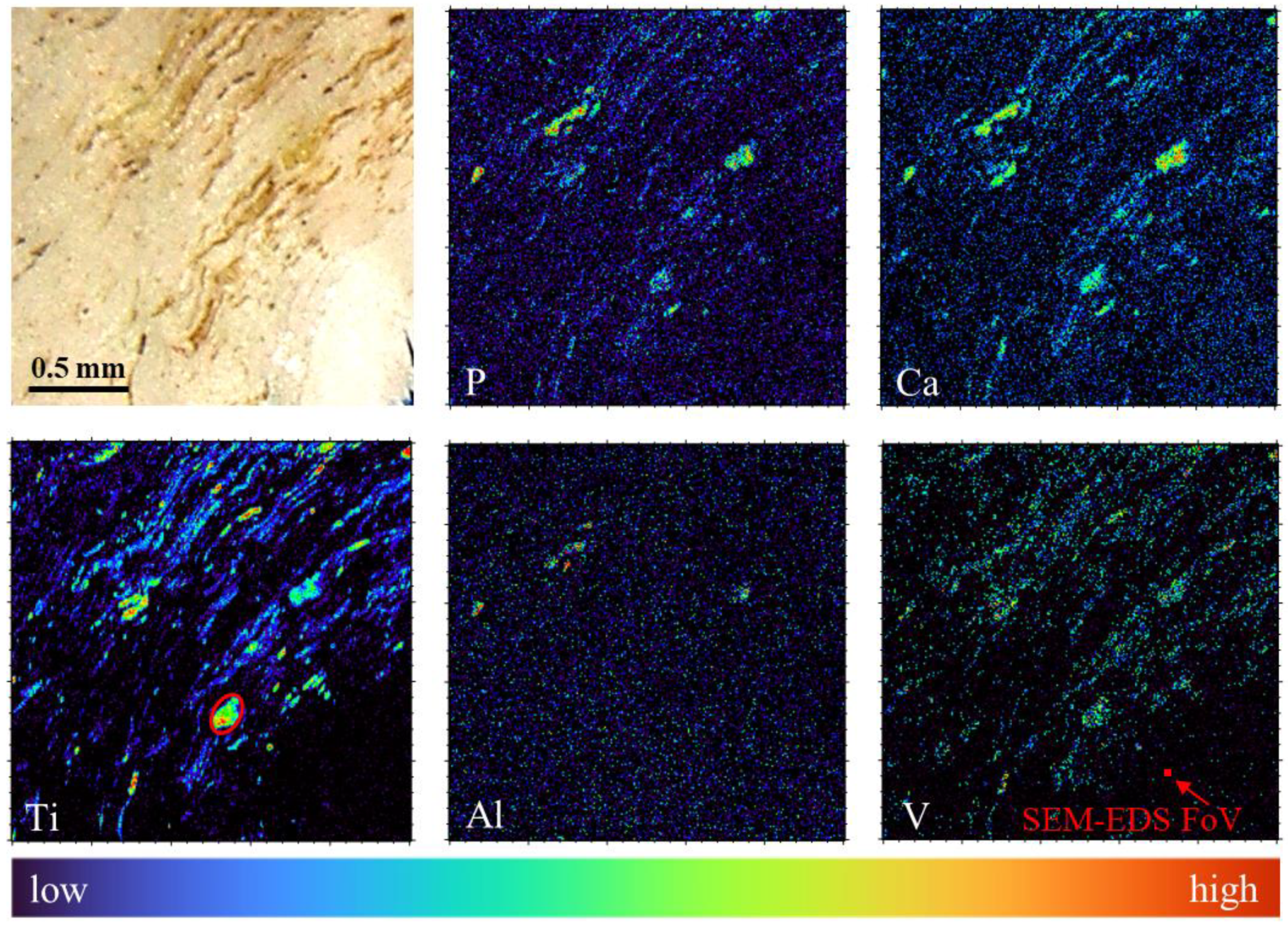
3.4. Confirmation of Strong Al and V Leaching from Wear Debris by Micro-PIXE
4. Conclusions
Supplementary Materials
Author Contributions
Funding
Institutional Review Board Statement
Informed Consent Statement
Data Availability Statement
Acknowledgments
Conflicts of Interest
References
- Cobelli, N.; Scharf, B.; Crisi, G.M.; Hardin, J.; Santambrogio, L. Mediators of the inflammatory response to joint replacement devices. Nat. Rev. Rheumatol. 2011, 7, 600–608. [Google Scholar] [CrossRef]
- Goodman, S.B.; Gallo, J.; Gibon, E.; Takagi, M. Diagnosis and management of implant debris-associated inflammation. Expert Rev. Med. Devices 2020, 17, 41–56. [Google Scholar] [CrossRef] [PubMed]
- Goodman, S.B.; Gibon, E.; Yao, Z. The basic science of periprosthetic osteolysis. Instr. Course Lect. 2013, 62, 201–206. [Google Scholar]
- Asri, R.I.M.; Harun, W.S.W.; Samykano, M.; Lah, N.A.C.; Ghani, S.A.C.; Tarlochan, F.; Raza, M.R. Corrosion and surface modification on biocompatible metals: A review. Mater. Sci. Eng. C 2017, 77, 1261–1274. [Google Scholar] [CrossRef]
- Xia, Z.; Ricciardi, B.F.; Liu, Z.; von Ruhland, C.; Ward, M.; Lord, A.; Hughes, L.; Goldring, S.R.; Purdue, E.; Murray, D.; et al. Nano-analyses of wear particles from metal-on-metal and non-metal-on-metal dual modular neck hip arthroplasty. Nanomed. Nanotechnol. Biol. Med. 2017, 13, 1205–1217. [Google Scholar] [CrossRef]
- Kovač, S.; Mavčič, B.; Kotnik, M.; Levašič, V.; Sirše, M.; Fokter, S.K. What factors are associated with neck fracture in one commonly used bimodular THA design? A multicenter, nationwide study in Slovenia. Clin. Orthop. Relat. Res. 2019, 477, 1324–1332. [Google Scholar] [CrossRef]
- Cooper, H.J.; Urban, R.M.; Wixson, R.L.; Meneghini, R.M.; Jacobs, J.J. Adverse local tissue reaction arising from corrosion at the femoral neck-body junction in a dual-taper stem with a cobalt-chromium modular neck. J. Bone Jt. Surg. Am. 2013, 95, 865–872. [Google Scholar] [CrossRef] [PubMed]
- Molloy, D.O.; Munir, S.; Jack, C.M.; Cross, M.B.; Walter, W.L.; Walter, W.K. Fretting and corrosion in modular-neck total hip arthroplasty femoral stems. J. Bone Jt. Surg. Am. 2014, 96, 488–493. [Google Scholar] [CrossRef]
- Miloŝev, L.; Antoliĉ, V.; Minoviĉ, A.; Cör, A.; Herman, S.; Pavlovcic, V.; Campbell, P. Extensive metallosis and necrosis in failed prostheses with cemented titanium-alloy stems and ceramic heads. J. Bone Jt. Surg. Br. 2000, 82, 352–357. [Google Scholar] [CrossRef]
- Polyzois, I.; Nikolopoulos, D.; Michos, I.; Patsouris, E.; Theocharis, S. Local and systemic toxicity of nanoscale debris particles in total hip arthroplasty. J. Appl. Toxicol. 2012, 32, 255–269. [Google Scholar] [CrossRef]
- Howie, D.W.; Neale, S.D.; Haynes, D.R.; Holubowycz, O.T.; McGee, M.A.; Solomon, L.B.; Callary, S.A.; Atkins, G.J.; Findlay, D.M. Periprosthetic osteolysis after total hip replacement: Molecular pathology and clinical management. Inflammopharmacology 2013, 21, 389–396. [Google Scholar] [CrossRef]
- Warashina, H.; Sakano, S.; Kitamura, S.; Yamauchi, K.I.; Yamaguchi, J.; Ishiguro, N.; Hasegawa, Y. Biological reaction to alumina, zirconia, titanium and polyethylene particles implanted onto murine calvaria. Biomaterials 2003, 24, 3655–3661. [Google Scholar] [CrossRef]
- Fokter, S.K.; Moličnik, A.; Kavalar, R.; Pelicon, P.; Rudolf, R.; Gubeljak, N. Why do some titanium-alloy total hip arthroplasty modular necks fail? J. Mech. Behav. Biomed. Mater. 2017, 69, 107–114. [Google Scholar] [CrossRef]
- Fokter, S.K.; Zajc, J.; Merc, M. Interchangeable neck failures of bi-modular femoral stems in primary total hip arthroplasty cannot be predicted from serum trace element analysis. Int. Orthop. 2020. [Google Scholar] [CrossRef]
- Fedarko, N.S. The biology of aging and frailty. Clin. Geriatr. Med. 2011, 27, 27–37. [Google Scholar] [CrossRef] [PubMed]
- Pisanu, F.; Doria, C.; Andreozzi, M.; Bartoli, M.; Saderi, L.; Sotgiu, G.; Tranquilli Leali, P. Pleomorphic clinical spectrum of metallosis in total hip arthroplasty. Int. Orthop. 2019, 43, 85–96. [Google Scholar] [CrossRef] [PubMed]
- Briant-Evans, T.W.; Lyle, N.; Barbur, S.; Hauptfleisch, J.; Amess, R.; Pearce, A.R.; Conn, K.S.; Stranks, G.J.; Britton, J.M. A longitudinal study of MARS MRI scanning of soft-tissue lesions around metal-on-metal total hip arthroplasties and disease progression. Bone Jt. J. 2015, 97-B, 1328–1337. [Google Scholar] [CrossRef] [PubMed]
- Koff, M.F.; Esposito, C.; Shah, P.; Miranda, M.; Baral, E.; Fields, K.; Bauer, T.; Padgett, D.E.; Wright, T.; Potter, H.G. MRI of THA correlates with implant wear and tissue reactions: A cross-sectional study. Clin. Orthop. Relat. Res. 2019, 477, 159–174. [Google Scholar] [CrossRef] [PubMed]
- Tam, H.H.; Bhaludin, B.; Rahman, F.; Weller, A.; Ejindu, V.; Parthipun, A. SPECT-CT in total hip arthroplasty. Clin. Radiol. 2014, 69, 82–95. [Google Scholar] [CrossRef]
- Drynda, A.; Singh, G.; Buchhorn, G.H.; Awiszus, F.; Ruetschi, M.; Feuerstein, B.; Kliche, S.; Lohmann, C.H. Metallic wear debris may regulate CXCR4 expression in vitro and in vivo. J. Biomed. Mater. Res. Part A 2015, 103, 1940–1948. [Google Scholar] [CrossRef]
- Goldstein, J.I.; Newbury, D.E.; Michael, J.R.; Ritchie, N.W.M.; Scott, J.H.J.; Joy, D.C. Energy dispersive X-ray spectrometry: Physical principles and user-selected parameters. In Scanning Electron Microscopy and X-ray Microanalysis; Goldstein, J.I., Newbury, D.E., Michael, J.R., Ritchie, N.W.M., Scott, J.H.J., Joy, D.C., Eds.; Springer: New York, NY, USA, 2018; pp. 209–234. ISBN 978-1-4939-6676-9. [Google Scholar]
- Verma, H.R. (Ed.) X-ray fluorescence (XRF) and particle-induced X-ray emission (PIXE). In Atomic and Nuclear Analytical Methods: XRF, Mössbauer, XPS, NAA and B63Ion-Beam Spectroscopic Techniques; Springer: Berlin/Heidelberg, Germany, 2007; pp. 1–90. ISBN 978-3-540-30279-7. [Google Scholar]
- Vavpetič, P.; Kelemen, M.; Jenčič, B.; Pelicon, P. Nuclear microprobe performance in high-current proton beam mode for micro-PIXE. Nucl. Instrum. Methods Phys. Res. Sect. B Beam Interact. Mater. At. 2017, 404, 69–73. [Google Scholar] [CrossRef]
- Caplan, J.; Niethammer, M.; Taylor, R.M.; Czymmek, K.J. The Power of Correlative Microscopy: Multi-Modal, Multi-Scale, Multi-Dimensional. Curr. Opin. Struct. Biol. 2011, 21, 686–693. [Google Scholar] [CrossRef] [PubMed]
- Ando, T.; Bhamidimarri, S.P.; Brending, N.; Colin-York, H.; Collinson, L.; Jonge, N.D.; de Pablo, P.J.; Debroye, E.; Eggeling, C.; Franck, C.; et al. The 2018 Correlative Microscopy Techniques Roadmap. J. Phys. D Appl. Phys. 2018, 51, 443001. [Google Scholar] [CrossRef]
- Bernhardt, M.; Nicolas, J.-D.; Osterhoff, M.; Mittelstädt, H.; Reuss, M.; Harke, B.; Wittmeier, A.; Sprung, M.; Köster, S.; Salditt, T. Correlative microscopy approach for biology using X-ray holography, X-ray scanning diffraction and STED microscopy. Nat. Commun. 2018, 9, 1–9. [Google Scholar] [CrossRef] [PubMed]
- Liu, S.; Hall, D.J.; McCarthy, S.M.; Jacobs, J.J.; Urban, R.M.; Pourzal, R. Fourier transform infrared spectroscopic imaging of wear and corrosion products within joint capsule tissue from total hip replacements patients. J. Biomed. Mater. Res. Part B Appl. Biomater. 2020, 108, 513–526. [Google Scholar] [CrossRef] [PubMed]
- Becker, W. The Bh TCSPC Handbook, 8th ed.; Becker & Hickl GmbH: Berlin, Germany, 2019. [Google Scholar]
- Siano, D.B.; Metzler, D.E. Band shapes of the electronic spectra of complex molecules. J. Chem. Phys. 1969, 51, 1856–1861. [Google Scholar] [CrossRef]
- Urbančič, I.; Arsov, Z.; Ljubetič, A.; Biglino, D.; Strancar, J. Bleaching-corrected fluorescence microspectroscopy with nanometer peak position resolution. Opt. Express 2013, 21, 25291–25306. [Google Scholar] [CrossRef] [PubMed]
- Klingner, N.; Heller, R.; Hlawacek, G.; von Borany, J.; Notte, J.; Huang, J.; Facsko, S. Nanometer scale elemental analysis in the helium ion microscope using time of flight spectrometry. Ultramicroscopy 2016, 162, 91–97. [Google Scholar] [CrossRef] [PubMed]
- Simičič, J.; Pelicon, P.; Budnar, M.; Šmit, Ž. The performance of the Ljubljana ion microprobe. Nucl. Instrum. Methods Phys. Res. Sect. B Beam Interact. Mater. At. 2002, 190, 283–286. [Google Scholar] [CrossRef]
- Pelicon, P.; Podaru, N.C.; Vavpetič, P.; Jeromel, L.; Ogrinc Potocnik, N.; Ondračka, S.; Gottdang, A.; Mous, D.J.M. A high brightness proton injector for the tandetron accelerator at Jožef Stefan Institute. Nucl. Instrum. Methods Phys. Res. Sect. B Beam Interact. Mater. At. 2014, 332, 229–233. [Google Scholar] [CrossRef]
- Ackerley, C.A.; Tilups, A. Correlative light microscopy is required to fully comprehend images of cryofractured surfaces of complex tissues in the cryo FESEM. Microsc. Microanal. 2004, 10, 1222–1223. [Google Scholar] [CrossRef]
- Mahendra, G.; Pandit, H.; Kliskey, K.; Murray, D.; Gill, H.S.; Athanasou, N. Necrotic and inflammatory changes in metal-on-metal resurfacing hip arthroplasties. Acta Orthop. 2009, 80, 653–659. [Google Scholar] [CrossRef]
- Guggenheim, E.J.; Khan, A.; Pike, J.; Chang, L.; Lynch, I.; Rappoport, J.Z. Comparison of confocal and super-resolution reflectance imaging of metal oxide nanoparticles. PLoS ONE 2016, 11, e0159980. [Google Scholar] [CrossRef]
- Patková, J.; Vojtíšek, M.; Tůma, J.; Vožeh, F.; Knotková, J.; Santorová, P.; Wilhelm, J. Evaluation of lipofuscin-like pigments as an index of lead-induced oxidative damage in the brain. Exp. Toxicol. Pathol. 2012, 64, 51–56. [Google Scholar] [CrossRef]
- Yang, F.; Tang, J.; Dai, K.; Huang, Y. Metallic wear debris collected from patients induces apoptosis in rat primary osteoblasts via reactive oxygen species-mediated mitochondrial dysfunction and endoplasmic reticulum stress. Mol. Med. Rep. 2019, 19, 1629–1637. [Google Scholar] [CrossRef] [PubMed]
- Kokot, H.; Kokot, B.; Sebastijanović, A.; Voss, C.; Podlipec, R.; Zawilska, P.; Berthing, T.; Ballester-López, C.; Danielsen, P.H.; Contini, C.; et al. Prediction of chronic inflammation for inhaled particles: The impact of material cycling and quarantining in the lung epithelium. Adv. Mater. 2020, 32, 2003913. [Google Scholar] [CrossRef] [PubMed]
- Moreno-García, A.; Kun, A.; Calero, O.; Medina, M.; Calero, M. An overview of the role of lipofuscin in age-related neurodegeneration. Front. Neurosci. 2018, 12. [Google Scholar] [CrossRef] [PubMed]
- Andersson, H.; Baechi, T.; Hoechl, M.; Richter, C. Autofluorescence of living cells. J. Microsc. 1998, 191, 1–7. [Google Scholar] [CrossRef]
- Yasuda, R. 3-Principle and application of fluorescence lifetime imaging for neuroscience: Monitoring biochemical signaling in single synapses using fluorescence lifetime imaging. In Neurophotonics and Biomedical Spectroscopy; Alfano, R.R., Shi, L., Eds.; Elsevier: Amsterdam, The Netherlands, 2019; pp. 53–64. ISBN 978-0-323-48067-3. [Google Scholar]
- Li, J.; Krasavin, A.V.; Webster, L.; Segovia, P.; Zayats, A.V.; Richards, D. Spectral variation of fluorescence lifetime near single metal nanoparticles. Sci. Rep. 2016, 6, 21349. [Google Scholar] [CrossRef]
- Schweitzer, D.; Schenke, S.; Hammer, M.; Schweitzer, F.; Jentsch, S.; Birckner, E.; Becker, W.; Bergmann, A. Towards metabolic mapping of the human retina. Microsc. Res. Tech. 2007, 70, 410–419. [Google Scholar] [CrossRef]
- Podlipec, R.; Mur, J.; Petelin, J.; Štrancar, J.; Petkovšek, R. Two-photon retinal theranostics by adaptive compact laser source. Appl. Phys. A 2020, 126, 405. [Google Scholar] [CrossRef]
- Croce, A.C.; Bottiroli, G. Autofluorescence spectroscopy and imaging: A tool for biomedical research and diagnosis. Eur. J. Histochem. 2014, 58. [Google Scholar] [CrossRef]
- Billi, F.; Campbell, P. Nanotoxicology of metal wear particles in total joint arthroplasty: A review of current concepts. J. Appl. Biomater. Biomech. 2018. [Google Scholar] [CrossRef]
- Joens, M.S.; Huynh, C.; Kasuboski, J.M.; Ferranti, D.; Sigal, Y.J.; Zeitvogel, F.; Obst, M.; Burkhardt, C.J.; Curran, K.P.; Chalasani, S.H.; et al. Helium ion microscopy (HIM) for the imaging of biological samples at sub-nanometer resolution. Sci. Rep. 2013, 3, 3514. [Google Scholar] [CrossRef]
- Rohde, F.; Braumann, U.-D.; Schmidt, M. Correlia: An ImageJ plugin to co-register and visualise multimodal correlative micrographs. J. Microsc. 2020, 280, 3–11. [Google Scholar] [CrossRef]
- Hlawacek, G.; Veligura, V.; van Gastel, R.; Poelsema, B. Helium ion microscopy. J. Vac. Sci. Technol. B 2014, 32, 020801. [Google Scholar] [CrossRef]
- Burch, M.J.; Ievlev, A.V.; Mahady, K.; Hysmith, H.; Rack, P.D.; Belianinov, A.; Ovchinnikova, O.S. Helium ion microscopy for imaging and quantifying porosity at the nanoscale. Anal. Chem. 2018, 90, 1370–1375. [Google Scholar] [CrossRef] [PubMed]
- Hlawacek, G.; Gölzhäuser, A. Helium Ion Microscopy; Springer: Berlin/Heidelberg, Germany, 2016; ISBN 978-3-319-41990-9. [Google Scholar]
- Lohmann, C.H.; Singh, G.; Willert, H.-G.; Buchhorn, G.H. Metallic debris from metal-on-metal total hip arthroplasty regulates periprosthetic tissues. World J. Orthop. 2014, 5, 660–666. [Google Scholar] [CrossRef]
- Fillingham, Y.A.; Della Valle, C.J.; Bohl, D.D.; Kelly, M.P.; Hall, D.J.; Pourzal, R.; Jacobs, J.J. Serum metal levels for diagnosis of adverse local tissue reactions secondary to corrosion in metal-on-polyethylene total hip arthroplasty. J. Arthroplast. 2017, 32, S272–S277. [Google Scholar] [CrossRef]
- Ehlert, M.; Radtke, A.; Roszek, K.; Jędrzejewski, T.; Piszczek, P. Assessment of titanate nanolayers in terms of their physicochemical and biological properties. Materials 2021, 14, 806. [Google Scholar] [CrossRef] [PubMed]
- Sundgren, J.-E.; Bodö, P.; Lundström, I. Auger electron spectroscopic studies of the interface between human tissue and implants of titanium and stainless steel. J. Colloid Interface Sci. 1986, 110, 9–20. [Google Scholar] [CrossRef]
- Serro, A.P.; Fernandes, A.C.; Saramago, B.; Lima, J.; Barbosa, M.A. Apatite deposition on titanium surfaces–The role of albumin adsorption. Biomaterials 1997, 18, 963–968. [Google Scholar] [CrossRef]
- Connor, P.A.; McQuillan, A.J. Phosphate adsorption onto TiO2 from aqueous solutions: An in situ internal reflection infrared spectroscopic study. Langmuir 1999, 15, 2916–2921. [Google Scholar] [CrossRef]
- Punzón-Quijorna, E.; Kelemen, M.; Vavpetič, P.; Kavalar, R.; Pelicon, P.; Fokter, S.K. Particle induced X-ray emission (PIXE) for elemental tissue imaging in hip modular prosthesis fracture case. Nucl. Instrum. Methods Phys. Res. Sect. B Beam Interact. Mater. At. 2020, 462, 182–186. [Google Scholar] [CrossRef]
- Rogers, S.D.; Howie, D.W.; Graves, S.E.; Pearcy, M.J.; Haynes, D.R. In vitro human monocyte response to wear particles of titanium alloy containing vanadium or niobium. J Bone Jt. Surg. Br. 1997, 79, 311–315. [Google Scholar] [CrossRef]
- Kim, K.T.; Eo, M.Y.; Nguyen, T.T.H.; Kim, S.M. General review of titanium toxicity. Int. J. Implant. Dent. 2019, 5, 10. [Google Scholar] [CrossRef] [PubMed]
- Ryan, C.G. Developments in dynamic analysis for quantitative PIXE true elemental imaging. Nucl. Instrum. Methods Phys. Res. Sect. B Beam Interact. Mater. At. 2001, 181, 170–179. [Google Scholar] [CrossRef]
- Lausmaa, J. Surface spectroscopic characterization of titanium implant materials. J. Electron. Spectrosc. Relat. Phenom. 1996, 81, 343–361. [Google Scholar] [CrossRef]
- Mujika, J.I.; Lopez, X.; Rezabal, E.; Castillo, R.; Marti, S.; Moliner, V.; Ugalde, J.M. A QM/MM study of the complexes formed by aluminum and iron with serum transferrin at neutral and acidic PH. J. Inorg. Biochem. 2011, 105, 1446–1456. [Google Scholar] [CrossRef]
- Kavalar, R.; Fokter, S.K.; Lamovec, J. Total hip arthroplasty-related osteogenic osteosarcoma: Case report and review of the literature. Eur. J. Med. Res. 2016, 21, 8. [Google Scholar] [CrossRef]
- Exley, C.; Clarkson, E. Aluminium in human brain tissue from donors without neurodegenerative disease: A comparison with Alzheimer’s disease, multiple Sclerosis and Autism. Sci. Rep. 2020, 10, 7770. [Google Scholar] [CrossRef] [PubMed]
- Schoon, J.; Hesse, B.; Rakow, A.; Ort, M.J.; Lagrange, A.; Jacobi, D.; Winter, A.; Huesker, K.; Reinke, S.; Cotte, M.; et al. Metal-specific biomaterial accumulation in human peri-implant bone and bone marrow. Adv. Sci. 2020, 7, 2000412. [Google Scholar] [CrossRef] [PubMed]
- Klein, G.L. Aluminum toxicity to bone: A multisystem effect? Osteoporos. Sarcopenia 2019, 5, 2–5. [Google Scholar] [CrossRef] [PubMed]
- Pascua-Maestro, R.; Diez-Hermano, S.; Lillo, C.; Ganfornina, M.D.; Sanchez, D. Protecting cells by protecting their vulnerable lysosomes: Identification of a new mechanism for preserving lysosomal functional integrity upon oxidative stress. PLoS Genet. 2017, 13, e1006603. [Google Scholar] [CrossRef] [PubMed]


| SEM-EDS | Micro-PIXE | |||
|---|---|---|---|---|
| Elements\Site | Flat Region A | Rough Region B | Rough Region C | Larger Debris |
| Analysed area (µm2) | 90 | 120 | 110 | 16,000 |
| Ti [wt% Total] | 7.0 ± 0.02 | 8.0 ± 0.02 | 9.0 ± 0.02 | 14.3 ± 0.2 |
| Ti [wt% Norm-Alloy] | 98.3 ± 0.3% | 98.0 ± 0.2% | 97.6 ± 0.2% | 97.3 ± 1.4% |
| Al [wt% Total] | 0.04 ± 0.01 | 0.03 ± 0.01 | 0.04 ± 0.01 | 0.19 ± 0.1 |
| Al [wt% Norm-Alloy] | 0.6 ± 0.2% | 0.4 ± 0.2% | 0.4 ± 0.1% | 1.3 ± 0.7% |
| V [wt% Total] | 0.08 ± 0.01 | 0.13 ± 0.01 | 0.18 ± 0.01 | 0.2 ± 0.1 |
| V [wt% Norm-Alloy] | 1.1 ± 0.2% | 1.6 ± 0.1% | 2.0 ± 0.2% | 1.4 ± 0.7% |
| P [wt% Total] | 0.8 ± 0.01 | 0.42 ± 0.01 | 0.48 ± 0.01 | 2.0 ± 0.1 |
| Ca [wt% Total] | 0.66 ± 0.01 | 0.33 ± 0.01 | 0.41 ± 0.01 | 0.87 ± 0.05 |
| O [wt% Total] | 7.2 ± 0.03 | 5.5 ± 0.03 | 6.7 ± 0.03 | - |
| TOTAL | 16.6 | 15 | 17.4 | 17.6 ± 0.6 |
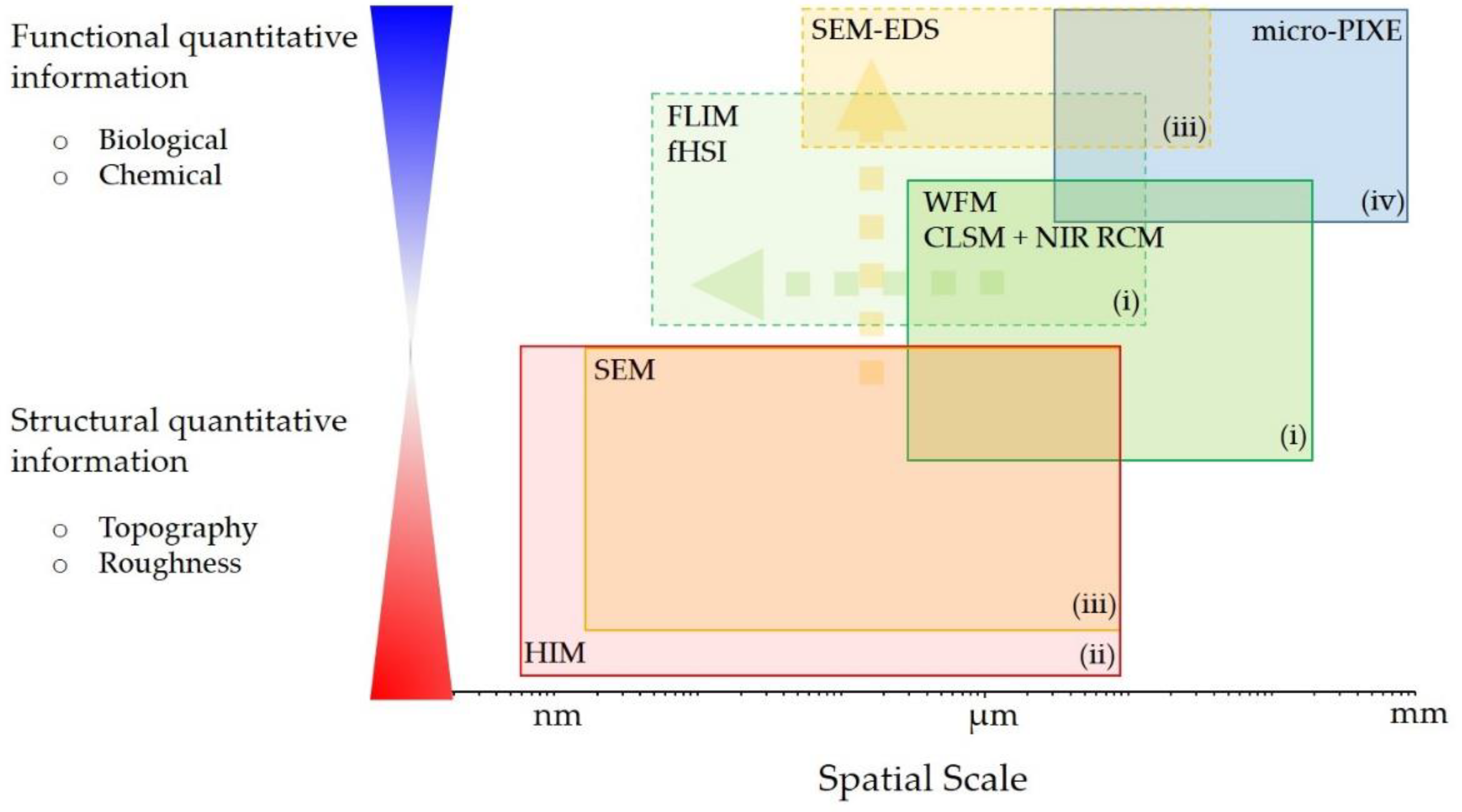
| Technique | Lateral Resolution | Technique Capability | Biomedical Observation |
|---|---|---|---|
| CLSM, NIR RCM | 250 nm | Wear debris inside the tissue can be directly detected by NIR RCM where their sizes vary from sub µm to >10 µm. | Elevated autofluorescence was observed in the direct surrounding of the wear debris indicating on the biological response. |
| fHSI, FLIM | 250 nm (*few nm—distinguish nearby molecular environment) | Wear debris was detected through the decrease in FLIM decay through the surface plasmon effect, [43] made possible by the presence of the lipopigment molecules in the direct vicinity. | Broad spectral width, the measured peak wavelength and the µm sized “granules” confirmed lipofuscin presence accumulated in lysosomes as a consequence of the biological oxidative stress response [69] which is induced by the nearby, micron-sized wear debris. |
| HIM | nm | HIM imaging showed high surface sensitivity with nm resolution that can easily differ between wear debris with different surface topographies. | The wide extent of morphologies can be attributed to the substantial wear damage due to fretting and corrosion that can induce inflammatory effect on the surrounding biological environment [53]. |
| SEM-EDS | Sub µm (EDS) | Elemental analysis was possible on individual micron sized wear debris and showed non-homogeneous P and Ca deposition on its surface with distinct topographical features on a nm scale; | A substantial leaching of the alloy constituents Al and V was measured in the individual wear debris; Regions with remaining Al coincide with regions rich in P and Ca indicating Al-dependent hydroxyapatite layer formation or that the calcification effect may play a role in the retention of Al. |
| Micro-PIXE | few µm | The wear debris characterization was possible on a wider field-of-view with approximately 10 times better sensitivity and with the quantitative data matching SEM-EDS analysis. | Debris from Ti alloy is spread throughout the whole tissue slice; Elemental mapping confirmed significant leaching of Al and V from wear debris into the body during the implant corrosion process; Analysis confirmed the EDS finding of Al regions on wear debris coinciding with hydroxyapatite-like layer formation; Observed V leaching could be the main cause of significantly elevated concentration of V in patient’s serum. |
Publisher’s Note: MDPI stays neutral with regard to jurisdictional claims in published maps and institutional affiliations. |
© 2021 by the authors. Licensee MDPI, Basel, Switzerland. This article is an open access article distributed under the terms and conditions of the Creative Commons Attribution (CC BY) license (https://creativecommons.org/licenses/by/4.0/).
Share and Cite
Podlipec, R.; Punzón-Quijorna, E.; Pirker, L.; Kelemen, M.; Vavpetič, P.; Kavalar, R.; Hlawacek, G.; Štrancar, J.; Pelicon, P.; Fokter, S.K. Revealing Inflammatory Indications Induced by Titanium Alloy Wear Debris in Periprosthetic Tissue by Label-Free Correlative High-Resolution Ion, Electron and Optical Microspectroscopy. Materials 2021, 14, 3048. https://doi.org/10.3390/ma14113048
Podlipec R, Punzón-Quijorna E, Pirker L, Kelemen M, Vavpetič P, Kavalar R, Hlawacek G, Štrancar J, Pelicon P, Fokter SK. Revealing Inflammatory Indications Induced by Titanium Alloy Wear Debris in Periprosthetic Tissue by Label-Free Correlative High-Resolution Ion, Electron and Optical Microspectroscopy. Materials. 2021; 14(11):3048. https://doi.org/10.3390/ma14113048
Chicago/Turabian StylePodlipec, Rok, Esther Punzón-Quijorna, Luka Pirker, Mitja Kelemen, Primož Vavpetič, Rajko Kavalar, Gregor Hlawacek, Janez Štrancar, Primož Pelicon, and Samo K. Fokter. 2021. "Revealing Inflammatory Indications Induced by Titanium Alloy Wear Debris in Periprosthetic Tissue by Label-Free Correlative High-Resolution Ion, Electron and Optical Microspectroscopy" Materials 14, no. 11: 3048. https://doi.org/10.3390/ma14113048
APA StylePodlipec, R., Punzón-Quijorna, E., Pirker, L., Kelemen, M., Vavpetič, P., Kavalar, R., Hlawacek, G., Štrancar, J., Pelicon, P., & Fokter, S. K. (2021). Revealing Inflammatory Indications Induced by Titanium Alloy Wear Debris in Periprosthetic Tissue by Label-Free Correlative High-Resolution Ion, Electron and Optical Microspectroscopy. Materials, 14(11), 3048. https://doi.org/10.3390/ma14113048