Review

Modifications of Dental Implant Surfaces at the Micro- and Nano-Level for Enhanced Osseointegration

Department of Prosthodontics, School of Dentistry and Dental Research Institute, Seoul National University, Seoul 03080, Korea
Materials 2020, 13(1), 89; https://doi.org/10.3390/ma13010089
Submission received: 30 October 2019 / Revised: 13 December 2019 / Accepted: 20 December 2019 / Published: 23 December 2019
(This article belongs to the Special Issue Dental Implant Materials 2019)

Abstract

:
This review paper describes several recent modification methods for biocompatible titanium dental implant surfaces. The micro-roughened surfaces reviewed in the literature are sandblasted, large-grit, acid-etched, and anodically oxidized. These globally-used surfaces have been clinically investigated, showing survival rates higher than 95%. In the past, dental clinicians believed that eukaryotic cells for osteogenesis did not recognize the changes of the nanostructures of dental implant surfaces. However, research findings have recently shown that osteogenic cells respond to chemical and morphological changes at a nanoscale on the surfaces, including titanium dioxide nanotube arrangements, functional peptide coatings, fluoride treatments, calcium–phosphorus applications, and ultraviolet photofunctionalization. Some of the nano-level modifications have not yet been clinically evaluated. However, these modified dental implant surfaces at the nanoscale have shown excellent in vitro and in vivo results, and thus promising potential future clinical use.

1. Introduction

The surface quality of titanium (Ti) dental implants, which replace missing teeth, is one of the keys to the long-term clinical success of implants in a patient’s mouth [1]. The bone response to the Ti implant surface depends on its surface characteristics: Contact (bone formation on the implant surface towards the bone) and distance osteogenesis occur around micro-roughened Ti surfaces while only distance osteogenesis (bone formation from the old bone toward the implant surface) appear around turned Ti [2]. Although contact osteogenesis seems to require other factors to be triggered, modification of the implant surface is very important to accelerate osseointegration [3].
Ti is known to be stable in biologic responses and not to trigger a foreign body reaction when inserted into the human body [4,5]. Therefore, osseointegration was originally defined as the direct contact between a loaded implant surface and bone at the microscopic level of resolution [1]. Recently, this term has been interpreted from a new point of view: Osseointegration is essentially a demarcation response to a foreign body of Ti when the Ti implant is immobile in bone [6]. This demarcation is immune-driven and is classified as a type IV hypersensitivity [7]. Based on the original definition, the modification of a Ti implant surface implies that the surface would be more biocompatible, thereby increasing the bioaffinity of the hard tissue and accelerating the bone response to the surface. The new standpoint on osseointegration suggests that the modified Ti surface would be recognized more sensitively by the hard tissue, which would isolate this foreign body with a faster and stronger accumulation of bone substances. Thus, the nature of osseointegration is under investigation at present [8]. The detection of the actual bond between the bone and implant surfaces could support the bioaffinitive nature of bone response to the surfaces [9,10]. Only friction and physical contact would exist at the interface if the bony demarcation hypothesis is correct.
To date, implant surfaces have been modified in various ways under the bioaffinity concept for osseointegration. Conventionally, the topography of the surface has been changed at the micro-level (1–10 μm). At present, some chemical features and nanotechnologies have been added to the surfaces. This review introduces several recent advancements of biocompatible implant surfaces with a few representative micro-roughened modified surfaces. Since most implant surfaces used in the global market have been made of commercially pure Ti (cp-Ti), especially grade 4 cp-Ti, this review is based on the modification of a grade 4 cp-Ti surface.

2. Micro-Roughened Modification

2.1. Sandblasted, Large-Grit, Acid-Etched (SLA) Surface

The computer numerical controlled milling of cp-Ti manufactures screw-shaped endosseous dental implants. The surface machined by this milling procedure, which is now called a turned Ti surface, shows many parallel grooves in scanning electron microscopy (SEM). The turned surface experiences no modification process, which has frequently served as a control to evaluate the biocompatibility of modified surfaces. When an implant is inserted into the bone and the implant surface becomes juxtaposed to the bone, bone healing (or osseointegration) on the surface is known to be fulfilled by two mechanisms: distance and contact osteogenesis [2,11]. In distance osteogenesis, new bone starts to be formed on the surfaces of bone. The direction of bone growth is from the bone towards the implant surface (Figure 1A). In contact osteogenesis, or de novo bone formation, new bone formation begins on the implant surface. The direction of bone growth is from the implant towards the bone, opposite to that for distance osteogenesis (Figure 1B). When an endosseous implant with a turned surface is placed into the jawbone, only distance osteogenesis occurs, which implies that more time is needed for sufficient osseointegration to withstand masticatory forces [2,12]. The necessity of reduction in the patient’s edentulous period has led the modification of an implant surface to accelerate bone healing.
The traditional approach to the surface modification of a Ti implant has been roughening at the micro-level. One of the most successful surfaces in clinical dentistry is the sandblasted, large-grit, and acid-etched (or SLA) surface. An SLA Ti surface is made by sandblasting the turned Ti surface with large-grit particles, the sizes of which range from 250 μm to 500 μm in general, and by acid-etching the blasted surface. The acids for etching are usually strong acids including hydrochloric, sulfuric, and nitric acids. SEM shows topographically changed irregularities on the SLA surface, with large dips, small micropits, sharp edges, and pointed tips. Sa, one of the surface parameters defined as the arithmetic mean height of the surface, is approximately 1.5 μm to 2 μm. Osteogenic cells migrate to the roughened Ti surface through the fibrin clot that is formed at the peri-implant site after bone drilling for implant insertion, and these cells appear to recognize the irregularities of the SLA surface as lacunae to be filled with bone materials [2,13]. Contact osteogenesis occurs as the osteogenic cells secrete a bone matrix. The occurrence of both contact and distance osteogenesis accelerates the osseointegration on the SLA surface compared to the turned surface.
The Ti surface of a dental implant is originally hydrophobic [14]. Water (H2O) is considered to have initial contact with the implant surface when the implant is inserted into the bone [15]. Therefore, there have been attempts to add hydrophilicity to an SLA surface, since hydrophilicity is expected to help accelerate the bone healing process [14,16]. A dental implant with a hydrophilic SLA surface, commercially called SLActive (Institute Straumann AG, Basel, Switzerland), is made with a water rinse of the original SLA implant in a nitrogen chamber and a packaging technique of storing the implant in an isotonic sodium chloride solution with no atmospheric contact, and this hydrophilic implant is being clinically used in the global market [17].
Regardless of whether an SLA surface is hydrophobic or hydrophilic, this dental implant surface has shown excellent long-term clinical results [18,19,20,21,22]. A previous 10-year retrospective study investigating more than 500 SLA Ti implants concluded that both the survival and success rates were 97% or higher [18]. The 10-year survival rate of SLA Ti implants was reported to be higher than 95%, even in periodontally compromised patients, although strict periodontal interventions were applied to these patients [20]. Similar results were found in 10-year prospective studies investigating the survival rates of dental implants with SLA surfaces [19,21,22]. This modified surface, roughened at the micro-scale, is one of the dental implant surfaces that has been most frequently tested in clinics for the longest period.

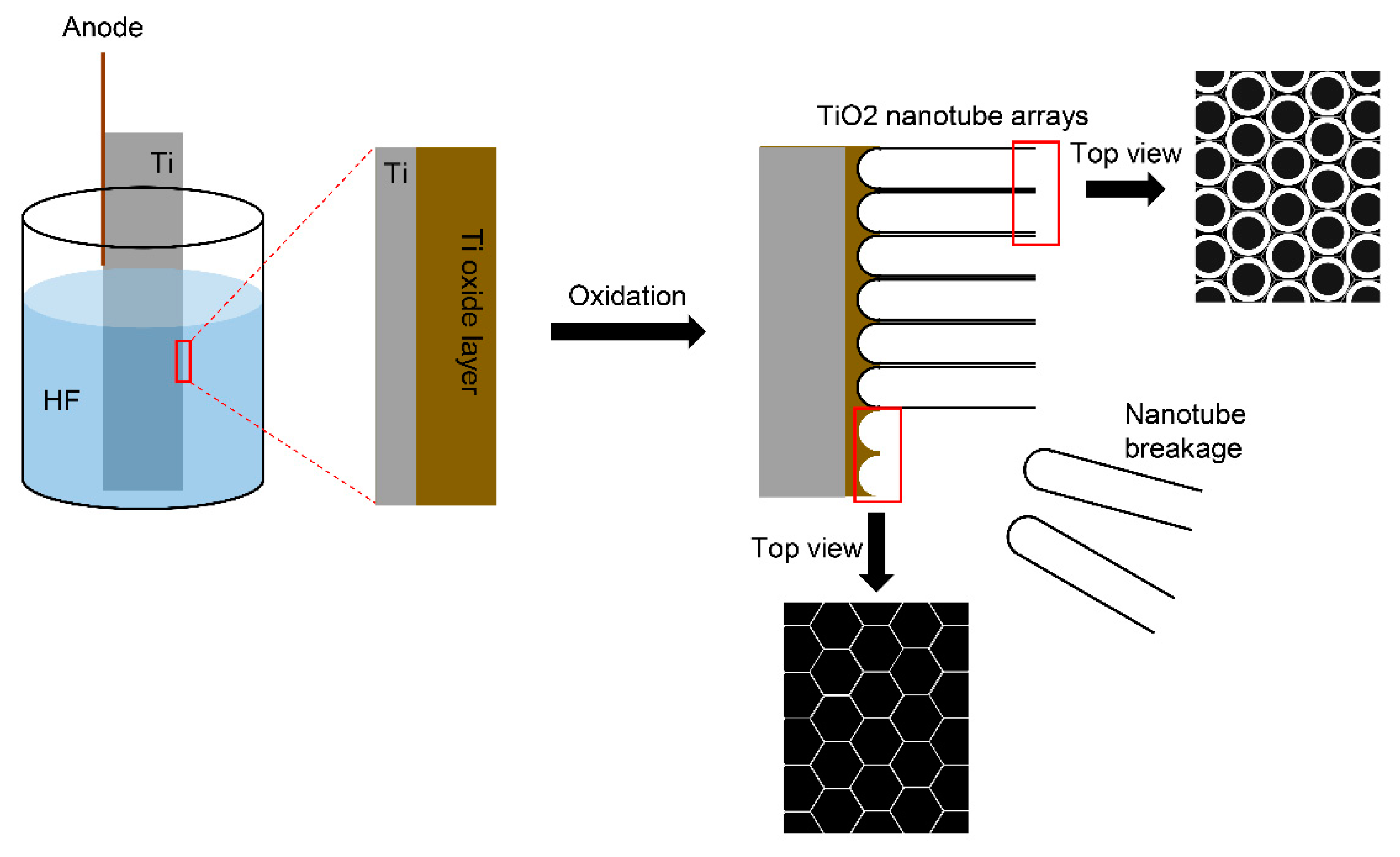
2.2. Anodic Oxidation

The genuine biocompatible surface on the Ti dental implant is Ti oxide (TiO2), not Ti itself, which is spontaneously formed when the Ti surface is exposed to the atmosphere. However, this Ti oxide layer is very thin (a few nm in thickness) and is imperfect with defects [23]. Also, chemically unstable Ti3+ and Ti2+ are known to exist in the oxide layer [24]. Therefore, there have been several techniques developed to thicken and stabilize the Ti oxide layer, which is considered to increase the biocompatibility of the surface [25,26,27]. When Ti becomes the anode under an electric potential in an electrochemical cell, Ti is oxidized to be Ti4+, and the TiO2 layer is able to be thickened and roughened [15]. Topographically, the oxidized Ti surface for a dental implant has many volcano-like micropores with various sizes, which are observed in SEM. The surface characteristics of the anodized Ti surface depend on the applied potential, surface treatment time, concentrations, and types of electrolytes [15,27]. The arithmetic mean height of this surface, or Sa, is evaluated to be approximately 1 to 1.5 μm for dental use [28,29,30,31].
Osteogenic cells appear to recognize the topography of a dental implant surface although we do not yet know which surface topography is more proper in bone healing, or if the irregularities of the SLA surface are more effective for the osteogenic cell response than the microporous structure of the anodized surface [32]. To date, no in vivo model has found any significant differences in bone responses to the microtopographies of Ti dental implant surfaces [33,34]. What is definitely known about implant surface topography is that the cp-Ti surfaces topographically modified at the microscale accelerate osseointegration more than the turned surface, and these modified surfaces show superior results to the turned surface during in vitro, in vivo, and clinical studies.
The anodically oxidized Ti surface has shown superior results to the turned surface in various in vitro tests and in vivo histomorphometry [31,34,35,36]. A previous meta-analytic study reported lower failure rates of the oxidized Ti implants than those of the turned implants from the included 38 clinical investigations [37]. A prior retrospective and a 10-year prospective study concluded that that success rates were higher than 95% for the TiUnite surface (Brånemark System, Nobel Biocare, Göteborg, Sweden), which is a trade name for the oxidized Ti surface [38,39]. However, a recent 20-year randomized controlled clinical trial notably reported a similar marginal bone loss between micro-roughened and turned Ti implants [40]. This clinical study used an identical implant design with an implant-abutment connection structure and internal friction connection [40]. Identifying which of the two factors (surface characteristics and implant design) is a major contributor to the long-term clinical success of dental implants needs to be thoroughly investigated, although higher success or survival rates have been steadily published for Ti dental implants with modified surfaces at the micro-scale, compared to the turned implant [19,41,42].

3. Molecular Modification

3.1. TiO2 Nanotube

Anodic oxidation is extended to the modification of a Ti dental implant at the nanoscale (1–100 nm). The electric current of the electrochemical cell, temperature, the pH values of electrolyte solutions, the electrolytes, oxidation voltage, and oxidation time affect the nanotopographies of the Ti surface [43,44]. In an electrochemical cell composed of Ti at the anode and platinum (or Ti) at the cathode, the TiO2 layer is normally formed on the Ti implant surface of the anode [43]. In an appropriate fluoride-based electrolyte, the nano-morphology of the TiO2 layer is changed, and the aligned TiO2 nanotube layer is developed (Figure 2) [43].
In the past, implant surface nanostructures were reported to have no effect on cell responses and bone responses to dental implant surfaces and were thought to depend on the microtopographies of the surfaces [45,46]. Optimal micro-roughness is known at present to be 1.5 μm in Sa and approximately 4 μm in diameter of the surface irregularities [30,47]. However, a previous review article noted that the microtopographies of the dental implant surfaces have a limited influence on the initial responses of the in vivo hard tissue environment [48]. Presently, the nanotopographical features of Ti implant surfaces have been known to be contributors to the initial biologic responses of the hard tissue, including osteoblast activities and osteoclast reactions [44,49].
This modified surface with TiO2 nanotube arrays is highly biocompatible [44,50,51]. Both osteoblasts and osteoclasts showed maximal cellular responses to Ti surfaces with TiO2 nanotubes that were 15 nm in diameter [52]. Interestingly, smaller TiO2 nanotubes, which were approximately 30 nm in diameter, were more effective in the adhesion and growth of mesenchymal stem cells than larger TiO2 nanotubes that ranged from 70 nm to 100 nm, while the latter TiO2 nanotubes were more inductive in the differentiation into osteoblast-like cells, although there is contrary to previous studies [52,53]. The modified TiO2 nanotubular surface showed excellent bone-to-implant contact in the osteoporotic bone in an in vivo study using ovariectomized rats [54].
Another characteristic of this nano-modified surface is a drug delivery effect [55,56,57,58]. Drug release from TiO2 nanotubes is associated with the dimensions of TiO2 nanotube arrays regardless of the direct release or indirect discharge by nanocarriers [59]. The diameter and length of TiO2 nanotubes generally increase as the voltage and duration of the oxidation process increase, and the drug release has been found to be effective when the diameter is larger than approximately 100 nm [56,59,60]. A combination of this nano-modified TiO2 surface and carrier molecules, including micelles, is being actively investigated for drug delivery at a constant rate, unrelated to the drug concentration and release period [57,58,60].
The nanotopography of the TiO2 nanotubular surface has antibacterial properties alongside delivering antibiotic drugs [61]. Streptococcus mutans, which are associated with the initial formation of biofilm in the oral cavity, were reported to adhere to the TiO2 nanotube arrays less than to a micro-roughened SLA surface [62]. The hydrophilic properties of TiO2 nanotubes seems to hinder bacterial adhesion to the nanotubular surface [62]. However, it is notable that many studies have described the wettability of the TiO2 nanotube arrays, showing conflicting results in cellular and bacterial responses to the nanotubular surface [61,63,64]. Although the hydrophilicity of the TiO2 nanotube arrays is adjustable, some studies reported that the reduction of bacterial adhesion was due to the hydrophilic properties of the surface, whereas other studies described that such a result was due to the hydrophobic properties [61,63,64]. Further investigation is required to determine the mechanism of bacterial and cellular responses to the wettability of Ti surfaces.
Despite that the modified surface with TiO2 nanotube arrays has very useful advantages (e.g., high biocompatibility, the capability of drug delivery, and antibacterial properties), this surface has been neither applied nor tested clinically. The mechanical strength between the TiO2 nanotubes and the base Ti surface is too weak for this surface to be applied to a dental implant [43]. Recently, the hexagonal nano-structure of the base Ti surface was evaluated to be adequate for biologic application when the TiO2 nanotube arrays are removed from the base surface in order to prevent the delamination of the TiO2 nanotube coating in an in vivo environment (Figure 2) [44]. The aligned TiO2 nanotube-layered surface has great potential in biologic and clinical applications [55,56,57,65]. However, it is necessary to overcome this delamination problem before this TiO2 nanotubular surface is clinically used in the field of dental implantology.

3.2. Functional Peptides

Water and ions have first contact with the implant surface when the bone is drilled for implant insertion and a screw-shaped endosseous dental implant is placed into the bone. Then, the plasma proteins adhere to the surface through ionic bridges (like a calcium ion linkage), and the fibrin clot is formed. During hemostasis, extracellular matrix (ECM) proteins gradually replace the plasma proteins [15]. The adhesion proteins, including fibronectin and vitronectin (which are also ECM proteins), are recognized by the transmembrane proteins of osteogenic cells like integrins. Through binding of the transmembrane proteins to the osteogenic cells, the cells interact with ECM, which controls the cellular activities for bone healing [66]. Therefore, the bone healing process starts from the adhesion of the osteogenic cells to surfaces, and these adhesion proteins can play a role in accelerating osseointegration into dental implants when the proteins are applied to the implant surfaces. Core amino acid sequences, which are extracted from the original adhesion proteins and still have binding activities to the transmembrane receptors, are very useful in rapid bone healing when the core sequences are treated on the implant surfaces. These core functional peptides are considered to be more promising candidates for implant surface treatment than the original proteins because of the lower antigenicity and simpler adjustability of the peptides [67].
A functional peptide derived from the fibronectin, arginyl-glycyl-aspartic acid sequence, revealed improved histomorphometric results when this peptide was coated on a Ti dental implant surface and when this peptide-treated surface was compared to the uncoated surface [68]. Two functional amino acid sequences derived from another adhesion protein, laminin, showed excellent results as accelerating modifiers for Ti implant surfaces for osseointegration [35,67]. These functional peptides based on the adhesion of osteogenic cells seem to surpass the effects of the microtopographical features of the underlying Ti implant surfaces in bone healing, although further studies are definitely needed [35,69]. The mechanism behind the superior bone cell responses has been tried to be explained, based on the hypothesized tunable allosteric control of the receptor proteins [67,70]. A recent investigation evaluating a functional peptide from vitronectin found a Janus effect of this peptide for bone formation, activating osteoblasts and inhibiting osteoclasts, that is, controlling the osteoporotic environment locally to be favorable for osseointegration [71].
Cytokines, particularly growth factors, are another class of bioactive proteins. Bone morphogenetic proteins (BMPs) are available for bone healing in the field of dental implantology. Human recombinant BMP-2 (rhBMP-2) is used in the global market for bone regeneration. BMP-2 is known to have a direct effect on osteogenic cells to promote bone formation with various interactions between this protein and other bioactive molecules, including osteogenic genes [72,73]. However, these growth factors have many problems to be solved before clinical application to Ti dental implant surfaces. BMP-2 has complicated biologic effects depending on its concentrations and surroundings; osteogenesis, adipogenesis, and chodrogenesis, but osteolysis also occurs [72,74,75]. The rhBMP-2-treated Ti surface was reported to make bone healing around a dental implant faster in an in vivo model [76,77]. However, it is recognizable that growth factors are usually active in free forms, not in bound forms. Therefore, these molecules are ineffective or, if any, limitedly active when the factors are bound or attached to implant surfaces [78]. The cell transmembrane proteins that recognize these growth factors are disengaged in the attachment of the cells [78]. Because of the multiple enigmatic effects of these growth factors on living tissues and the growth factor receptors’ lack of involvement in cell adhesion, growth factor-treated implant surfaces have not been used clinically until now.
Although these bioactive molecules, including the adhesion molecules and growth factors, have the potential to be applied to dental implants for accelerated osseointegration, the Ti dental implants on which these molecules are coated have not been clinically tested; there have been no published clinical trials to report the results of such implants. The functional peptides from the adhesion molecules are to be clinically tried and applied in dental implantology in the near future due to the simplicity in their biologic effects and their low probability of side effects. For growth factors, it seems to be necessary to find core amino acid sequences from growth factors to increase the clinical applicability of these factors. Before these derived peptides are clinically tried, further studies are required on release strategies for the molecules from the implant surfaces and on the biologic activities of the core peptides.

3.3. Fluoride Treatment (Cathodic Reduction)

When a Ti implant is a cathode in the hydrofluoric acid solution of an electrochemical cell, a fluoride ion gives an electron to the cathode, where the reduction of a Ti ion occurs. As a result, a trace amount of fluoride ions adheres to the Ti implant surface when the concentration of hydrogen fluoride is low in the solution. This trace amount of fluoride ions is known to primarily affect osteoprogenitor cells and undifferentiated osteoblasts to enhance bone formation, rather than highly differentiated osteoblasts [79,80]. Furthermore, fluoride is helpful for bone mineralization because of its properties that are attractive for calcium [78]. However, fluoride ions are thought to become cytotoxic as the number of ions increases on the Ti implant surface.
Clinically, a modified surface is used as a dental implant surface (Osseospeed, Astra Tech, Dentsply, Waltham, MA, USA), which is fluoride-treated after the grade 4 cp-Ti is sandblasted with TiO2 particles. This fluoride-modified surface has a very low amount of fluoride, which is difficult to find by energy dispersive spectroscopy, while x-ray photoelectron spectroscopy is able to detect this trace amount [81,82]. The average mean height of this marketed surface has been investigated to be approximately 1.5 μm [30,82]. The fluoride-treated Ti surface has shown stronger binding between the bone and this surface than the control Ti surface without fluoride-treatment [83,84]. However, finding any significant differences in the histomorphometric results has been very rare when the fluoride-treated dental implants have been compared in vivo to other modified implants, including SLA implants, while some previous studies have been found to show more favorable results in bone responses to the fluoride-treated surface than those to its predecessor with no application of fluoride [78,82,85,86].
Dental implants with a fluoride-treated surface have exhibited high rates of success and survival rates in clinical trials. These fluoride-treated implants have supported prosthodontic restorations in edentulous mandibles with a 100% survival rate for ten years [87]. Regardless of the maxilla or the mandible, high survival rates of over 95% have been reported for the surface-modified dental implants in the prospective clinical studies, the observation periods of which are longer than 5 years [87,88,89]. It is notable and very interesting that these previous clinical studies have consistently reported the vertical loss of bone surrounding the implants of less than 0.5–1 mm, which is interpreted as almost no change of the bone level [40,87,89]. Importantly, these clinical studies used fluoride-treated dental implants with the same implant macro-design, including an identical thread shape and internal friction implant–abutment connection, so care must be taken when interpreting data in comparison studies of the biologic responses between the dental implant systems [82]. It remains uncertain which factor (surface chemistry in fluoride treatment, surface topography, implant–abutment connection tectonics) is a major contributor to the biologic responses in humans to this marketed fluoride-treated dental implant.

3.4. Hydroxyapatite and Other Calcium–Phosphorus Compounds

This idea of hydroxyapatite (HA) coating on a Ti dental implant surface is based on the fact that the main component of bone is HA. HA (Ca10(PO4)6(OH)2) is still the most commonly-utilized coating material for Ti dental implant surfaces [90]. HA and other calcium-phosphorus coating materials are basically osteoconductive to the surrounding bone. The biologic features of these materials, such as their biodegradation properties and foreign body reactions, seem to depend on calcium/phosphorus ratios, crystallinities, and coating thicknesses [31,90,91,92,93]. Plasma spraying (a conventional atmospheric plasma-spray method) is one of the most widely used methods to coat HA on a Ti implant surface [90]. HA particles that are contained and heated in a plasma flame whose temperature is approximately 15,000 to 20,000 Kelvin are sprayed on the Ti surface, resulting in a HA coating layer that is 50–100 μm in thickness [94]. The spray parameters, including the flame combination and spraying flow rate, affect the chemical and physical features of the HA coating [92].
The HA coating is biocompatible with the hard tissue, showing direct contact with bone and the attachment of osteoblasts on the coating surface [95,96]. Many studies have reported enhanced bone apposition and the prevention of metal-ion release into the bone from metal implants with an HA coated surface [97,98,99,100,101]. However, the HA coating layer has some critical issues to be addressed. Like the TiO2 nanotube arrays, the delamination of the coating layer from the Ti dental implant surface is one of the problems (adhesive failure) [92]. Delaminated or worn HA particles hinder bone healing and provoke inflammation around the implant inserted into the bone [92,102]. The thick coating layer is able to make a breakage inside the layer, especially at the implant in a load-bearing area (cohesive failure) [92]. Recently, a thin calcium–phosphorus coating layer has been achieved and investigated using various coating techniques [76,93,101,103]. Compared to the plasma sprayed HA coating, however, the other calcium–phosphorus coatings are considered to lack long term clinical results [104,105].
The five-year clinical success rate of the HA coated implant has been evaluated to be approximately 95% [106]. However, this success rate has dropped markedly to below 80% after 10 years of implant placement [106,107,108]. Such a low success rate may result from the above-mentioned problems of the HA coating layer. It is notable, however, that these clinical evaluations resulted from the data of cylindrical implants [107,108]. A previous study using HA coated screw-shaped implants (MicroVent, Zimmer Dental, Carlsbad, CA, USA) reported that the long-term clinical success rate (> 10 years) was higher than 90% [109]. Nevertheless, clinical trials are certainly necessary to evaluate the calcium–phosphorus coating more precisely.

3.5. Photofunctionalization

In 1997, it was determined that the wettability of the TiO2 surface is increased by ultraviolet (UV) radiation [110]. Originally, the UV-induced TiO2 surface is amphiphilic—both hydrophilic and oleophilic [110]. However, the enhanced biologic effect of this surface is considered to be caused by the hydrophilic properties. Such hydrophilicity and elimination of hydrocarbon contamination on the TiO2 surface are known to be the mechanisms behind further activated bone responses to a dental implant in UV-mediated photofunctionalization. The hard tissue affinity drops for an aged Ti surface that has been stored for longer than two weeks [111]. UV irradiation on the Ti implant surface appears to make the Ti surface reactivate, as the implant is freshly made.
UV radiation is subcategorized into three types according to its wavelengths and dermal biologic reactions to the electromagnetic waves: UVA, UVB, and UVC [112]. The wavelengths of UVA range from 320 to 400 nm, and those of UVC range from 200 to 280 nm [113]. Both UVA and UVC contribute to increasing the hydrophilicity of the Ti surface. However, considering the fact that some reports show the promoted osteogenic activities on hydrophobic surfaces, the removal of carbon from the Ti surface, which is caused by UVC, is likely a fundamental mechanism behind excellent osseointegration [114,115,116,117]. Strictly, neither UVA nor UVC appears to make a topographic change at the nano-scale on the Ti surface [115,117]. Friction force microscopy shows a nano-scale modification that UV irradiation may produce by converting Ti4+ to Ti3+ [110]. UV treatment on the Ti surface enhances the adsorption of proteins, such as albumin and fibronectin, which are plasma proteins in the human body [118]. UV-photofunctionalized implant surfaces show improved osteogenic cell attachment, spreading, and proliferation [117]. The antibacterial effects are described for the UV activation of the Ti surface [112]. Faster bone responses to UV-treated Ti surfaces are reported in various in vivo studies, some of which show almost 100% bone-to-implant contact [117,118,119,120].
A previous clinical study showed that the stability of implants inserted into the patients’ jaw bones increased more rapidly when the implants were UV-photofunctionalized [121]. The retrospective clinical studies concluded that UV-mediated photofunctionalization reduced early implant failure, and the success rate of the photofunctionalized implants was 97.6% during the functional loading period of approximately 2.5 years [122,123]. No prospective long-term clinical study (published in English) evaluating UV-mediated photofunctionalization has yet been found in the field of implant dentistry. However, a prospective clinical evaluation of UV-treated implants over more than 5 years is expected to be published shortly.

3.6. Laser Ablation

For laser ablation, an implant whose collar, or neck area, was treated by laser micromachining to generate nano-channels is used (Laser-Lok, BioHorizons, Birmingham, AL, USA) [124,125]. Laser ablation is also able to produce micro-scale patterns by controlling laser processing parameters [126]. This approach was intended to promote not only fast osseointegration, but also connective tissue attachment [124,127]. The connective tissue fiber direction in the soft tissue attachment is known to be perpendicular to the laser-microtextured Ti implant surface, which is characteristically different from the general orientation of the fibers parallel to implant surfaces [127,128]
This marketed laser-modified surface (Laser-Lok) showed significantly improved bone-to-implant contact in a previous in vivo study, compared to a turned Ti surface [129]. The survival rate was evaluated to be 95.6% in a two-year retrospective multicenter study and to be 94% in another 5-year retrospective controlled study [127,130]. Recently, the prospective three-year results of a randomized clinical trial were reported for single implant-supported restorations with the laser-modified Ti implant surface, where the survival rate was estimated to be 96.1% [131]. Both the hard and soft tissue responses to the laser-modified Ti surface appear to be favorable [127,130,131,132]. However, long-term prospective clinical results of laser micromachining are still needed.

4. Concluding Remarks

When the bone is prepared for implant placement, surgical trauma provokes bleeding and hemostasis. Moreover, this surgical trauma activates the growth and differentiation factors released from the bone debris and matrix [15]. Surface modification of the Ti dental implant focuses on improving such initial biologic responses to the implant surface. Researchers and dental clinicians anticipate the best performance of the implant surfaces during these initial events and more readily establish these events by changing the physical and chemical properties of the surfaces, thereby boosting the speed and strengthening the quality of the healing process [133]. However, as the long-term clinical studies show, implant-supported prostheses have been used for a long time in patients’ mouths. Therefore, the modified surfaces also need to harmonize with the bone remodeling process, which has not yet been investigated. This paper reviews several modified surfaces of dental implants that are widely used in the global market or are highly possible to be clinically used. All these reviewed surfaces are targeted to accelerate early bone responses. The late responses of the hard tissue to the surfaces, including bone remodeling, need to be investigated. Moreover, long-term clinical trials are still required for these implant surfaces.

Funding

This research was supported by a grant from the Korea Health Technology R&D Project through the Korea Health Industry Development Institute (KHIDI), which is funded by the Ministry of Health and Welfare of the Republic of Korea (grant number: HI15C1535).

Conflicts of Interest

The authors report no conflict of interest related to this study.

References

  1. Albrektsson, T.; Branemark, P.I.; Hansson, H.A.; Lindstrom, J. Osseointegrated titanium implants. Requirements for ensuring a long-lasting, direct bone-to-implant anchorage in man. Acta Orthop. Scand. 1981, 52, 155–170. [Google Scholar] [CrossRef] [PubMed] [Green Version]
  2. Davies, J.E. Mechanisms of endosseous integration. Int. J. Prosthodont. 1998, 11, 391–401. [Google Scholar] [PubMed]
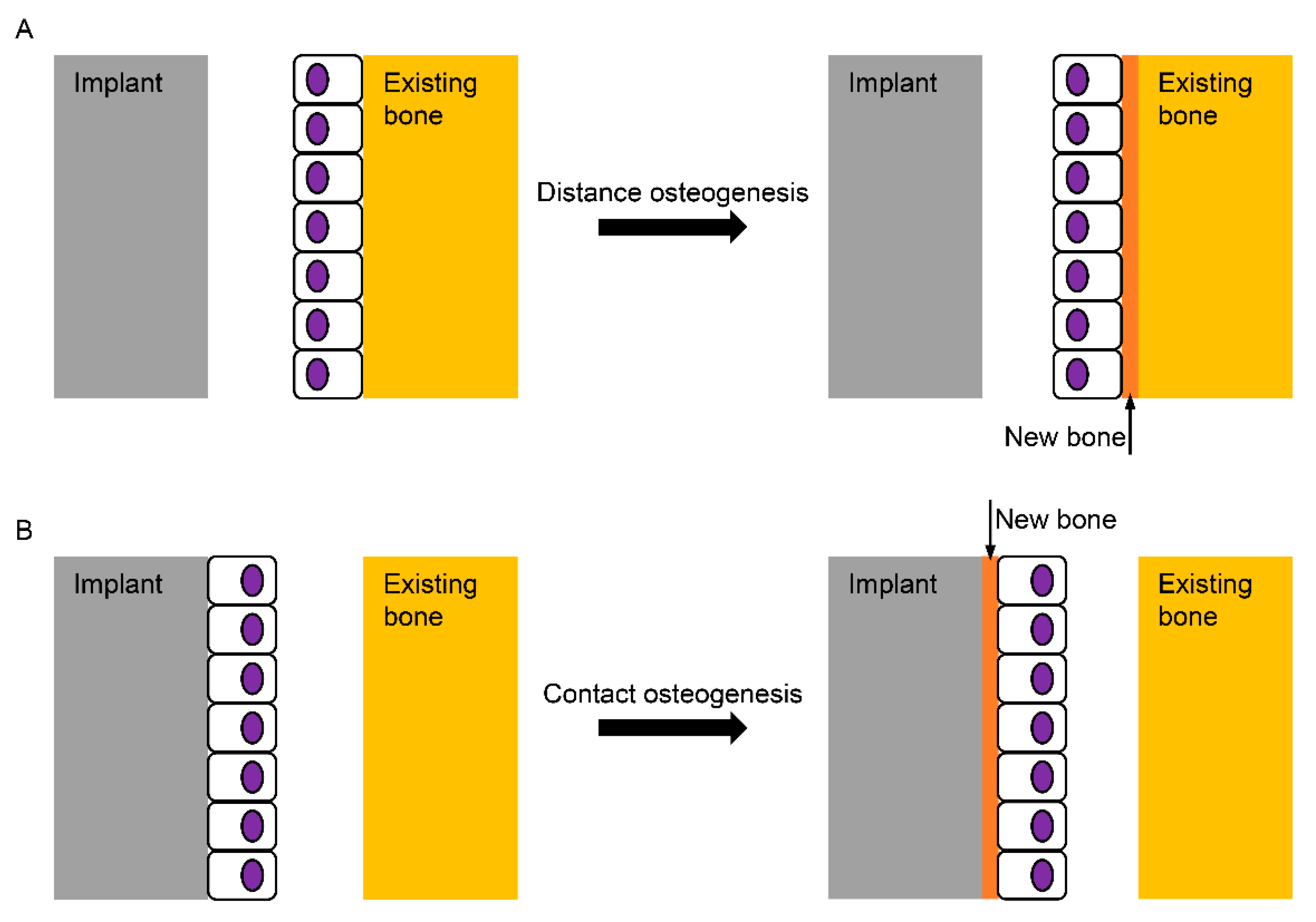
  3. Choi, J.Y.; Sim, J.H.; Yeo, I.L. Characteristics of contact and distance osteogenesis around modified implant surfaces in rabbit tibiae. J. Periodontal Implant Sci. 2017, 47, 182–192. [Google Scholar] [CrossRef] [PubMed] [Green Version]
  4. Albrektsson, T.; Wennerberg, A. Oral implant surfaces: Part 1--review focusing on topographic and chemical properties of different surfaces and in vivo responses to them. Int. J. Prosthodont. 2004, 17, 536–543. [Google Scholar] [PubMed]
  5. Kulkarni, M.; Mazare, A.; Gongadze, E.; Perutkova, S.; Kralj-Iglic, V.; Milosev, I.; Schmuki, P.; Iglic, A.; Mozetic, M. Titanium nanostructures for biomedical applications. Nanotechnology 2015, 26, 062002. [Google Scholar] [CrossRef]
  6. Albrektsson, T.; Jemt, T.; Molne, J.; Tengvall, P.; Wennerberg, A. On inflammation-immunological balance theory-A critical apprehension of disease concepts around implants: Mucositis and marginal bone loss may represent normal conditions and not necessarily a state of disease. Clin. Implant Dent. Relat. Res. 2019, 21, 183–189. [Google Scholar] [CrossRef] [Green Version]
  7. Albrektsson, T.; Chrcanovic, B.; Molne, J.; Wennerberg, A. Foreign body reactions, marginal bone loss and allergies in relation to titanium implants. Eur. J. Oral Implantol. 2018, 11, S37–S46. [Google Scholar]
  8. Davies, J.E. INVITED COMMENTARY: Is Osseointegration a Foreign Body Reaction? Int. J. Prosthodont. 2019, 32, 133–136. [Google Scholar] [CrossRef]
  9. Brunski, J. On Implant Prosthodontics: One Narrative, Twelve Voices - 2. Int. J. Prosthodont. 2018, 31, s15–s22. [Google Scholar]
  10. Kwon, T.K.; Choi, J.Y.; Park, J.I.; Yeo, I.L. A Clue to the Existence of Bonding between Bone and Implant Surface: An In Vivo Study. Mater. 2019, 12, 1187. [Google Scholar] [CrossRef] [Green Version]
  11. Osborn, J.; Newesely, H. Dynamic aspects of the implant-bone interface. Dental implants 1980, 111, 123. [Google Scholar]
  12. Branemark, P.I.; Adell, R.; Breine, U.; Hansson, B.O.; Lindstrom, J.; Ohlsson, A. Intra-osseous anchorage of dental prostheses. I. Experimental studies. Scand. J. Plast. Reconstr. Surg. 1969, 3, 81–100. [Google Scholar] [CrossRef] [PubMed]
  13. Sims, N.A.; Gooi, J.H. Bone remodeling: Multiple cellular interactions required for coupling of bone formation and resorption. Semin. Cell Dev. Biol. 2008, 19, 444–451. [Google Scholar] [CrossRef] [PubMed]
  14. Rupp, F.; Scheideler, L.; Olshanska, N.; de Wild, M.; Wieland, M.; Geis-Gerstorfer, J. Enhancing surface free energy and hydrophilicity through chemical modification of microstructured titanium implant surfaces. J. Biomed. Mater. Res. A. 2006, 76, 323–334. [Google Scholar] [CrossRef] [PubMed]
  15. Yeo, I.-S. Surface modification of dental biomaterials for controlling bone response. In Bone Response to Dental Implant Materials; Woodhead Publishing: Cambridge, UK, 2017; pp. 43–64. [Google Scholar]
  16. Rupp, F.; Scheideler, L.; Rehbein, D.; Axmann, D.; Geis-Gerstorfer, J. Roughness induced dynamic changes of wettability of acid etched titanium implant modifications. Biomaterials 2004, 25, 1429–1438. [Google Scholar] [CrossRef] [PubMed]
  17. Wall, I.; Donos, N.; Carlqvist, K.; Jones, F.; Brett, P. Modified titanium surfaces promote accelerated osteogenic differentiation of mesenchymal stromal cells in vitro. Bone 2009, 45, 17–26. [Google Scholar] [CrossRef]
  18. Buser, D.; Janner, S.F.; Wittneben, J.G.; Bragger, U.; Ramseier, C.A.; Salvi, G.E. 10-year survival and success rates of 511 titanium implants with a sandblasted and acid-etched surface: A retrospective study in 303 partially edentulous patients. Clin. Implant Dent. Relat. Res. 2012, 14, 839–851. [Google Scholar] [CrossRef]
  19. Nicolau, P.; Guerra, F.; Reis, R.; Krafft, T.; Benz, K.; Jackowski, J. 10-year outcomes with immediate and early loaded implants with a chemically modified SLA surface. Quintessence Int. 2018, 50, 2–12. [Google Scholar]
  20. Roccuzzo, M.; Bonino, L.; Dalmasso, P.; Aglietta, M. Long-term results of a three arms prospective cohort study on implants in periodontally compromised patients: 10-year data around sandblasted and acid-etched (SLA) surface. Clin. Oral Implants Res. 2014, 25, 1105–1112. [Google Scholar] [CrossRef]
  21. Rossi, F.; Lang, N.P.; Ricci, E.; Ferraioli, L.; Baldi, N.; Botticelli, D. Long-term follow-up of single crowns supported by short, moderately rough implants-A prospective 10-year cohort study. Clin. Oral Implants Res. 2018, 29, 1212–1219. [Google Scholar] [CrossRef]
  22. Van Velzen, F.J.; Ofec, R.; Schulten, E.A.; Ten Bruggenkate, C.M. 10-year survival rate and the incidence of peri-implant disease of 374 titanium dental implants with a SLA surface: A prospective cohort study in 177 fully and partially edentulous patients. Clin. Oral Implants Res. 2015, 26, 1121–1128. [Google Scholar] [CrossRef] [PubMed]
  23. Li, S.M.; Yao, W.H.; Liu, J.H.; Yu, M.; Wu, L.; Ma, K. Study on anodic oxidation process and property of composite film formed on Ti-10V-2Fe-3Al alloy in SiC nanoparticle suspension. Surf. Coat Tech. 2015, 277, 234–241. [Google Scholar] [CrossRef]
  24. Hanawa, T. Metal ion release from metal implants. Mat. Sci. Eng. C-Bio. S. 2004, 24, 745–752. [Google Scholar] [CrossRef]
  25. Manhabosco, T.M.; Tamborim, S.M.; dos Santos, C.B.; Muller, I.L. Tribological, electrochemical and tribo-electrochemical characterization of bare and nitrided Ti6Al4V in simulated body fluid solution. Corros. Sci. 2011, 53, 1786–1793. [Google Scholar] [CrossRef] [Green Version]
  26. Wang, J.W.; Ma, Y.; Guan, J.; Zhang, D.W. Characterizations of anodic oxide films formed on Ti6A14V in the silicate electrolyte with sodium polyacrylate as an additive. Surf. Coat Tech. 2018, 338, 14–21. [Google Scholar] [CrossRef]
  27. Zhang, L.; Duan, Y.; Gao, R.; Yang, J.; Wei, K.; Tang, D.; Fu, T. The Effect of Potential on Surface Characteristic and Corrosion Resistance of Anodic Oxide Film Formed on Commercial Pure Titanium at the Potentiodynamic-Aging Mode. Materials 2019, 12, 370. [Google Scholar] [CrossRef] [Green Version]
  28. Kwon, T.K.; Lee, H.J.; Min, S.K.; Yeo, I.S. Evaluation of early bone response to fluoride-modified and anodically oxidized titanium implants through continuous removal torque analysis. Implant Dent. 2012, 21, 427–432. [Google Scholar] [CrossRef] [Green Version]
  29. Lee, H.J.; Yang, I.H.; Kim, S.K.; Yeo, I.S.; Kwon, T.K. In vivo comparison between the effects of chemically modified hydrophilic and anodically oxidized titanium surfaces on initial bone healing. J. Periodontal. Implant Sci. 2015, 45, 94–100. [Google Scholar] [CrossRef] [Green Version]
  30. Wennerberg, A.; Albrektsson, T. On implant surfaces: A review of current knowledge and opinions. Int. J. Oral Maxillofac. Implants. 2010, 25, 63–74. [Google Scholar]
  31. Yeo, I.S.; Han, J.S.; Yang, J.H. Biomechanical and histomorphometric study of dental implants with different surface characteristics. J. Biomed. Mater. Res. B. Appl. Biomater. 2008, 87, 303–311. [Google Scholar] [CrossRef]
  32. Cooper, L.F. A role for surface topography in creating and maintaining bone at titanium endosseous implants. J. Prosthet. Dent. 2000, 84, 522–534. [Google Scholar] [CrossRef] [PubMed]
  33. Koh, J.W.; Kim, Y.S.; Yang, J.H.; Yeo, I.S. Effects of a calcium phosphate-coated and anodized titanium surface on early bone response. Int. J. Oral Maxillofac. Implants. 2013, 28, 790–797. [Google Scholar] [CrossRef] [PubMed] [Green Version]
  34. Yeo, I.S. Reality of dental implant surface modification: A short literature review. Open Biomed. Eng. J. 2014, 8, 114–119. [Google Scholar] [CrossRef] [PubMed]
  35. Kang, H.K.; Kim, O.B.; Min, S.K.; Jung, S.Y.; Jang, D.H.; Kwon, T.K.; Min, B.M.; Yeo, I.S. The effect of the DLTIDDSYWYRI motif of the human laminin alpha2 chain on implant osseointegration. Biomaterials 2013, 34, 4027–4037. [Google Scholar] [CrossRef]
  36. Min, S.K.; Kang, H.K.; Jang, D.H.; Jung, S.Y.; Kim, O.B.; Min, B.M.; Yeo, I.S. Titanium surface coating with a laminin-derived functional peptide promotes bone cell adhesion. Biomed Res Int 2013, 2013, 638348. [Google Scholar] [CrossRef] [Green Version]
  37. Chrcanovic, B.R.; Albrektsson, T.; Wennerberg, A. Turned versus anodised dental implants: A meta-analysis. J. Oral Rehabil. 2016, 43, 716–728. [Google Scholar] [CrossRef] [Green Version]
  38. Degidi, M.; Nardi, D.; Piattelli, A. 10-year follow-up of immediately loaded implants with TiUnite porous anodized surface. Clin. Implant. Dent. Relat. Res. 2012, 14, 828–838. [Google Scholar] [CrossRef]
  39. Shibuya, Y.; Kobayashi, M.; Takeuchi, J.; Asai, T.; Murata, M.; Umeda, M.; Komori, T. Analysis of 472 Branemark system TiUnite implants:a retrospective study. Kobe J. Med. Sci. 2010, 55, E73–E81. [Google Scholar]
  40. Donati, M.; Ekestubbe, A.; Lindhe, J.; Wennstrom, J.L. Marginal bone loss at implants with different surface characteristics - A 20-year follow-up of a randomized controlled clinical trial. Clin. Oral Implants Res. 2018, 29, 480–487. [Google Scholar] [CrossRef]
  41. Adell, R.; Lekholm, U.; Rockler, B.; Branemark, P.I. A 15-year study of osseointegrated implants in the treatment of the edentulous jaw. Int. J. Oral Surg. 1981, 10, 387–416. [Google Scholar] [CrossRef]
  42. Rocci, A.; Rocci, M.; Rocci, C.; Scoccia, A.; Gargari, M.; Martignoni, M.; Gottlow, J.; Sennerby, L. Immediate loading of Branemark system TiUnite and machined-surface implants in the posterior mandible, part II: A randomized open-ended 9-year follow-up clinical trial. Int. J. Oral Maxillofac. Implants. 2013, 28, 891–895. [Google Scholar] [CrossRef] [PubMed] [Green Version]
  43. Li, T.; Gulati, K.; Wang, N.; Zhang, Z.; Ivanovski, S. Understanding and augmenting the stability of therapeutic nanotubes on anodized titanium implants. Mater. Sci. Eng. C. Mater. Biol. Appl. 2018, 88, 182–195. [Google Scholar] [CrossRef] [PubMed] [Green Version]
  44. Shin, Y.C.; Pang, K.M.; Han, D.W.; Lee, K.H.; Ha, Y.C.; Park, J.W.; Kim, B.; Kim, D.; Lee, J.H. Enhanced osteogenic differentiation of human mesenchymal stem cells on Ti surfaces with electrochemical nanopattern formation. Mater. Sci. Eng. C. Mater. Biol. Appl. 2019, 99, 1174–1181. [Google Scholar] [CrossRef] [PubMed]
  45. Rice, J.M.; Hunt, J.A.; Gallagher, J.A.; Hanarp, P.; Sutherland, D.S.; Gold, J. Quantitative assessment of the response of primary derived human osteoblasts and macrophages to a range of nanotopography surfaces in a single culture model in vitro. Biomaterials 2003, 24, 4799–4818. [Google Scholar] [CrossRef]
  46. Wennerberg, A.; Albrektsson, T. Suggested guidelines for the topographic evaluation of implant surfaces. Int. J. Oral Maxillofac. Implants. 2000, 15, 331–344. [Google Scholar]
  47. Hansson, S.; Norton, M. The relation between surface roughness and interfacial shear strength for bone-anchored implants. A mathematical model. J. Biomech. 1999, 32, 829–836. [Google Scholar] [CrossRef]
  48. Mendonca, G.; Mendonca, D.B.; Aragao, F.J.; Cooper, L.F. Advancing dental implant surface technology--from micron- to nanotopography. Biomaterials 2008, 29, 3822–3835. [Google Scholar] [CrossRef]
  49. Liu, H.; Webster, T.J. Nanomedicine for implants: A review of studies and necessary experimental tools. Biomaterials 2007, 28, 354–369. [Google Scholar] [CrossRef]
  50. Ahn, T.K.; Lee, D.H.; Kim, T.S.; Jang, G.C.; Choi, S.; Oh, J.B.; Ye, G.; Lee, S. Modification of Titanium Implant and Titanium Dioxide for Bone Tissue Engineering. Adv. Exp. Med. Biol. 2018, 1077, 355–368. [Google Scholar]
  51. Awad, N.K.; Edwards, S.L.; Morsi, Y.S. A review of TiO2 NTs on Ti metal: Electrochemical synthesis, functionalization and potential use as bone implants. Mater. Sci. Eng. C. Mater. Biol. Appl. 2017, 76, 1401–1412. [Google Scholar] [CrossRef]
  52. Park, J.; Bauer, S.; Schlegel, K.A.; Neukam, F.W.; von der Mark, K.; Schmuki, P. TiO2 nanotube surfaces: 15 nm--an optimal length scale of surface topography for cell adhesion and differentiation. Small 2009, 5, 666–671. [Google Scholar] [CrossRef] [PubMed]
  53. Oh, S.; Brammer, K.S.; Li, Y.S.; Teng, D.; Engler, A.J.; Chien, S.; Jin, S. Stem cell fate dictated solely by altered nanotube dimension. Proc. Natl. Acad. Sci. USA 2009, 106, 2130–2135. [Google Scholar] [CrossRef] [Green Version]
  54. Jiang, N.; Du, P.; Qu, W.; Li, L.; Liu, Z.; Zhu, S. The synergistic effect of TiO2 nanoporous modification and platelet-rich plasma treatment on titanium-implant stability in ovariectomized rats. Int. J. Nanomed. 2016, 11, 4719–4733. [Google Scholar]
  55. Gulati, K.; Ivanovski, S. Dental implants modified with drug releasing titania nanotubes: Therapeutic potential and developmental challenges. Expert Opin. Drug Deliv. 2017, 14, 1009–1024. [Google Scholar] [CrossRef] [PubMed]
  56. Kwon, D.H.; Lee, S.J.; Wikesjo, U.M.E.; Johansson, P.H.; Johansson, C.B.; Sul, Y.T. Bone tissue response following local drug delivery of bisphosphonate through titanium oxide nanotube implants in a rabbit model. J. Clin. Periodontol. 2017, 44, 941–949. [Google Scholar] [CrossRef] [PubMed]
  57. Wang, Q.; Huang, J.Y.; Li, H.Q.; Zhao, A.Z.; Wang, Y.; Zhang, K.Q.; Sun, H.T.; Lai, Y.K. Recent advances on smart TiO2 nanotube platforms for sustainable drug delivery applications. Int. J. Nanomed. 2017, 12, 151–165. [Google Scholar] [CrossRef] [Green Version]
  58. Yang, W.; Deng, C.; Liu, P.; Hu, Y.; Luo, Z.; Cai, K. Sustained release of aspirin and vitamin C from titanium nanotubes: An experimental and stimulation study. Mater. Sci. Eng. C. Mater. Biol. Appl. 2016, 64, 139–147. [Google Scholar] [CrossRef]
  59. Hamlekhan, A.; Sinha-Ray, S.; Takoudis, C.; Mathew, M.T.; Sukotjo, C.; Yarin, A.L.; Shokuhfar, T. Fabrication of drug eluting implants: Study of drug release mechanism from titanium dioxide nanotubes. J. Phys. D. Appl. Phys. 2015, 48, 275401. [Google Scholar] [CrossRef]
  60. Aw, M.S.; Gulati, K.; Losic, D. Controlling drug release from titania nanotube arrays using polymer nanocarriers and biopolymer coating. J. Biomater. Nanobiotechnol. 2011, 2, 477. [Google Scholar] [CrossRef] [Green Version]
  61. Kunrath, M.F.; Leal, B.F.; Hubler, R.; de Oliveira, S.D.; Teixeira, E.R. Antibacterial potential associated with drug-delivery built TiO2 nanotubes in biomedical implants. AMB Express. 2019, 9, 51. [Google Scholar] [CrossRef]
  62. Miao, X.; Wang, D.; Xu, L.; Wang, J.; Zeng, D.; Lin, S.; Huang, C.; Liu, X.; Jiang, X. The response of human osteoblasts, epithelial cells, fibroblasts, macrophages and oral bacteria to nanostructured titanium surfaces: A systematic study. Int. J. Nanomed. 2017, 12, 1415–1430. [Google Scholar] [CrossRef] [PubMed] [Green Version]
  63. Gittens, R.A.; Scheideler, L.; Rupp, F.; Hyzy, S.L.; Geis-Gerstorfer, J.; Schwartz, Z.; Boyan, B.D. A review on the wettability of dental implant surfaces II: Biological and clinical aspects. Acta Biomater. 2014, 10, 2907–2918. [Google Scholar] [CrossRef] [PubMed] [Green Version]
  64. Kulkarni, M.; Patil-Sen, Y.; Junkar, I.; Kulkarni, C.V.; Lorenzetti, M.; Iglic, A. Wettability studies of topologically distinct titanium surfaces. Colloids Surf. B Biointerfaces 2015, 129, 47–53. [Google Scholar] [CrossRef] [PubMed]
  65. Kaur, G.; Willsmore, T.; Gulati, K.; Zinonos, I.; Wang, Y.; Kurian, M.; Hay, S.; Losic, D.; Evdokiou, A. Titanium wire implants with nanotube arrays: A study model for localized cancer treatment. Biomaterials 2016, 101, 176–188. [Google Scholar] [CrossRef]
  66. Stephansson, S.N.; Byers, B.A.; Garcia, A.J. Enhanced expression of the osteoblastic phenotype on substrates that modulate fibronectin conformation and integrin receptor binding. Biomaterials 2002, 23, 2527–2534. [Google Scholar] [CrossRef]
  67. Yeo, I.S.; Min, S.K.; Kang, H.K.; Kwon, T.K.; Jung, S.Y.; Min, B.M. Identification of a bioactive core sequence from human laminin and its applicability to tissue engineering. Biomaterials 2015, 73, 96–109. [Google Scholar] [CrossRef]
  68. Ryu, J.J.; Park, K.; Kim, H.S.; Jeong, C.M.; Huh, J.B. Effects of anodized titanium with Arg-Gly-Asp (RGD) peptide immobilized via chemical grafting or physical adsorption on bone cell adhesion and differentiation. Int. J. Oral Maxillofac. Implants. 2013, 28, 963–972. [Google Scholar] [CrossRef] [Green Version]
  69. Kim, S.; Choi, J.Y.; Jung, S.Y.; Kang, H.K.; Min, B.M.; Yeo, I.L. A laminin-derived functional peptide, PPFEGCIWN, promotes bone formation on sandblasted, large-grit, acid-etched titanium implant surfaces. Int. J. Oral Maxillofac. Implants. 2019, 34, 836–844. [Google Scholar] [CrossRef]
  70. Motlagh, H.N.; Wrabl, J.O.; Li, J.; Hilser, V.J. The ensemble nature of allostery. Nature 2014, 508, 331–339. [Google Scholar] [CrossRef] [Green Version]
  71. Min, S.K.; Kang, H.K.; Jung, S.Y.; Jang, D.H.; Min, B.M. A vitronectin-derived peptide reverses ovariectomy-induced bone loss via regulation of osteoblast and osteoclast differentiation. Cell Death Differ. 2018, 25, 268–281. [Google Scholar] [CrossRef]
  72. Rogers, M.B.; Shah, T.A.; Shaikh, N.N. Turning Bone Morphogenetic Protein 2 (BMP2) on and off in Mesenchymal Cells. J. Cell Biochem. 2015, 116, 2127–2138. [Google Scholar] [CrossRef] [PubMed] [Green Version]
  73. Song, R.; Wang, D.; Zeng, R.; Wang, J. Synergistic effects of fibroblast growth factor-2 and bone morphogenetic protein-2 on bone induction. Mol. Med. Rep. 2017, 16, 4483–4492. [Google Scholar] [CrossRef] [PubMed]
  74. Kang, J.D. Another complication associated with rhBMP-2? Spine J. 2011, 11, 517–519. [Google Scholar] [CrossRef] [PubMed]
  75. Kawaguchi, H.; Jingushi, S.; Izumi, T.; Fukunaga, M.; Matsushita, T.; Nakamura, T.; Mizuno, K.; Nakamura, T.; Nakamura, K. Local application of recombinant human fibroblast growth factor-2 on bone repair: A dose-escalation prospective trial on patients with osteotomy. J. Orthop. Res. 2007, 25, 480–487. [Google Scholar] [CrossRef]
  76. Choi, J.Y.; Jung, U.W.; Kim, C.S.; Jung, S.M.; Lee, I.S.; Choi, S.H. Influence of nanocoated calcium phosphate on two different types of implant surfaces in different bone environment: An animal study. Clin. Oral Implants Res. 2013, 24, 1018–1022. [Google Scholar] [CrossRef]
  77. Kim, J.E.; Kang, S.S.; Choi, K.H.; Shim, J.S.; Jeong, C.M.; Shin, S.W.; Huh, J.B. The effect of anodized implants coated with combined rhBMP-2 and recombinant human vascular endothelial growth factors on vertical bone regeneration in the marginal portion of the peri-implant. Oral Surg. Oral Med. Oral Pathol. Oral Radiol. 2013, 115, e24–e31. [Google Scholar] [CrossRef]
  78. Ellingsen, J.E.; Thomsen, P.; Lyngstadaas, S.P. Advances in dental implant materials and tissue regeneration. Periodontol. 2000. 2006, 41, 136–156. [Google Scholar] [CrossRef]
  79. Bellows, C.G.; Heersche, J.N.; Aubin, J.E. The effects of fluoride on osteoblast progenitors in vitro. J. Bone Miner. Res. 1990, 5, S101–S105. [Google Scholar] [CrossRef]
  80. Kassem, M.; Mosekilde, L.; Eriksen, E.F. Effects of fluoride on human bone cells in vitro: Differences in responsiveness between stromal osteoblast precursors and mature osteoblasts. Eur. J. Endocrinol. 1994, 130, 381–386. [Google Scholar] [CrossRef]
  81. Choi, J.Y.; Lee, H.J.; Jang, J.U.; Yeo, I.S. Comparison between bioactive fluoride modified and bioinert anodically oxidized implant surfaces in early bone response using rabbit tibia model. Implant Dent. 2012, 21, 124–128. [Google Scholar] [CrossRef] [Green Version]
  82. Choi, J.Y.; Kang, S.H.; Kim, H.Y.; Yeo, I.L. Control variable implants improve interpretation of surface modification and implant design effects on early bone responses: An in vivo study. Int. J. Oral Maxillofac. Implants. 2018, 33, 1033–1040. [Google Scholar] [CrossRef] [PubMed]
  83. Ellingsen, J.E. Pre-treatment of titanium implants with fluoride improves their retention in bone. J. Mater. Sci-Mater. M. 1995, 6, 749–753. [Google Scholar] [CrossRef]
  84. Ellingsen, J.E.; Johansson, C.B.; Wennerberg, A.; Holmen, A. Improved retention and bone-to-implant contact with fluoride-modified titanium implants. Int. J. Oral Max. Impl. 2004, 19, 659–666. [Google Scholar]
  85. Hong, Y.S.; Kim, M.J.; Han, J.S.; Yeo, I.S. Effects of hydrophilicity and fluoride surface modifications to titanium dental implants on early osseointegration: An in vivo study. Implant Dent. 2014, 2, 529–533. [Google Scholar] [CrossRef] [PubMed]
  86. Taxt-Lamolle, S.F.; Rubert, M.; Haugen, H.J.; Lyngstadaas, S.P.; Ellingsen, J.E.; Monjo, M. Controlled electro-implementation of fluoride in titanium implant surfaces enhances cortical bone formation and mineralization. Acta Biomater. 2010, 6, 1025–1032. [Google Scholar] [CrossRef]
  87. Windael, S.; Vervaeke, S.; Wijnen, L.; Jacquet, W.; De Bruyn, H.; Collaert, B. Ten-year follow-up of dental implants used for immediate loading in the edentulous mandible: A prospective clinical study. Clin. Implant Dent. Relat. Res. 2018, 20, 515–521. [Google Scholar] [CrossRef]
  88. Mertens, C.; Steveling, H.G. Early and immediate loading of titanium implants with fluoride-modified surfaces: Results of 5-year prospective study. Clin. Oral Implants Res. 2011, 22, 1354–1360. [Google Scholar] [CrossRef]
  89. Oxby, G.; Oxby, F.; Oxby, J.; Saltvik, T.; Nilsson, P. Early loading of fluoridated implants placed in fresh extraction sockets and healed bone: A 3- to 5-year clinical and radiographic follow-up study of 39 consecutive patients. Clin. Implant Dent. Relat. Res. 2015, 17, 898–907. [Google Scholar] [CrossRef]
  90. Xuereb, M.; Camilleri, J.; Attard, N.J. Systematic review of current dental implant coating materials and novel coating techniques. Int. J. Prosthodont. 2015, 28, 51–59. [Google Scholar] [CrossRef] [Green Version]
  91. Alizadeh-Osgouei, M.; Li, Y.; Wen, C. A comprehensive review of biodegradable synthetic polymer-ceramic composites and their manufacture for biomedical applications. Bioact. Mater. 2019, 4, 22–36. [Google Scholar] [CrossRef]
  92. Sun, L.; Berndt, C.C.; Gross, K.A.; Kucuk, A. Material fundamentals and clinical performance of plasma-sprayed hydroxyapatite coatings: A review. J. Biomed. Mater. Res. 2001, 58, 570–592. [Google Scholar] [CrossRef]
  93. You, C.; Yeo, I.S.; Kim, M.D.; Eom, T.K.; Lee, J.Y.; Kim, S. Characterization and in vivo evaluation of calcium phosphate coated cp-titanium by dip-spin method. Curr. Appl. Phys. 2005, 5, 501–506. [Google Scholar] [CrossRef]
  94. Gupta, A.; Dhanraj, M.; Sivagami, G. Status of surface treatment in endosseous implant: A literary overview. Indian J. Dent. Res. 2010, 21, 433–438. [Google Scholar] [CrossRef] [PubMed]
  95. Geesink, R.G.; de Groot, K.; Klein, C.P. Bonding of bone to apatite-coated implants. J. Bone Joint Surg. Br. 1988, 70, 17–22. [Google Scholar] [CrossRef] [PubMed]
  96. Manero, J.M.; Salsench, J.; Nogueras, J.; Aparicio, C.; Padros, A.; Balcells, M.; Gil, F.J.; Planell, J.A. Growth of bioactive surfaces on dental implants. Implant Dent. 2002, 11, 170–175. [Google Scholar] [CrossRef] [PubMed]
  97. Dalton, J.E.; Cook, S.D. In vivo mechanical and histological characteristics of HA-coated implants vary with coating vendor. J. Biomed. Mater. Res. 1995, 29, 239–245. [Google Scholar] [CrossRef]
  98. Ducheyne, P.; Healy, K.E. The effect of plasma-sprayed calcium phosphate ceramic coatings on the metal ion release from porous titanium and cobalt-chromium alloys. J Biomed Mater Res 1988, 22, 1137–1163. [Google Scholar] [CrossRef]
  99. Ducheyne, P.; Hench, L.L.; Kagan, A., II; Martens, M.; Bursens, A.; Mulier, J.C. Effect of hydroxyapatite impregnation on skeletal bonding of porous coated implants. J. Biomed. Mater. Res. 1980, 14, 225–237. [Google Scholar] [CrossRef]
  100. Oonishi, H.; Yamamoto, M.; Ishimaru, H.; Tsuji, E.; Kushitani, S.; Aono, M.; Ukon, Y. The effect of hydroxyapatite coating on bone growth into porous titanium alloy implants. J. Bone Joint Surg. Br. 1989, 71, 213–216. [Google Scholar] [CrossRef] [Green Version]
  101. Yeo, I.S.; Min, S.K.; An, Y. Influence of bioactive material coating of Ti dental implant surfaces on early healing and osseointegration of bone. J. Korean Phys. Soc. 2010, 57, 1717–1720. [Google Scholar] [CrossRef]
  102. Yeung, W.K.; Reilly, G.C.; Matthews, A.; Yerokhin, A. In vitro biological response of plasma electrolytically oxidized and plasma-sprayed hydroxyapatite coatings on Ti-6Al-4V alloy. J. Biomed. Mater. Res. B Appl. Biomater. 2013, 101, 939–949. [Google Scholar] [CrossRef]
  103. Wennerberg, A.; Jimbo, R.; Allard, S.; Skarnemark, G.; Andersson, M. In vivo stability of hydroxyapatite nanoparticles coated on titanium implant surfaces. Int. J. Oral Maxillofac. Implants. 2011, 26, 1161–1166. [Google Scholar]
  104. Ostman, P.O.; Wennerberg, A.; Ekestubbe, A.; Albrektsson, T. Immediate occlusal loading of NanoTite tapered implants: A prospective 1-year clinical and radiographic study. Clin. Implant Dent. Relat. Res. 2013, 15, 809–818. [Google Scholar] [CrossRef]
  105. Oztel, M.; Bilski, W.M.; Bilski, A. Risk factors associated with dental implant failure: A study of 302 implants placed in a regional center. J. Contemp. Dent. Pract. 2017, 18, 705–709. [Google Scholar] [CrossRef]
  106. Van Oirschot, B.A.; Bronkhorst, E.M.; van den Beucken, J.J.; Meijer, G.J.; Jansen, J.A.; Junker, R. A systematic review on the long-term success of calcium phosphate plasma-spray-coated dental implants. Odontology 2016, 104, 347–356. [Google Scholar] [CrossRef]
  107. Artzi, Z.; Carmeli, G.; Kozlovsky, A. A distinguishable observation between survival and success rate outcome of hydroxyapatite-coated implants in 5–10 years in function. Clin. Oral Implants Res. 2006, 17, 85–93. [Google Scholar] [CrossRef]
  108. Binahmed, A.; Stoykewych, A.; Hussain, A.; Love, B.; Pruthi, V. Long-term follow-up of hydroxyapatite-coated dental implants--a clinical trial. Int. J. Oral Maxillofac. Implants. 2007, 22, 963–968. [Google Scholar]
  109. Schwartz-Arad, D.; Mardinger, O.; Levin, L.; Kozlovsky, A.; Hirshberg, A. Marginal bone loss pattern around hydroxyapatite-coated versus commercially pure titanium implants after up to 12 years of follow-up. Int. J. Oral Maxillofac. Implants. 2005, 20, 238–244. [Google Scholar]
  110. Wang, R.; Hashimoto, K.; Fujishima, A.; Chikuni, M.; Kojima, E.; Kitamura, A.; Shimohigoshi, M.; Watanabe, T. Light-induced amphiphilic surfaces. Nature 1997, 388, 431–432. [Google Scholar] [CrossRef]
  111. Att, W.; Hori, N.; Takeuchi, M.; Ouyang, J.; Yang, Y.; Anpo, M.; Ogawa, T. Time-dependent degradation of titanium osteoconductivity: An implication of biological aging of implant materials. Biomaterials 2009, 30, 5352–5363. [Google Scholar] [CrossRef]
  112. Flanagan, D. Photofunctionalization of dental implants. J. Oral Implantol. 2016, 42, 445–450. [Google Scholar] [CrossRef]
  113. Clydesdale, G.J.; Dandie, G.W.; Muller, H.K. Ultraviolet light induced injury: Immunological and inflammatory effects. Immunol. Cell Biol. 2001, 79, 547–568. [Google Scholar] [CrossRef]
  114. Aita, H.; Att, W.; Ueno, T.; Yamada, M.; Hori, N.; Iwasa, F.; Tsukimura, N.; Ogawa, T. Ultraviolet light-mediated photofunctionalization of titanium to promote human mesenchymal stem cell migration, attachment, proliferation and differentiation. Acta Biomater. 2009, 5, 3247–3257. [Google Scholar] [CrossRef]
  115. Jain, S.; Williamson, R.S.; Marquart, M.; Janorkar, A.V.; Griggs, J.A.; Roach, M.D. Photofunctionalization of anodized titanium surfaces using UVA or UVC light and its effects against Streptococcus sanguinis. J. Biomed. Mater. Res. B Appl. Biomater. 2018, 106, 2284–2294. [Google Scholar] [CrossRef]
  116. Jansen, E.J.; Sladek, R.E.; Bahar, H.; Yaffe, A.; Gijbels, M.J.; Kuijer, R.; Bulstra, S.K.; Guldemond, N.A.; Binderman, I.; Koole, L.H. Hydrophobicity as a design criterion for polymer scaffolds in bone tissue engineering. Biomaterials 2005, 26, 4423–4431. [Google Scholar] [CrossRef]
  117. Ogawa, T. Ultraviolet photofunctionalization of titanium implants. Int. J. Oral Maxillofac. Implants. 2014, 29, e95–e102. [Google Scholar] [CrossRef] [Green Version]
  118. Aita, H.; Hori, N.; Takeuchi, M.; Suzuki, T.; Yamada, M.; Anpo, M.; Ogawa, T. The effect of ultraviolet functionalization of titanium on integration with bone. Biomaterials 2009, 30, 1015–1025. [Google Scholar] [CrossRef]
  119. Park, K.H.; Koak, J.Y.; Kim, S.K.; Han, C.H.; Heo, S.J. The effect of ultraviolet-C irradiation via a bactericidal ultraviolet sterilizer on an anodized titanium implant: A study in rabbits. Int. J. Oral Maxillofac. Implants. 2013, 28, 57–66. [Google Scholar] [CrossRef] [Green Version]
  120. Lee, J.B.; Jo, Y.H.; Choi, J.Y.; Seol, Y.J.; Lee, Y.M.; Ku, Y.; Rhyu, I.C.; Yeo, I.L. The effect of ultraviolet photofunctionalization on a titanium dental implant with machined surface: An in vitro and in vivo study. Materials 2019, 12, 2078. [Google Scholar] [CrossRef] [Green Version]
  121. Hirota, M.; Ozawa, T.; Iwai, T.; Ogawa, T.; Tohnai, I. Implant stability development of photofunctionalized implants placed in regular and complex cases: A case-control study. Int. J. Oral Maxillofac. Implants. 2016, 31, 676–686. [Google Scholar] [CrossRef]
  122. Funato, A.; Yamada, M.; Ogawa, T. Success rate, healing time, and implant stability of photofunctionalized dental implants. Int. J. Oral Maxillofac. Implants. 2013, 28, 1261–1271. [Google Scholar] [CrossRef]
  123. Hirota, M.; Ozawa, T.; Iwai, T.; Ogawa, T.; Tohnai, I. Effect of photofunctionalization on early implant failure. Int. J. Oral Maxillofac. Implants. 2018, 33, 1098–1102. [Google Scholar] [CrossRef]
  124. Asensio, G.; Vazquez-Lasa, B.; Rojo, L. Achievements in the Topographic Design of Commercial Titanium Dental Implants: Towards Anti-Peri-Implantitis Surfaces. J. Clin. Med. 2019, 8, 1982. [Google Scholar] [CrossRef] [Green Version]
  125. Smeets, R.; Stadlinger, B.; Schwarz, F.; Beck-Broichsitter, B.; Jung, O.; Precht, C.; Kloss, F.; Grobe, A.; Heiland, M.; Ebker, T. Impact of Dental Implant Surface Modifications on Osseointegration. Biomed. Res. Int. 2016, 2016, 6285620. [Google Scholar] [CrossRef] [Green Version]
  126. Souza, J.C.M.; Sordi, M.B.; Kanazawa, M.; Ravindran, S.; Henriques, B.; Silva, F.S.; Aparicio, C.; Cooper, L.F. Nano-scale modification of titanium implant surfaces to enhance osseointegration. Acta Biomater. 2019, 94, 112–131. [Google Scholar] [CrossRef]
  127. Iorio-Siciliano, V.; Matarasso, R.; Guarnieri, R.; Nicolo, M.; Farronato, D.; Matarasso, S. Soft tissue conditions and marginal bone levels of implants with a laser-microtextured collar: A 5-year, retrospective, controlled study. Clin. Oral Implants Res. 2015, 26, 257–262. [Google Scholar] [CrossRef]
  128. Degidi, M.; Piattelli, A.; Scarano, A.; Shibli, J.A.; Iezzi, G. Peri-implant collagen fibers around human cone Morse connection implants under polarized light: A report of three cases. Int. J. Periodontics Restorative Dent. 2012, 32, 323–328. [Google Scholar]
  129. Nevins, M.; Kim, D.M.; Jun, S.H.; Guze, K.; Schupbach, P.; Nevins, M.L. Histologic evidence of a connective tissue attachment to laser microgrooved abutments: A canine study. Int. J. Periodontics Restorative Dent. 2010, 30, 245–255. [Google Scholar]
  130. Guarnieri, R.; Placella, R.; Testarelli, L.; Iorio-Siciliano, V.; Grande, M. Clinical, radiographic, and esthetic evaluation of immediately loaded laser microtextured implants placed into fresh extraction sockets in the anterior maxilla: A 2-year retrospective multicentric study. Implant Dent. 2014, 23, 144–154. [Google Scholar] [CrossRef]
  131. Guarnieri, R.; Grande, M.; Ippoliti, S.; Iorio-Siciliano, V.; Riccitiello, F.; Farronato, D. Influence of a Laser-Lok Surface on Immediate Functional Loading of Implants in Single-Tooth Replacement: Three-Year Results of a Prospective Randomized Clinical Study on Soft Tissue Response and Esthetics. Int. J. Periodontics Restorative Dent. 2015, 35, 865–875. [Google Scholar] [CrossRef] [Green Version]
  132. Farronato, D.; Mangano, F.; Briguglio, F.; Iorio-Siciliano, V.; Riccitiello, F.; Guarnieri, R. Influence of Laser-Lok surface on immediate functional loading of implants in single-tooth replacement: A 2-year prospective clinical study. Int. J. Periodontics Restorative Dent. 2014, 34, 79–89. [Google Scholar] [CrossRef]
  133. Kunrath, M.F.; Hubler, R. A bone preservation protocol that enables evaluation of osseointegration of implants with micro- and nanotextured surfaces. Biotech. Histochem. 2019, 94, 261–270. [Google Scholar] [CrossRef]
Figure 1. Schematic diagram for the healing mechanisms of the bone surrounding an implant. (A) In distance osteogenesis, the direction of bone formation is from the existing bone to the implant; (B) in contact osteogenesis, however, the direction is opposite, from the implant to the existing bone, which is known not to occur on the turned Ti (Titanium) surface without any modification.
Materials 13 00089 g001
Figure 2. Schematic diagram showing the formation of TiO2 nanotube arrays. In the electrolyte solution containing hydrogen fluoride (HF), regular tube structures are formed on the Ti surface of the anode at a nanoscale. When the structures are viewed on top, the circular forms of the tubules are found via scanning electron microscopy. The binding between the nanotube arrays and Ti surface is generally weak, and breakdown is frequent at the interface. The morphology underneath the tubes is hexagonal.
Materials 13 00089 g002

