Green Synthesis and Characterization of Pullulan Mediated Silver Nanoparticles through Ultraviolet Irradiation

 ,
, 
Abstract
1. Introduction
2. Materials and Methods
2.1. Materials
2.2. Green Synthesis of Pullulan Mediated Silver Nanoparticles
2.3. Characterization of Pullulan Mediated Silver Nanoparticles
3. Results and Discussion
4. Conclusions
Abbreviations List
| NPs | Nanoparticles |
| UV | Ultraviolet |
| P-AgNPs | Pullulan mediated silver nanoparticles |
| FESEM | Field emission scanning electron microscopy |
| HRTEM | High resolution transmission electron microscopy |
| SAED | Selected area electron diffraction |
| PXRD | Powder X-ray diffraction |
| FT-IR | Fourier transform infrared spectroscopy |
| fcc | Face centered cubic |
| Rpm | Revolution per minute |
| w/v | Weight/volume |
| M solution | Molar solution |
Author Contributions
Funding
Acknowledgments
Conflicts of Interest
References
- Alexis, F.; Pridgen, E.; Molnar, L.K.; Farokhzad, O.C. Factors Affecting the Clearance and Biodistribution of Polymeric Nanoparticles. Mol. Pharm. 2008, 5, 505–515. [Google Scholar] [CrossRef] [PubMed]
- Wyser, Y.; Adams, M.; Avella, M.; Carlander, D.; Garcia, L.; Pieper, G.; Weiss, J. Outlook and challenges of nanotechnologies for food packaging. Packag. Technol. Sci. 2016, 29, 615–648. [Google Scholar] [CrossRef]
- Huang, Y.; Mei, L.; Chen, X.; Wang, Q. Recent Developments in Food Packaging Based on Nanomaterials. Nanomaterials 2018, 8, 830. [Google Scholar] [CrossRef]
- Mittal, V. Polymer Layered Silicate Nanocomposites: A Review. Materials 2009, 2, 992–1057. [Google Scholar] [CrossRef]
- Salarbashi, D.; Mortazavi, S.A.; Noghabi, M.S.; Fazly-Bazzaz, B.S.; Sedaghat, N.; Ramezani, M.; Shahabi-Ghahfarrokhi, I. Development of new active packaging film made from a soluble soybean polysaccharide incorporating ZnO nanoparticles. Carbohydr. Polym. 2016, 140, 220–227. [Google Scholar] [CrossRef] [PubMed]
- Cushen, M.; Kerry, J.; Morris, M.; Cruz-Romero, M.; Cummins, E. Migration and exposure assessment of silver from a PVC nanocomposite. Food Chem. 2013, 139, 389–397. [Google Scholar] [CrossRef]
- Lara, H.H.; Ayala-Núñez, N.V.; Ixtepan-Turrent, L.; RodríguezPadilla, C. Bactericidal effect of silver nanoparticles against multidrug-resistant bacteria. World J. Microbiol. Biotechnol. 2010, 26, 615–621. [Google Scholar] [CrossRef]
- Lara, H.H.; Garza-Treviño, E.N.; Ixtepan-Turrent, L.; Singh, D.K. Silver nanoparticles are broad-spectrum bactericidal and virucidal compounds. J. Nanobiotechnol. 2011, 9, 30. [Google Scholar] [CrossRef]
- Zhang, K.; Ai, S.; Xie, J.; Xu, J. Comparison of direct synthesis of silver nanoparticles colloid using pullulan under conventional heating and microwave irradiation. Inorg. Nano-Met. Chem. 2017, 47, 938–945. [Google Scholar] [CrossRef]
- Raveendran, P.; Fu, J.; Wallen, S.L. Completely “green” synthesis and stabilization of metal nanoparticles. J. Am. Chem. Soc. 2003, 125, 13940–13941. [Google Scholar] [CrossRef]
- Dubiel, M.; Heinz, M.; Srabionyan, V.V.; Pryadchenko, V.V.; Avakyan, L.A.; Zubavichus, Y.V.; Bugaev, L.A. Silver nanoparticles in silicate glass prepared by UV laser irradiation: Dependences of size and atomic structure of particles upon irradiation parameters. J. Phys. Conf. Ser. 2016, 712, 8–12. [Google Scholar] [CrossRef]
- Darroudi, M.; Ahmad, M.B.; Zak, A.K.; Zamiri, R.; Hakimi, M. Fabrication and characterization of gelatin stabilized silver nanoparticles under UV-Light. Int. J. Mol. Sci. 2011, 12, 6346–6356. [Google Scholar] [CrossRef] [PubMed]
- Shameli, K.; Ahmad, M.B.; Yunus, W.M.Z.W.; Rustaiyan, A.; Ibrahim, N.A.; Zargar, M.; Abdollahi, Y. Green synthesis of silver/montmorillonite/chitosan bionanocomposites using the UV irradiation method and evaluation of antibacterial activity. Int. J. Nanomed. 2010, 5, 875–887. [Google Scholar] [CrossRef] [PubMed]
- Aceh, B.; Saleha, S. Preparation and characterization edible film packaging from carrageenan. Proc. Ann. Int. Conf. Syiah Kuala Univ. 2013, 3, 44–50. [Google Scholar]
- Varaprasad, K.; Vimala, K.; Ravindra, S.; Narayana-Reddy, N.; VenkataSubbaReddy, G.; MohanaRaju, K. Fabrication of silver nanocomposite films impregnated with curcumin for superior antibacterial applications. J. Mater. Sci. Mater. Med. 2011, 22, 1863–1872. [Google Scholar] [CrossRef] [PubMed]
- Chen, X.; Zou, L.Q.; Niu, J.; Liu, W.; Peng, S.F.; Liu, C.M. The stability, sustained release and cellular antioxidant activity of curcumin nanoliposomes. Molecules 2015, 20, 14293–14311. [Google Scholar] [CrossRef] [PubMed]
- Bankura, K.P.; Maity, D.; Mollick, M.M.R.; Mondal, D.; Bhowmick, B.; Bain, M.K. Synthesis, characterization and antimicrobial activity of dextran stabilized silver nanoparticles in aqueous medium. Carbohydr. Polym. 2012, 89, 1159–1165. [Google Scholar] [CrossRef] [PubMed]
- Bindhu, M.R.; Umadevi, M. Synthesis of monodispersed silver nanoparticles using Hibiscus cannabinusleaf extract and its antimicrobial activity. Spectrochim. Acta Part A Mol. Biomol. Spectrosc. 2013, 101, 184–190. [Google Scholar] [CrossRef] [PubMed]
- Kalishwaralal, K.; BarathManiKanth, S.; RamKumarPandian, S.; Deepak, V.; Gurunathan, S. Silver nanoparticles impede the biofilm formation by Pseudomonas aeruginosa and Staphylococcus epidermidis. Colloids Surf. B Biointerface 2010, 79, 340–344. [Google Scholar] [CrossRef]
- Roopan, S.M.; RohitMadhumitha, G.; Abdul-Rahumanb, A.; Kamaraj, C.; Bharathi, A.; Surendra, T.V. Low-cost and eco-friendly phytosynthesis of silver nanoparticles using Cocos nuciferacoir extract and its larvicidal activity. Ind. Crops Prod. 2013, 43, 631–635. [Google Scholar] [CrossRef]
- Wei, X.; Luo, M.; Li, W.; Yang, L.; Liang, X.; Xu, L. Synthesis of silver nanoparticles by solar irradiation of cell-free Bacillus amyloliquefaciens extracts and AgNO3. Bioresour. Technol. 2012, 103, 273–278. [Google Scholar] [CrossRef] [PubMed]
- Chen, X.; Yan, J.K.; Wu, J.Y. Characterization and antibacterial activity of silver nanoparticles prepared with a fungal exopolysaccharide in water. Food Hydrocoll. 2016, 53, 69–74. [Google Scholar] [CrossRef]
- Vasquez, R.D.; Apostol, J.G.; de-Leon, J.D.; Mariano, J.D.; Mirhan, C.M.C.; Pangan, S.; Zamora, E.T. Polysaccharide-mediated green synthesis of silver nanoparticles from Sargassumsiliquosum J.G. Agardh: Assessment of toxicity and hepatoprotective activity. OpenNano 2016, 1, 16–24. [Google Scholar] [CrossRef]
- Saravanan, C.; Rajesh, R.; Kaviarasan, T. Synthesis of silver nanoparticles using bacterial exopolysaccharide and its application for degradation of azo-dyes. Biotechnol. Rep. 2017, 15, 33–40. [Google Scholar] [CrossRef] [PubMed]
- Cheng, K.C.; Demirci, A.; Catchmark, J.M. Pullulan: Biosynthesis, production, and applications. Appl. Microbiol. Biotechnol. 2011, 92, 29. [Google Scholar] [CrossRef] [PubMed]
- Duncan, T.V. Applications of nanotechnology in food packaging and food safety: Barrier materials, antimicrobials and sensors. J. Colloid Interface Sci. 2011, 363, 1–24. [Google Scholar] [CrossRef] [PubMed]
- Kavya, A.; Du, X.; Liu, Z.; Lu, J.W.; Morris, J.R.; Glasser, W.G. Surfaceplasmon resonance studies of pullulan and pullulan cinnamate adsorption ontocellulose. Biomacromolecules 2008, 10, 2451–2459. [Google Scholar]
- Wu, J.; Zhong, F.; Li, Y.; Shoemaker, C.F.; Xia, W. Preparation and characterization of pullulan–chitosan and pullulan–carboxymethyl chitosan blended films. Food Hydrocoll. 2013, 30, 82–91. [Google Scholar] [CrossRef]
- Spatareanu, A.; Bercea, M.; Budtova, T.; Harabagiu, V.; Sacarescu, L.; Coseri, S. Synthesis, characterization and solution behaviour of oxidized Pullulan. Carbohydr. Polym. 2014, 111, 63–71. [Google Scholar] [CrossRef]
- Coseri, S.; Spatareanu, A.; Sacarescu, L.; Rimbu, C.; Suteu, D.; Spirk, S.; Harabagiu, V. Green synthesis of the silver nanoparticles mediated by pullulan and 6-carboxypullulan. Carbohydr. Polym. 2015, 116, 9–17. [Google Scholar] [CrossRef]
- Ganduri, V.S.R.K.; Mangamuri, U.; Muvva, V.; Poda, S. Pullulan-Stabilized Silver Nanoparticles—Their Synthesis, Characterization and Application as Bactericidal Agents. J. Appl. Pharm. Sci. 2016, 6, 027–037. [Google Scholar] [CrossRef]
- Kanmani, P.; Lim, S.T. Synthesis and characterization of pullulan-mediated silver nanoparticles and its antimicrobial activities. Carbohydr. Polym. 2013, 97, 421–428. [Google Scholar] [CrossRef] [PubMed]
- Trinetta, V.; Cutter, C.N. Pullulan: A Suitable Biopolymer for Antimicrobial Food Packaging Applications. Antimicrob. Food Packag. 2016, 385–397. [Google Scholar] [CrossRef]
- Pandey, S.; Goswami, G.K.; Nanda, K.K. Green synthesis of biopolymer–silver nanoparticle nanocomposite: An optical sensor for ammonia detection. Int. J. Biol. Macromol. 2012, 51, 583–589. [Google Scholar] [CrossRef] [PubMed]
- Esumi, K.; Hosoya, T.; Suzuki, A.; Torigoe, K. Formation of gold and silver nanoparticles in aqueous solution of sugar-persubstitutedpoly (amidoamine) dendrimers. J. Colloid Interface Sci. 2000, 226, 346–352. [Google Scholar] [CrossRef]
- Kuthirummal, N.; Deana, A.; Yao, C.; Risen, J.W. Photo-formation of gold nanoparticles: Photoacoustic studies on solid monoliths of Au(III)–chitosan–silica aerogels. Spectrochim. Acta Part A Mol. Biomol. Spectrosc. 2008, 70, 700–703. [Google Scholar] [CrossRef] [PubMed]
- Huang, L.; Zhai, M.; Peng, J.; Xu, L.; Li, J.; Wei, G. Synthesis, size control and fluorescence studies of gold nanoparticles in carboxymethylated chitosan aqueous solutions. Colloid Interface Sci. 2007, 316, 398–404. [Google Scholar] [CrossRef]
- Dubey, M.; Bhadauria, S.; Kushwah, B.S. Green synthesis of nano silver particles from extract of Eucalyptus hybrida (Safeda) leaf. Dig. J. Nanomater. Biostruct. 2009, 4, 537–543. [Google Scholar]
- Quester, K.; Avalos-borja, M.; Castro-longoria, E. Controllable Biosynthesis of Small Silver Nanoparticles Using Fungal Extract. J. Biomater. Nanobiotechnol. 2016, 7, 118–125. [Google Scholar] [CrossRef]
- Wang, C.; Gao, X.; Chen, Z.; Chen, Y.; Chen, H. Preparation, characterization and application of polysaccharide-based metallic nanoparticles: A review. Polymers 2017, 9, 689. [Google Scholar] [CrossRef]
- Kurian, M.; Varghese, B.; Athira, T.S.; Krishna, S. Novel and Efficient Synthesis of Silver Nanoparticles Using Curcuma Longa and Zingiber Officinale Rhizome Extracts. Int. J. Nanosci. Nanotechnol. 2016, 12, 175–181. [Google Scholar]
- Kumar, B.; Smita, K.; Cumbal, L.; Debut, A.; Pathak, R.N. Sonochemical Synthesis of Silver Nanoparticles Using Starch: A comparison. Bioinorg. Chem. Appl. 2014, 2014, 784268. [Google Scholar] [CrossRef] [PubMed]


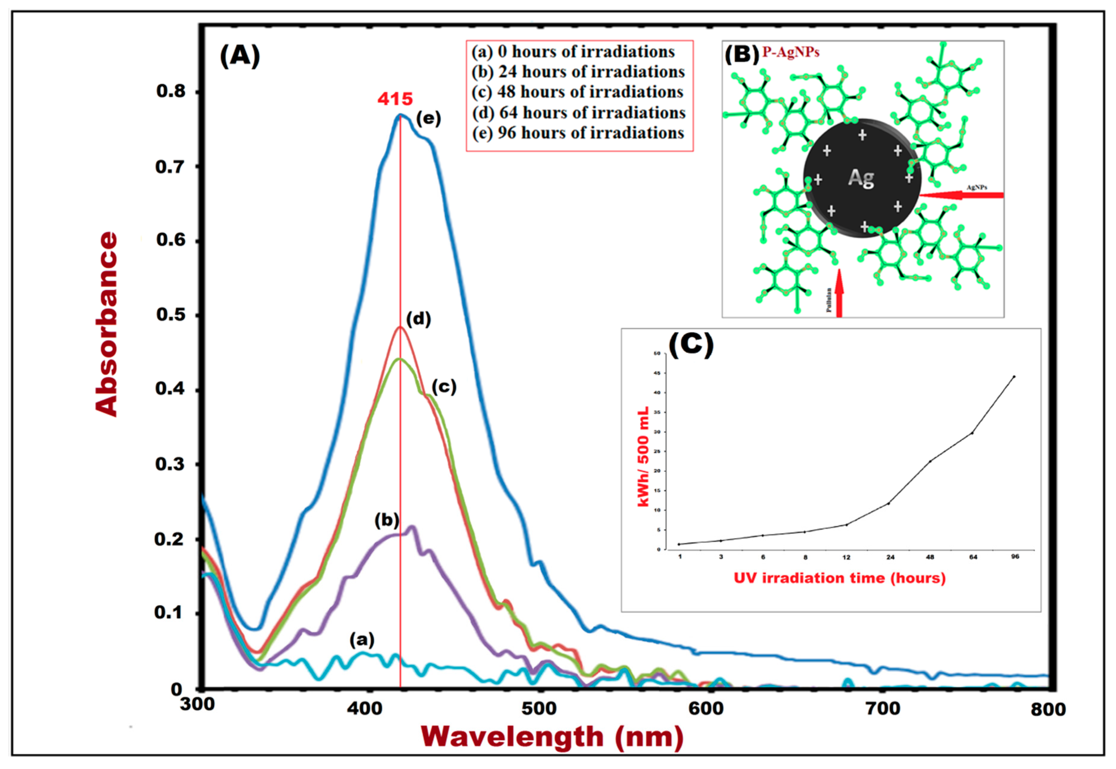
| Samples | UV Irradiation Time (h) | Kilo Watt Hour (kWh)/500 mL |
|---|---|---|
| A1 | 1 | 0.45 |
| A2 | 3 | 1.35 |
| A3 | 6 | 2.70 |
| A4 | 8 | 3.60 |
| A5 | 12 | 5.40 |
| A6 | 24 | 10.80 |
| A7 | 48 | 21.60 |
| A8 | 64 | 28.80 |
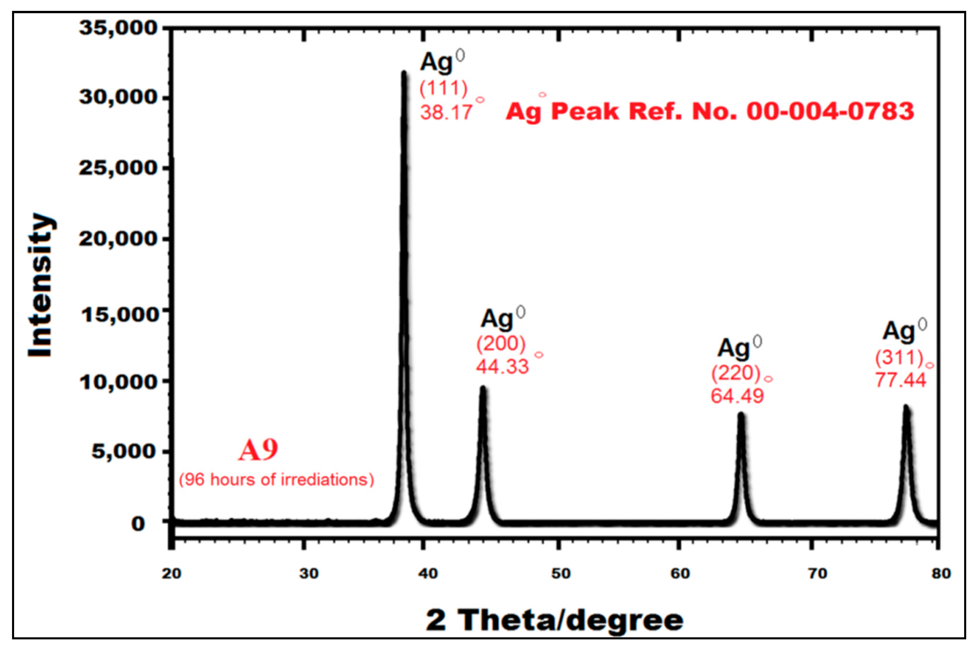
| A9 | 96 | 43.20 |
© 2019 by the authors. Licensee MDPI, Basel, Switzerland. This article is an open access article distributed under the terms and conditions of the Creative Commons Attribution (CC BY) license (http://creativecommons.org/licenses/by/4.0/).
Share and Cite
Khan, M.J.; Kumari, S.; Shameli, K.; Selamat, J.; Sazili, A.Q. Green Synthesis and Characterization of Pullulan Mediated Silver Nanoparticles through Ultraviolet Irradiation. Materials 2019, 12, 2382. https://doi.org/10.3390/ma12152382
Khan MJ, Kumari S, Shameli K, Selamat J, Sazili AQ. Green Synthesis and Characterization of Pullulan Mediated Silver Nanoparticles through Ultraviolet Irradiation. Materials. 2019; 12(15):2382. https://doi.org/10.3390/ma12152382
Chicago/Turabian StyleKhan, Muhammad Jamshed, Suriya Kumari, Kamyar Shameli, Jinap Selamat, and Awis Qurni Sazili. 2019. "Green Synthesis and Characterization of Pullulan Mediated Silver Nanoparticles through Ultraviolet Irradiation" Materials 12, no. 15: 2382. https://doi.org/10.3390/ma12152382
APA StyleKhan, M. J., Kumari, S., Shameli, K., Selamat, J., & Sazili, A. Q. (2019). Green Synthesis and Characterization of Pullulan Mediated Silver Nanoparticles through Ultraviolet Irradiation. Materials, 12(15), 2382. https://doi.org/10.3390/ma12152382