Article

Facile Fabrication of Dumbbell-Like β-Bi2O3/Graphene Nanocomposites and Their Highly Efficient Photocatalytic Activity

1
Chongqing Key Laboratory of Environmental Materials & Remediation Technologies, Chongqing University of Arts and Sciences, Yongchuan 402160, China
2
Chongqing Key Laboratory of Extraordinary Bond Engineering and Advanced Materials Technology (EBEAM), Yangtze Normal University, Chongqing 408100, China
3
State Key Laboratory of Coal Mine Disaster Dynamics and Control, Chongqing University, Chongqing 400044, China
4
College of Chemistry and Chemical Engineering, Chongqing University, Chongqing 401331, China
*
Authors to whom correspondence should be addressed.
The two authors contributed equally to this work.
Materials 2018, 11(8), 1359; https://doi.org/10.3390/ma11081359
Submission received: 10 July 2018 / Revised: 3 August 2018 / Accepted: 3 August 2018 / Published: 6 August 2018
(This article belongs to the Special Issue Photocatalysis for Wastewater Treatment)

Abstract

:
β-Bi2O3 decorated graphene nanosheets (β-Bi2O3/GN) were prepared by a facile solution mixing method. The crystal structure, surface morphology, and photo absorbance properties of the products were characterized by XRD, SEM, and UV-VIS diffuse reflection, respectively. Moreover, the effect of graphene content on photocatalytic activity was systematically investigated, and the results indicated that these composites possessed a high degradation rate of Rhodamine B (RhB), which was three times higher than that of bare β-Bi2O3 when graphene content was 1 wt %. This high photocatalytic activity was attributed predominantly to the presence of graphene, which served as an electron collector and transporter to efficiently lengthen the lifetime of the photogenerated charge carriers from β-Bi2O3.

1. Introduction

The semiconductor can be used as a photocatalyst due to its unique electronic component structure (a valence band full of electrons and an empty conduction band). Bi2O3 is a common important semiconductor material, which is widely used in various fields, such as electronic ceramics, sensors, and high-temperature superconductivity, to name a few [1,2]. As a photocatalyst, it has gained more and more attention [3]. Bi2O3 mainly has four crystal structures, including α, β, γ, and δ. Due to a lower band gap and a unique electronic structure, the β-Bi2O3 has higher photocatalytic activity than the other configurations of Bi2O3 [4,5]. However, the photocatalytic activity of pure Bi2O3 is still restricted. In order to further enhance its activity, researchers have made efforts, such as using Pt, Au, Ag, and other noble metals, to improve the conductivity of the electrons and reduce the recombination probability of charge carrier [6,7]. Chai et al. [8] used Bi2O3 as a precursor to prepare the BiOCl/Bi2O3 complex and finally form a heterojunction so as to significantly improve its activity in the photocatalytic degradation of pollutants.
In recent years, graphene-based semiconductor photocatalysts have gained great attention due to their highly efficient electronic conduction force, larger specific surface area, and good adsorption performance [9]. To date, various graphene–semiconductor composites with enhanced photocatalytic performance have been designed, such as graphene–TiO2 nanocomposite [10], graphene/zirconium oxide [11] and RGO–Bi2O3 nanocomposite [12]. As far as we know, only a few research studies with graphene-based Bi2O3 used as a photocatalyst have been reported. For example, Som et al. [13] and Maruthamani et al. [14] introduced a co-precipitation route to prepare GO/α-Bi2O3 or rGO/Bi2O3 rods. Cao et al. [15] explored an organic electrolyte-assisted method to prepare the GR/β-Bi2O3 composites. Therefore, in this paper, we consider it important to undertake the green chemical synthesis of the composites; a simple method of solution mixing and thermal reduction was used to prepare β-Bi2O3 decorated graphene nanosheets (β-Bi2O3/GN) in one step. Rhodamine B (RhB) is used as the model organic dye to investigate the activity of as prepared samples, as it is an important factor in environmental pollution and its degradation mechanism has been studied quite well [16,17]. In addition, the effect of the content of graphene was also studied systematically, and finally, a possible photocatalytic mechanism of β-Bi2O3/GN composite was proposed.

2. Experimental

2.1. Synthesis of Bi2O3/GN

Graphite oxide was prepared by a modified Hummers method [18]; then, the aqueous solution of graphene oxide (GO) could be obtained by ultrasonic stripping from graphite oxide for 1 h. β-Bi2O3-decorated graphene nanosheets (β-Bi2O3/GN) were prepared by a facile solution mixing method and thermal reduction. In a typical process, 8 mmol of Bi(NO3)3·5H2O was dissolved in 20 cm3 of nitric acid (1 mol/dm3); then, a different volume of GO (2 mg/dm3) was added dropwise into the solution, which was then continuously stirred for 30 min. Then, the above solution was added dropwise into 80 cm3 (0.6 mol/dm3) of saturated sodium carbonate solution. The reaction mixture was stirred for 5 h before filtration. After being washed by water and ethanol several times respectively, the whole product was dried at 60 °C for 10 h. The dried product was then transferred into the muffle furnace after being ground. Finally, the dried product was roasted at 360 °C for 10 min under the nitrogen atmosphere protection, then, it was natural cooled to room temperature to obtain the resultant product. Here, it was worth noting that the heating rate was 4 °C/min. During the reaction, GO was reduced to GN. Pure β-Bi2O3 was synthesized by the same experimental process, except that GO was not added.

2.2. Materials’ Characterization

The samples’ crystal structure was characterized by XRD (Bruker Advance D8, Cu Kα irradiation, Bruker, Germany). Scanning electron microscopy (SEM, JSM-7800F, Japan electronics, Japan) was used to observe the morphology of the prepared samples. The Fourier transform infrared spectroscopy (FTIR) spectra of samples were recorded on a 5DX FTIR (5DX, Nicolet. Co., Rhinelander, WI, USA) spectrometer using KBr powder-pressed pellets. The Brunauer–Emmett–Teller (BET) special surface area was determined through N2 adsorption at 77 K using an adsorption instrument (ASAP-2020, Micromeritics, Norcross, GA, USA). The UV-VIS diffuse reflectance spectra (UV-vis DRS) of samples were measured using a UV-VIS spectrophotometer (TU1901, Beijing Purkinje, Beijing, China).

2.3. Test of Photocatalytic Activity

The photodegradation test was carried out by using a 300-W xenon lamp (the corresponding emission spectrum see Figure S1) (CEL-HXF300, AULTT, Beijing, Country), the self-made circulating water system maintained the temperature of the reaction system at 25 ± 5 °C. RhB solution (50 cm3, 10 mg/dm3) containing 50 mg of catalyst was put in a glass beaker and stirred in the dark overnight to ensure adsorption–desorption equilibrium. After light illumination at regular time intervals, the absorbance of the RhB solution was monitored by a UV-VIS spectrophotometer.

3. Results and Discussion

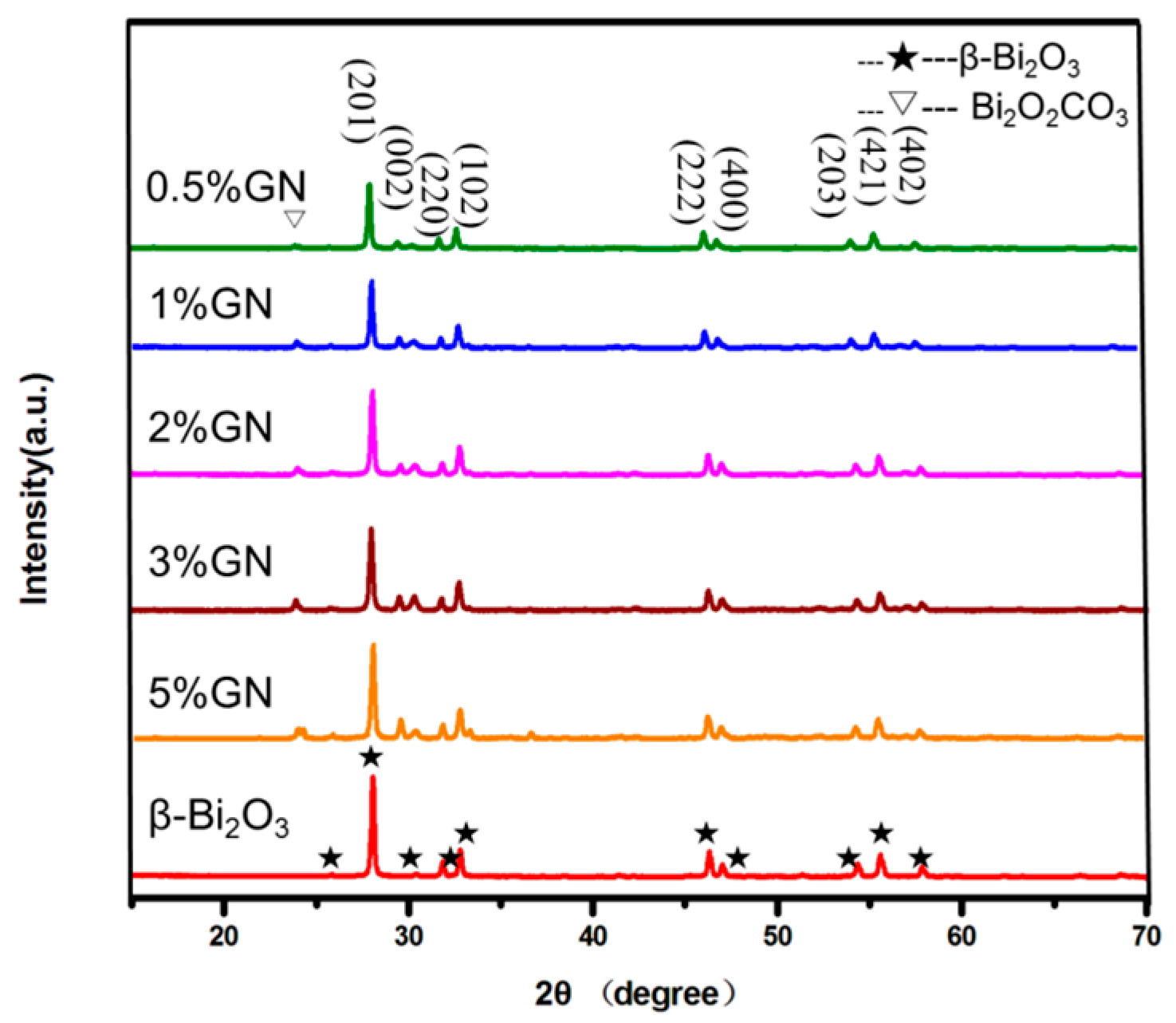
3.1. Crystal Structure Characterization
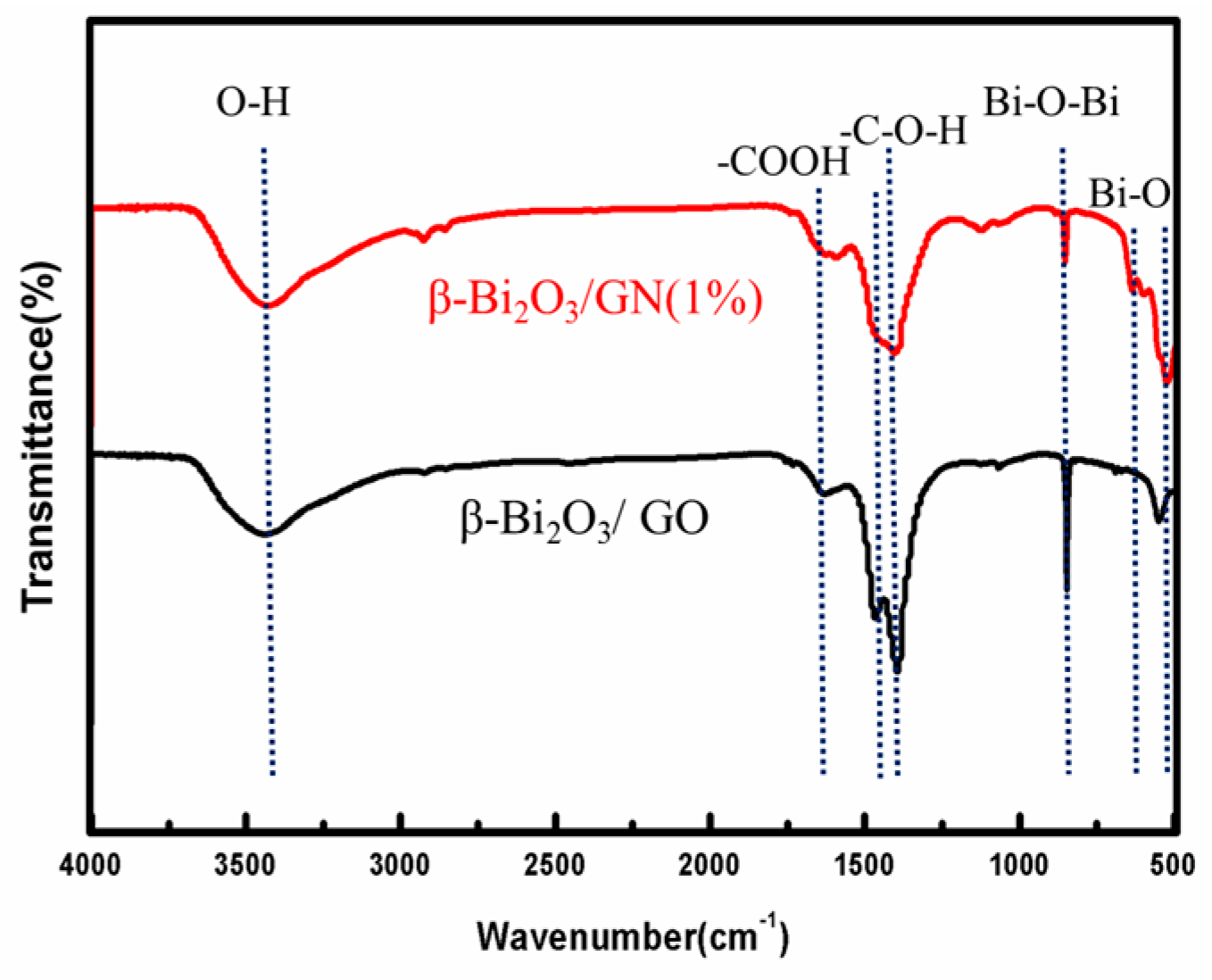
Figure 1 shows the XRD patterns of pure β-Bi2O3 and composites with different masses of GN. The peak positions of 27.9°, 31.7°, 32.6°, and 33.8° correspond to the crystal plane diffractions of (201), (002), (220), and (102) of the tetragonal β-Bi2O3 (JCPDS 27-0050), respectively. The diffraction peaks of the β-Bi2O3/GN sample and β-Bi2O3 are essentially the same, except for a few diffraction peaks of Bi2O2CO3 (the inverted triangle as shown in Figure 1. This may be due to the coating effect of GN, which caused Bi2O2CO3 to not be completely converted into β-Bi2O3 in the thermal decomposition process. However, Bi2O2CO3 has a similar electronic structure to Bi2O3, which is also a well-known photocatalyst. When Bi2O2CO3 was used as the photocatalytic material, it can play a synergistic effect. For example, Chai et al. reported that one β-Bi2O3/Bi2O2CO3 nanosheet composite exhibits much higher photodegradation activity than single phase [5]. It was worth noting that there were no significant carbon-related diffraction peaks in XRD, which is because of the low GN content and low diffraction intensity [19].

3.2. FTIR Spectra

In order to better analyze the state of GN on the surface of β-Bi2O3, the FTIR of β-Bi2O3/GN (1%) and β-Bi2O3/GO are displayed in Figure 2. It can be seen that the composites both have a peak at 500–700 cm−1, which belongs to the telescopic vibration of the Bi–O bond of BiO6 octahedron, and the telescopic vibration peak at 840 cm−1 belongs to Bi–O–Bi [20]. In addition, the O–H stretching vibration of adsorbed water corresponds to 3450 cm−1, and the O–H stretching vibration peak in C–O–H corresponds to 1408 cm−1 [21,22]. The peaks at 1440–1630 cm−1 can be attributed to the antisymmetric stretching vibration and symmetry stretching vibration of the C=O bond in –COOH, respectively. It is worth noting that the C=O antisymmetric stretching vibration at 1450 cm−1 has disappeared after the thermal reduction, and what’s more, the peak intensity becomes weaker after reduction, indicating the effectiveness of the thermal reduction. The results of FTIR show the existence of GO and Bi2O3 in the composites, and GO is effectively reduced to GN after thermal reduction.

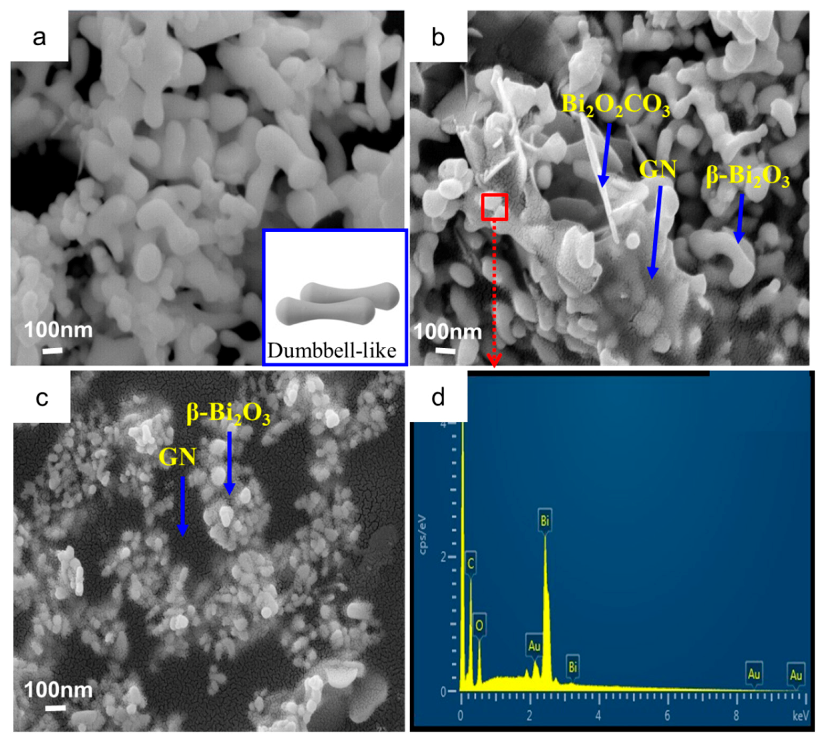
3.3. Surface Morphology Characterization

Surface morphology of the samples was characterized by SEM (Figure 3). The pure β-Bi2O3 is as shown in Figure 3a. β-Bi2O3 has a dumbbell-like morphology (the insert is dumbbell). After the 1% GN was introduced, it can be clearly seen that the β-Bi2O3 was coated by GN (Figure 3b) or embedded in the GN sheets (Figure 3c). In Figure 3b, it can also be seen there is very little Bi2O2CO3, which was consistent with the XRD results. The elemental mapping (Figure 3d) of the ternary as-prepared β-Bi2O3/GN obtained by EDS (the mapped region highlighted with red frame in Figure 3b) indicates that the weight ratios of the elements are close to the Bi2O3 molar mass ratios. Meanwhile, only the peaks of C, O, Bi, Au were detected, which means that the as-prepared composites are composed of graphene and Bi2O3.

3.4. Surface Areas and Pore Size Distributions

In general, the specific surface area of catalysts and its surface structure have significant influences on catalytic activity. Therefore, the specific surface analyzer is utilized to implement further research on the specific surface area and the pore diameter distribution of pure β-Bi2O3 and β-Bi2O3/GN (1%). Figure 4 shows the N2 adsorption–desorption isotherms and the corresponding curves of the pore size distribution (inset) for samples β-Bi2O3 and β-Bi2O3/GN (1%). According to the Brunauer–Deming–Deming–Teller (BDDT) classification, pure β-Bi2O3 isotherms can be categorized as type III (Figure 4a), which is convex to the P/P0 axis over its entire range, indicating that the pure β-Bi2O3 belong to a nonporous structure. Meanwhile, samples of β-Bi2O3/GN (1%) have isotherms of type IV, suggesting the presence of mesopores. Upon observing the pore diameter distribution diagram obtained from desorption isotherms, pore diameter mainly distributes in 5.28 nm (Figure 4b inset). The specific surface area of β-Bi2O3 and β-Bi2O3/GN (1%) are given by BET measurement as 3.37 m2/g, 4.53 m2/g, respectively, which conforms to the results. In other words, when graphene is introduced, mesopores begin to appear in samples and the specific surface area increases, which is because the prepared nanocomposite is composed of sheet-like graphene decorated with β-Bi2O3, similar to the previous report [23]. Consequently, the introduction of graphene can increase the specific surface area, and graphene may play a role in enhancing the photocatalytic activity.

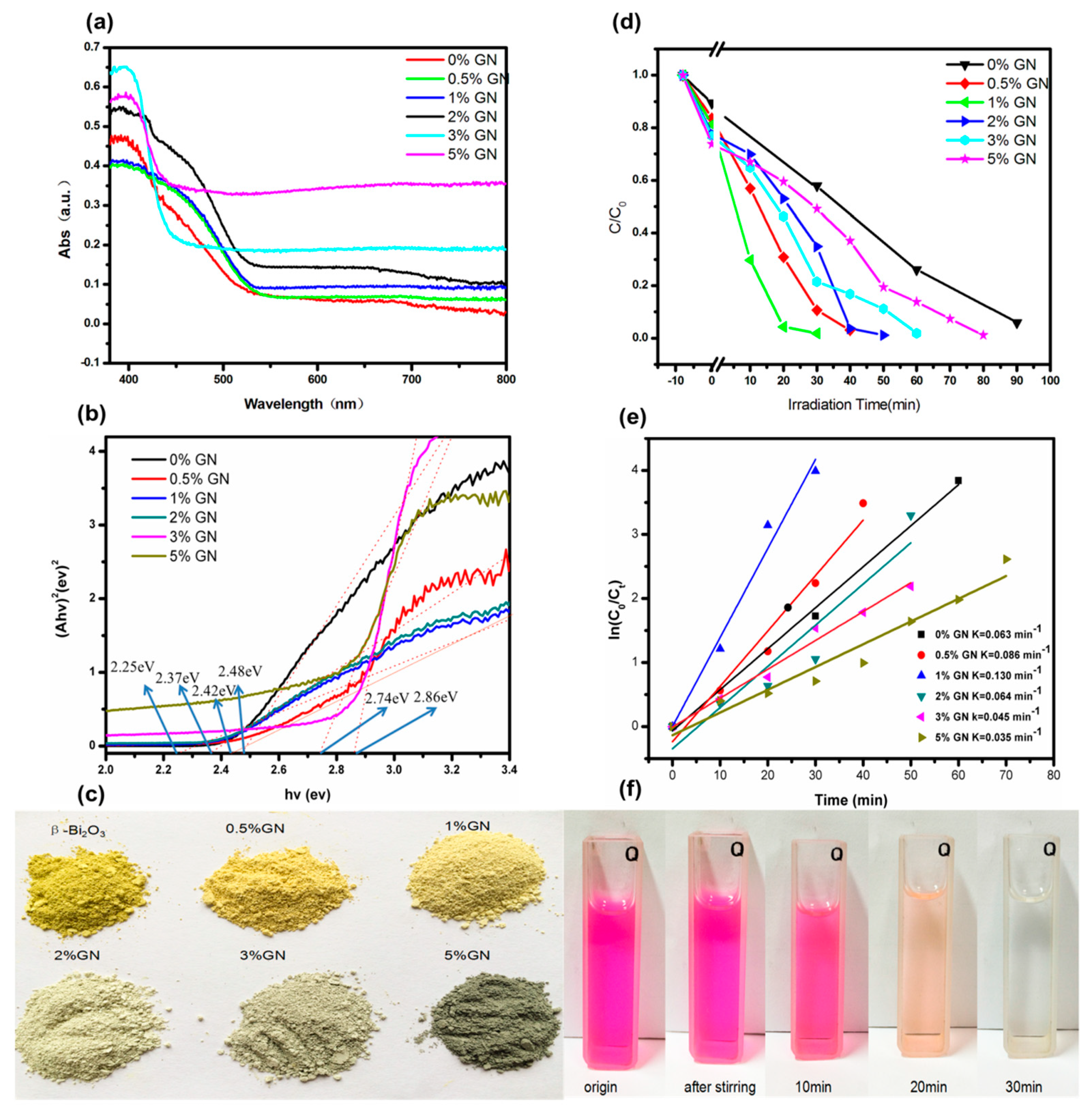
3.5. UV-Vis Diffuse Reflectance and Photocatalytic Activity

The photocatalytic activity tests are shown in Figure 5. From Figure 5a, the absorption of visible light by the samples can be seen as the content of GN increases, and the sample color gradually changing from yellow to deeper brown can also be seen in Figure 5c. This is another proof to the increasing absorption of visible light. The band gap of calculated β-Bi2O3 and composites are shown in Figure 5b. Although the 5% GN is not narrowest band gap, the absorbed threshold value of sample’s visible light region is significantly greater than the other samples. Therefore, it is hypothesized that the introduction of graphene can better absorb the visible light and enhance the photocatalytic activity.
The tests of photocatalytic degradation of RhB (Figure 5d) show that all of the composites have higher degradation rates than bare β-Bi2O3, suggesting that there is a synergistic effect between the GN sheet and β-Bi2O3 nanoparticles. The sequential dye degradation rates are as follows: 1% GN > 0.5% GN > 2% GN > 3% GN > 5% GN > bare β-Bi2O3. It seen that only the low content (1%) of GN is introduced, the dye degradation rate can be significantly increased by three times in comparison with the bare β-Bi2O3. The kinetics of photodegradation reaction are investigated in Figure 5e, and the results show that 1% GN composite has the highest constant photodegradation reaction rate, which is 0.130 min−1. With the increase of GN content, the dye degradation rate did not always increase; the activity of 5% GN was significantly less than that of 1% GN. The reason why the degradation ratio decreased as the content of graphene increased was that the introduction of a large mass of black graphene would result in a rapid decrease of the light absorption of the reaction solution [23,24]. Thus, in order to achieve an optimal photocatalytic performance, it is crucial to control the composition ratio in the nanocomposite of β-Bi2O3/GN.

3.6. Possible Mechanisms Speculation

According to the above experimental results, the possible photocatalytic mechanisms of β-Bi2O3/GN were estimated, as shown in Figure 6. Under simulated sunlight irradiation, the excited electrons of the β-Bi2O3 semiconductor divert a part of the electrons out due to the good conductivity of the graphene layers in the course of being conducted to the conduction band. In other words, in the system of β-Bi2O3/GN, the graphene, as the receiving body and conductor of the electrons, effectively separated the photogenerated electron-hole pairs to avoid the recombination of electrons and holes. It’s similarly compared with a previously studied system in which the GO embedded into TiO2 nanofiber and served as a conduit of electron transfer [25]. Posa et al. [10] and Li et al. [23] prepared graphene–TiO2 nanocomposite and CdS-cluster-decorated graphene nanosheets respectively; the similar mechanisms were proposed, too. Furthermore, the unique features of graphene also contribute to the improvement of photocatalytic activity, which allow photocatalytic reactions to take place not only on the surface of semiconductor catalysts, but also on the graphene sheet, greatly enlarging the reaction space.

4. Conclusions

With the simple method of solution mixing, β-Bi2O3/GN composite photocatalysts with different qualities were successfully prepared. In the test of degradation rate, it was found that the sample with 1% GN on its surface had the highest photocatalytic activity, with its dye degradation efficiency being three times higher than the pure β-Bi2O3. However, when the content of GN increased, the degradation rate decreased. This was because the introduction of more graphene may decrease the light absorption of the reaction solution. Overall, the introduction of appropriate quantity of graphene could significantly increase the photocatalytic degradation rate of the catalyst.

Supplementary Materials

The following are available online at https://www.mdpi.com/1996-1944/11/8/1359/s1, Figure S1: The emission spectrum of a 300-W Xenon lamp (CEL-HXF300, AULTT, Beijing, China).

Author Contributions

Writing-Original Draft Preparation, J.Y. and T.X.; Writing-Review & Editing, J.Y. and T.X.; Supervision, C.L. and L.X.; Funding Acquisition, J.Y.

Funding

This research was funded by Scientific and Technological Research Program of Chongqing Municipal Education Commission (KJ1711286) and Chongqing Basic Science and Advanced Technology Research Program (CSTC2015jcyjBX0015).

Acknowledgments

We want to thank the financial support from Scientific and Technological Research Program of Chongqing Municipal Education Commission (KJ1711286) and Chongqing Basic Science and Advanced Technology Research Program (CSTC2015jcyjBX0015).

Conflicts of Interest

The authors declare no conflict of interest.

References

  1. Maeder, T. Review of Bi2O3 based glasses for electronics and related applications. Int. Mater. Rev. 2013, 58, 3–40. [Google Scholar] [CrossRef]
  2. Cabot, A.; Marsal, A.; Arbiol, J. Bi2O3 as a selective sensing material for NO detection. Sens. Actuators B Chem. 2004, 99, 74–89. [Google Scholar] [CrossRef]
  3. Zhou, L.; Wang, W.Z.; Xu, H.L.; Sun, S.M.; Shang, M. Bi2O3 hierarchical nanostructures: Controllable synthesis, growth mechanism and their application in photocatalysis. Chem. Eur. J. 2009, 15, 1776–1782. [Google Scholar] [CrossRef] [PubMed]
  4. Cheng, H.F.; Huang, B.B.; Lu, J.B.; Wang, Z.Y.; Xu, B.; Dai, Y. Synergistic effect of crystal and electronic structures on the visible-light-driven photocatalytic performances of Bi2O3 polymorphs. Phys. Chem. Chem. Phys. 2010, 12, 15468–15475. [Google Scholar] [CrossRef] [PubMed]
  5. Cai, G.Y.; Xu, L.L.; Wei, B.; Che, J.X.; Gao, H.; Sun, W.J. Facile synthesis of β-Bi2O3/Bi2O2CO3 nanocomposite with high visible-light photocatalytic activity. Mater. Lett. 2014, 120, 1–4. [Google Scholar] [CrossRef]
  6. Li, R.H.; Chen, W.X.; Kobay, H.; Ma, C.X. Platinum-nanoparticle-loaded bismuth oxide: An efficient plasmonic photocatalyst active under visible light. Green Chem. 2010, 12, 212–215. [Google Scholar] [CrossRef]
  7. Anandan, S.; Lee, G.J.; Chen, P.K.; Fan, C.; Wu, J. Removal of Orange II Dye in Water by Visible Light Assisted Photocatalytic Ozonation Using Bi2O3 and Au/ Bi2O3 Nanorods. Ind. Eng. Chem. Res. 2010, 49, 9729–9737. [Google Scholar] [CrossRef]
  8. Chai, S.Y.; Kim, Y.J.; Jung, M.H.; Chakraborty, A.K.; Jung, D.; Lee, W.I. Heterojunctioned BiOCl/Bi2O3, a new visible light photocatalyst. J. Catal. 2009, 262, 144–149. [Google Scholar] [CrossRef]
  9. Xiang, Q.; Yu, J.; Jaroniec, M. Graphene-based semiconductor photocatalysts. Chem. Soc. Rev. 2012, 41, 782–796. [Google Scholar] [CrossRef] [PubMed]
  10. Posa, V.R.; Annavaram, V.; Koduru, J.R.; Bobbala, P.; Madhavi, V.; Somala, A.R. Preparation of graphene-TiO2 nanocomposite and photocatalytic degradation of Rhodamine B under solar light irradiation. J. Exp. Nano Sci. 2016, 11, 722–736. [Google Scholar] [CrossRef]
  11. Rania, S.; Aggarwal, M.; Kumarc, M.; Sharma, S.; Kumard, D. Removal of methylene blue and rhodamine B from water byzirconium oxide/graphene. Water Sci. 2016, 30, 51–60. [Google Scholar] [CrossRef]
  12. Suresh, M.; Sivasamy, A. Bismuth oxide nanoparticles decorated raphene layers for the degradation of Methylene blue dye under visible light irradiations and antimicrobial activities. J. Environ. Chem. Eng. 2018, 6, 3745–3756. [Google Scholar] [CrossRef]
  13. Tirtha, S.T.; Troppenz, G.V.; Wendt, R.R.; Wollgarten, M. Graphene Oxide/α-Bi2O3 Composites for Visible-Light Photocatalysis Chemical Catalysis and Solar Energy Conversion. ChemSusChem 2014, 7, 854–865. [Google Scholar] [CrossRef] [Green Version]
  14. Maruthamania, D.; Vadivela, S.; Kumaravela, M.; Saravanakumarb, B.; Paulc, B.; Sankar, D.S.; Habibi-Yangjehd, A.; Manikandane, A.; Ramadoss, G. Fine cutting edge shaped Bi2O3 rods/reduced graphene oxide (RGO)composite for supercapacitor and visible-light photocatalytic applications. J. Colloid Interface Sci. 2017, 498, 449–459. [Google Scholar] [CrossRef]
  15. Cao, S.; Chen, C.; Xie, X. Hypothermia-controlled Co-precipitation route to deposit well-dispersed β-Bi2O3 nanospheres on polymorphic graphene flakes. Vacuum 2014, 102, 1–4. [Google Scholar] [CrossRef]
  16. Hu, X.F.; Mohamood, T.; Ma, W.H.; Chen, C.C.; Zhao, J.C. Oxidative Decomposition of Rhodamine B Dye in the Presence of VO2+ and/or Pt(IV) under Visible Light Irradiation: N-Deethylation, Chromophore Cleavage, and Mineralization. J. Phys. Chem. B 2006, 110, 26012–26018. [Google Scholar] [CrossRef] [PubMed]
  17. Patrick, W.; Stephan, D. Photodegradation of rhodamine B in aqueous solution via SiO2@TiO2 nano-spheres. J. Photochem. Photobiol. A 2007, 185, 19–25. [Google Scholar] [CrossRef]
  18. Daniela, C.M.; Dmitry, V.K.; Jacob, M.B.; Alexander, S.; Sun, Z.Z.; Slesarev, A.; Alemany, L.B.; Lu, W.; Tour, J.M. Improved Synthesis of Graphene Oxide. ACS Nano 2010, 4, 4806–4814. [Google Scholar] [CrossRef] [Green Version]
  19. Adivel, S.; Keerthi, V.M.; Muthukrishnaraj, A.; Balasubramanian, N. Solvothermal synthesis of Sm-doped BiOBr/RGO composite as an efficient photocatalytic material, for methyl orange degradation. Mater. Lett. 2014, 128, 287–290. [Google Scholar] [CrossRef]
  20. Lu, Y.G.; Yang, Y.C.; Ye, Z.X.; Liu, S.Y. Preparation and Visible Light Responsive Photocatalytic Activity of Nitrogen-doped Bi2O3 Phocatalyst. J. Inorg. Mater. 2012, 6, 643–648. [Google Scholar] [CrossRef]
  21. Chi, S.; Kim, B. Nuclear Graphites (I): Oxidation Behaviors. Carbon Lett. 2009, 10, 239–249. [Google Scholar] [CrossRef]
  22. Chen, C.; Long, M.; Xia, M.; Cai, W. Reduction of Graphene Oxide by An in-Situ Photoelectrochemical Method in a Dye-Sensitized Solar Cell Assembly. Nanoscale Res. Lett. 2012, 7, 1–5. [Google Scholar] [CrossRef] [PubMed]
  23. Li, Q.; Guo, B.; Yu, J.; Ran, J.; Zhang, B.; Yan, H.; Gong, JR. Highly Efficient Visible-Light-Driven Photocatalytic Hydrogen Production of CdS Cluster Decorated Graphene Nanosheets. J. Am. Chem. Soc. 2011, 133, 10878–10884. [Google Scholar] [CrossRef] [PubMed]
  24. Yu, J.; Ma, T.; Liu, S. Enhanced photocatalytic activity of mesoporous TiO2 aggregates by embedding carbon nanotubes as electron-transfer channel. Phys. Chem. Chem. Phys. 2011, 13, 3491–3501. [Google Scholar] [CrossRef] [PubMed]
  25. Kim, H.L.; Kim, S.; Kang, J.K.; Choi, W.Y. Graphene oxide embedded into TiO2 nanofiber: Effective hybrid photocatalyst for solar conversion. J. Catal. 2014, 309, 49–57. [Google Scholar] [CrossRef]
Figure 1. Diffraction patterns of pure β-Bi2O3 and hybrid composites with different mass ratios of graphene nanosheets (GN).
Materials 11 01359 g001
Figure 2. Fourier transform infrared (FTIR) spectra of β-Bi2O3/GN (1%) and β-Bi2O3/GO.
Materials 11 01359 g002
Figure 3. (a) SEM images of pure β-Bi2O3; (b,c) β-Bi2O3/GN(1%); and (d) EDS pattern of β-Bi2O3/GN.
Materials 11 01359 g003
Figure 4. N2 adsorption–desorption isotherm of (a) β-Bi2O3 and (b) β-Bi2O3/GN (1%). Inset: the corresponding pore size distribution.
Materials 11 01359 g004
Figure 5. (a) The spectrogram of UV-VIS diffuse reflectance of GN with different mass ratios; and (b) corresponding band gap energy; (c) the chart of the corresponding sample color change; (d) the Rhodamine B (RhB) degradation rates of samples with different quantity ratios of GN; (e) kinetic curve of photodegradation; (f) color change chart of RhB solution by 1% GN degradation.
Materials 11 01359 g005
Figure 6. Speculation schematic of β-Bi2O3/GN photocatalytic degradation mechanism of dye.
Materials 11 01359 g006

