Article

Structural Characterization and Molecular Model Construction of Lignite: A Case of Xianfeng Coal

1
Key Laboratory of Coalbed Methane Resources and Reservoir Formation Process, Ministry of Education, China University of Mining and Technology, Xuzhou 221008, China
2
School of Resources and Geosciences, China University of Mining and Technology, Xuzhou 221116, China
*
Author to whom correspondence should be addressed.
Energies 2024, 17(5), 1049; https://doi.org/10.3390/en17051049
Submission received: 7 January 2024 / Revised: 24 January 2024 / Accepted: 19 February 2024 / Published: 23 February 2024
(This article belongs to the Section H3: Fossil)

Abstract

:
The object of the study is lignite. Analytical testing techniques, such as elemental analysis, 13C nuclear magnetic resonance (13C NMR) spectroscopy, Fourier transform infrared spectroscopy (FTIR), X-ray photoelectron spectroscopy (XPS), and high-resolution transmission electron microscopy (HRTEM), were used to acquire information on the structural parameters of lignite. The aromaticity of Xianfeng lignite is 43.57%, and the aromatic carbon structure is mainly naphthalene and anthracene/phenanthrene. The aliphatic carbon structure is dominated by cycloalkanes, alkyl side chains, and hydrogenated aromatics. Oxygen is mainly present in ether oxygen, carboxyl, and carbonyl groups. Nitrogen is mainly in the form of pyrrole nitrogen and quaternary nitrogen. Sulfur is mainly thiophene sulfur. According to the analysis results, the molecular structure model of XF lignite was constructed. The molecular formula is C184H172O39N6S2. The 2D structure was converted to a 3D structure using computer simulation software and optimized. The optimized model has a remarkable stereoconfiguration, and the aromatic lamellae are irregularly arranged in space. The aromatic rings were mainly connected by methylene, hypomethylene, methoxy, and aliphatic rings. In addition, the simulated 13C NMR spectra are in good agreement with the experimental spectra. This shows the rationality of the 3D chemical structure model.

1. Introduction

Lignite is a macromolecular aggregate formed via the condensation of functional groups, such as aromatic rings and aliphatic chains. It is characterized by good chemical reactivity [1]. Understanding the chemical structure of lignite is the core content of coal chemistry, which is the basis for exploring the chemical reaction activity and properties of lignite and has important theoretical value [2].
The purpose of the construction of coal structure models is to study the properties of coal at the molecular level. The structural evolution of coal has been the focus and difficulty of research in the fields of coal geology and oil and gas geology, and it has a long research history and more research results [3,4]. Okolo et al. [5] revealed the structural characteristics of bituminous coal via 13C NMR spectroscopy. Yuan et al. [6] studied the structural characteristics of Inner Mongolia lignite and obtained detailed information on various heteroatom clusters via photoelectron spectroscopy. However, the analysis of 2D surface functional groups cannot meet the understanding of the coal macromolecular structure. Lin et al. [7] characterized Indonesian lignite and constructed its model using 13C NMR, FTIR, XPS, and XRD (X-ray Diffraction). Wang et al. [8] obtained the aromatic structural compositions and fatty side chains using HRTEM and, in combination with XPS, manually constructed a molecular model of the lignite. Song et al. [9] used 13C NMR to investigate the carbon structure types of four low-rank coals. It can be seen from previous studies that different spectral techniques can be used to obtain coal macromolecular structure information from different angles. Based on the joint characterization of various technical means, more comprehensive coal structure information can be obtained, and the coal macromolecular structure can be more fully understood [10].
Although there is a certain understanding of coal structural evolution based on various experimental test methods, there is less knowledge on the evolution of the three-dimensional spatial structure of coal. Moreover, due to the complexity, diversity, and heterogeneity of lignite, the study of lignite structure lags much behind compared to bituminous coal [11]. And there are few reports on the establishment of three-dimensional molecular structure models of lignite. In this study, the structure and properties of Xianfeng (XF) lignite were studied. Modern analytical techniques, such as elemental analysis, 13C NMR, FTIR, XPS, XRD, Raman, and HRTEM, were used in the study. Then, the molecular average structure model of the XF lignite was constructed based on these techniques. Using computer-aided optimization, the conformational optimization and spectral simulation of the molecular model were performed. Finally, the accuracy of the constructed molecular model is verified by comparing the agreement of the simulated spectra with the experimental spectra. Then, an accurate scientific lignite molecular structure model was constructed.

2. Coal Sample and Experimental Method

2.1. Characteristics of Coal Sample

The samples were taken from the fresh working face of the Xianfeng Opencast Coal Mine in Yunnan Province, China. The reflectance (Ro) of coal samples was determined according to GB/T6948-2008 [12]. The specular group reflectance Ro of the sample is 0.321%. The raw coal samples were crushed and sieved with a sample crusher at a particle size range of 150–200 mesh. The proximate analysis tests of the samples were based on GB/T212-2008 [13]. The ultimate analysis tests of the samples were based on GB/T476-2008 [14], GB/T19227-2008 [15], and GB/T214-2007 [16] (Table 1). The proportion of C, H, O, N, and S atoms in the molecular structure of coal is the basic parameter for constructing the model. The molecular ratio can be calculated based on the content of each element (content below 0.5% modeling is not considered) [17].

2.2. Experiments

13C NMR spectra were acquired using a Bruker (Karlsruhe, Germany) AVANCE II 400M spectrometer and a 4 mm MAS probe. The NMR spectra were obtained via CPMAS (Cross-polarized Magic Angle Spinning Technique) pulses at a spin rate of 10 kHz. The cycle delay was 4 s.
The FTIR spectra were tested using a Thermo Scientific Nicolet iS20 instrument, Waltham, MA, USA. During the test, we first collected the background and then collected the infrared spectrum of the sample. The resolution was 4 cm−1, the number of scans was 32, and the test wavenumber range was 400–4000 cm−1.
The XPS experiments were performed on a Thermo Scientific (Waltham, MA, USA) K-Alpha type X-ray photoelectron spectrometer. The pass energy of the full spectrum scan was 150 eV with a step size of 1 eV. The pass energy of the scan was 50 eV in steps of 0.1 eV. Also, the XPS experiments were performed five times to reduce the influence of instrument noise.
Laser Raman spectroscopic characterization was performed using a LabRAM HR Evolution confocal Raman microspectrometerr, produced by HORIBA company in Paris, France. The laser wavelength was 532 nm, the power was 20 mW, the scanning time was 2 s, the cumulative number of times was 10, and the resolution was 9–18 cm−1. The Raman spectra were recorded in the range of 50–4000 cm−1.
HRTEM experiments were performed on a JEOL JEM 2100F transmission electron microscope in Tokyo, Japan. The crushed sample was put into anhydrous ethanol, and the powder was well dispersed in the anhydrous ethanol using ultrasonic waves. In this paper, the structural features of aromatic groups based on the lengths of the edges of HRTEM images were analyzed according to the method of Niekerk and Mathews [18].
The instrument used in the XRD experiment was the Bruker D8 Advance X-ray diffractometer, produced by Brooke Company in Werther, Germany. The wavelength of the X-ray used was 1.54059 Å (Cu). The tube current was 10–60 mA. The angle scanning range was 5°–70°, and the scanning speed was 2°/min. Powder samples were used in the experiment, and the particles passed a 325-mesh sieve.

2.3. Lignite Structure Model Construction Method

The 3D macromolecular structure of coal in the thesis was optimized on top of the 2D chemical planar structure. The construction of the 2D planar chemical structure was mainly based on 13C NMR, XPS, FTIR, Raman, HRTEM, and XRD. The specific steps of the model construction are shown in Figure 1 [19].
Step 1: Analysis of key experimental data.
Information about the morphology and relative content of the target coal structural fragments was obtained through Step 1. The 13C NMR data of the sample provide carbon skeleton information and provide partial information on aromatic carbon and fatty carbon for model construction [20]. FTIR spectroscopy reflects the presence of functional groups in coal and is the basis for constructing oxygen atoms in the model [21]. XPS spectroscopy was used to qualitatively and quantitatively study the existing forms of surface elements. It mainly provides the existing forms of nitrogen and sulfur elements on the surface of the coal structure [22]. Molecular structure orderliness information was analyzed using the Raman test. HRTEM was used for aromatic structure and morphology. The microcrystalline structural characteristics of coal were analyzed via XRD [23].
Step 2: Establish a 2D structure model of coal macromolecules.
The type and number of major aromatic structures in the model were determined based on 13C NMR, XPS, and HRTEM experiments in the first step. Calculate the number of aromatic carbon atoms and determine the molecular formula of the sample based on the relative proportions of atoms obtained from elemental analysis. Determine the type of fatty carbon structure, and build an initial planar molecular model.
Step 3: 2D structure model adjustment.
Calculate the NMR spectra of the initial built model; compare the experimental NMR spectra with the calculated NMR spectra. Readjust the structural model according to the difference between the experimental and calculated values until the experimental and calculated values are basically the same.
Step 4: Establish a 3D coal macromolecule structure model.
The 3D macromolecular structure of lignite was built using the Materials Studio 2017 software. Then, geometry optimization and energy optimization were performed using the Foricte module. An AC module was used to set the density. The final 3D structural model obtained had good agreement among the chemical composition, spatial density, and experimental values.

3. Analysis and Results

3.1. 13C NMR

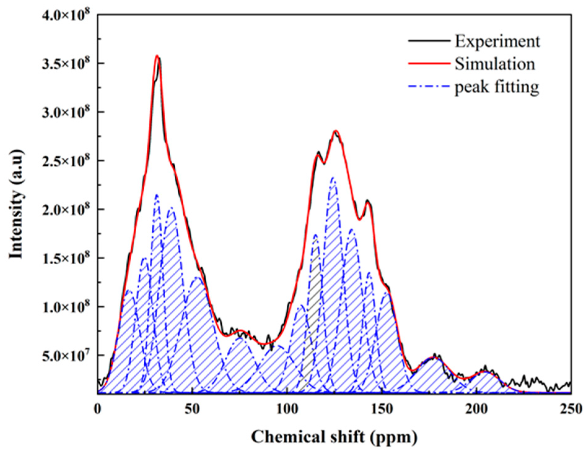
In the NMR spectrum of coal, there are three main chemical regions: the aliphatic fraction (0–90), the aromatic fraction (90–165), and the carbonyl fraction (165–240). In this study, the XF-lignite samples were analyzed via 13C NMR, and the carbon skeleton structure was analyzed based on peak fitting of its carbon spectrum. As shown in Figure 2, the spectrum is divided into 15 peaks using PeakFit v4.12 software, which represent different carbon types. Among them, the 13C NMR peak positions and chemical shifts (δ) are attributed, with reference, to the relevant literature (Table 2) [17,24,25,26].
Different chemical shifts correspond to different carbon atom attributions. The peaks of NMR are mainly concentrated at 0–165 (Figure 2). This indicates that the carbon skeleton of the pioneer coal macromolecule is mainly aliphatic carbon and aromatic carbon. The chemical structure parameters of the carbon atoms at the coal samples were calculated from Table 1 and Figure 2. The results are shown in Table 3. The aliphatic carbon rate (fal = fal3 + fala + fal2 + fal1 + falO) is 51.29%, which is the main part of the molecular structure. The aromatic carbon rate (fa = faH + faB + faS + faO) is 43.57%. The carboxyl carbon content (faC = faCC1 + faCC2) is 5.13%. In the aliphatic carbon region, the oxygenated aliphatic carbon, represented by the peak in the 50–95 spectral band, is the most abundant. Its relative content is 19.45%. The quaternary carbon, represented by the shoulder peak at δ 38.85, is the next most abundant, with a relative content of 12.17%. And the methylene carbon was represented by the main peak at δ of 31.28, with 6.96%. Among the aromatic carbons, the most abundant is the protonated aromatic carbon, with a relative content of 23.25%. The aromatic bridge carbon has a relative content of 9.83%.
The ratio of bridged aromatic carbon to pericarbon in sample XBP can be calculated to be 0.29 using Equation (1) [27]. The average methylene chain length (Cn = fal2/faS) is 1.58. And the degree of aromatic ring substitution (σ = (faH + faS + faO)/fa) is 77%. All of the above parameters are important indicators for the construction of the structural model of pioneer coal [28].
X B P = f a B f a H + f a S + f a O

3.2. FTIR

Different characteristic frequencies in FTIR maps reflect different functional group structures and different vibrational forms (including stretching, bending, and deformation vibrations) [29]. The structural composition of coal is very complex, and the vibration peaks of functional groups with similar frequencies are easy to overlap with each other, which affects the quantitative analysis of the spectrum. On the basis of previous research results, the Fourier transform infrared image of coal was segmented based on the computer peak separation fitting method (Figure 3). The Gaussian model is used in the fitting process, and the second derivative of the spectrum is selected to determine the approximate position and quantity of the initial peaks [27]. The main characteristic absorption peaks were attributed, and the results are shown in Table 4.
The 900–700 cm−1 region is the out-of-plane bending vibration of the C–H bond of the aromatic ring. As shown in Figure 3a, the relevant absorption peaks are located near 730 cm−1 and 790 cm−1. The intensity of these peaks is low, reflecting that the sample has a low degree of aromatic condensation.
Figure 3b shows that 1200–1000 cm−1 corresponds to the antisymmetric stretching vibrations of C–O–C. The absorption peaks at 1600–1300 cm−1 (1488.09 cm−1, 1447.09 cm−1, 1390.60 cm−1, 1316.16 cm−1) are skeletal vibrations of aromatic compounds. The absorption peak at 1706.93 cm−1 reflects the strong C=O stretching vibration and hydrogen bond resonance formed by C=O and hydroxyl groups in carboxyl group.
The region of wave number 3000–2800 cm−1 is the region of C–H stretching vibrations in saturated hydrocarbons (Figure 3c). The absorption peaks near 2800–2900 cm−1 indicate the presence of –CH3 and –CH2 symmetric stretching vibrations. The absorption peaks near 2900–3000 cm−1 (2965.95 cm−1, 2951.15 cm−1, 2923.52 cm−1) belong to the –CH3 antisymmetric stretching vibration. This region shows that the content of methyl and methylene structures in coal samples is high. It shows that there are more fat structures in coal samples, which is also consistent with the properties of lignite [30].
The absorption peak in the range of 3700–3400 cm−1 indicates the stretching vibration of N–H and O–H bonds. The absorption peak near 3300–3000 cm−1 corresponds to the stretching vibration of the C–H bond and C–H in the aromatic ring. At the same time, it is observed here that the C–H structure has the potential to connect various functional groups [31,32].

3.3. XPS

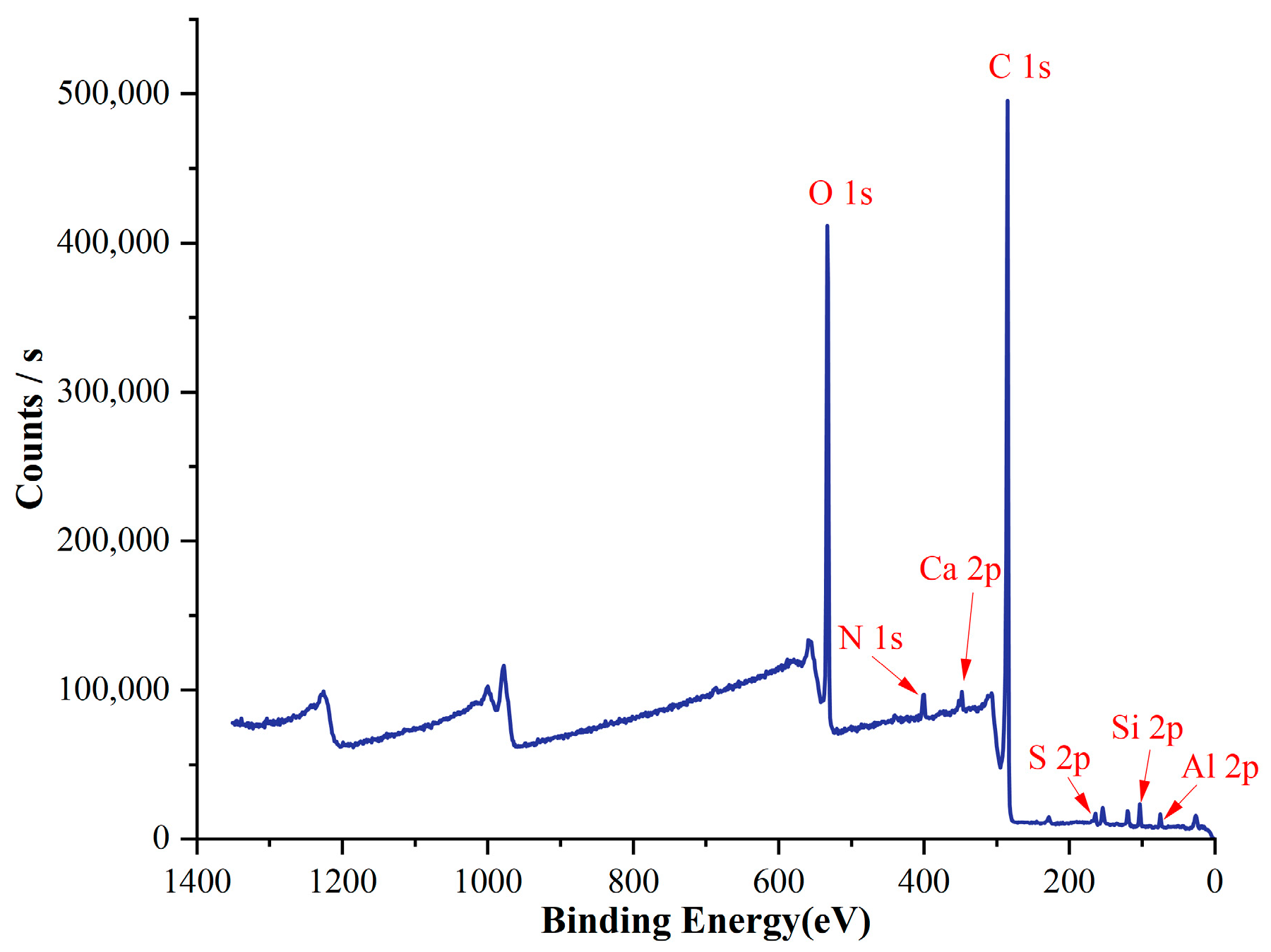
XPS is mainly used to analyze the forms and chemical environments of heteroatom groups in coal molecules, such as nitrogen and sulfur (Figure 4). The relative content of carbon atoms is 68.90%. The oxygen content is 23.222%. The nitrogen content is 2.38%. The sulfur content is low, at 0.69% (Table 5). These results are similar to those obtained through elemental analysis (Table 1). In addition, a small amount of Si, Al, and Ca atoms can be found in Table 5. Those could be clay minerals [33]. We do not consider inorganic minerals in the construction of the organic macromolecular structure model of coal.
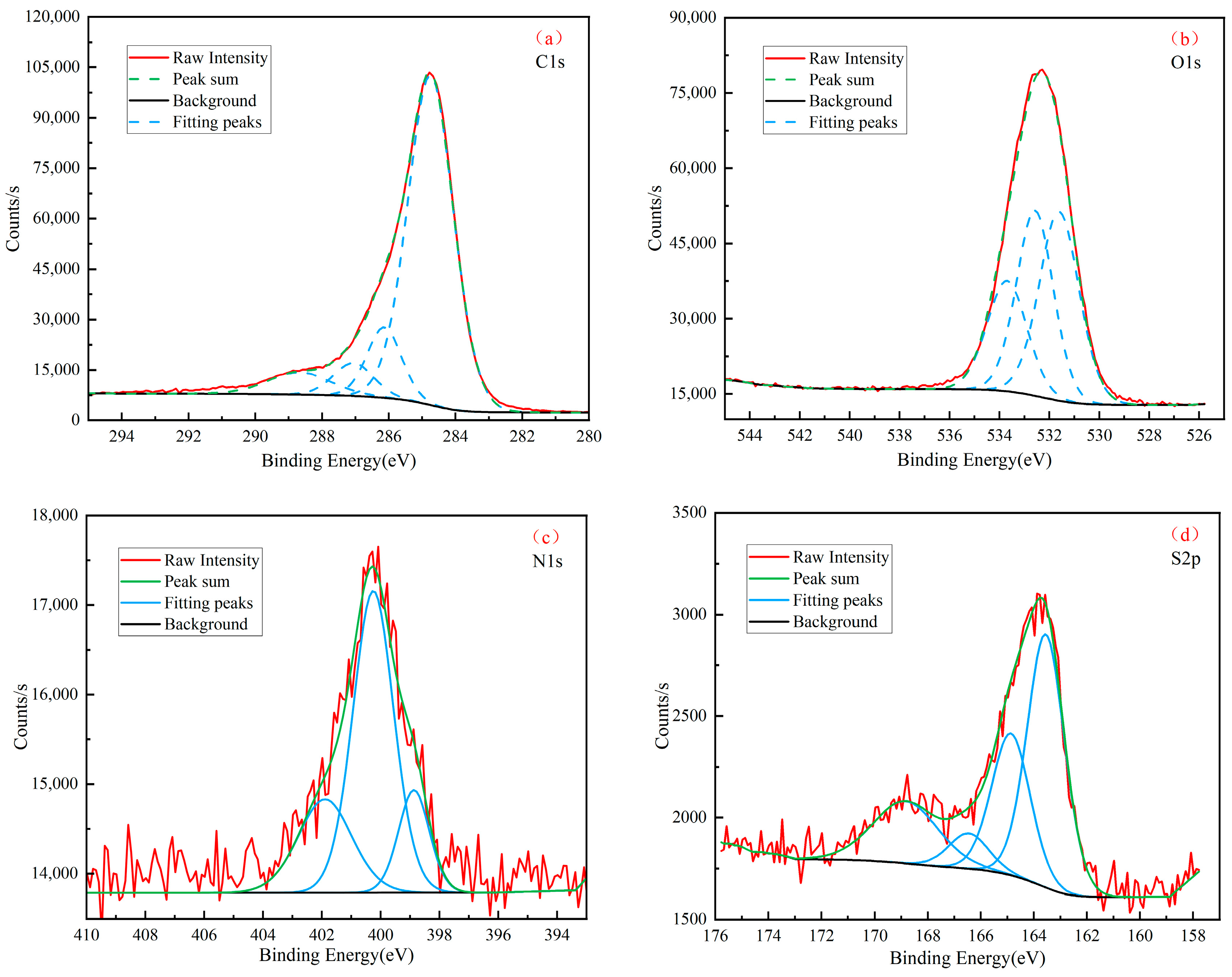
In this study, we applied split-peak fitting to the XPS spectra of the coal samples (Figure 5a–d). Specifically, four forms of C1s, three forms of O1s, three forms of N1s, and four forms of S2p were identified. The results of the different chemical bond binding energies are presented in Table 6 [8,34].
The XPS C1s spectrum showed a predominant presence of C–C and C–H contents, constituting 82.13% of the coal structure. This finding suggests the dominance of aromatic carbons and substituted alkanes in the coal composition. The O1s spectrum indicated the prevalence of organic oxygen in the form of C=O bonds. Notably, pyrrole-type nitrogen accounted for 60.16%. This indicates that the nitrogen in the lignite structure primarily existed in the form of pyrrole, followed by quaternary nitrogen and pyridine. Peak-fit quantitative analysis of the S2p scan revealed that thiophene sulfur constituted the highest percentage of the total sulfur content, approximately 46.47%. Additionally, sulfoxide, sulfate, and sulfur sulfide accounted for 26.90%, 19.75%, and 6.88% of the total sulfur atoms, respectively.

3.4. Raman

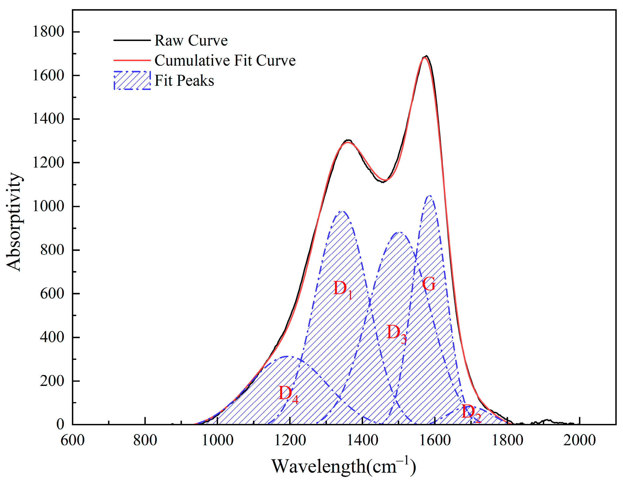
The entire laser Raman spectrum is usually divided into the primary mode region (1000–1800 cm−1) and the secondary mode region (2300–3000 cm−1). Combining with previous studies [32,35], the primary mode region mainly characterizes the order of two-dimensional coal structure, while the secondary mode region can better reflect the graphitization process of coal and the three-dimensional order of hexagonal graphite produced in the process. Among them, the Raman parameters of the D and G bands are closely related to the composition and order of the molecular structure. Therefore, in this study, the spectra in the range of 800–2000 cm−1 were selected for fitting analysis [36]. The primary mode region of the laser Raman spectrum of lignite can be fitted into five main peaks, which are the G, D1, D2, D3, and D4 peaks (Figure 6).
There are mainly two main peaks in the range of Raman shifts from 800 cm−1 to 2000 cm−1, which are located at 1330 cm−1 to 1360 cm−1 (D1 peak) and 1570 cm−1 to 1600 cm−1 (G peak). The peak height of the G peak is significantly larger than that of the D1 peak, but the peak width of the G peak is smaller than that of the D1 peak. The G peak is mainly associated with C=C stretching vibrations in in-plane aromatic rings. The D1 peak band is mainly associated with disorder in the sp2 carbon network. The D2 peak is mainly associated with disordered sp2. The D3 peak is associated with amorphous carbon. And the D4 peak band is associated with sp3 or C–C and C=C stretching vibrations [37].

3.5. HRTEM

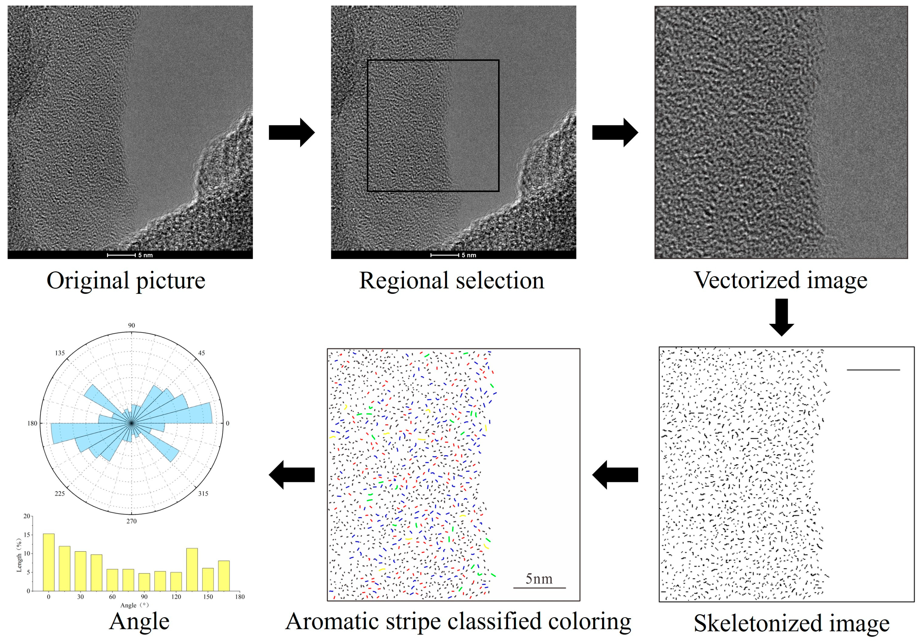
The length of the grid stripes in the transmission electron microscope data represents the size of the aromatic ring. The orientation of the grid stripes can characterize the spatial arrangement of aromatic rings [38]. The quantitative processing steps of transmission electron microscope images are shown in Figure 7. For low-rank coal samples, the aromatic core structure corresponding to the lattice fringes was assigned in this study. The results are shown in Table 7 [5,39].
The analysis results show that the aromatic nucleus structures corresponding to the lattice stripes of XF samples are mainly benzene, naphthalene, and anthracene/phenanthrene with a low condensation degree. The overall length is short. The length within 4.4 accounted for 90.53%. Meanwhile, the aromatic structure of XF coal samples is mainly naphthalene rings, accounting for 49.03%. It is in good agreement with the results of 13C NMR.
We can construct a rose chart to visualize the direction of aromatic stripes relative to the bedding plane. Using the stripe direction extracted from the skeleton image, statistics are made according to the angular interval of 15. The rose diagram can visually display the angular distribution of the lattice fringes (Figure 8). The proportion of aromatic rings in each direction was quantitatively calculated in the figure. It can be seen from the picture that the direction of the aromatic stripes is biased. The aromatic ring content in the main direction is 45 (total range is 180), accounting for 47.63% of the total. This is consistent with the previous view that the aromatic rings of low-rank coal are not randomly distributed [38,40].

3.6. XRD

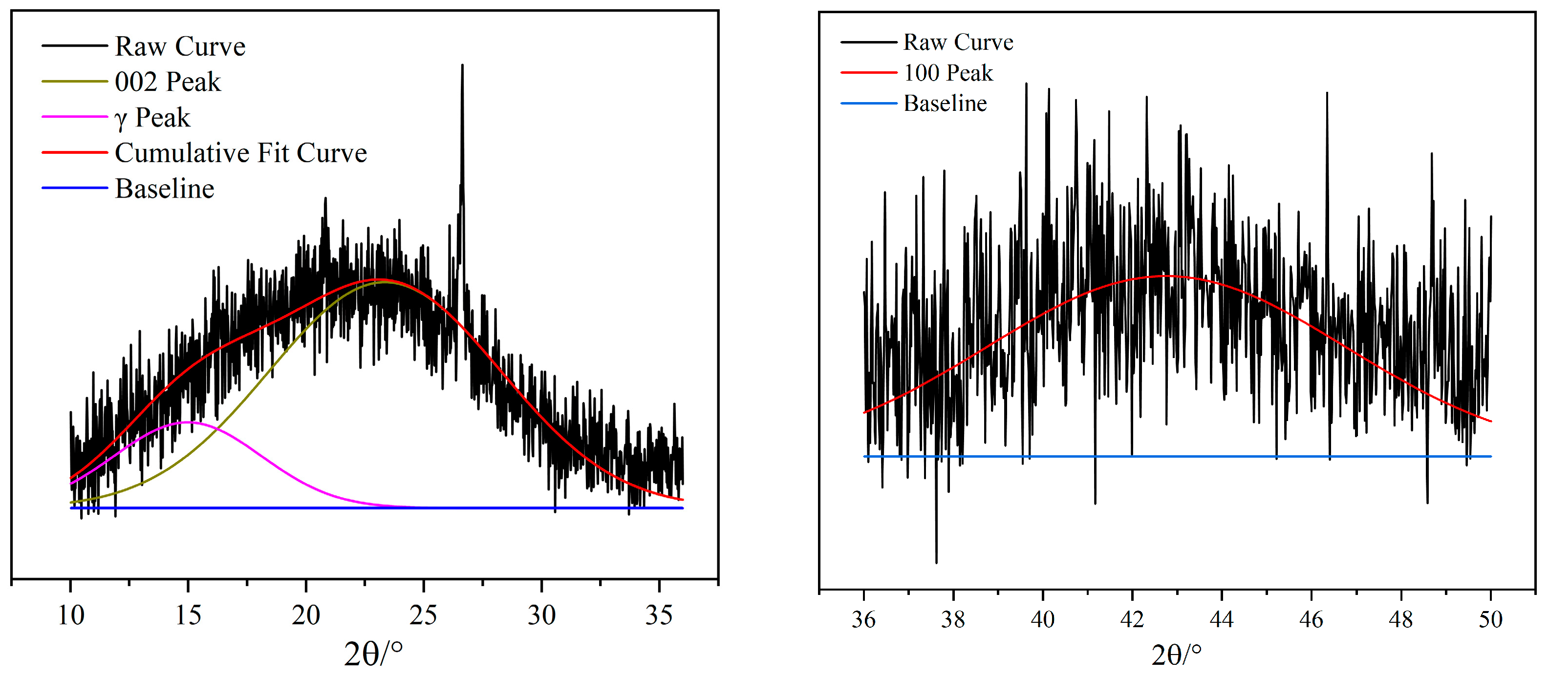
Figure 9 shows the XRD pattern of XF coal. There are two diffraction peaks with wide peak shapes, located around 20° and 40°. They correspond to peaks 002 and 100 of graphite crystals, respectively [41]. The study was carried out to fit the split peaks of the 002 and 100 peaks of the XRD spectra separately (Figure 10). The parameters of each diffraction peak were obtained based on the information of the split peaks (Table 8).
The aromatic carbon layer 002 surface mesh spacing (d002), aromatic carbon layer ductility (La), aromatic layer longitudinal stacking degree (Lc), and effective stacking layer number Nave (Equations (1)–(4)) can be calculated based on the Bragges and Scherrer formula [42]. The results are presented in Table 9.
d002 (Å) = λ/2sinθ002
La (Å) = K2λ/(β100cosθ100)
Lc (Å) = K1λ/(β002cosθ002)
Nave = Lc/d002
where θ002 and θ100 are the center positions of the 002 and 100 peaks, respectively. λ is the X-ray wavelength (1.78901 Å). β00 and β100 are the half-peak widths of the 002 and 100 peaks, respectively. K1 and K2 are the microcrystal shape factors; K1 = 0.94, K2 = 1.84.

4. Construction and Model Validation of Lignite Molecular Structure Model

4.1. Construction of a Molecular Structure Model of Lignite

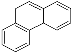
It is necessary to understand the type and number of aromatic skeletons and aliphatic hydrocarbons in order to construct a microscopic molecular structure model of coal [43]. In addition, we used computer-aided structural optimization to determine the relationship between them. Aromatic cluster formation was primarily inferred from 13C NMR data, while aliphatic structures were identified based on 13C NMR and FTIR data. Information about the N and S atoms in the molecule was obtained from XPS data. By adjusting the number of benzene, naphthalene, pyrrole, and thiophene units to achieve a value close to 0.29 for XBP, the resulting species and the number of aromatic structures were determined, as outlined in Table 10.
The total number of aromatic carbon atoms was calculated at 80. Based on the 13C NMR data, the aromaticity was 43.57%. Therefore, the total number of carbon atoms was determined to be 184. Based on the relative proportions of C, H, O, N, and S atoms in the samples (Table 1), the molecular formula of the sample was identified as C184H172O39N6S2. Aliphatic carbon structures primarily comprised cycloalkanes, alkyl side chains, and hydrogenated aromatics.
These findings were integrated into a computer-aided molecular design process, resulting in the construction of both 2D and 3D macromolecular models. Initially, the aromatic backbone and side-chain groups of the coal molecules were systematically linked to form an initial planar molecular model (Figure 11), focusing on the primary hydrogen binding modes during this construction [44].
The 13C NMR spectra, calculated using the constructed macromolecular model of XF lignite, were compared with the experimental spectra (Figure 12). Remarkably, the calculated 13C NMR spectra of the XF lignite macromolecular structure model agreed well with the experimental values, indicating that the established structures accurately represented the studied samples. Notably, the experimental 13C NMR spectra were multifaceted, reflecting the complex nature of low-order coal samples, which inherently consist of diverse mixtures and lack a fixed chemical structure. The average molecular structural model of the samples is shown in Figure 11.

4.2. Optimization and Validation of Coal Molecular Structure Model

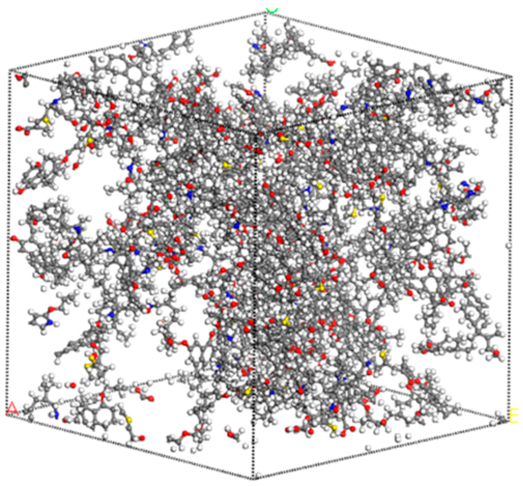
In its natural state, matter is stable in its lowest-energy form. However, there is often a large difference between the constructed model and the actual molecular structure of the matter. To minimize this difference and approach the equilibrium state structure, it is necessary for the system to reach the local energy minimum. Therefore, the geometric configuration of the system must be optimized using the principle of minimum energy, as established in prior research [34]. Figure 13 shows the three-dimensional macromolecular structure of the XF sample. The three-dimensional macromolecular structure was built on the basis of the already obtained two-dimensional molecular model using Materials Studio 2017 software after density correction and geometry optimization steps.
The Forcite module and the Amorphous Cell module were used in the simulation. The Forcite module was used to obtain the lowest energy-stable model of the coal macromolecule structure through geometric optimization and annealing optimization. In this study, the COMPASS force field in the Forcite module and the Amorphous Cell module were used to study the energy of the coal sample at different densities to find the lowest energy corresponding to the density value [45]. A cell containing 15 molecules was constructed, ensuring a calculated system density of 1.14 g/cm3. The resulting molecular formula was [C162H123O7N4S(OCH3)(OHphenol.)6(OHaliph.)5(OOHCOOH)3(C4H5Npyrrole)(C4H4Sthiophene)]15. That is C2565H2235N75O375S30. The dimensions of the individual 3D macromolecular structure model were 3.9 nm × 3.9 nm × 3.9 nm.

5. Conclusions

In this study, the macromolecular structure of XF lignite was established. Information about coal structure fragments, such as the carbon skeleton structure, aromatic structure, and heteroatom, was obtained through experimental tests (elemental analysis, FTIR, 13C NMR, HRTEM, XPS, XRD, and Raman). Two-dimensional and three-dimensional molecular models of lignite were established by integrating functional groups.
The aromatic carbon rate of XF lignite is 43.57%. Naphthalene and anthracene are the main aromatic compounds in coal molecules. The optimization and validation of the 3D model were carried out with the principle of energy minimalism. The constructed molecular structure of XF lignite contains 5270 atoms (C2565H2235N75O375S30) with a size of 3.9 nm × 3.9 nm × 3.9 nm. The XF molecular model has a significant three-dimensional effect, with many side chain groups, and the aromatic lamellae are irregularly arranged in space. This study demonstrates a systematic method to construct a model of a low-order coal macromolecule structure.

Author Contributions

Conceptualization, methodology, writing—original draft preparation, Y.S. (Ying Shi); Funding acquisition, Supervision, Y.Z.; writing—review and editing, investigation, and visualization, S.C., Y.W. and Y.S. (Yu Song). All authors have read and agreed to the published version of the manuscript.

Funding

This research was funded by the National Natural Science Foundation of China (No. 42072199).

Data Availability Statement

Data is contained within the article.

Acknowledgments

The authors would like to sincerely thank various organizations for the financial support.

Conflicts of Interest

The authors declare no conflicts of interest.

References

  1. Wu, L.; Guan, Y.; Li, C.; Shi, L.; Yang, S.; Rajasekhar Reddy, B.; Ye, G.; Zhang, Q.; Liew, R.K.; Zhou, J.; et al. Free-radical behaviors of co-pyrolysis of low-rank coal and different solid hydrogen-rich donors: A critical review. Chem. Eng. J. 2023, 474, 145900. [Google Scholar] [CrossRef]
  2. Meng, X.; Gao, M.; Chu, R.; Miao, Z.; Wu, G.; Bai, L.; Liu, P.; Yan, Y.; Zhang, P. Construction of a macromolecular structural model of Chinese lignite and analysis of its low-temperature oxidation behavior. Chin. J. Chem. Eng. 2017, 25, 1314–1321. [Google Scholar] [CrossRef]
  3. Bian, J.; Li, X.; Zeng, F.; Wang, X. Construction and Evaluation of a Medium-Rank Coal Molecular Model Using a Hybrid Experimental–Simulation–Theoretical Method. Energy Fuels 2019, 33, 12905–12915. [Google Scholar] [CrossRef]
  4. Landais, P.; Gerard, L.; Chyi, L.L.; Medlin, J.H.; Coleman, S.L. Coalification stages from confined pyrolysis of an immature humic coal. Int. J. Coal Geol. 1996, 30, 285–301. [Google Scholar] [CrossRef]
  5. Okolo, G.N.; Neomagus, H.W.J.P.; Everson, R.C.; Roberts, M.J.; Bunt, J.R.; Sakurovs, R.; Mathews, J.P. Chemical-structural properties of South African bituminous coals: Insights from wide angle XRD-carbon fraction analysis, ATR-FTIR, solid state 13C NMR, and HRTEM techniques. Fuel 2015, 158, 779–792. [Google Scholar] [CrossRef]
  6. Yuan, S.; Liu, J.; Zhu, J.; Zhou, Q.; Wang, Z.; Zhou, J.; Cen, K. Effect of microwave irradiation on the propensity for spontaneous combustion of Inner Mongolia lignite. J. Loss Prevent. Proc. 2016, 44, 390–396. [Google Scholar] [CrossRef]
  7. Lin, H.; Li, K.; Zhang, X.; Wang, H. Structure Characterization and Model Construction of Indonesian Brown Coal. Energy Fuels 2016, 30, 3809–3814. [Google Scholar] [CrossRef]
  8. Wang, J.; He, Y.; Li, H.; Yu, J.; Xie, W.; Wei, H. The molecular structure of Inner Mongolia lignite utilizing XRD, solid state 13C NMR, HRTEM and XPS techniques. Fuel 2017, 203, 764–773. [Google Scholar] [CrossRef]
  9. Song, H.; Liu, G.; Wu, J. Pyrolysis characteristics and kinetics of low rank coals by distributed activation energy model. Energy Convers. Manag. 2016, 126, 1037–1046. [Google Scholar] [CrossRef]
  10. Jia, J.; Yang, Q.; Liu, B.; Wang, D. Structural characterization and macromolecular structure construction of non-caking coal in Chicheng Mine. Sci. Rep. 2023, 13, 16931. [Google Scholar] [CrossRef]
  11. Yin, N.; Song, Y.; Wang, C.; Zhang, X.; Wu, L.; Jing, X.; Zhou, J.; Lan, X. Molecular structure model construction and pyrolysis characterization of four typical coals: A case study. Energy Sources Part A Recovery Util. Environ. Eff. 2022, 44, 6837–6855. [Google Scholar] [CrossRef]
  12. GB/T6948-2008; Method of Determining Microscopically the Reflectance of Vitrinite in Coal. China Standard Press: Beijing, China, 2008.
  13. GB/T212-2008; Proximate Analysis of Coal. China Standard Press: Beijing, China, 2008.
  14. GB/T476-2008; Determination of Carbon and Hydrogen in Coal. China Standard Press: Beijing, China, 2008.
  15. GB/T19227-2008; Determination of Nitrogen in Coal. China Standard Press: Beijing, China, 2008.
  16. GB/T214-2007; Determination of Total Sulfur in Coal. China Standard Press: Beijing, China, 2008.
  17. Cui, X.; Yan, H.; Zhao, P.; Yang, Y.; Xie, Y. Modeling of molecular and properties of anthracite base on structural accuracy identification methods. J. Mol. Struct. 2019, 1183, 313–323. [Google Scholar] [CrossRef]
  18. Niekerk, D.V.; Mathews, J.P. Molecular representations of Permian-aged vitrinite-rich and inertinite-rich South African coals. Fuel 2010, 89, 73–82. [Google Scholar] [CrossRef]
  19. Liu, Y.; Liu, S.; Zhang, R.; Zhang, Y. The molecular model of Marcellus shale kerogen: Experimental characterization and structure reconstruction. Int. J. Coal Geol. 2021, 246, 103833. [Google Scholar] [CrossRef]
  20. Li, Y.; Cao, X.; Zhu, D.; Chappell, M.A.; Miller, L.F.; Mao, J. Characterization of coals and their laboratory-prepared black carbon using advanced solid-state 13C nuclear magnetic resonance spectroscopy. Fuel Process. Technol. 2012, 96, 56–64. [Google Scholar] [CrossRef]
  21. Jiang, J.; Zhang, S.; Longhurst, P.; Yang, W.; Zheng, S. Molecular structure characterization of bituminous coal in Northern China via XRD, Raman and FTIR spectroscopy. Spectrochim. Acta Part A Mol. Biomol. Spectrosc. 2021, 255, 119724. [Google Scholar] [CrossRef] [PubMed]
  22. Xia, W.; Yang, J.; Liang, C. Investigation of changes in surface properties of bituminous coal during natural weathering processes by XPS and SEM. Appl. Surf. Sci. 2014, 293, 293–298. [Google Scholar] [CrossRef]
  23. Takagi, H.; Maruyama, K.; Yoshizawa, N.; Yamada, Y.; Sato, Y. XRD analysis of carbon stacking structure in coal during heat treatment. Fuel 2004, 83, 2427–2433. [Google Scholar] [CrossRef]
  24. Jia, J.; Wang, D.; Li, B. Study on molecular structure characteristic and optimization of molecular model construction of coal with different metamorphic grade. J. Mol. Struct. 2024, 1295, 136655. [Google Scholar] [CrossRef]
  25. Yan, J.; Lei, Z.; Li, Z.; Wang, Z.; Ren, S.; Kang, S.; Wang, X.; Shui, H. Molecular structure characterization of low-medium rank coals via XRD, solid state 13C NMR and FTIR spectroscopy. Fuel 2020, 268, 117038. [Google Scholar] [CrossRef]
  26. Ding, D.; Liu, G.; Fu, B. Influence of carbon type on carbon isotopic composition of coal from the perspective of solid-state 13C NMR. Fuel 2019, 245, 174–180. [Google Scholar] [CrossRef]
  27. Zhu, B.; Dong, X.; Fan, Y.; Ma, X.; Yao, S.; Fu, Y.; Chen, R.; Chang, M. Structural Characterization and Molecular Model Construction of High-Ash Coal from Northern China. Molecules 2023, 28, 5593. [Google Scholar] [CrossRef]
  28. Wang, Y.; Wei, X.; Xie, R.; Liu, F.; Li, P.; Zong, Z. Structural Characterization of Typical Organic Species in Jincheng No. 15 Anthracite. Energy Fuels 2015, 29, 595–601. [Google Scholar] [CrossRef]
  29. Dun, W.; Guijian, L.; Ruoyu, S.; Xiang, F. Investigation of Structural Characteristics of Thermally Metamorphosed Coal by FTIR Spectroscopy and X-ray Diffraction. Energy Fuels 2013, 27, 5823–5830. [Google Scholar] [CrossRef]
  30. Jiang, B.; Huang, J.; Yu, C.; Wang, X.; Zhou, Y.; Zheng, Y.; Ji, B.; Zhang, Q. Experimental and theoretical study on molecular structure construction of Hongliulin coal. Fuel 2023, 349, 128708. [Google Scholar] [CrossRef]
  31. Xia, W.; Zhang, W. Characterization of surface properties of Inner Mongolia coal using FTIR and XPS. Energy Sources Part A Recovery Util. Environ. Eff. 2017, 39, 1190–1194. [Google Scholar] [CrossRef]
  32. He, X.; Liu, X.; Nie, B.; Song, D. FTIR and Raman spectroscopy characterization of functional groups in various rank coals. Fuel 2017, 206, 555–563. [Google Scholar] [CrossRef]
  33. Wang, C.; Xing, Y.; Shi, K.; Wang, S.; Xia, Y.; Li, J.; Gui, X. Chemical Structure Characteristics and Model Construction of Coal with Three Kinds of Coalification Degrees. ACS Omega 2024, 9, 1881–1893. [Google Scholar] [CrossRef]
  34. Zhang, S.; Zhang, C.; Zhang, M.; Liu, X.; Xue, S. Model construction and optimization of coal molecular structure. J. Mol. Struct. 2023, 1290, 135960. [Google Scholar] [CrossRef]
  35. Marshall, C.P.; Edwards, H.G.M.; Jehlicka, J. Understanding the application of Raman spectroscopy to the detection of traces of life. Astrobiology 2010, 10, 229–243. [Google Scholar] [CrossRef] [PubMed]
  36. Gao, B.; Wu, C.; Song, Y.; Zhou, D.; Niu, Q.; Zhou, H.; Jiang, X. Structural characterization of high fidelity for bituminous and semi-anthracite: Insights from spectral analysis and modeling. Fuel 2022, 315, 123183. [Google Scholar] [CrossRef]
  37. Baysal, M.; Yürüm, A.; Yıldız, B.; Yürüm, Y. Structure of some western Anatolia coals investigated by FTIR, Raman, 13C solid state NMR spectroscopy and X-ray diffraction. Int. J. Coal Geol. 2016, 163, 166–176. [Google Scholar] [CrossRef]
  38. Mathews, J.P.; Sharma, A. The structural alignment of coal and the analogous case of Argonne Upper Freeport coal. Fuel 2012, 95, 19–24. [Google Scholar] [CrossRef]
  39. Pan, J.; Lv, M.; Bai, H.; Hou, Q.; Li, M.; Wang, Z. Effects of Metamorphism and Deformation on the Coal Macromolecular Structure by Laser Raman Spectroscopy. Energy Fuels 2017, 31, 1136–1146. [Google Scholar] [CrossRef]
  40. Li, S.; Zhu, Y.; He, R. Mechanistic analysis of chemical structure evolution for coals under igneous intrusion. Fuel 2024, 357, 130055. [Google Scholar] [CrossRef]
  41. Wang, C.; Zeng, F. Molecular Structure Characterization of CS2–NMP Extract and Residue for Malan Bituminous Coal via Solid-State 13C NMR, FTIR, XPS, XRD, and CAMD Techniques. Energy Fuels 2020, 34, 12142–12157. [Google Scholar] [CrossRef]
  42. Saikia, B.K.; Boruah, R.K.; Gogoi, P.K. FT-IR and XRD analysis of coal from Makum coalfield of Assam. J. Earth Syst. Sci. 2007, 116, 575–579. [Google Scholar] [CrossRef]
  43. Qian, L.; Tao, C.; Ma, C.; Xue, J.; Guo, F.; Jia, X.; Yan, W. Construction of a Macromolecular Structure Model for Zhundong Subbituminous Coal. J. Mol. Struct. 2022, 1248, 131496. [Google Scholar] [CrossRef]
  44. Liu, H.; Jiang, B.; Song, Y.; Hou, C. The tectonic stress-driving alteration and evolution of chemical structure for low- to medium-rank coals–by molecular simulation method. Arab. J. Geosci. 2019, 12, 726. [Google Scholar] [CrossRef]
  45. Feng, W.; Li, Z.; Gao, H.; Wang, Q.; Bai, H.; Li, P. Understanding the molecular structure of HSW coal at atomic level: A comprehensive characterization from combined experimental and computational study. Green Energy Environ. 2021, 6, 150–159. [Google Scholar] [CrossRef]
Figure 1. Flow chart for 3D coal macromolecular structure modeling.
Energies 17 01049 g001
Figure 2. 13C NMR peak fitting spectra of coal sample.
Energies 17 01049 g002
Figure 3. FTIR spectrum fitting curve of coal samples. (a) 900–700 cm−1, (b) 1200–1000 cm−1, (c) 3000–2800 cm−1, (d) 3700–3400 cm−1.
Energies 17 01049 g003
Figure 4. XPS wide-scan spectra of coal sample.
Energies 17 01049 g004
Figure 5. Curve-fitted XPS C1s scan (a), O1s scan (b), N1s scan (c), and S2p scan (d) of coal sample.
Energies 17 01049 g005
Figure 6. Peak fitting of Raman spectrum.
Energies 17 01049 g006
Figure 7. Steps of HRTEM aromatic stripe image analysis of XF coal.
Energies 17 01049 g007
Figure 8. Angle distribution of lattice fringes of coal samples.
Energies 17 01049 g008
Figure 9. XRD spectra of coal sample.
Energies 17 01049 g009
Figure 10. Partition-fitting XRD spectrum of coal sample.
Energies 17 01049 g010
Figure 11. The 2D model of the XF coal molecular structure.
Energies 17 01049 g011
Figure 12. Comparison of 13C NMR spectra between experimental and model.
Energies 17 01049 g012
Figure 13. The 3D model of the XF coal molecular structure. (Carbon atoms are gray, hydrogen atoms are white, oxygen atoms are red, nitrogen atoms are blue, and sulfur atoms are yellow).
Energies 17 01049 g013
Table 1. Essential features of the coal sample.
Proximate Analysis/%Ultimate Analysis/%Atomic Ratio
MadAdVdafFCdCdafHdafOdafNdafSt,dH/CO/CN/CS/C
4.225.6649.4747.6770.315.4719.731.902.440.9330.2100.0230.013
Table 2. Assignment of different types of carbons based on 13C NMR data.
NumberChemical Shift/δFWHMRelative AreaSymbolCarbon Type
116.6413.120.0617fal3Aliphatic methyl
224.7210.490.0653falaAromatic methyl
331.287.690.0696fal2Methylene
438.8514.350.1217fal1Methine
552.6319.570.1036falOOxy-aliphatic carbons
675.1617.850.0445falOOxy-aliphatic carbons
794.8321.620.0464falOOxy-aliphatic carbons
8107.3712.410.0497faHProtonated aromatic carbons
9115.078.900.0644faHProtonated aromatic carbons
10124.3212.040.1185faHProtonated aromatic carbons
11134.1913.150.0983faBBridged carbons
12143.408.030.0441faSAlkylated aromatic carbons
14177.1421.070.0341faCC1Carboxyl
15204.6518.280.0173faCC2Carboxyl
Table 3. 13C NMR structural parameters of coal sample.
falfal3falafal2fal1falO
0.51290.06170.06530.06960.12170.1945
fafaHfaBfaSfaOfaC
0.43570.23250.09830.04410.06080.0513
Table 4. Peak assignments for FTIR spectra of coal sample.
Wavenumber/cm−1Group
3300hydrogen bond
3030hydrogen bond–C–H–
2950–CH3
1735, 1690–1720, 1650–1630, 1600, 1560–1590C=O
2920, 2850Fatty bond–CH, CH2, –CH3
1600Aromatic ring stretching vibration
1490Aromatic ring stretching vibration
1450–CH2 and –CH3 bending vibration
1375–CH3
1300–1110C–O stretching vibration, O–H bending vibration
1100–1000Aliphatic ethers
700–900Aromatic C–H bending vibration
Table 5. Analysis results of XPS spectra.
NamePeak BEFWHM eVArea (P) CPS.eVAtomic %
C1s284.292.821,381,579.0468.90
O1s531.853.271,124,898.6523.22
N1s399.833.6573,904.702.38
Si2p102.092.7242,064.262.09
Al2p74.052.6827,376.632.15
S2p163.363.1527,832.190.69
Ca2p347.212.9965,965.510.57
Table 6. XPS C1s, O1s, N1s, and S2p data of coal sample.
ElementalFunctionalityBinding Energy/eVMolar Content%
C1sC–C, C–H284.7882.13
C–O286.487.51
O–C–O287.384.32
COO–288.586.03
O1sC=O531.6140.21
–OH532.5836.28
COO–533.6923.51
N1sPyridinic nitrogen398.8715.42
Pyrrolic nitrogen400.2560.16
Quaternary nitrogen401.8924.42
S2pThiophenes163.5546.47
Sulphoxides164.8726.90
Sulphones166.466.88
Sulphates168.8419.75
Table 7. Lattice fringe classification of HRTEM.
Aromatic SheetGroupingMinL (Å)MaxL (Å)Mean (Å)Freq (%)
Noise<0.25 Å////
Benzene0.25 Å–3.0 Å2.512.992.7341.50
Naphthalene3.0 Å–4.4 Å3.004.373.5649.03
Phenanthrene/Anthracene4.4 Å–5.9 Å4.425.874.936.69
2 × 25.9 Å–7.4 Å5.987.366.482.79
Table 8. XRD fitting parameter results.
Peak2θ/°FWHMPeak Shape
γ14.987.39Gaussian
00223.3511.58Gaussian
10042.759.45Gaussian
Table 9. Aromatic microcrystalline structure parameters of XF coal.
d002LcLaNave
4.420.150.370.03
Table 10. Types and number of aromatic structure units.
TypeAromatic Unit StructureNumberType
NaphthaleneEnergies 17 01049 i0013Naphthalene
AnthraceneEnergies 17 01049 i0022Anthracene
PhenanthreneEnergies 17 01049 i0031Phenanthrene
PyrroleEnergies 17 01049 i0041Pyrrole
ThiopheneEnergies 17 01049 i0051Thiophene
TypeAromatic unit structureNumberType
Disclaimer/Publisher’s Note: The statements, opinions and data contained in all publications are solely those of the individual author(s) and contributor(s) and not of MDPI and/or the editor(s). MDPI and/or the editor(s) disclaim responsibility for any injury to people or property resulting from any ideas, methods, instructions or products referred to in the content.