Maxillary Sinus Mucocele as a Late Complication in Zygomatic-Orbital Complex Fracture
Abstract
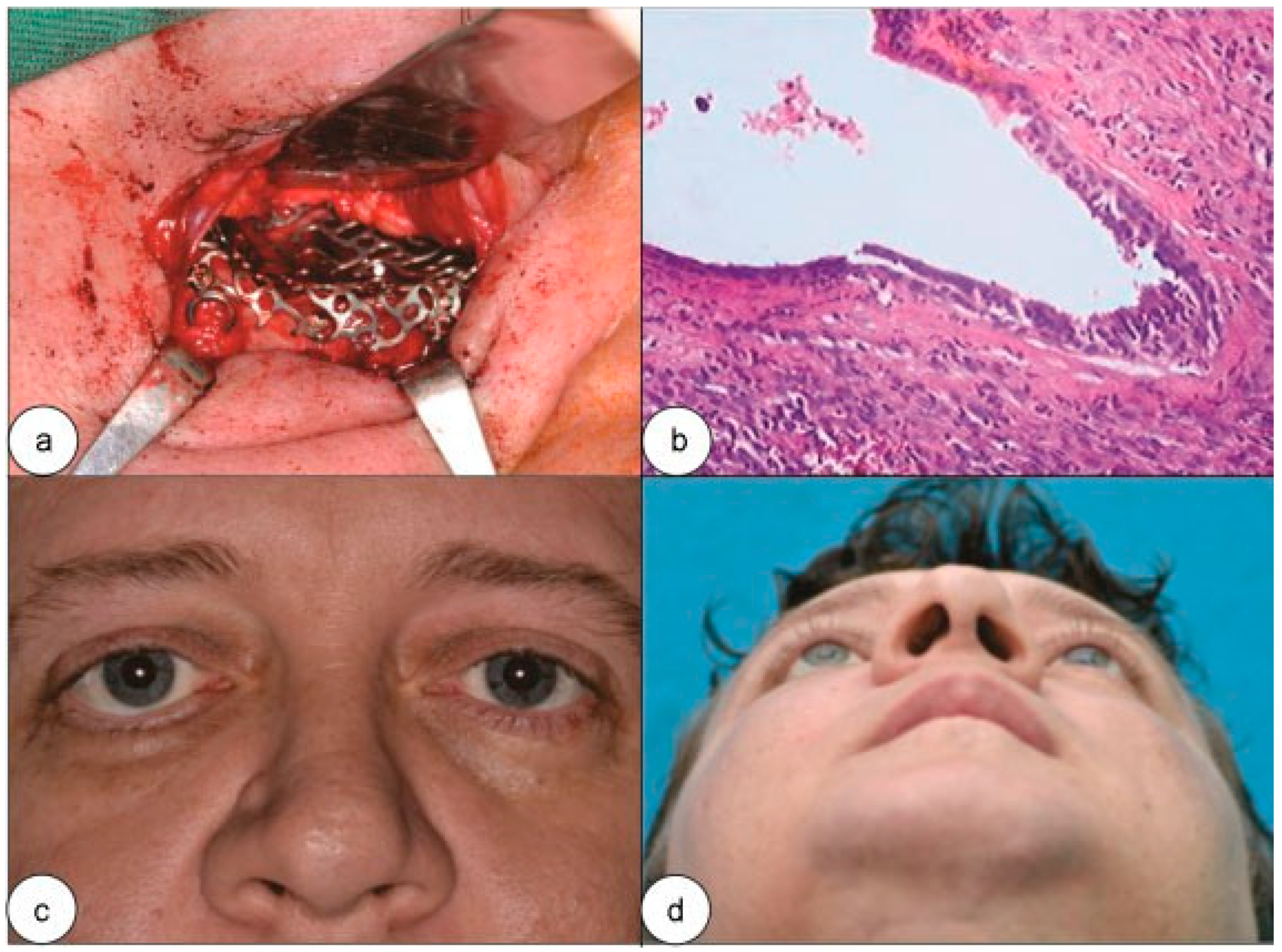
:Case Report

Discussion
Conclusion
References
- Gardner, D.G.; Gullane, P.J. Mucoceles of the maxillary sinus. Oral Surg Oral Med Oral Pathol 1986, 62, 538–543. [Google Scholar] [PubMed]
- Johnson, J.T.; Ferguson, B.J. Infection. In Otolaryngology, Head and Neck Surgery, 3rd ed.; Cummings, C.W., Fredrickson, J.M., Harker, L.A., Krause, C.J., Schuller, D.E., Richardson, M.A., Eds.; Mosby: St. Louis, MI, YSA, 1998; pp. 1115–1116. [Google Scholar]
- Koudstaal, M.J.; van der Wal, K.G.; Bijvoet, H.W.; Vincent, A.J.; Poublon, R.M. Post-trauma mucocele formation in the frontal sinus; a rationale of follow-up. Int J Oral Maxillofac Surg 2004, 33, 751–754. [Google Scholar] [PubMed]
- East, D. Mucocoeles of the maxillary antrum. Description, case reports and review of the literature. J Laryngol Otol 1985, 99, 49–56. [Google Scholar] [CrossRef] [PubMed]
- Hasegawa, M.; Saito, Y.; Watanabe, I.; Kern, E.B. Postoperative mucoceles of the maxillary sinus. Rhinology 1979, 17, 253–256. [Google Scholar] [PubMed]
- Lai, P.C.; Liao, S.L.; Jou, J.R.; Hou, P.K. Transcaruncular approach for the management of frontoethmoid mucoceles. Br J Ophthalmol 2003, 87, 699–703. [Google Scholar] [PubMed]
- Smoot, E.C.I.I.I.; Bowen, D.G.; Lappert, P.; Ruiz, J.A. Delayed development of an ectopic frontal sinus mucocele after pediatric cranial trauma. J Craniofac Surg 1995, 6, 327–331. [Google Scholar] [PubMed]
- Palmer-Hall, A.M.; Anderson, S.F. Paraocular sinus mucoceles. J Am Optom Assoc 1997, 68, 725–733. [Google Scholar] [PubMed]
- Bhandary, S.K.; Bhat, V.S.; Khanna, R.A. Frontal sinus mucocele following a trivial facial trauma, presenting as a pyocele a case report. NUJHS 2013, 3, 90–92. [Google Scholar]
- Curtin, H.D.; Rabinov, J.D. Extension to the orbit from paraorbital disease. The sinuses. Radiol Clin North Am 1998, 36, 1201–1213. [Google Scholar] [CrossRef] [PubMed]
- Caylakli, F.; Yavuz, H.; Cagici, A.C.; Ozluoglu, L.N. Endoscopic sinus surgery for maxillary sinus mucoceles. Head Face Med 2006, 2, 29. [Google Scholar] [PubMed]
- Salam, M.A.; Whitehead, E. Large maxillary antral mucocele presenting with facial asymmetry. J Laryngol Otol 1993, 107, 451–452. [Google Scholar] [CrossRef] [PubMed]
- Ormerod, L.D.; Weber, A.L.; Rauch, S.D.; Feldon, S.E. Ophthalmic manifestations of maxillary sinus mucoceles. Ophthalmology 1987, 94, 1013–1019. [Google Scholar] [PubMed]
- Sheth, H.G.; Goel, R. Diplopia due to maxillary sinus mucocoele. Int Ophthalmol 2007, 27, 365–367. [Google Scholar] [PubMed]
- Dispenza, C.; Saraniti, C.; Caramanna, C.; Dispenza, F. Endoscopic treatment of maxillary sinus mucocele. Acta Otorhinolaryngol Ital 2004, 24, 292–296. [Google Scholar] [PubMed]
- Har-El, G. Endoscopic management of 108 sinus mucoceles. Laryngoscope 2001, 111, 2131–2134. [Google Scholar] [CrossRef] [PubMed]
- Moriyama, H.; Hesaka, H.; Tachibana, T.; Honda, Y. Mucoceles of ethmoid and sphenoid sinus with visual disturbance. Arch Otolaryngol Head Neck Surg 1992, 118, 142–146. [Google Scholar] [CrossRef] [PubMed]
- Shimo-Oku, M.; Miyazaki, S.; Shiraki, K.; Sugimoto, T.; Sotani, H. Optic nerve involvement in posterior paranasal sinus diseases. Neuroophthalmology 1989, 9, 147–155. [Google Scholar]
- Marks, S.C.; Latoni, J.D.; Mathog, R.H. Mucoceles of the maxillary sinus. Otolaryngol Head Neck Surg 1997, 117, 18–21. [Google Scholar] [CrossRef] [PubMed]
© 2016 by the author. The Author(s) 2016.
Share and Cite
da Silva de Menezes, J.D.; Moura, L.B.; Pereira-Filho, V.A.; Hochuli-Vieira, E. Maxillary Sinus Mucocele as a Late Complication in Zygomatic-Orbital Complex Fracture. Craniomaxillofac. Trauma Reconstr. 2016, 9, 342-344. https://doi.org/10.1055/s-0036-1582453
da Silva de Menezes JD, Moura LB, Pereira-Filho VA, Hochuli-Vieira E. Maxillary Sinus Mucocele as a Late Complication in Zygomatic-Orbital Complex Fracture. Craniomaxillofacial Trauma & Reconstruction. 2016; 9(4):342-344. https://doi.org/10.1055/s-0036-1582453
Chicago/Turabian Styleda Silva de Menezes, Juliana Dreyer, Lucas Borin Moura, Valfrido Antonio Pereira-Filho, and Eduardo Hochuli-Vieira. 2016. "Maxillary Sinus Mucocele as a Late Complication in Zygomatic-Orbital Complex Fracture" Craniomaxillofacial Trauma & Reconstruction 9, no. 4: 342-344. https://doi.org/10.1055/s-0036-1582453
APA Styleda Silva de Menezes, J. D., Moura, L. B., Pereira-Filho, V. A., & Hochuli-Vieira, E. (2016). Maxillary Sinus Mucocele as a Late Complication in Zygomatic-Orbital Complex Fracture. Craniomaxillofacial Trauma & Reconstruction, 9(4), 342-344. https://doi.org/10.1055/s-0036-1582453