A Modified Technique for Placing Prebent Plates During a Le Fort I Osteotomy: A Technical Note
Abstract
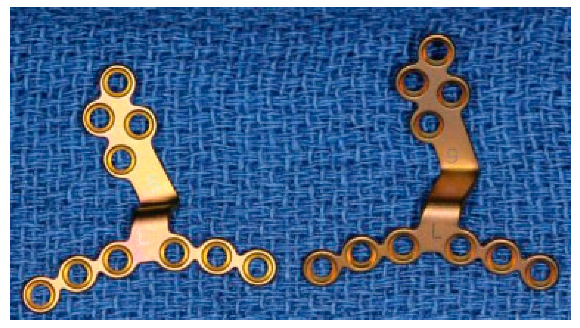
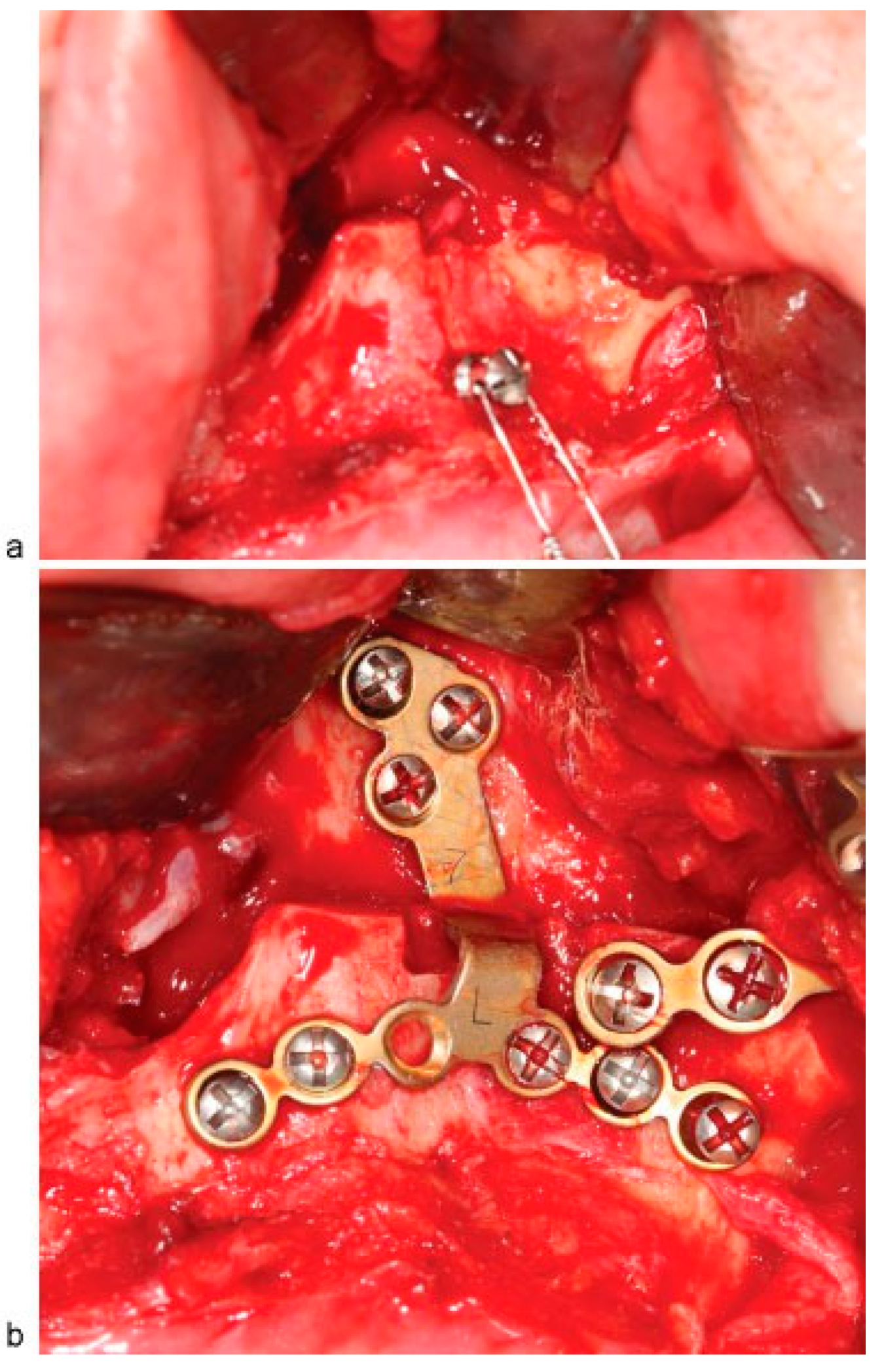
:Surgical Technique
Discussion
Conclusion
References
- Bothur, S.; Blomqvist, J.E.; Isaksson, S. Stability of Le Fort I osteotomy with advancement: a comparison of single maxillary surgery and a two-jaw procedure. J Oral Maxillofac Surg 1998, 56, 1029–1033. [Google Scholar] [CrossRef] [PubMed]
- Van Sickels, J.E.; Jeter, T.D.; Aragon, S.B. Rigid fixation of maxillary osteotomies: a preliminary report and technique article. Oral Surg Oral Med Oral Pathol 1985, 60, 262–265. [Google Scholar] [PubMed]
- Proffit, W.R.; Turvey, T.A.; Phillips, C. The hierarchy of stability and predictability in orthognathic surgery with rigid fixation: an update and extension. Head Face Med 2007, 3, 21. [Google Scholar] [PubMed]
- Bell, W.H.; Jacobs, J.D.; Quejada, J.G. Simultaneous repositioning of the maxilla, mandible, and chin. Treatment planning and analysis of soft tissues. Am J Orthod 1986, 89, 28–50. [Google Scholar] [PubMed]
- Lye, K.W.; Waite, P.D.; Wang, D.; Sittitavornwong, S. Predictability of prebent advancement plates for use in maxillomandibular advancement surgery. J Oral Maxillofac Surg 2008, 66, 1625–1629. [Google Scholar] [PubMed]
- Coskunses, F.M.; Kan, B.; Mutlu, I.; Cilasun, U.; Celik, T. Evaluation of prebent miniplates in fixation of Le Fort I advancement osteotomy with the finite element method. J Craniomaxillofac Surg 2015, 43, 1505–1510. [Google Scholar] [PubMed]
- Araujo, M.M.; Waite, P.D.; Lemons, J.E. Strength analysis of Le Fort I osteotomy fixation: titanium versus resorbable plates. J Oral Maxillofac Surg 2001, 59, 1034–1039. [Google Scholar] [PubMed]
- Ragaey, M.; Fan, L.; Burchett, W.; Van Sickels, J. Stability of maxillary advancements using a combination of prebent and conventional plates for fixation. In Proceedings of the 11th Annual CCTS Spring Conference, Lexington, KY; 2016. [Google Scholar]
© 2016 by the author. The Author(s) 2016
Share and Cite
Ragaey, M.; Van Sickels, J. A Modified Technique for Placing Prebent Plates During a Le Fort I Osteotomy: A Technical Note. Craniomaxillofac. Trauma Reconstr. 2016, 9, 297-298. https://doi.org/10.1055/s-0036-1592092
Ragaey M, Van Sickels J. A Modified Technique for Placing Prebent Plates During a Le Fort I Osteotomy: A Technical Note. Craniomaxillofacial Trauma & Reconstruction. 2016; 9(4):297-298. https://doi.org/10.1055/s-0036-1592092
Chicago/Turabian StyleRagaey, Marwa, and Joseph Van Sickels. 2016. "A Modified Technique for Placing Prebent Plates During a Le Fort I Osteotomy: A Technical Note" Craniomaxillofacial Trauma & Reconstruction 9, no. 4: 297-298. https://doi.org/10.1055/s-0036-1592092
APA StyleRagaey, M., & Van Sickels, J. (2016). A Modified Technique for Placing Prebent Plates During a Le Fort I Osteotomy: A Technical Note. Craniomaxillofacial Trauma & Reconstruction, 9(4), 297-298. https://doi.org/10.1055/s-0036-1592092