Next Article in Journal
Tomographic Evaluation of Mandibular Thickness on Premolar and Molar Regions Related to Monocortical Screws
Previous Article in Journal
Guided Bone Regeneration of an Atrophic Mandible with a Heterologous Bone Block
 
 
Craniomaxillofacial Trauma & Reconstruction is published by MDPI from Volume 18 Issue 1 (2025). Previous articles were published by another publisher in Open Access under a CC-BY (or CC-BY-NC-ND) licence, and they are hosted by MDPI on mdpi.com as a courtesy and upon agreement with Sage.
Case Report

Nevoid Basal Cell Carcinoma Syndrome: A Long-Term Study in a Family

by
Thiago de Santana Santos
1,*,
André Vajgel
2,
Paulo Ricardo Saquete Martins-Filho
3,
AlmirWalter de Albuquerque Maranhao Filho
2,
Ricardo José De Holanda Vasconcellos
4,
Riedel Frota
2 and
José Rodrigues Laureano Filho
2
1
Hospital Universitário, Universidade Federal de Sergipe, Rua Claudio Batista, Aracaju, Sergipe, Brazil
2
Department of Oral and Maxillofacial Surgery, Pernambuco School of Dentistry, Camaragibe, Pernambuco, Brazil
3
Department of Dentistry, Universidade Federal de Sergipe, Aracaju, Sergipe, Brazil
4
Department of Oral and Maxillofacial Surgery, University of Pernambuco, Recife, Pernambuco, Brazil
*
Author to whom correspondence should be addressed.
Craniomaxillofac. Trauma Reconstr. 2016, 9(1), 94-104; https://doi.org/10.1055/s-0035-1558454 (registering DOI)
Submission received: 27 December 2014 / Revised: 22 February 2015 / Accepted: 22 February 2015 / Published: 3 August 2015

Abstract

We present a family case series with 10 individuals having nevoid basal cell carcinoma syndrome (NBCCS) with a 10-year follow-up. All articles published in the literature between 1967 and 2011 on familial Gorlin–Goltz syndrome in any language were surveyed to determine the mapping of cases per country of occurrence of this disease. All patients in the present series were presented with calcification of the falx cerebri, mild hypertelorism, and frontal bossing. Odontogenic keratocystic tumors, palmar and plantar pits, and multiple basal cell carcinomas occurred in 90, 40, and 20%, respectively, of the patients. One of the patients died of skin cancer. Diagnosis of odontogenic keratocyst tumors was confirmed by histopathological examination. NBCCS is a rare autosomal dominant cancer predisposition syndrome; it is important to recognize it when a patient has multiple odontogenic keratocyst tumors because life-long monitoring is essential for patient management..

Nevoid basal cell carcinoma syndrome (NBCCS), also known as Gorlin–Goltz syndrome, is a rare autosomal dominant disorder with two distinct phenotypic features: predisposition to develop malignant tumors and developmental defects. [1,2,3,4] The disease was described in the 1960s by Gorlin and Goltz, [5] who established the classic triad of multiple basal cell carcinomas (BCCs), keratocystic odontogenic tumors (KCOTs), and bifid ribs. The disease has a high penetrance, but variable expressivity, including the possible presence of other abnormalities, such as palmar and plantar pits, bilamellar calcification of the falx cerebri, macrocephaly, fusion of vertebrae, spina bifida, polydactyly, cleft lip or palate, hypertelorism, ovarian fibroma, and medulloblastoma (MB). [6]
In recent decades, it has been demonstrated that NBCCS is a chromosome instability syndrome, characterized by spontaneous chromosomal rearrangements, increased frequency of sister chromatid exchanges, and slowing of the cell cycle. [7] The human homolog of Drosophila patched (PTCH) is a tumor suppressor gene localized at chromosome 9q22.3-q31 (GenBank accession numbers: U43148 and U59464), [8,9] member of the Hedgehog/ Patched (or sonic hedgehog [SHH]/smoothened [SMO]/PTCH) signaling pathway, which seems to have a fundamental role during embryogenic development and appears to underlie many disease states when dysregulated. [10] Increased PTCH expression has been detected immunohistochemically in sporadic and Gorlin syndrome–related KCOTs. [11]
Few studies in the English language literature include all members of the same family. Thus to better understand this syndrome, we present a familial case series of 10 family members with a 10-year follow-up, with the development of clinical signals years from the first observations. In addition, we also discuss the recurrence potential of KCOT and the efficacy of photodynamic therapy (PDT) as a treatment for multiple BCCs.

Methods

A retrospective study of a family with NBCCS was performed (Figure 1). The sample was obtained after the first individual attended the hospital for treatment in the period between January 2004 and January 2014. The clinical, radiographic, and histological data of a 10-year study of 10 cases in a Brazilian population with NBCCS were recorded and analyzed. NBCCS was diagnosed with two major or one major and two minor criteria; the diagnostic criteria for NBCCS proposed by Evans et al, [12] as modified by Kimonis et al, [6] were used (Table 1).
This study was approved by the ethics committee (CEP/UPE: 135717/07). All participants provided written informed consent.
After treatment of each case, data on gender, age, site, the presence of associated systemic disorders, time elapsed since onset of the disease, and treatment were gathered. In all cases, the specimens were sent for histopathological analyses to confirm the diagnosis.
All of the patients with KCOT underwent operations soon after admission to the emergency ward. Under general anesthesia, peripheral osteotomy (PO) with curettage was employed.

Literature Review

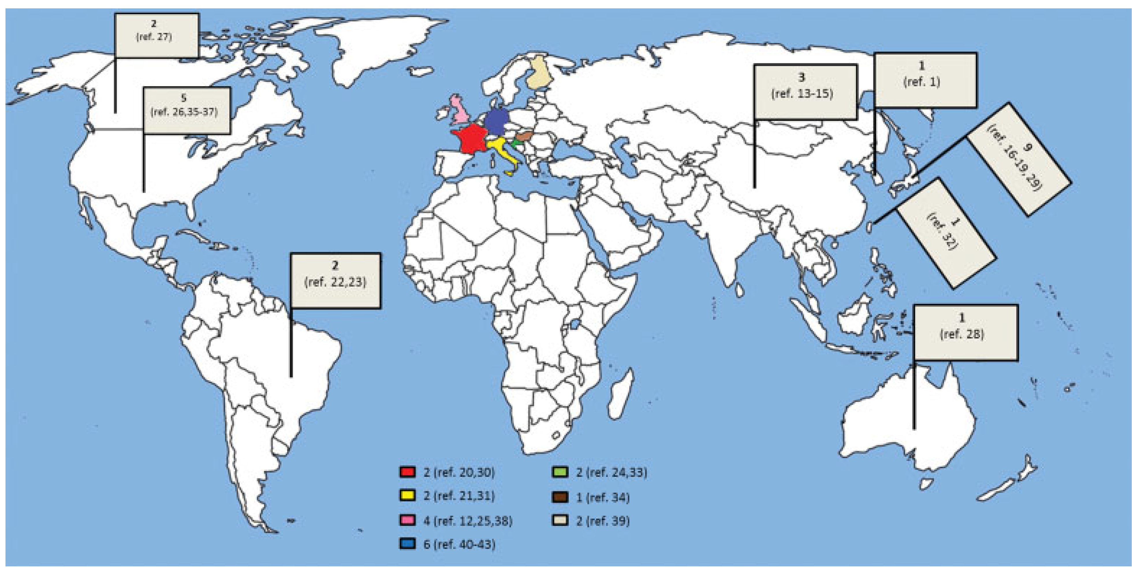
All articles published between 1967 and 2014 on familial NBCCS were surveyed to determine the mapping of cases per country of the occurrence of the disease. The Medline (PubMed) database was used for this search, using the following descriptors (basal cell nevus syndrome or Gorlin syndrome or Gorlin–Goltz syndrome) and (familial or family). Articles that did not have familial reports (cyto)genetic studies of families that had not provided complete clinical information on affected members, suspected cases (that were not confirmed), articles without abstracts, and those performed in countries that no longer exist were excluded. After defining the sample, the familial cases were counted and separated by country. Among a total of 232 articles found in the search, 175 were excluded because they did not represent familial reports, as were 11 due to the absence of an abstract, 9 genetic studies, and 2 suspected cases. Two were excluded because the origin of the article could not be identified. Thus, in total, 199 articles were excluded and 33 articles satisfied the inclusion criteria. The familial cases per country [1,12,13,14,15,16,17,18,19,20,21,22,23,24,25,26,27,28,29,30,31,32,33,34,35,36,37,38,39,40,41,42,43] included in this study are distributed on the map in Figure 2.

Results

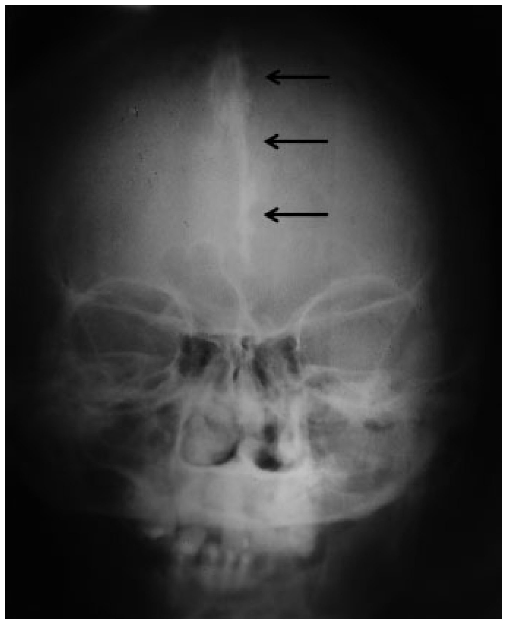
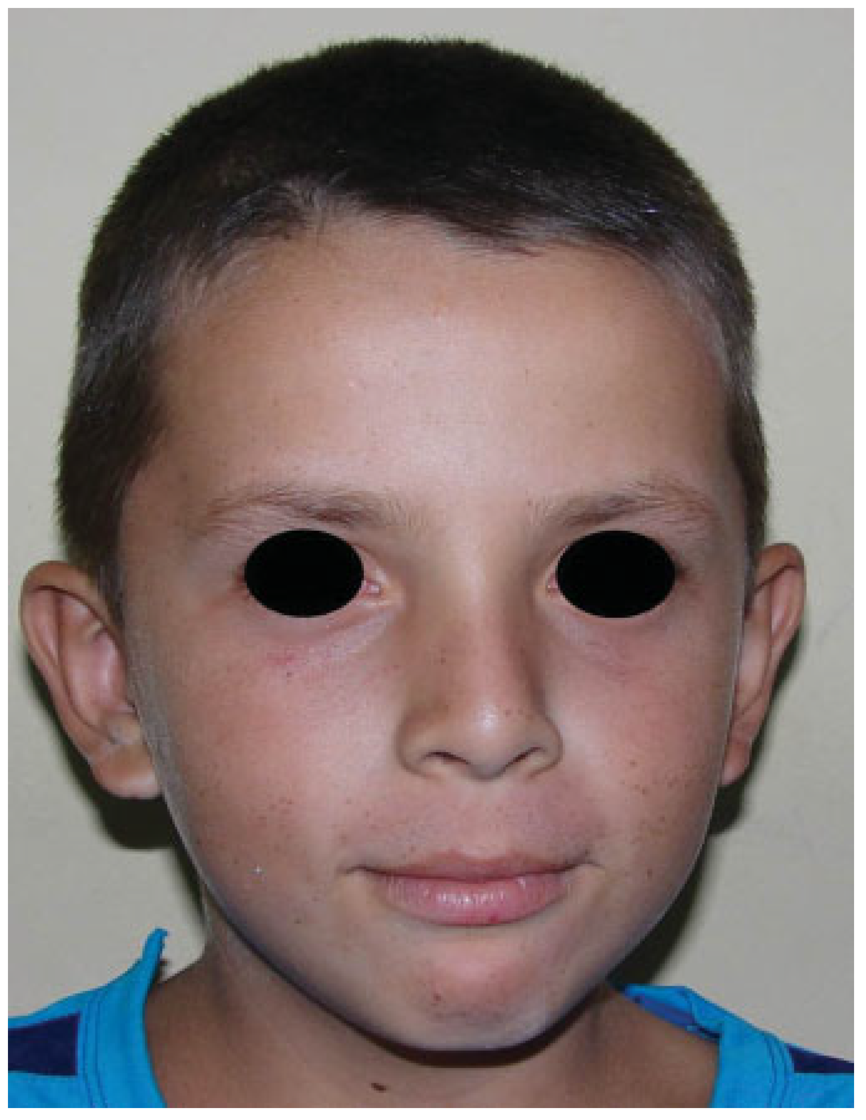
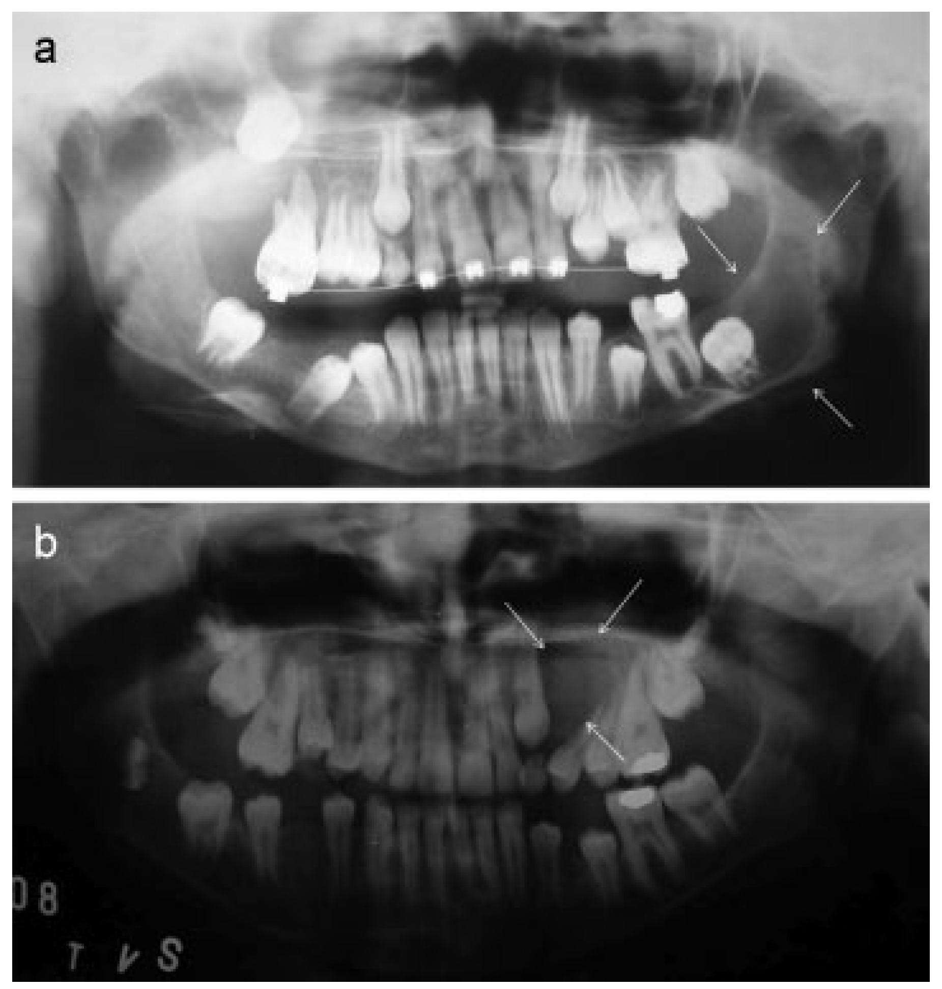
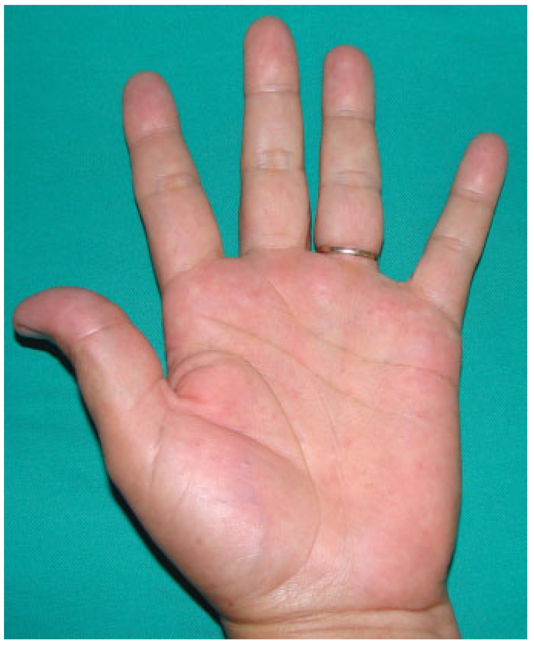
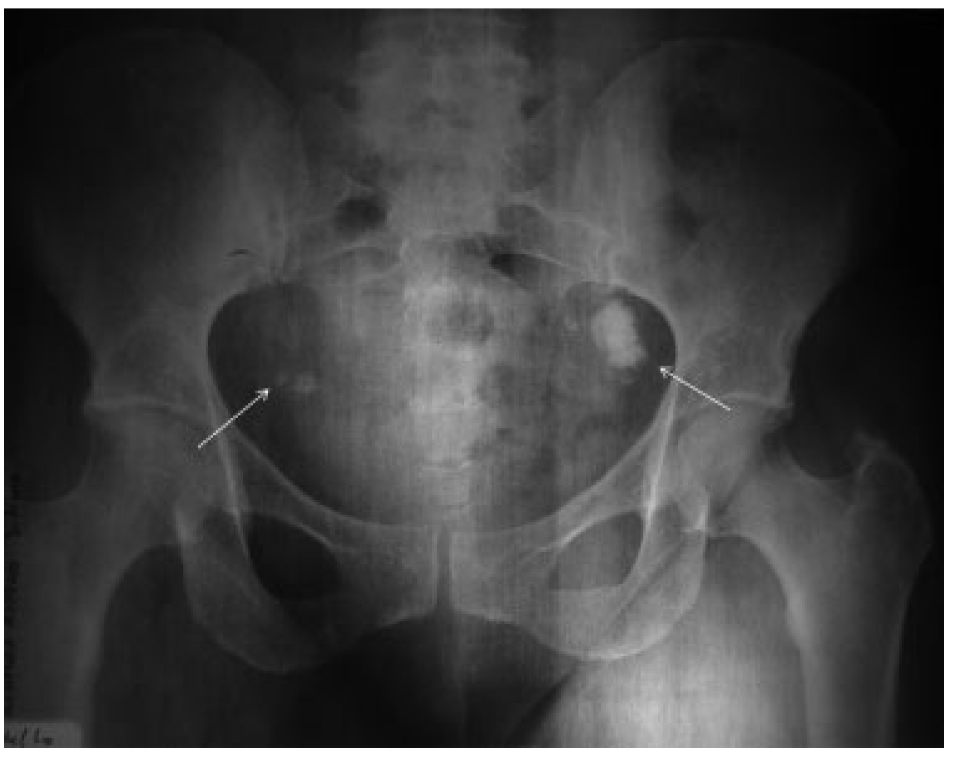
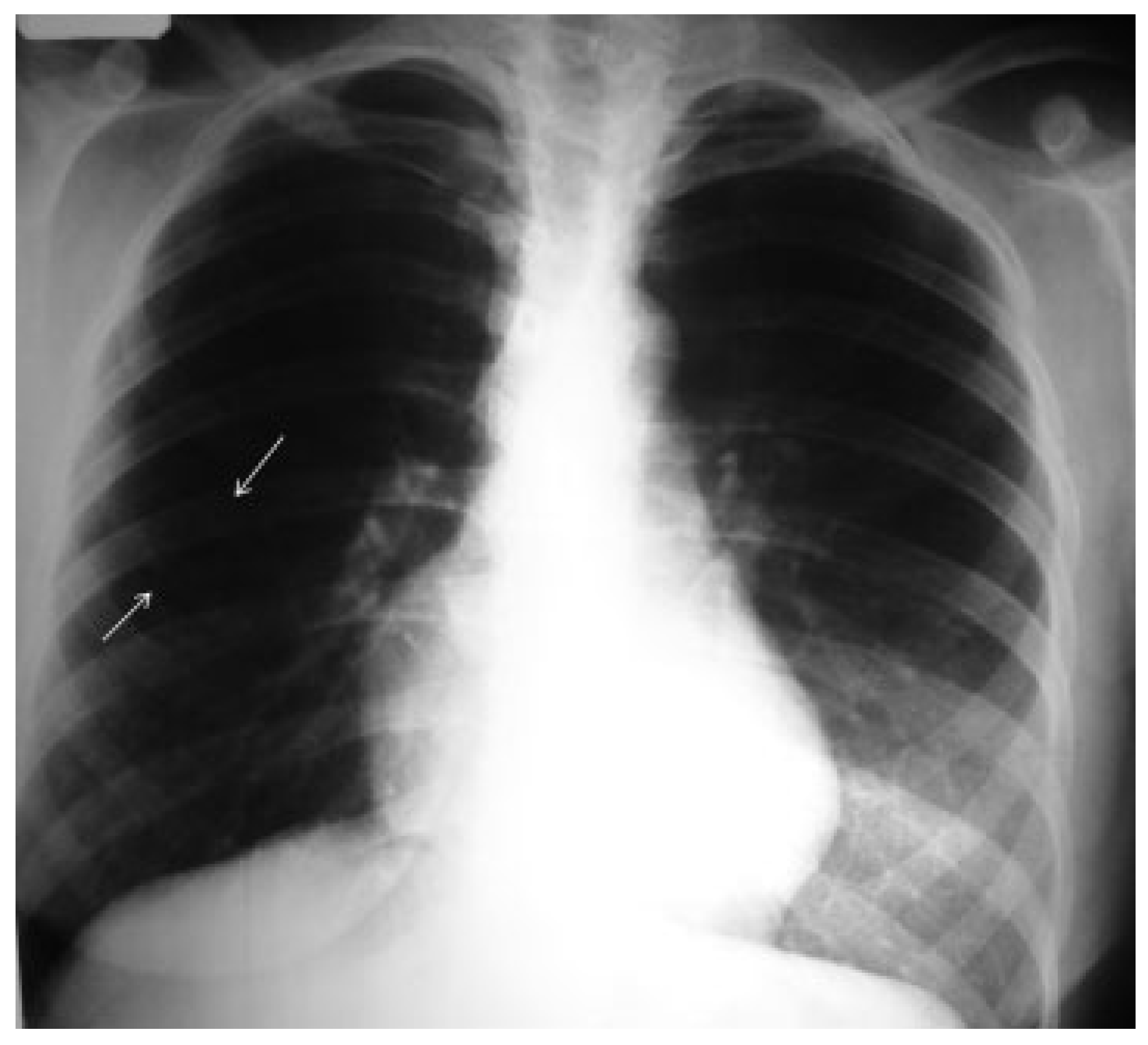
Since 2002, we have reviewed the findings in 10 affected individuals with NBCCS in a Brazilian population (mean age ± standard deviation: 23.5 ± 11.39 years, range: 11–45 years). The patients consisted of four (40%) males and six (60%) females. All patients (100%) presented calcification of the falx cerebri (Figure 3), mild hypertelorism, and macrocephaly (Figure 4). KCOTs (Figure 5) had occurred in 90% of the patients. The number of total jaw lesions ranged from 0 to 3. Palmar and plantar pits were seen in 40% of the patients (Figure 6). Multiple BCCs (Figure 7) occurred in 20% of the patients; one was treated with topical PDT with satisfactory cosmetic outcome and no recurrence, and another was later diagnosed with a large facial tumor and died within a few months. Only one patient, a 41-year-old, had been affected by ovarian calcified fibroma (Figure 8). The same patient also had a bifid rib in a chest X-ray (Figure 9; Table 2).

Discussion

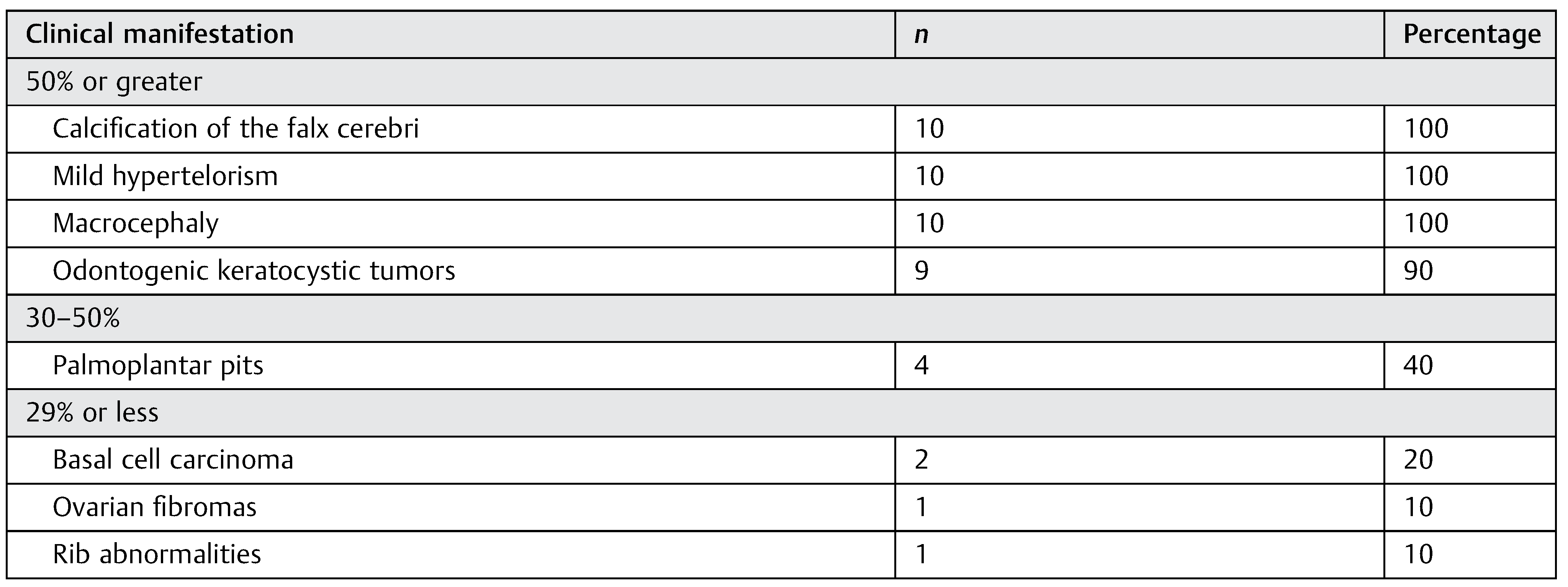
In this study, we reviewed the clinical and radiological manifestations in 10 Brazilian individuals with NBCCS from the same family. Characteristics seen in more than 90% of the patients were frontal bossing, mild hypertelorism, calcification of the falx cerebri, and the presence of multiple KCOTs. On the contrary, BCCs were diagnosed in 20% of the patients (Table 3). These frequencies were similar to some previous reports, [13,15,16,17,26] but quite different from others, [1,12,14,18,19,20,21,24,25,27,28] including another Brazilian series [22,23] indicating the wide phenotypic heterogeneity of this syndrome even in the same ethnic background. The exact incidence of this syndrome and its different manifestations are unrecorded and it is typically difficult to identify the condition clinically until jaw cysts or skin cancers become apparent. Nevertheless, this syndrome probably occurs in all ethnic groups, although few cases have been published in certain races. [35] The relative lack, for example, of BCCs in African Americans may partially retect ultraviolet radiation protection, resulting from increased skin pigmentation. [35] The presence of melanin reduces the absorption of ultraviolet radiation in the skin and, consequently, DNA damage. This may explain the lack of familial reports in Africa (Figure 1) because BCCs are the most evident clinical sign of the syndrome. Also, we believe that the wide variation in NBCCS phenotypes may be associated with genetic mutations, especially in the PTCH gene, [13,44,45] in addition to environmental variation. [46]
As in the present series, literature reports show that skeletal anomalies in the craniofacial complex are the most common features in patients with NBCCS. However, although characteristic facies are part of the syndrome, not every patient has the typical skull and facial features (Table 2), which consist basically of macrocephaly (due to frontal and temporoparietal bossing) combined with prominent supraorbital ridges as well as a broad nasal root, resulting in an appearance of hypertelorism. Increased dural calcification, especially in the falx region, is a quite common radiological manifestation in patients with NBCCS. According to Pritchard et al, [47] the radiographic appearance is that of one or more flat sheets extending more widely (> 2.0 mm) than in the normal person. Although Kimonis et al [6] reported that calcification of the falx cerebri is more common in patients older than 20 years, our series showed that at least 60% of patients < 20 years already had signs of dural calcification at first attendance. These data suggest that children with a familial history of NBCCS should undergo cranial imaginological exams in searching for the first signs of the syndrome.
Other craniofacial skeletal anomalies, such as bridge sella turcica, high-arched palate, and cleft palate, are less frequent [39,48] but should not be overlooked. In contrast to the present series, the occurrence of mental retardation, short metacarpals, and low phosphorus excretion have been described in adult and child patients with NBCCS, suggesting the presence of pseudohypoparathyroidism, but this endocrine aberration seems not be linked to deformity of the pituitary fossa. However, the skeletal anomalies and the mandibular prognathism are suggestive of a growth disturbance. [49] Thus, the clinical importance of these findings remains open to discussion. In addition, earlier studies have described skeletal anomalies in the thoracic cage, including bifid ribs and fusion of the thoracic vertebrae. [50] Although bifid ribs are of little consequence to the patient, as in case 1, vertebral deformities may cause scoliosis or compression of the spinal dorsal roots, leading to peripheral pain and disability. In this series, no patient was diagnosed with vertebral anomalies.
KCOTs are the second most common features associated with NBCCS, with a peak occurrence in the second and third decades and with a predilection for the third molar region, [51] as in the present cases. In children and adolescents, the lesions may cause displacement of developing teeth and delay dental development. [22] Although most KCOTs present as solitary lesions, multiple lesions are a well-recognized feature of NBCCS. In fact, there are authors who state that the presence of multiple keratocysts—even just keratocysts if there is a family association—is sufficient for its establishment. [25] However, most authors make the diagnosis if two major criteria (KCOTs of the jaws, nevoid BCCs, palmar and plantar pits, cerebral falx calcifications, family history, and skeletal anomalies) or one major and two minor criteria (macrocephalus, hypertelorism, MB, ovarian calcification) are presented. [2] Early diagnosis and treatment is essential, as is family detection and genetic counseling. It is important to recognize when a patient has multiple KCOTs because life-long monitoring is essential for patient management.
Recently, the WHO Working Group defined this lesion as “a benign uni- or multicystic, intraosseous tumor of odontogenic origin, with a characteristic lining of parakeratinized stratified squamous epithelium and potential for aggressive, infiltrative behavior.” When the syndrome is present, the tumor can be larger and may be associated with painful symptoms, swelling, or drainage. KCOTs in patients with this syndrome tend to have more satellite cysts, solid islands of epithelial proliferation, and odontogenic epithelial rests within the fibrous capsule than do isolated tumors. [52] These characteristics lead to a great rate of recurrence. [53,54] In addition, it has been suggested that recurrence is higher in multilocular large lesions with high SMO [55] and podoplanin expression. [56]
A range of surgical treatment approaches have been described in the literature for KCOTs, including simples enucleation (with or without curettage), enucleation with adjunctive liquid nitrogen therapy, or the application of Carnoy solution, marsupialization, cryotherapy, electrocautery, removal with peripheral ostectomy, and localized or segmental resection. According to Borgonovo et al, [57] the goals of treatment should involve eliminating the potential for recurrence, while also minimizing the morbidity and a two-stage treatment protocol was suggested for young patients with NBCCS, involving conservative surgical treatment, based on marsupialization and later enucleation with peripheral ostectomy. In our series, all cases were treated with PO and curettage, with a recurrence rate of 28.5% (two cases). The two cases of recurrence occurred 6 and 2 years after the surgery, indicating the need for long-term follow-up. Unfortunately, in a recent systematic review, [58] it was observed a lack of evidence regarding the optimal surgical treatment for KCOTs in NBCCS patients, although enucleation and marsupialization appeared to be the most common treatments in the literature.
Recent advances have linked Shh pathway signaling activation to a variety of human cancers, including BCCs of the skin and MBs, common malignancies diagnosed in patients with Gorlin syndrome. It has been hypothesized that loss-of-function mutations of PTCH-1, a critical membrane protein for embryonic development and a human tumor suppressor gene, at human chromosome 9q22 repress constitutive signaling activity of a second membrane protein, known as SMO, leading to activation of the Shh pathway. [59] It has been demonstrated experimentally that conditional ablation of PTCH-1 with UV irradiation [60] and consequently the overexpression of Shh [61] are needed for the occurrence of BCCs in mice, similar to that found in NBCCS patients, suggesting that exposure to solar radiation is important in the development of these tumors, even in patients with NBCCS.
An oncogenic two-hit model has been proposed that states that normal cells require two mutagenic hits to produce a cancer. Patients with the syndrome have a germline defect in the DNA sequence in one of the two copies of the PTCH-1 gene; this is insufficient to cause malignant transformation. However, if loss of the normal allele occurs at the same locus, a second hit, as a consequence of mitotic nondisjunction, deletion, or mitotic recombination, the cell may become malignant. [62,63] According to Gorlin, [64] in cases of BCCs, both hits come from the somatic type of mutation, needing only one additional hit, due to solar radiation, for tumor development. In addition, it was noted that 70% of KCOTs exhibit loss of heterozygosity of one or more of the common tumor suppressor genes, indicating that the behavior of these lesions could resemble low-grade tumors more than congenital malformations. [65]
In clinical practice, the incidence of BCCs related to Gorlin syndrome is higher in fair-skinned patients, probably due to the lack of melanin, a complex polymeric pigment that absorbs all wavelengths of damaging ultraviolet radiation, reducing or preventing its penetration further into the body. Dark-skinned patients with NBCCS only rarely have more than a few BCCs over a life time. Thus, because skin tumors in NBCCS tend to appear earlier in life than their nonsyndromal counterparts, the introduction of a specific disease prevention program should be instituted for these patients and should include information about risks related to sun exposure, regular dermatological examinations, and use of sunscreens and long-brimmed hats. These measures are important especially in tropical countries, such as Brazil, most of the territory of which is subject to strong solar radiation, and activities in the primary sector of the economy, such as agriculture, livestock, and fisheries, are common.
In the present series, educational measures and preventive strategies against solar radiation have been effective because, so far, no family member younger than 30 years has developed skin cancer. In contrast, two patients older than 30 years were diagnosed with BCCs, located on the face. One patient (case 3) was diagnosed too late and died 1 year after the diagnosis. Case 1 was treated with PDT, with satisfactory outcome (Figure 7).
The treatment of BCCs in patients with Gorlin syndrome is complex. Currently, inhibitors and modulators of the Shh pathway are being tested with promising results. In a randomized, double-blind, placebo-controlled trial, it was demonstrated that vismodegib, a new orally administered hedgehog pathway inhibitor, reduced the basal cell carcinoma tumor burden and blocked growth of new basal cell carcinomas in patients with NBCCS. [66] However, while a curative breakthrough for BCCs may occur in the foreseeable future, most cases have been treated with surgical excision and cryotherapy. However, these methods can result in significant disfigurement due to scarring. Topical PDT has been used as an alternative to surgery for patients with NBCCS, especially for facial lesions which are < 2 mm thick on ultrasound assessment. [67] Photodynamic inactivation involves the photoactivation of a photosensitizer agent, such as cationic phenothiazinium dyes (methylene blue and toluidine blue), porphyrins, crystal violet, indocyanine green, and erythrosine, by visible light, generating singlet oxygen (1O2). [67] 1O2 is an excited form of molecular oxygen characterized by the opposite spin of a pair of electrons, and is less stable and more reactive than the normal triplet oxygen (O2). This reactive oxygen molecule causes irreparable damage to cellular components, leading to cell death. [68,69,70] Although in most cases photosensitizers are activated by red light, from low-power lasers, the choice of light source should be based on photosensitizer absorption (fluorescence excitation and action spectra), disease features, cost, and size. On the contrary, the clinical efficacy of PDT is dependent on complex dosimetry that includes total light dose, light exposure time, and light delivery mode. [70]
In addition to cosmetic appearance, topical PDT appears to have a series of advantages versus surgery, as follows: the possibility of treating several BCCs at the same time, the need for only one visit for each treatment, minimally invasive treatment, the possibility of retreating a previously treated area, minimal discomfort, and temporary epilation following treatment. [67,71] In a study by Loncaster et al, [71] the local control rate at 12 months after PDT was 56.3%, and the overall average percentage reduction in lesion thickness was 68% at 12 months and 65% at 24 months. Itkin and Gilchrest [72] used topical PDT for the treatment of multiple lesions on two patients with NBCCS, resulting in an 89% complete clinical response of superficial tumors in contrast to 31% of nodular BCCs. Thus, although there are no existing guidelines about the use of topical PDT in NBCCS patients, this method seems to be adequate for superficial BCCs. For lesions > 3 mm thick, a debulking curettage can be performed 6 weeks before PDT. Extensive lesions that preclude the application of a topical sensitizer should be treated with an intravenous photosensitizer, such as porfimer sodium. [71]
A wide range of reproductive anomalies has been reported in patients with NBCCS, including uterine and ovarian fibromas, calcified ovarian cysts, supernumerary nipples, hypogonadism, and cryptorchidism. In this familial series, only one case of reproductive anomaly was observed; a bilateral ovarian fibroma was diagnosed in a 31-year-old woman (case 1).
Ovarian fibromas are uncommon benign neoplasms of gonadal cell stroma origin, composed of spindly fibroblastic cells that produce collagen. The tumors appear as unilateral masses in 90% of cases, generally in older, menopausal patients, but a peak frequency between 20 and 40 years has been described. [73] Although most ovarian fibromas appear sporadically, some tumors may be associated with Meigs syndrome (ovarian fibroma, hydrothorax, ascites), familial polyposis (such as Gardner and Richard syndrome and Peutz– Jeghers syndrome), and NBCCS.
According to Gorlin, [74] approximately 2 to 25% of females with NBCCS will develop ovarian fibromas, leading to challenges in gynecological care and counseling. In patients with NBCCS, the tumors are often bilateral and calcified, [75] as in case 1 here. In rare cases, the ovarian fibromas may be related to endocrine manifestations, with the presence of pelvic pain, metrorrhagia, and abdominal distension. [73]
In most cases, the treatment of these lesions is based on surgery because there is some risk of malignancy. [76] Tumorectomy is indicated for young and bilateral salpingo-oophorectomy for menopausal patients. [74] When counseling patients, the clinician should consider the patient’s desire for future fertility and attempt to conserve normal ovarian tissue if fertility is desired. In addition, factors such as age, ovarian reserve, presence of comorbidities, partner availability, and financial resources or insurance coverage for fertility preservation should be considered. [76] Although no data exist about the fertility potential of females with NBCCS, a patient diagnosed with ovarian fibromas in this series had four children with the syndrome and did not undergo surgery for personal reasons. In pediatric patients, the development of ovarian fibromas is rare, [77] but regular gynecological follow-up is required.
Also, as part of the interdisciplinary investigation in patients with NBCCS, other anomalies should not be overlooked, such as the presence of hand alterations, intracranial tumors, eye alterations, and cardiac fibromas. Hand alterations include the presence of palmar pits, polydactyly, syndactyly, and shortening of the metacarpal bones. Of these, palmar pits are the most common alterations, affecting 85% of patients with NBCCS over the age of 20 years. [46] Clinically, the lesions appear as punctiform depressions in the skin of the palms, but in some cases, the soles of the feet, the back, the face, and the chest may also be affected. [78] In the present series, some cases of palmar pits were observed in patients younger than 20 years. Although difficult to detect, the presence of three or more palmar/plantar pits constitutes a major diagnostic criterion in NBCCS. Thus, when lesions are discovered, a careful physical and radiological examination is mandatory, especially in children with a positive familial history of NBCCS.
Tumors of the central nervous system are not uncommon in patients with NBCCS. MB is the brain tumor most frequently associated with the syndrome and is one of the features included in the minor criteria for its diagnosis. It is estimated that 5% of all NBCCS patients will develop cranial tumors, with a high incidence in those younger than 5 years. [75] Desmoplastic MB is the most common histological type associated with NBCCS, and seems to have a better prognosis than other histological variants, with a decreased propensity for metastasis and higher sensitivity to chemotherapy. [79,80] In addition, some authors [80,81,82] have shown that patients with syndromic MBs treated with craniospinal irradiation often develop late-onset neoplasms, increasing morbidity and mortality in this population. Thus, it has been suggested that syndromic children < 5 years old with desmoplastic MB should be treated with surgical excision, followed by chemotherapy, to prevent sequelae secondary to radiation. [80] In the present series, although no case of MB had been diagnosed, all patients continue to be closely monitored by neurologists because the development of MB is not restricted to the first years of life.
Ophthalmic lesions and cardiac fibromas are minor diagnostic criteria for NBCCS and affect 26 and 3% of these patients, respectively. [75] Ophthalmic lesions include the presence of hypertelorism (as in the present cases), strabismus, nystagmus, cataract, coloboma, and microphthalmos. [5] Recently, four cases of BCC of the eyelid associated with the syndrome were reported [83] and, thus, ophthalmic evaluation should be mandatory in patients with NBCCS. In contrast, no case of cardiac fibromas was found in the present series. Most cases of these tumors arise in the left ventricle, are asymptomatic, [84] and are discovered after an echocardiography or magnetic resonance examination. In some circumstances, patients may present episodes of syncope, [85] fatigue, chest pain, and dyspnea, [86] as well as ventricular tachycardia. [87] The presence of dysrhythmia, conduction defects, or congestive heart failure is rare but may lead to death. [88] For this reason, any cardiac symptom in a patient with NBCCS should be evaluated promptly by a cardiologist to rule out a cardiac tumor. In summary, it is important that families with NBCCS are carefully informed about the genetic alterations responsible for the disease, prognosis, risks of having other children with the problem, and what can be done to improve the quality of life for everyone living with this condition.

Note

This article has been checked by at least two professional editors, both native speakers of English. For a certificate, please see: http://www.textcheck.com/ certificate/18GWy3.

References

  1. Lee, Y.W.; Roh, B.H.; Ki, C.S.; et al. Identification of a novel mutation in the PTCH gene in a Korean family with naevoid basal cell carcinoma syndrome. Clin Exp Dermatol 2007, 32, 202–203. [Google Scholar] [CrossRef] [PubMed]
  2. Evans, D.G.; Farndon, P.A.; Burnell, L.D.; Gattamaneni, H.R.; Birch, J.M. The incidence of Gorlin syndrome in 173 consecutive cases of medulloblastoma. Br J Cancer 1991, 64, 959–961. [Google Scholar] [CrossRef]
  3. Gundlach, K.K.; Kiehn, M. Multiple basal cell carcinomas and keratocysts—the Gorlin and Goltz syndrome. J Maxillofac Surg 1979, 7, 299–307. [Google Scholar] [CrossRef]
  4. Habibi, A.; Jafarzadeh, H. Nevoid basal cell carcinoma syndrome: a 17-year study of 19 cases in Iranian population (1991-2008). J Oral Pathol Med 2010, 39, 677–680. [Google Scholar] [CrossRef] [PubMed]
  5. Gorlin, R.J.; Goltz, R.W. Multiple nevoid basal-cell epithelioma, jaw cysts and bifid rib. A syndrome. N Engl J Med 1960, 262, 908–912. [Google Scholar] [CrossRef] [PubMed]
  6. Kimonis, V.E.; Goldstein, A.M.; Pastakia, B.; et al. Clinical manifestations in 105 persons with nevoid basal cell carcinoma syndrome. Am J Med Genet 1997, 69, 299–308. [Google Scholar] [CrossRef]
  7. Shafei-Benaissa, E.; Savage, J.R.; Babin, P.; et al. The naevoid basal-cell carcinoma syndrome (Gorlin syndrome) is a chromosomal instability syndrome. Mutat Res 1998, 397, 287–292. [Google Scholar] [CrossRef]
  8. Hahn, H.; Wicking, C.; Zaphiropoulous, P.G.; et al. Mutations of the human homolog of Drosophila patched in the nevoid basal cell carcinoma syndrome. Cell 1996, 85, 841–851. [Google Scholar] [CrossRef]
  9. Roudgari, H.; Farndon, P.A.; Murray, A.D.; Hardy, C.; Miedzybrodzka, Z. Is PATCHED an important candidate gene for neural tube defects? Cranial and thoracic neural tube defects in a family with Gorlin syndrome: a case report. Clin Genet 2012, 82, 71–76. [Google Scholar] [CrossRef]
  10. Saldanha, G. The Hedgehog signalling pathway and cancer. J Pathol 2001, 193, 427–432. [Google Scholar] [CrossRef]
  11. Zedan, W.; Robinson, P.A.; High, A.S. A novel polymorphism in the PTC gene allows easy identification of allelic loss in basal cell nevus syndrome lesions. Diagn Mol Pathol 2001, 10, 41–45. [Google Scholar] [PubMed]
  12. Evans, D.G.; Sims, D.G.; Donnai, D. Family implications of neonatal Gorlin’s syndrome. Arch Dis Child 1991, 66, 1162–1163. [Google Scholar] [CrossRef]
  13. Zhang, T.; Chen, M.; Lü, Y.; Xing, Q.; Chen, W. A novel mutation of the PTCH1 gene activates the Shh/Gli signaling pathway in a Chinese family with nevoid basal cell carcinoma syndrome. Biochem Biophys Res Commun 2011, 409, 166–170. [Google Scholar]
  14. Fan, Z.; Li, J.; Du, J.; et al. A missense mutation in PTCH2 underlies dominantly inherited NBCCS in a Chinese family. J Med Genet 2008, 45, 303–308. [Google Scholar] [CrossRef] [PubMed]
  15. Lü, Y.; Zhu, H.G.; Ye, W.M.; Zhang, M.B.; He, D.; Chen, W.T. A new mutation of PTCH gene in a Chinese family with nevoid basal cell carcinoma syndrome. Chin Med J (Engl) 2008, 121, 118–121. [Google Scholar]
  16. Fujii, M.; Noguchi, K.; Urade, M.; et al. Novel PTCH1 mutations in Japanese Nevoid basal cell carcinoma syndrome patients: two familial and three sporadic cases including the first Japanese patient with medulloblastoma. J Hum Genet 2011, 56, 277–283. [Google Scholar] [CrossRef]
  17. Abe, S.; Kabashima, K.; Sakabe, J.; et al. Coincident two mutations and one single nucleotide polymorphism of the PTCH1 gene in a family with naevoid basal cell carcinoma syndrome. Acta Derm Venereol 2008, 88, 635–636. [Google Scholar] [CrossRef] [PubMed]
  18. Otsubo, S.; Honma, M.; Asano, K.; Takahashi, H.; Iizuka, H. A novel germline mutation of PTCH1 gene in a Japanese family of nevoid basal cell carcinoma syndrome: are the palmoplantar pits associated with true basal cell carcinoma? J Dermatol Sci 2008, 51, 144–146. [Google Scholar] [CrossRef][Green Version]
  19. Matsuzawa, N.; Nagao, T.; Shimozato, K.; Niikawa, N.; Yoshiura, K.I. Patched homologue 1 mutations in four Japanese families with basal cell nevus syndrome. J Clin Pathol 2006, 59, 1084–1086. [Google Scholar] [CrossRef]
  20. Le Brun Keris, Y.; Jouk, P.S.; Saada-Sebag, G.; et al. Prenatal manifestation in a family affected by nevoid basal cell carcinoma syndrome. Eur J Med Genet 2008, 51, 472–478. [Google Scholar]
  21. Pastorino, L.; Ghiorzo, P.; Nasti, S.; et al. Identification of a SUFU germline mutation in a family with Gorlin syndrome. Am J Med Genet A 2009, 149A, 1539–1543. [Google Scholar] [PubMed]
  22. Melo, E.S.; Kawamura, J.Y.; Alves, C.A.; Nunes, F.D.; Jorge, W.A.; Cavalcanti, M.G. Imaging modality correlations of an odontogenic keratocyst in the nevoid basal cell carcinoma syndrome: a family case report. Oral Surg Oral Med Oral Pathol Oral Radiol Endod 2004, 98, 232–236. [Google Scholar] [PubMed]
  23. Jose Tincani, A.; Santos Martins, A.; Gomes Andrade, R.; Franco Mello, E.J., Jr.; Camargo Bueno, M.A. Nevoid basal-cell syndrome: literature review and case report in a family. Sao Paulo Med J 1995, 113, 917–921. [Google Scholar]
  24. Musani, V.; Gorry, P.; Basta-Juzbasic, A.; Stipic, T.; Miklic, P.; Levanat, S. Mutation in exon 7 of PTCH deregulates SHH/PTCH/SMO signaling: possible linkage to WNT. Int J Mol Med 2006, 17, 755–759. [Google Scholar] [CrossRef] [PubMed][Green Version]
  25. Totten, J.R. The multiple nevoid basal cell carcinoma syndrome. Report of its occurrence in four generations of a family. Cancer 1980, 46, 1456–1462. [Google Scholar]
  26. Heimler, A.; Friedman, E.; Rosenthal, A.D. Naevoid basal cell carcinoma syndrome and Charcot-Marie-Tooth disease: two autosomal dominant disorders segregating in a family. J Med Genet 1978, 15, 288–291. [Google Scholar]
  27. Fitzpatrick, P.J.; Thompson, G.A. Gorlin’s syndrome, or nevoid basal cell carcinoma syndrome. Can Med Assoc J 1982, 127, 465–470. [Google Scholar]
  28. Wallace, D.C.; Murphy, K.J.; Kelly, L.; Ward, W.H. The basal cell naevus syndrome. Report of a family with anosmia and a case of hypogonadotrophic hypopituitarism. J Med Genet 1973, 10, 30–33. [Google Scholar]
  29. Fujii, K.; Sugita, K.; Watanabe, T.; Takanashi, J.; Niimi, H. A familial case of Gorlin syndrome [in Japanese]. No To Hattatsu 1996, 28, 257–260. [Google Scholar]
  30. Huault, M.; Bayonne, E.; Laxenaire, A.; Blanchard, P.; Levy, J.D. Basocellular nevomatosis. Follow-up of 3 generations [in French]. Rev Stomatol Chir Maxillofac 1998, 99, 244–249. [Google Scholar]
  31. Mazzola, G.; Galioto, V.; Mazzola, U. Gorlin-Gotz syndrome in 3 patients from the same family monitored from 1993 to 1999 [in Italian]. Minerva Stomatol 2000, 49, 197–203. [Google Scholar]
  32. Chung, C.H.; Wong, T.Y.; Shieh, T.Y.; Shieh, D.B.; Chao, S.C. Nevoid basal cell carcinoma syndrome—clinical manifestations and mutation analysis of a Taiwanese family. J Formos Med Assoc 2003, 102, 793–797. [Google Scholar] [PubMed]
  33. Zmak, M.; Dobric, P.; Medica, I. Gorlin syndrome: a report on a family. Minerva Pediatr 1994, 46, 217–219. [Google Scholar]
  34. Ujpál, M.; Szabó, G. Familial occurrence of Gorlin-Goltz syndrome [in Hungarian]. Fogorv Sz 1992, 85, 209–212. [Google Scholar] [PubMed]
  35. Goldstein, A.M.; Pastakia, B.; DiGiovanna, J.J.; et al. Clinical findings in two African-American families with the nevoid basal cell carcinoma syndrome (NBCC). Am J Med Genet 1994, 50, 272–281. [Google Scholar] [CrossRef] [PubMed]
  36. McLoughlin, P.M.; Dickenson, A.J.; Avery, B.S. Gorlin’s syndrome and von Recklinghausen’s disease occurring in one family. Br J Oral Maxillofac Surg 1991, 29, 189–193. [Google Scholar] [CrossRef]
  37. Lovin, J.D.; Talarico, C.L.; Wegert, S.L.; Gaynor, L.F.; Sutley, S.S. Gorlin’s syndrome with associated odontogenic cysts. Pediatr Radiol 1991, 21, 584–587. [Google Scholar] [CrossRef]
  38. Evans, D.G.; Birch, J.M.; Orton, C.I. Brain tumours and the occurrence of severe invasive basal cell carcinoma in first degree relatives with Gorlin syndrome. Br J Neurosurg 1991, 5, 643–646. [Google Scholar] [CrossRef]
  39. Tasanen, A.; Lamberg, M.A.; Nordling, S. Skeletal anomalies and keratocysts in the basal cell nevus syndrome. Int J Oral Surg 1975, 4, 225–235. [Google Scholar] [CrossRef]
  40. Brumme, S.; Löwicke, G. Kinship studies in Gorlin-Goltz syndrome (basal cell nevus syndrome) [in German]. Dermatol Monatsschr 1990, 176, 487–492. [Google Scholar]
  41. Gartmann, H.; Groth, W.; Quinkler, C. Multiple basaloid follicular hamartomas in 2 members of a family with Gorlin-Goltz syndrome [in German]. Z Hautkr 1989, 64, 915–918. [Google Scholar] [PubMed]
  42. Rausch, E.; Trödhan, A.; Porteder, H. Multiple keratocysts suggesting the presence of Gorlin-Goltz syndrome: report of 3 patients in a family [in German]. Z Stomatol 1989, 86, 275–277. [Google Scholar] [PubMed]
  43. Lorenz, R.; Fuhrmann, W. Familial basal cell nevus syndrome. Hum Genet 1978, 44, 153–163. [Google Scholar] [PubMed]
  44. Aszterbaum, M.; Rothman, A.; Johnson, R.L.; et al. Identification of mutations in the human PATCHED gene in sporadic basal cell carcinomas and in patients with the basal cell nevus syndrome. J Invest Dermatol 1998, 110, 885–888. [Google Scholar] [CrossRef]
  45. Sasaki, R.; Miyashita, T.; Matsumoto, N.; Fujii, K.; Saito, K.; Ando, T. Multiple keratocystic odontogenic tumors associated with nevoid basal cell carcinoma syndrome having distinct PTCH1 mutations: a case report. Oral Surg Oral Med Oral Pathol Oral Radiol Endod 2010, 110, e41–e46. [Google Scholar]
  46. Lo Muzio, L. Nevoid basal cell carcinoma syndrome (Gorlin syndrome). Orphanet J Rare Dis 2008, 3, 32. [Google Scholar]
  47. Pritchard, L.J.; Delfino, J.J.; Ivey, D.M.; Sclaroff, A.; Giglio, J.A. Variable expressivity of the multiple nevoid basal cell carcinoma syndrome. J Oral Maxillofac Surg 1982, 40, 261–269. [Google Scholar]
  48. Manfredi, M.; Vescovi, P.; Bonanini, M.; Porter, S. Nevoid basal cell carcinoma syndrome: a review of the literature. Int J Oral Maxillofac Surg 2004, 33, 117–124. [Google Scholar]
  49. Rayne, J. The multiple basal cell naevi syndrome. Br J Oral Surg 1971, 9, 65–71. [Google Scholar] [CrossRef]
  50. Gorlin, R.J.; Vickers, R.A.; Kellen, E.; Williamson, J.J. Multiple basal-cell nevi syndrome. An analysis of a syndrome consisting of multiple nevoid basal-cell carcinoma, jaw cysts, skeletal anomalies, medulloblastoma, and hyporesponsiveness to parathormone. Cancer 1965, 18, 89–104. [Google Scholar]
  51. Shand, J.M.; Heggie, A.A. Cysts of the jaws and advances in the diagnosis and management of nevoid Basal cell carcinoma syndrome. Oral Maxillofac Surg Clin North Am 2005, 17, 403–414. [Google Scholar] [CrossRef]
  52. Woolgar, J.A.; Rippin, J.W.; Browne, R.M. A comparative histological study of odontogenic keratocysts in basal cell naevus syndrome and control patients. J Oral Pathol 1987, 16, 75–80. [Google Scholar] [CrossRef] [PubMed]
  53. Avelar, R.L.; Antunes, A.A.; Santos Tde, S.; Andrade, E.S.; Dourado, E. Odontogenic tumors: clinical and pathology study of 238 cases. Braz J Otorhinolaryngol 2008, 74, 668–673. [Google Scholar] [PubMed]
  54. Morgan, T.A.; Burton, C.C.; Qian, F. A retrospective review of treatment of the odontogenic keratocyst. J Oral Maxillofac Surg 2005, 63, 635–639. [Google Scholar] [CrossRef] [PubMed]
  55. Yagyuu, T.; Kirita, T.; Sasahira, T.; Moriwaka, Y.; Yamamoto, K.; Kuniyasu, H. Recurrence of keratocystic odontogenic tumor: clinicopathological features and immunohistochemical study of the Hedgehog signaling pathway. Pathobiology 2008, 75, 171–176. [Google Scholar] [CrossRef]
  56. Friedrich, R.E.; Scheuer, H.A.; Zustin, J. Expression of podoplanin in nevoid basal cell carcinoma syndrome-associated keratocystic odontogenic tumours. Anticancer Res 2012, 32, 2125–2127. [Google Scholar] [PubMed]
  57. Borgonovo, A.E.; Di Lascia, S.; Grossi, G.; Maiorana, C. Two-stage treatment protocol of keratocystic odontogenic tumour in young patients with Gorlin-Goltz syndrome: marsupialization and later enucleation with peripheral ostectomy. A 5-year-follow-up experience. Int J Pediatr Otorhinolaryngol 2011, 75, 1565–1571. [Google Scholar] [CrossRef]
  58. Antonoglou, G.N.; Sándor, G.K.; Koidou, V.P.; Papageorgiou, S.N. Non-syndromic and syndromic keratocystic odontogenic tumors: systematic review and meta-analysis of recurrences. J Craniomaxillofac Surg 2014, 42, e364–e371. [Google Scholar] [CrossRef] [PubMed]
  59. Li, C.; Chi, S.; Xie, J. Hedgehog signaling in skin cancers. Cell Signal 2011, 23, 1235–1243. [Google Scholar] [CrossRef]
  60. Mancuso, M.; Pazzaglia, S.; Tanori, M.; et al. Basal cell carcinoma and its development: insights from radiation-induced tumors in Ptch1-deficient mice. Cancer Res 2004, 64, 934–941. [Google Scholar] [CrossRef]
  61. Oro, A.E.; Higgins, K.M.; Hu, Z.; Bonifas, J.M.; Epstein, E.H., Jr.; Scott, M.P. Basal cell carcinomas in mice overexpressing sonic hedgehog. Science 1997, 276, 817–821. [Google Scholar] [PubMed]
  62. Levanat, S.; Gorlin, R.J.; Fallet, S.; Johnson, D.R.; Fantasia, J.E.; Bale, A.E. A two-hit model for developmental defects in Gorlin syndrome. Nat Genet 1996, 12, 85–87. [Google Scholar] [PubMed]
  63. Oliveira, D.M.; Silveira, M.M.F.; Andrade, E.S.S.; et al. Immunohistochemical Analysis of Human Homologue of Drosophila Patched (PTCH) in Dental Follicles of Impacted Third Molars. Int J Morphol 2012, 30, 105–109. [Google Scholar] [CrossRef]
  64. Gorlin, R.J. Nevoid basal cell carcinoma (Gorlin) syndrome: unanswered issues. J Lab Clin Med 1999, 134, 551–552. [Google Scholar] [CrossRef] [PubMed]
  65. Agaram, N.P.; Collins, B.M.; Barnes, L.; et al. Molecular analysis to demonstrate that odontogenic keratocysts are neoplastic. Arch Pathol Lab Med 2004, 128, 313–317. [Google Scholar]
  66. Tang, J.Y.; Mackay-Wiggan, J.M.; Aszterbaum, M.; et al. Inhibiting the hedgehog pathway in patients with the basal-cell nevus syndrome. N Engl J Med 2012, 366, 2180–2188. [Google Scholar] [CrossRef]
  67. Girard, C.; Debu, A.; Bessis, D.; Blatière, V.; Dereure, O.; Guillot, B. Treatment of Gorlin syndrome (nevoid basal cell carcinoma syndrome) with methylaminolevulinate photodynamic therapy in seven patients, including two children: interest of tumescent anesthesia for pain control in children. J Eur Acad Dermatol Venereol 2013, 27, e171–e175. [Google Scholar]
  68. Kurek, A.; Grudniak, A.M.; Kraczkiewicz-Dowjat, A.; Wolska, K.I. New antibacterial therapeutics and strategies. Pol J Microbiol 2011, 60, 3–12. [Google Scholar] [CrossRef]
  69. Levy, J.G. Photosensitizers in photodynamic therapy. Semin Oncol 1994, 21 (Suppl. 15), 4–10. [Google Scholar]
  70. Agostinis, P.; Berg, K.; Cengel, K.A.; et al. Photodynamic therapy of cancer: an update. CA Cancer J Clin 2011, 61, 250–281. [Google Scholar] [CrossRef]
  71. Loncaster, J.; Swindell, R.; Slevin, F.; Sheridan, L.; Allan, D.; Allan, E. Efficacy of photodynamic therapy as a treatment for Gorlin syndrome-related basal cell carcinomas. Clin Oncol (R Coll Radiol) 2009, 21, 502–508. [Google Scholar] [PubMed]
  72. Itkin, A.; Gilchrest, B.A. delta-Aminolevulinic acid and blue light photodynamic therapy for treatment of multiple basal cell carcinomas in two patients with nevoid basal cell carcinoma syndrome. Dermatol Surg 2004, 30, 1054–1061. [Google Scholar]
  73. Chechia, A.; Attia, L.; Temime, R.B.; Makhlouf, T.; Koubaa, A. Incidence, clinical analysis, and management of ovarian fibromas and fibro-thecomas. Am J Obstet Gynecol 2008, 199, 473.e1–473.e4. [Google Scholar] [PubMed]
  74. Gorlin, R.J. Nevoid basal cell carcinoma (Gorlin) syndrome. Genet Med 2004, 6, 530–539. [Google Scholar]
  75. Evans, D.G.; Ladusans, E.J.; Rimmer, S.; Burnell, L.D.; Thakker, N.; Farndon, P.A. Complications of the naevoid basal cell carcinoma syndrome: results of a population based study. J Med Genet 1993, 30, 460–464. [Google Scholar]
  76. Morse, C.B.; McLaren, J.F.; Roy, D.; Siegelman, E.S.; Livolsi, V.A.; Gracia, C.R. Ovarian preservation in a young patient with Gorlin syndrome and multiple bilateral ovarian masses. Fertil Steril 2011, 96, e47–e50. [Google Scholar]
  77. Ball, A.; Wenning, J.; Van Eyk, N. Ovarian fibromas in pediatric patients with basal cell nevus (Gorlin) syndrome. J Pediatr Adolesc Gynecol 2011, 24, e5–e7. [Google Scholar] [PubMed]
  78. Pereyra-Rodríguez, J.J.; Bernabeu-Wittel, J.; Conejo-Mir, J. Multiple palmar pits and basal cell carcinomas. CMAJ 2010, 182, E533. [Google Scholar] [CrossRef]
  79. Provias, J.P.; Becker, L.E. Cellular and molecular pathology of medulloblastoma. J Neurooncol 1996, 29, 35–43. [Google Scholar]
  80. Amlashi, S.F.; Riffaud, L.; Brassier, G.; Morandi, X. Nevoid basal cell carcinoma syndrome: relation with desmoplastic medulloblastoma in infancy. A population-based study and review of the literature. Cancer 2003, 98, 618–624. [Google Scholar] [CrossRef]
  81. Campbell, R.M.; Mader, R.D.; Dufresne, R.G., Jr. Meningiomas after medulloblastoma irradiation treatment in a patient with basal cell nevus syndrome. J Am Acad Dermatol 2005, 53 (Suppl 1), S256–S259. [Google Scholar] [CrossRef] [PubMed]
  82. Wallin, J.L.; Tanna, N.; Misra, S.; Puri, P.K.; Sadeghi, N. Sinonasal carcinoma after irradiation for medulloblastoma in nevoid basal cell carcinoma syndrome. Am J Otolaryngol 2007, 28, 360–362. [Google Scholar] [PubMed]
  83. Honavar, S.G.; Shields, J.A.; Shields, C.L.; Eagle, R.C., Jr.; Demirci, H.; Mahmood, E.Z. Basal cell carcinoma of the eyelid associated with Gorlin-Goltz syndrome. Ophthalmology 2001, 108, 1115–1123. [Google Scholar] [CrossRef]
  84. Harris, S.A.; Large, D.M. Gorlin’s syndrome with a cardiac lesion and jaw cysts with some unusual histological features. A case report and review of the literature. Int J Oral Surg 1984, 13, 59–64. [Google Scholar]
  85. Kluger, N.; Marco-Baertich, I.; Guillot, B. Late onset of cardiac tumour in naevoid Basal cell carcinoma (Gorlin) syndrome. Acta Derm Venereol 2007, 87, 272–273. [Google Scholar] [CrossRef] [PubMed]
  86. Yan, A.T.; Coffey, D.M.; Li, Y.; et al. Images in cardiovascular medicine. Myocardial fibroma in Gorlin syndrome by cardiac magnetic resonance imaging. Circulation 2006, 114, e376–e379. [Google Scholar] [CrossRef] [PubMed]
  87. Bossert, T.; Walther, T.; Vondrys, D.; Gummert, J.F.; Kostelka, M.; Mohr, F.W. Cardiac fibroma as an inherited manifestation of nevoid basal-cell carcinoma syndrome. Tex Heart Inst J 2006, 33, 88–90. [Google Scholar]
  88. Coffin, C.M. Congenital cardiac fibroma associated with Gorlin syndrome. Pediatr Pathol 1992, 12, 255–262. [Google Scholar] [CrossRef]
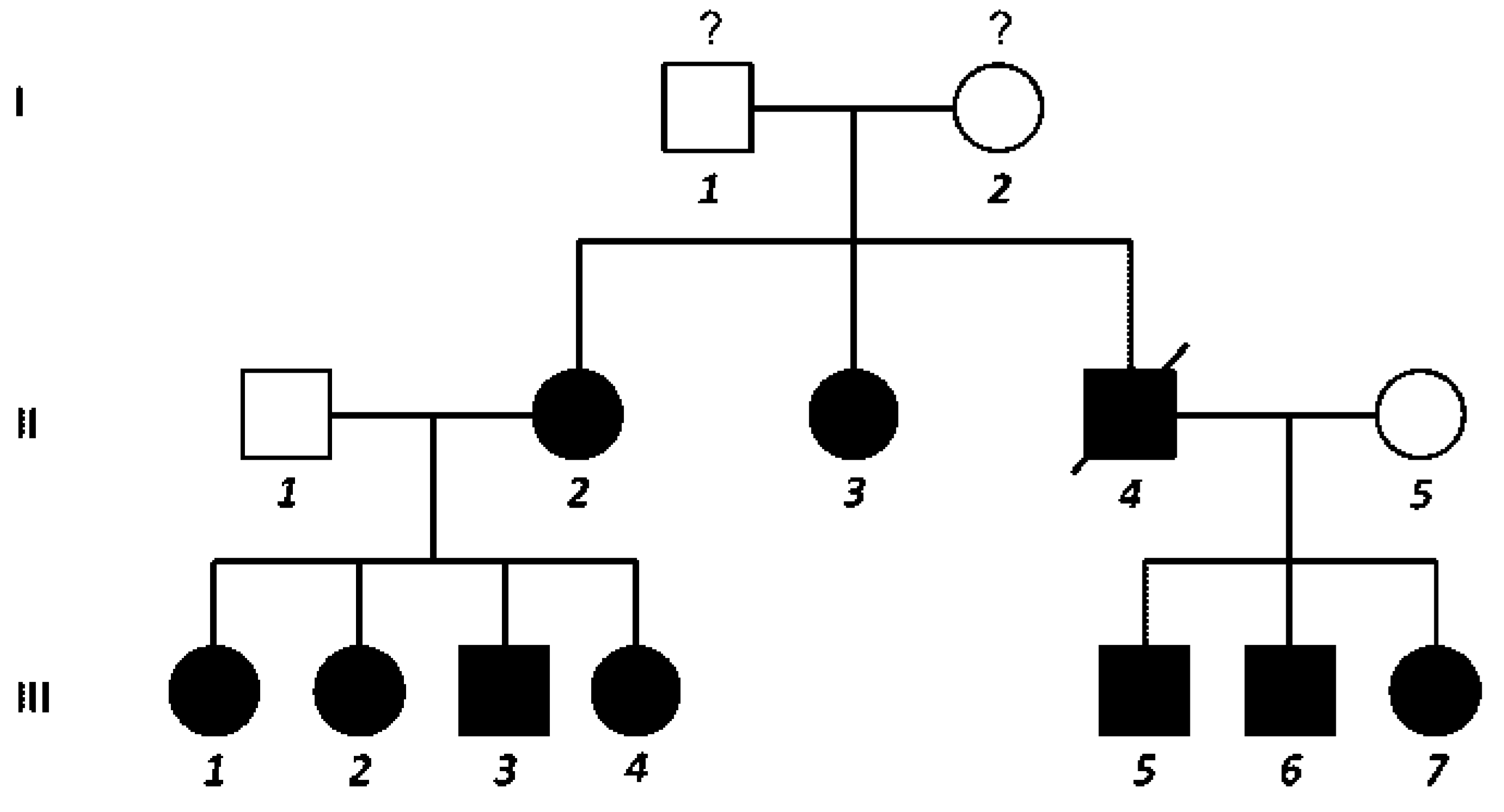
Figure 1. Pedigree of the patient family. Filled symbols indicate affected individuals.
Cmtr 09 00012 g001
Figure 2. Number of families per country affected by NBCCS. NBCCS, nevoid basal cell carcinoma syndrome.
Cmtr 09 00012 g002
Figure 3. Calcification of the falx cerebri (patient II-2).
Cmtr 09 00012 g003
Figure 4. Macrocephaly, hypertelorism, and the presence of multiple basal cell nevi in the face (patient III-3).
Cmtr 09 00012 g004
Figure 5. (a) KCOT in the body of the mandible associated with unerupted tooth (patient III-1). (b) Radiolucent lesion, diagnosed as KCOT, associated with impacted permanent maxillary canine (patient III-2). KCOT, keratocystic odontogenic tumor.
Cmtr 09 00012 g005
Figure 6. Palmar pits (patient II-2).
Cmtr 09 00012 g006
Figure 7. (a) Multiple basal cell carcinomas on the face. (b) Post-PDT treatment. PDT, photodynamic therapy.
Cmtr 09 00012 g007
Figure 8. Calcified ovarian masses are noted in pelvic Rx (patient II-2).
Cmtr 09 00012 g008
Figure 9. Posteroanterior chest radiograph showing a bifid rib (patient II-3).
Cmtr 09 00012 g009
Table 1. Diagnostic criteria for NBCCS.
Cmtr 09 00012 i001
Abbreviations: BCC, basal cell carcinoma; KCOT, keratocystic odontogenic tumor; NBCCS, nevoid basal cell carcinoma syndrome.
Table 2. Characteristics of study patients.
Cmtr 09 00012 i002
Abbreviations: BCC, basal cell carcinoma; KCOT, keratocystic odontogenic tumor; PO, peripheral osteotomy.
Table 3. Frequencies of clinical findings.
Cmtr 09 00012 i003