Circummandibular Wires for Treatment of Dentoalveolar Fractures Adjacent to Edentulous Areas: A Report of Two Cases
Abstract
:1. Case Reports
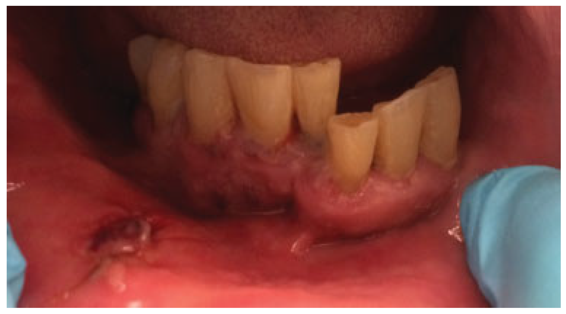
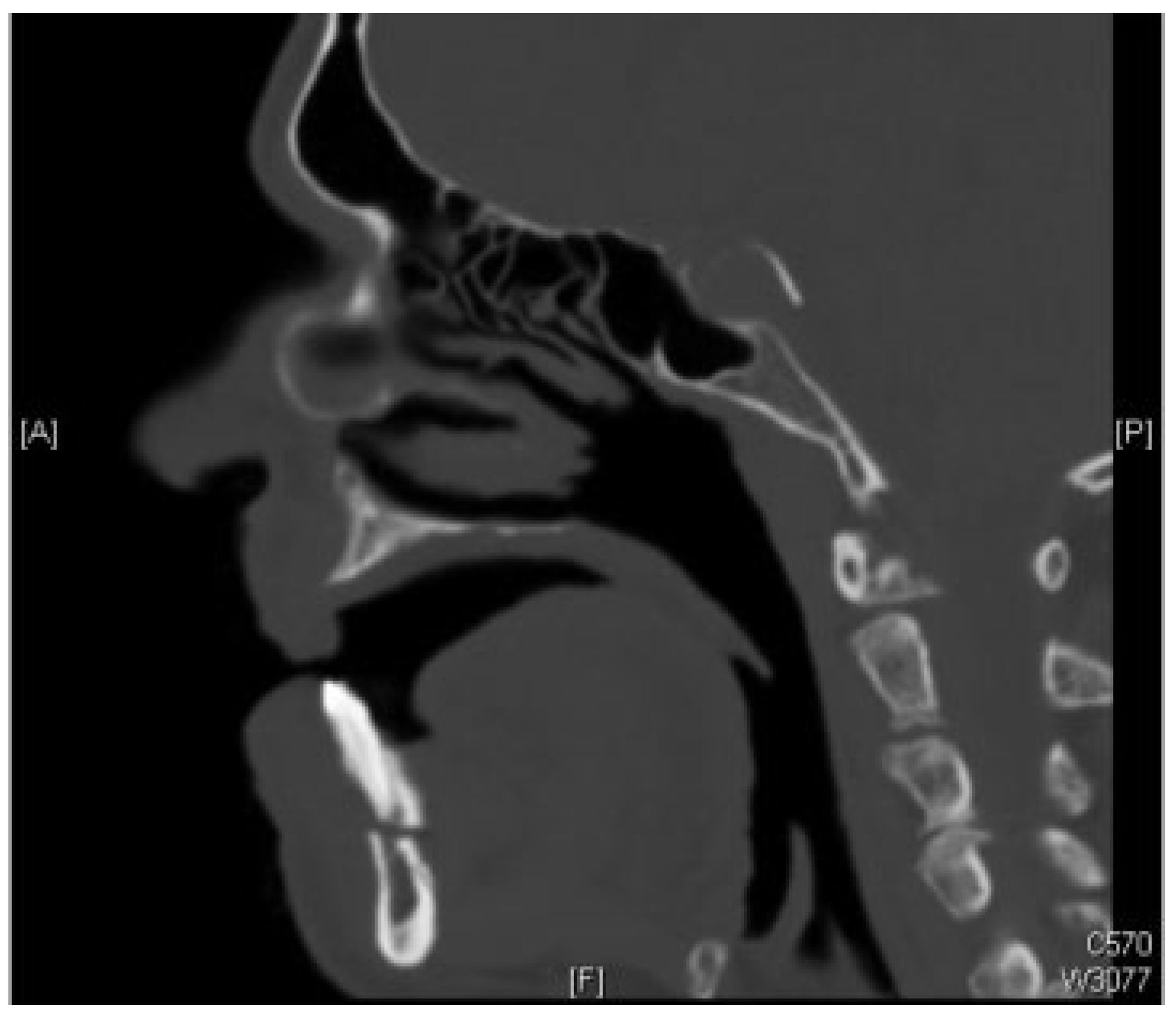
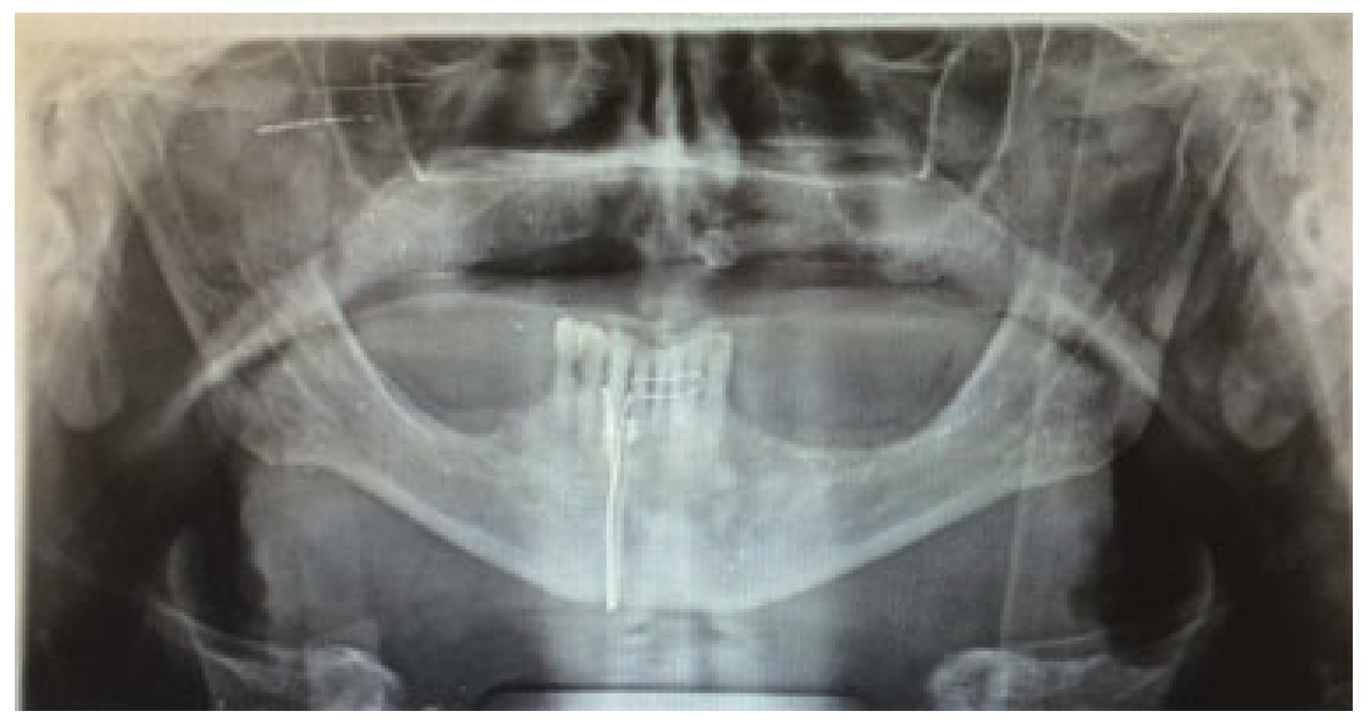
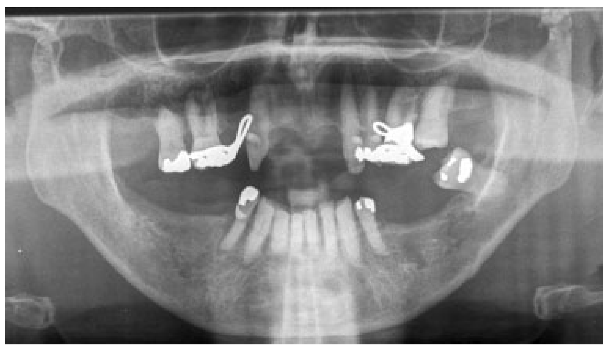
2.1. Case 1
2.2. Case 2
3. Discussion
Conflicts of Interest
Please State Any Sources of Funding for Your Research
References
- Powers, M.P.; Quereshy, F.A. Diagnosis and management of dentoalveolar injuries. In Oral and Maxillofacial Trauma, 2nd ed.; Fonseca, J.R., Walker, R.V., Betts, N.J., Barber, H.D., Eds.; WB Saunders: Philadelphia, PA, USA, 1997; Volume I. [Google Scholar]
- James, D. Maxillofacial injuries in children. In Maxillofacial Injuries, 1st ed.; Rowe, N.L., Williams, J.L.L., Eds.; Churchill Living-stone: Edinburg, UK, 1985. [Google Scholar]
- Lephart, S.M.; Fu, F.H. Emergency treatment of athletic injuries. Dent. Clin. N. Am. 1991, 35, 707–717. [Google Scholar] [CrossRef] [PubMed]
- Heintz, W.D. Mouth protection for athletics today. In The Relationship of Internal Protection Devices to Athletic Injuries and Athletic Performance; Godwin, W.D., Long, B.R., Cartwright, C.B., Eds.; University of Michigan: Ann Arbor, MI, USA, 1982. [Google Scholar]
- Ellis, E., III; Moos, K.F.; el-Attar, A. Ten years of mandibular fractures: an analysis of 2,137 cases. Oral Surg. Oral Med. Oral Pathol. 1985, 59, 120–129. [Google Scholar] [CrossRef] [PubMed]
- Andreasen, J.O. Etiology and pathogenesis of traumatic dental injuries. A clinical study of 1,298 cases. Scand. J. Dent. Res. 1970, 78, 329–342. [Google Scholar] [CrossRef] [PubMed]
- Wright, R.B.; Manfield, F.F. Damage to teeth during the administration of general anaesthesia. Anesth. Analg. 1974, 53, 405–408. [Google Scholar] [CrossRef] [PubMed]
- Star, E.G. Damage of teeth by oral airways. Prakt. Anaesth. 1976, 11, 347–348. (In German) [Google Scholar] [PubMed]
- Andreasen, J.O. Fractures of the alveolar process of the jaw. A clinical and radiographic follow-up study. Scand. J. Dent. Res. 1970, 78, 263–272. [Google Scholar] [CrossRef] [PubMed]
- Kupfer, S.R. Fracture of the maxillary alveolus. Oral Surg. Oral Med. Oral Pathol. 1954, 7, 830–836. [Google Scholar] [CrossRef] [PubMed]
- Bernstein, L.; Keyes, K.S. Dental and alveolar fractures. Otolaryngol. Clin. N. Am. 1972, 5, 273–281. [Google Scholar] [CrossRef]
- Abubaker, A.O.; Papadopoulos, H.; Giglio, J.A. Diagnosis and management of dentoalveolar injuries. In Oral and Maxillofacial Surgery, 2nd ed.; Fonseca, R., Marciani, R., Turvey, T, Eds.; Elsevier: St. Louis, MI, USA, 2009; Volume II. [Google Scholar]
- Andreasen, J.O.; Andreasen, F.M. Textbook and Color Atlas of Traumatic Injuries to the Teeth, 3rd ed.; Munksgaard: Copenhagen, Denmark, 1994. [Google Scholar]
- Kehoe, J.C. Splinting and replantation after traumatic avulsion. J. Am. Dent. Assoc. 1986, 112, 224–230. [Google Scholar] [CrossRef] [PubMed]
- Leathers, R.D.; Gowans, R.E. Management of alveolar and dental fractures. In Peterson’s Principals of Oral and Maxillofacial Surgery, 2nd ed.; Miloro, M., et al., Eds.; B.C. Decker Incorporated: Hamilton, ON, Canada, 2004. [Google Scholar]
- Gilmer, T.L. Lectures on Oral Surgery; Northwestern University Dental School: Chicago, IL, USA, 1901. [Google Scholar]
- Thoma, K.H. Oral Surgery, 3rd ed.; The C.V. Mosby Company: St. Louis, MI, USA, 1958. [Google Scholar]
- Obwegeser, H.L. Die Totale Mundbodenplastik. Schweiz. Mschwr. Zahnheilk 1963, 73, 565–572. [Google Scholar]
- Kushner, G.M.; Tiwana, P.S. Fractures of the growing mandible. Atlas Oral Maxillofac. Surg. Clin. N. Am. 2009, 17, 81–91. [Google Scholar] [CrossRef] [PubMed]
- Schilli, W.; Stoll, P.; Bähr, W.; et al. Mandibular fractures. In Manual of Internal Fixation in the Cranio-Facial Skeleton: Techniques Recommended by the AO/ASIF Maxillofacial Group; Prein, J., Ed.; Springer: New York, NY, USA, 1998. [Google Scholar]
- Novelli, G.; Sconza, C.; Ardito, E.; Bozzetti, A. Surgical treatment of the atrophic mandibular fractures by locked plates systems: our experience and a literature review. Craniomaxillofac. Trauma Reconstr. 2012, 5, 65–74. [Google Scholar] [CrossRef] [PubMed]
- Madsen, M.J.; Kushner, G.M.; Alpert, B. Failed Fixation in Atrophic Mandibular Fractures: The Case against Miniplates. Craniomaxillofac. Trauma Reconstr. 2011, 4, 145–150. [Google Scholar] [CrossRef] [PubMed]


© 2015 by the author. The Author(s) 2015.
Share and Cite
Maloney, K. Circummandibular Wires for Treatment of Dentoalveolar Fractures Adjacent to Edentulous Areas: A Report of Two Cases. Craniomaxillofac. Trauma Reconstr. 2015, 8, 246-250. https://doi.org/10.1055/s-0034-1399801
Maloney K. Circummandibular Wires for Treatment of Dentoalveolar Fractures Adjacent to Edentulous Areas: A Report of Two Cases. Craniomaxillofacial Trauma & Reconstruction. 2015; 8(3):246-250. https://doi.org/10.1055/s-0034-1399801
Chicago/Turabian StyleMaloney, Karl. 2015. "Circummandibular Wires for Treatment of Dentoalveolar Fractures Adjacent to Edentulous Areas: A Report of Two Cases" Craniomaxillofacial Trauma & Reconstruction 8, no. 3: 246-250. https://doi.org/10.1055/s-0034-1399801
APA StyleMaloney, K. (2015). Circummandibular Wires for Treatment of Dentoalveolar Fractures Adjacent to Edentulous Areas: A Report of Two Cases. Craniomaxillofacial Trauma & Reconstruction, 8(3), 246-250. https://doi.org/10.1055/s-0034-1399801