Next Article in Journal
Condylar Osteochondroma Treated with Total Condylectomy and Preservation of the Articular Disc: A Case Report
Previous Article in Journal
Remarkable Triple Pleomorphic Adenoma Affecting Both Parotid and Submandibular Glands
 
 
Craniomaxillofacial Trauma & Reconstruction is published by MDPI from Volume 18 Issue 1 (2025). Previous articles were published by another publisher in Open Access under a CC-BY (or CC-BY-NC-ND) licence, and they are hosted by MDPI on mdpi.com as a courtesy and upon agreement with Sage.
Case Report

Ellis–van Creveld Syndrome with Sagittal Craniosynostosis

by
Andrew S. Fischer
1,
William M. Weathers
1,
Erik M. Wolfswinkel
1,
Robert J. Bollo
2,
Larry H. Hollier, Jr.
1 and
Edward P. Buchanan
1,*
1
Division of Plastic Surgery, Michael E. Debakey Department of Surgery, Baylor College of Medicine, Houston, TX 77030, USA
2
Department of Neurosurgery, Baylor College of Medicine, Houston, TX, USA
*
Author to whom correspondence should be addressed.
Craniomaxillofac. Trauma Reconstr. 2015, 8(2), 132-135; https://doi.org/10.1055/s-0034-1393733
Submission received: 19 February 2014 / Revised: 29 March 2014 / Accepted: 29 March 2014 / Published: 27 October 2014

Abstract

:
Ellis–van Creveld syndrome (EVC) is a rare disorder (the incidence is estimated at around 7/1,000,000) characterized by the clinical tetrad of chondrodystrophy, polydactyly, ectodermal dysplasia, and cardiac anomalies. Sagittal synostosis is characterized by a dolichocephalic head shape resulting from premature fusion of the sagittal suture. Both are rare disorders, which have never been reported together. We present a case of EVC and sagittal synostosis. We report the clinical features of a Hispanic boy with EVC and sagittal craniosynostosis who underwent cranial vault remodeling. The presentation of this patient is gone over in detail. A never before reported case of EVC and sagittal synostosis is presented in detail.

Ellis–van Creveld syndrome (EVC) is an inherited, autosomal recessive disorder characterized by the clinical tetrad of chondrodystrophy, polydactyly, ectodermal dysplasia, and cardiac anomalies.[1,2,3] Affected individuals manifest clinically with disproportionate short-limb dwarfism, short ribs, postaxial polydactyly, and dysplastic nails and teeth. Cardiac defects occur in approximately 50 to 60% of cases and often involve the atrial septum.[2,4] Cognitive and motor development is unaffected.
Richard W. B. Ellis of Edinburgh and Simon van Creveld of Amsterdam first described the condition in 1940.[1] Since then, only 300 cases reported as of 2000.[5] The incidence is estimated at around 7/1,000,000.[2] The condition is more common among the American Amish, but it is known to affect all races with equal sex predilection.[2,3] Consanguinity is implicated in approximately one-third of the cases.[5] EVC has been linked to mutations in the EVC1 and EVC2 genes on the short arm of chromosome 4.[5] Individuals with mutations in either EVC1 or EVC2 are phenotypically indistinguishable, and one-third of individuals clinically diagnosed with EVC are not found to have either mutation.[6,7] Definitive diagnosis is made based on mutations in EVC1 and EVC2.
Sagittal craniosynostosis is characterized by the premature fusion of the sagittal suture. Growth is inhibited perpendicular to the sagittal suture, leading to dolichocephaly. Sagittal craniosynostosis occurs in approximately 1/2,000 to 1/7,000 births, and is the most common craniosynstosis.[8,9]
The purpose of this article is to present a case of EVC and sagittal synostosis, the combination of which has never been reported in the literature.

Case Report

A Hispanic male was found to have polydactyly on prenatal ultrasound in a mother with polyhydramnios. A subsequent fetal MRI showed body wall edema, mesomelic shortening and mild bowing of the long bones, bilateral postaxial polydactyly, short ribs, premature ossification of the femoral epiphysis, a dolichocephalic skull, and a possible lumbosacral vertebral anomaly. Fetal echo showed mild right ventricular hypertrophy and dilation with systolic function. Amniocentesis was done for amnioreduction and genetic testing at 33 weeks. Routine karyotype was 46,XY, normal male. The chromosomal microarray analysis was normal, but the whole-exome sequencing revealed a heterozygous mutation in the IFT140 gene. The mother’s medications included prenatal vitamins and steroids before amnioreduction.
The patient’s older sister carries a clinical diagnosis of EVC. She underwent sequencing and deletion/duplication analysis, and no mutation was found in EVC1 or EVC2, which can occur in one-third of cases. A third-generation pedigree was collected and there was no known consanguinity—both the mother and father’s families were from the same region of Mexico but from different cities.
At birth, the patient was diffusely edematous, particularly pronounced in the lower face and eyes, with a dolichocephalic head, small anterior fontanelle, narrow temporal region, and overriding sutures. Ears were pointed, low set, and posteriorly rotated. Right ear measured in the 3rd percentile at 3 cm. There was unfolding of the helices bilaterally with attached ear lobes. The palate was intact with a posteriorly set tongue. Excessive nuchal skin was noted. The nipples were widely spaced at 12 cm. The right hand, palm, and finger all measured approximately in the 3rd percentile (5.25, 3.25, and 2 cm, respectively), consistent with disproportionate dwarfism. Upper and lower extremities showed a predominance of rhizomelic to mesomelic shortening and excessive skin folds. Postaxial polydactyly of the upper and lower extremities was present. Hypoplastic nails of the toes bilaterally were seen as well. Extremities exhibited decreased tone.
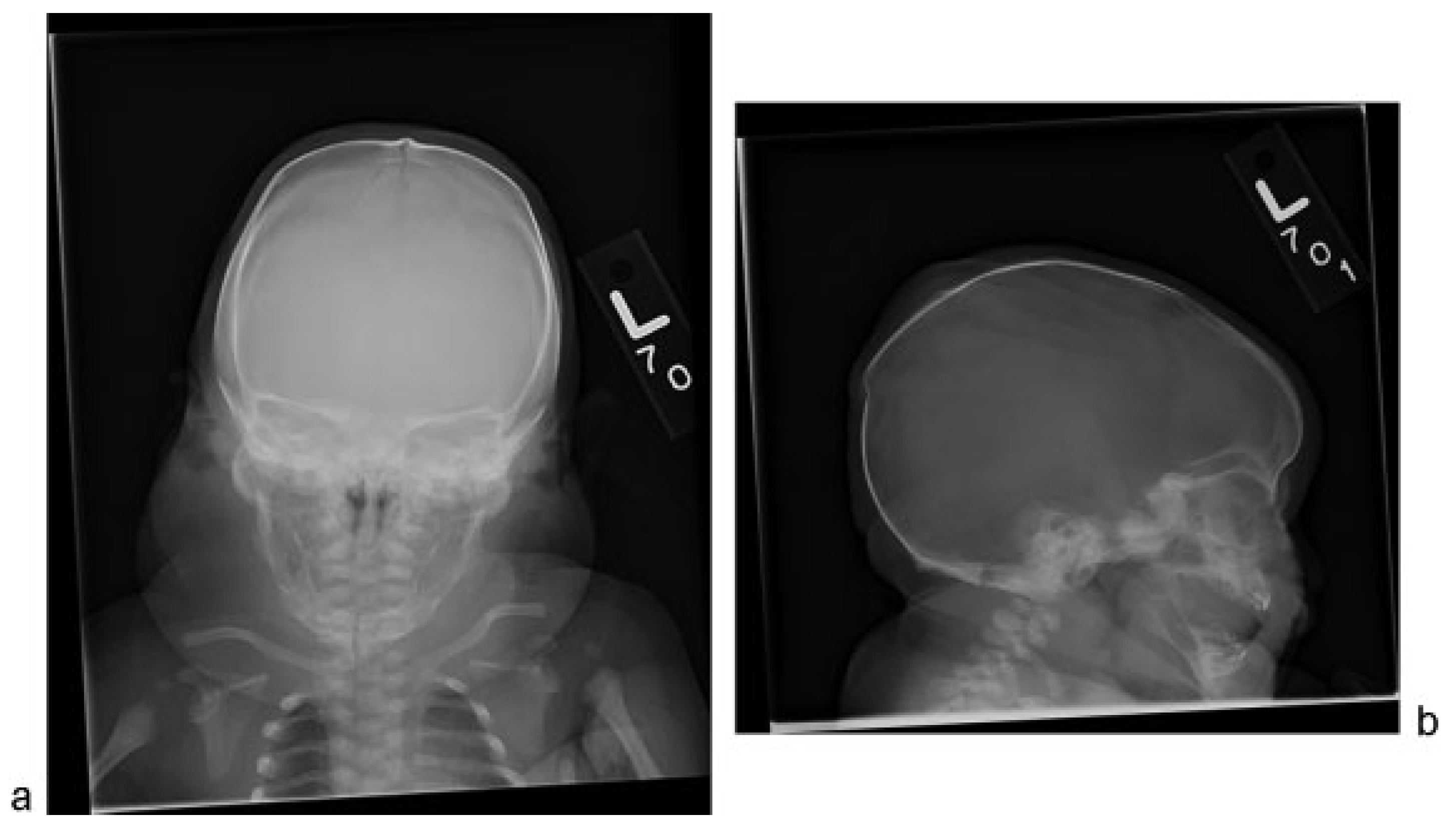
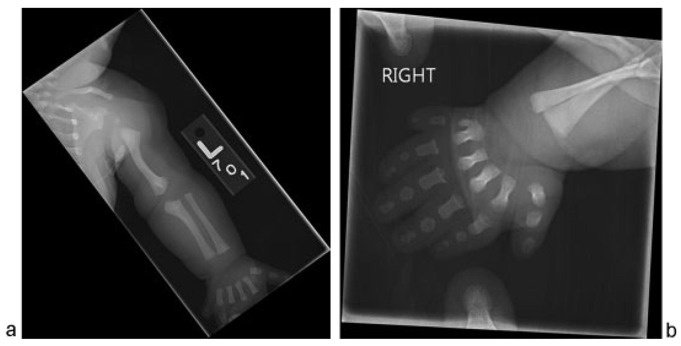
Skeletal survey confirmed sagittal craniosynostosis and a posterior parietal buckle fracture (Figure 1). Shortening and mild angular deformities of the bilateral ribs were noted. Upper extremities showed bilateral valgus deformities of the humeri, varus deformities of the proximal ulnas, radial head dislocations, and postaxial polydactyly exhibiting fusion at the base of the fifth and sixth metacarpals (Figure 2). Lower extremities showed bilateral mild varus deformities of the femurs, meso-melic shortening, relative overgrowth of the fibulas, and mild brachydactyly (Figure 3). An echocardiogram confirmed a moderately dilated and hypertrophied right ventricle with normal systolic function and revealed a small patent ductus arteriosus and a small secundum atrial septal defect.
The patient underwent surgery for cranial vault remodeling at 9 months to correct the scaphocephalic deformity. Preoperative computed tomographic scan demonstrated mild cervical kyphosis and a hypoplastic dens in addition to shortened mandibular rami and scaphocephaly with complete fusion of the anterior suture and frontal bossing (Figure 4). Thorax was narrowed as well as the cervical vertebral bodies with a more focal narrowing at the level of C1 secondary to an unfused Kerckring ossicle. The patient underwent middle posterior cranial vault remodeling. A sagittal craniectomy and multiple parietal barrel stave osteotomies were performed via the modified Pi technique for sagittal suture craniosynostosis. The patient tolerated surgery well without complications.

Discussion

EVC is an autosomal recessive skeletal dysplasia. Recently, EVC has been included in a group of human genetic disorders known as ciliopathies characterized by a dysfunctional molecular mechanism in the primary cilia.[10] Mutations in the EVC1 and EVC2 genes have been found in approximately two-thirds of the EVC cases.[5,6] No known mutation has been identified in the other one-third.[7] In this case, the patient and his older sister both carried a clinical diagnosis of the EVC, without mutation in either EVC1 or EVC2.
The cardinal features of EVC—short-limb dwarfism, polydactyly, ectodermal dysplasia of the nails, hair, and teeth, and congenital heart defects—are all present in our patient.[1,2,3]
Polydactyly of the ulnar side of the hands is seen in all individuals with EVC. Polydactyly of the feet, a condition reported in only 10% of EVC cases, was also present in our patient.[11] Cardiac anomalies are reported in roughly half of EVC cases, most commonly involving the atrium.[2,4] Cardiac defects are associated with a higher morbidity in the EVC.[12] Sagittal craniosynostosis is not known to be associated with EVC, and this case appears to be the first report of sagittal synostosis in a patient with EVC. It was first believed that this case may have been a presentation of cranioectodermal dysplasia. Cranioectodermal dysplasia is a rare developmental disorder that resembles EVC. Affected individuals often have pronounced dolichocephaly with sagittal suture synostosis, narrowed thorax, ciliopathy, rhizomelic shortening of the long bones, and congenital heart disease. However, polydactyly, as was seen in this patient, is not characteristic of this disorder.[13] It is possible that this case represents a variant of cranioectodermal dysplasia with polydactyly, but given the sister’s clinical diagnosis of EVC it was considered more consistent with EVC. Cranioectodermal dysplasia and EVC have significant overlap in their phenotypes and can be difficult to distinguish. This is the first known case of EVC and sagittal synostosis.

Products/Devices/Drugs

None.

Financial Disclosures/Commercial Associations

None.

References

  1. Ellis, R.W.; van Creveld, S. A syndrome characterized by ectodermal dysplasia, polydactyly, chondro-dysplasia and congenital morbius cordis: Report of three cases. Arch Dis Child 1940, 15, 65–84. [Google Scholar] [CrossRef] [PubMed]
  2. McKusick, V.A.; Egeland, J.A.; Eldridge, R.; Krusen, D.E. Dwarfism in the Amish I. The Ellis-van Creveld syndrome. Bull Johns Hopkins Hosp 1964, 115, 306–336. [Google Scholar] [PubMed]
  3. McKusick, V.A. Ellis-van Creveld syndrome and the Amish. Nat Genet 2000, 24, 203–204. [Google Scholar] [CrossRef] [PubMed]
  4. Digilio, M.C.; Marino, B.; Ammirati, A.; Borzaga, U.; Giannotti, A.; Dallapiccola, B. Cardiac malformations in patients with oral-facialskeletal syndromes: Clinical similarities with heterotaxia. Am J Med Genet 1999, 84, 350–356. [Google Scholar] [CrossRef]
  5. Ruiz-Perez, V.L.; Ide, S.E.; Strom, T.M.; et al. Mutations in a new gene in Ellis-van Creveld syndrome and Weyers acrodental dysostosis. Nat Genet 2000, 24, 283–286. [Google Scholar] [CrossRef] [PubMed]
  6. Ruiz-Perez, V.L.; Tompson, S.W.; Blair, H.J.; et al. Mutations in two nonhomologous genes in a head-to-head configuration cause Ellis-van Creveld syndrome. Am J Hum Genet 2003, 72, 728–732. [Google Scholar] [CrossRef] [PubMed]
  7. Tompson, S.W.; Ruiz-Perez, V.L.; Blair, H.J.; et al. Sequencing EVC and EVC2 identifies mutations in two-thirds of Ellis-van Creveld syndrome patients. Hum Genet 2007, 120, 663–670. [Google Scholar] [CrossRef] [PubMed]
  8. Fearon, J.A.; McLaughlin, E.B.; Kolar, J.C. Sagittal craniosynostosis: Surgical outcomes and long-term growth. Plast Reconstr Surg 2006, 117, 532–541. [Google Scholar] [CrossRef] [PubMed]
  9. Tatum, S.A.; Jones, L.R.; Cho, M.; Sandhu, R.S. Differential management of scaphocephaly. Laryngoscope 2012, 122, 246–253. [Google Scholar] [CrossRef] [PubMed]
  10. Badano, J.L.; Mitsuma, N.; Beales, P.L.; Katsanis, N. The ciliopathies: An emerging class of human genetic disorders. Annu Rev Genomics Hum Genet 2006, 7, 125–148. [Google Scholar] [CrossRef] [PubMed]
  11. Bhat, Y.J.; Baba, A.N.; Manzoor, S.; Qayoom, S.; Javed, S.; Ajaz, H. Ellis-van Creveld syndrome with facial hemiatrophy. Indian J Dermatol Venereol Leprol 2010, 76, 266–269. [Google Scholar] [CrossRef] [PubMed]
  12. Baujat, G.; Le Merrer, M. Ellis-van Creveld syndrome. Orphanet J Rare Dis 2007, 2, 27. [Google Scholar] [CrossRef] [PubMed]
  13. Young, I.D. Cranioectodermal dysplasia (Sensenbrenner’s syndrome). J Med Genet 1989, 26, 393–396. [Google Scholar] [CrossRef] [PubMed]
Figure 1. (a) Frontal view of skull demonstrating sagittal suture ridging, significant soft tissue swelling about the neck and short, and angular ribs. (b) Lateral view of the skull shows dolichocephalic head shape and a posterior parietal buckle fracture.
Cmtr 08 i2f132 g001
Figure 2. Skeletal survey. (a) Left upper extremity demonstrates valgus deformity of the humerus, varus deformity of the proximal ulna, radial head dislocation and postaxial polydactyly, similar findings were seen on the right. (b) Right hand demonstrates postaxial polydactyly with fusion at the base of the 5th and 6th metacarpals.
Cmtr 08 i2f132 g002
Figure 3. Radiograph of the right leg demonstrates mild varus deformity of the femur, mesomelic shortening, relative overgrowth of the fibula and mild brachydactyly, similar findings on the left.
Cmtr 08 i2f132 g003
Figure 4. Preoperative computed tomographic three-dimensional reconstruction (a) top-down view demonstrates anterior sagittal suture fusion. (b) oblique view demonstrates frontal bossing.
Cmtr 08 i2f132 g004