Abstract
The objective of this article is to present the first case reported in the literature of metachronous pleomorphic adenoma of bilateral parotid glands and submaxillary gland. The authors report the case of a 27-year-old female with metachronous mixed tumors in her right parotid and submandibular glands. The patient has no history of previous radiotherapy. All three lesions were diagnosed by fine-needle aspiration. The histopathologic evaluation of all three major salivary gland masses demonstrated pleomorphic adenomas, with no occult malignancy observed on serial sections. The presentation of pleomorphic adenomas in the parotids and submandibular glands probably represents three unrelated primary sites of tumor, yet the possibility of metastasis from one gland to the other cannot be excluded.
Multiple salivary gland tumors are extremely uncommmon.[1] Although the most usual tumor with bilateral development is the Warthin tumor, pleomorphic adenomas have been reported as occurring synchronously and metachronously within major salivary glands,[2,3,4,5] but occurrence in both parotid gland and submandibular gland is extremely rare, and in fact, as far as the authors know there is no previous reported cases in the international literature of mixed tumor of bilateral parotid and submaxillary gland not as multiple foci of the same disease.
We report the case of a female patient with metachronous bilateral mixed tumors in her right and left parotid and right submandibular glands.
Materials and Methods
At the first time of diagnosis in May 2005, a 22-year-old female presented with a 1-year history of a right preauricular mass. Past medical history and family medical records were unremarkable. No toxic habits.
On head and neck exam, a mildly tender preauricular mass of 2 cm diameter was identified with cutaneous overlying pathology. There was no associated facial weakness, paresthesia, or otalgia. The patient denied fever, otalgia, or otorrhea. Intraoral pathology or trismus was absent.
Following further investigation with fine-needle aspiration (FNA) cytology, the lesion was found to be consistent with a pleomorphic adenoma. Complete excision as an extracapsular dissection followed.
The follow-up was uneventful but 2 years later in October 2007, she presented complaining of swelling slightly painful on the right side of his neck. FNA cytology of the cervical mass suggested mixed tumor of submaxillary gland. Computed tomography (CT) revealed postsurgical changes in right parotid region without suggestive signs of recurrence. In right submaxillary cell, a well-defined mass of 2 cm surrounded by a fibrous pseudocapsule of varying appears (Figure 1). The patient was operated and discharged from hospital 2 days later. The final diagnosis was pleomorphic adenoma of submaxillary gland. Histopathological investigation reported that the lesion had typical features of pleomorphic adenomas with ducts and cords of cells lying within chondromyxoid stroma.
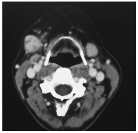
Figure 1.
In right submaxillary cell, a well-defined mass of 2 cm surrounded by a fibrous pseudocapsule of varying appears.
After 4 years of follow-up, the patient remains in good health but in February 2012, she reported a small node in contralateral retromandibular area.
The mass was asymptomatic, and their size did not change during meals. On clinical examination,a firm well-defined mass was palpated measuring 2 cm diameter and with no fixation to the adjacent anatomic structures. FNA examination revealed for third time that the mass was pleomorphic adenoma of left parotid gland. CT scan detected a unique left parotid lesion, well defined of 1.2 × 1.8 cm of glandular tissue (Figure 2).
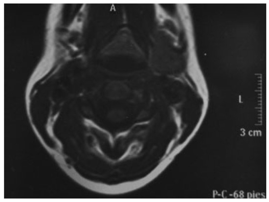
Figure 2.
Left parotid tumor of 12 × 18 mm. Postsurgical changes in right parotid gland.
Intraparotid nodes encountered appeared normal and were negative for malignancy on frozen analysis.
After superficial left parotidectomy, the mass was submitted for histologic analysis with final diagnosis of pleomorphic adenoma (Figure 3).
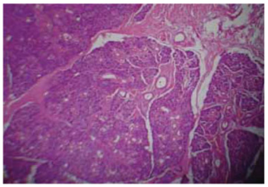
Figure 3.
Histologic analysis after left superficial parotidectomy: epithelial elements are arranged in a variable background myxoid stroma and duct-like structures that consist of stellate-shaped cells of squamous metaplasia.
One-year follow-up was successful with no radiological signs of recurrence and asymptomatic. No Frey syndrome or facial weakness was developed. The patient will go on in close long-term follow-up.
Discussion
The most likely etiologic factor associated with the development of two pleomorphic adenomas in the parotid and submandibular glands is the irradiation delivered to the head and neck region in childhood. Irradiation of the head and neck is a well-established predisposing condition that increases the risk for developing subsequent salivary neoplasms. This was particularly noteworthy as an increased incidence of salivary gland tumors after the atomic bomb explosions in Japan.[6]
The exposure effects have been reported to be both cumulative and dose dependent.[6]
Yu et al reported that 69.6% of the multiple salivary gland tumors of their series were synchronous, affecting men six times more than women.[7]
An interesting question is the chronology of the tumor development, which is ultimately linked to its diagnosis. Usually, the patient identifies the mass regarding the dimension and the location of the tumor (superficial or deep lobe of the gland), and overall, depending the presence or not of symptoms.
Early tumors located in the salivary gland deep lobe could pass unperceived if the dimension is small and symptoms are absent. In the absence of clinical evidence, imaging analyses of contralateral glands are not routinely used, which explains why there is no detailed information regarding the development of bilateral tumors.[8]
In fact, the present case report is an interesting example of this situation. In the first appointment, the patient presented with a visible well-defined growth on the right parotid gland. At that time, no swelling or superficial alterations on the right submaxillary area or left parotid gland could be observed by inspection or palpation.
These aspects explain the absence of any kind of radiologic investigation for the lesions at that time, once the cytologic diagnosis was obtained by FNA. Right parotid tumor was then assumed to be a unilateral tumor. Because of this, the metachronous designation was admitted when an ipsilateral lesion was identified in the submaxillary gland. Nevertheless, an ultrasonography, CT, or magnetic resonance imaging examination at the first time of presentation could have shown a smaller lesion deeply seated in the right submaxillary parenchyma or even in left parotid gland that was not perceptible on palpation. Thus, although the present case was considered metachronous based on the chronological identification of the clinical signs of the lesions, this case report also highlights the need for careful evaluation with the use of imaging investigation to detect early and unsuspected lesions. In conclusion, the present report describes a remarkable case. Presentation of pleomorphic adenomas in the parotids and submandibular glands probably represents two unrelated primary sites of tumor, yet the possibility of metastasis from one gland to the other cannot be excluded. Further work to understand the behavior of salivary gland tumors more fully would be an interesting area for future research.
Funding
No financial disclosures.
Conflicts of Interest
None.
References
- Bradley, P.J. Pleomorphic salivary adenoma of the parotid gland: which operation to perform? Curr Opin Otolaryngol Head Neck Surg 2004, 12, 69–70. [Google Scholar] [CrossRef] [PubMed]
- Keldahl, M.L.; Zarif, A.; Gattuso, P. Bilateral synchronous pleomorphic adenoma diagnosed by fine-needle aspiration. Diagn Cytopathol 2004, 30, 356–358. [Google Scholar] [CrossRef] [PubMed]
- Nagler, R.M.; Laufer, D. Synchronous pleomorphic adenomas of the major salivary glands: a case report. Oral Surg Oral Med Oral Pathol Oral Radiol Endod 1999, 87, 735–737. [Google Scholar] [CrossRef] [PubMed]
- Pistorio, V.; Teggi, R.; Bussi, M. Simultaneous pleomorphic adenoma of the parapharyngeal space and contralateral submandibular gland. Case report. Acta Otorhinolaryngol Ital 2008, 28, 257–260. [Google Scholar] [PubMed]
- Silva, S.J.; Costa Junior, G.T.; Brant Filho, A.C.; Faria, P.R.; Loyola, A.M. Metachronous bilateral pleomorphic adenoma of the parotid gland. Oral Surg Oral Med Oral Pathol Oral Radiol Endod 2006, 101, 333–338. [Google Scholar] [CrossRef] [PubMed]
- Yajin, K.; Harada, Y.; Omura, R.; et al. Simultaneous pleomorphic adenomas of the right parotid and ipsilateral submandibular glands. Auris Nasus Larynx 1987, 14, 47–55. [Google Scholar] [CrossRef] [PubMed]
- Yu, G.Y.; Ma, D.Q.; Zhang, Y.; et al. Multiple primary tumours of the parotid gland. Int J Oral Maxillofac Surg 2004, 33, 531–534. [Google Scholar] [CrossRef] [PubMed]
- Suzuki, S.; Okamura, H.; Ohtani, I. Bilateral parotid gland basal cell adenomas. Case report. ORL J Otorhinolaryngol Relat Spec 2000, 62, 278–281. [Google Scholar] [CrossRef] [PubMed]
© 2014 by the author. The Author(s) 2014.