Next Article in Journal
Spontaneous Bone Healing After Cysts Enucleation Without Bone Grafting Materials: A Randomized Clinical Study
Previous Article in Journal
Fracture of the Coronoid Process Associated with Frontosphenoidal Fractures
 
 
Craniomaxillofacial Trauma & Reconstruction is published by MDPI from Volume 18 Issue 1 (2025). Previous articles were published by another publisher in Open Access under a CC-BY (or CC-BY-NC-ND) licence, and they are hosted by MDPI on mdpi.com as a courtesy and upon agreement with Sage.
Review

Facial Nerve Trauma: Evaluation and Considerations in Management

by
Eli Gordin
1,
Thomas S. Lee
2,
Yadranko Ducic
3,4,* and
Demetri Arnaoutakis
4
1
Department of Otolaryngology–Head and Neck Surgery, SUNY Downstate Medical Center, Brooklyn, NY, USA
2
Department of Otolaryngology–Head and Neck Surgery, Virginia Commonwealth University Medical Center, Richmond, VA, USA
3
Otolaryngology and Facial Plastic Surgery Associates, Fort Worth, TX 76104, USA
4
Department of Otolaryngology–Head and Neck Surgery, University of Texas Southwestern Medical Center, Dallas, TX, USA
*
Author to whom correspondence should be addressed.
Craniomaxillofac. Trauma Reconstr. 2015, 8(1), 1-13; https://doi.org/10.1055/s-0034-1372522
Published: 8 December 2014

Abstract

:
The management of facial paralysis continues to evolve. Understanding the facial nerve anatomy and the different methods of evaluating the degree of facial nerve injury are crucial for successful management. When the facial nerve is transected, direct coaptation leads to the best outcome, followed by interpositional nerve grafting. In cases where motor end plates are still intact but a primary repair or graft is not feasible, a nerve transfer should be employed. When complete muscle atrophy has occurred, regional muscle transfer or free flap reconstruction is an option. When dynamic reanimation cannot be undertaken, static procedures offer some benefit. Adjunctive tools such as botulinum toxin injection and biofeedback can be helpful. Several new treatment modalities lie on the horizon which hold potential to alter the current treatment algorithm.

Facial paralysis can be a devastating consequence resulting from blunt and penetrating trauma to the head and neck, as well as surgical injury, either accidental or due to involvement by tumor. In addition, the etiology can be attributed to a variety of other causes, ranging from infectious to metabolic and is frequently idiopathic in nature. Treatment of the injured intratemporal facial nerve is historically controversial. Reanimation of the paralyzed face is an interesting and frequently evolving field. In this review, we will discuss the anatomy of the facial nerve, the etiology of facial nerve injury, the management of the traumatized facial nerve, and the assessment and treatment strategies for the patient suffering from facial paralysis.

Anatomy

The course of the facial nerve is divided into six segments (Figure 1). Originating within the pons in the facial nucleus, the motor fibers of cranial nerve VII are joined by those of the nervus intermedius before entering the temporal bone through the internal auditory meatus. The narrowest segment of the intratemporal facial nerve, the labyrinthine segment, extends from the internal auditory meatus to the geniculate ganglion, where the cell bodies of special visceral afferent neurons carrying taste from the anterior two-thirds of the tongue are located. It is at the geniculate where the nervus intermedius joins the facial nerve proper, and the greater superficial petrosal and lesser petrosal nerves exit the facial nerve.
The tympanic segment of the nerve extends posteriorly from the geniculate ganglion to the second genu, where the facial nerve turns inferiorly, transitioning to the vertical or mastoid segment. Bony dehiscence is most common in the tympanic segment, in the area immediately adjacent to the oval window. The nerve to the stapedius and the chorda tympani can be found exiting the vertical segment. The nerve exits the temporal bone through the stylomastoid foramen, yielding the extratemporal portion of the nerve, which proceeds to innervate all muscles of facial expression from their deep surfaces except for the levator anguli oris, buccinator, and mentalis, which are innervated from their superficial aspects.[1]
Surgical landmarks that aid in identification of the main trunk of the facial nerve are the tympanomastoid suture, the tragal pointer, the posterior belly of the digastric muscle, and the styloid process. The tympanomastoid suture line is located approximately 6 to 8 mm lateral to the stylomastoid foramen. The nerve lies inferior and medial to the tragal pointer, and its depth can be approximated by that of the posterior belly of the digastric muscle, lying lateral to the styloid process.
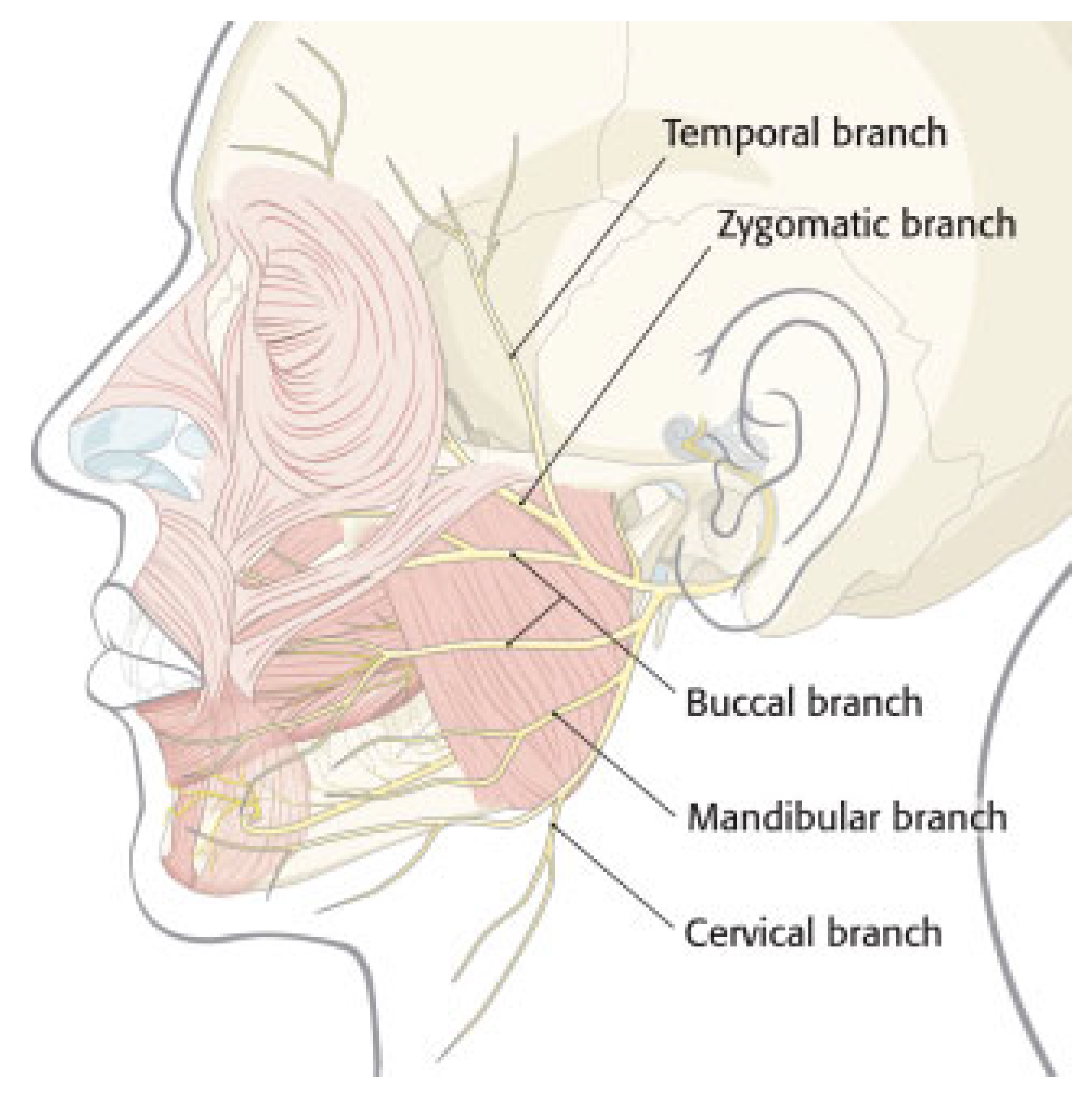
The facial nerve then enters the parotid gland, where the main trunk branches into the upper and lower divisions at the pes anserinus (Figure 2). The nerve further divides into five main branches: the temporal, zygomatic, buccal, marginal mandibular, and cervical.
The temporal branch lies within the superficial muscular aponeurotic system (SMAS) at the level of the zygomatic arch. It usually consists of several branches rather than one lone nerve, the anterior most of which can be approximated at a point midway between the anterior aspect of the zygomatic arch and the helical root.[2] The marginal mandibular branch usually lies deep to the layer of the platysma and SMAS along the body of the mandible, or within 1 to 2 cm of its inferior border.

Etiology

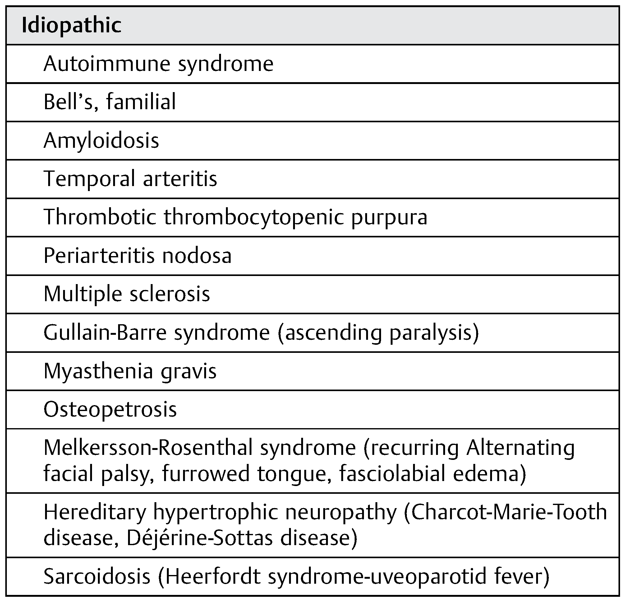
Facial paralysis can result from a wide range of causes. A list of causes can be found in Table 1.

Consequences

Facial paralysis can result in significant disfigurement, having severe implications for both the patient’s emotional and physical well-being. Ophthalmologic consequences include diminished effectiveness of lacrimation, brow ptosis, ectropion, epiphora, and lagophthalmos. These may lead to corneal damage from exposure keratopathy, potentially proceeding to blindness or even globe rupture.[3,4] Loss of muscular support to the nasal valve may lead to nasal obstruction. Ineffective contraction of the perioral musculature can result in insufficient oral competence, poor swallowing function, dysarthria, and ptyalism.[5]

Assessment

Precise characterization of facial nerve paralysis must be accomplished to provide proper patient counseling regarding prognosis and treatment. The House–Brackmann 6-point scale of facial nerve function is the most commonly used standardized tool for assessing the degree of facial weakness.[6,7] This scale is deficient, however, in terms of characterizing facial paralysis localized to one particular facial distribution. Other scales include the Terzis-Noah, Burres-Fisch, Nottingham, and Sunnybrook.[8,9,10,11]
Careful assessment and documentation of facial nerve function should include a detailed description of the status of motion of the upper, middle, and lower face. Special attention should be paid to the eye not only in terms of eyelid closure, lower lid laxity, and brow height but also to visual acuity, presence of Bell phenomenon, and corneal irritation. Aside from overall symmetry, other important functional considerations include nasal valve collapse and oral competence.
Injury to the intratemporal portion of the facial nerve can result in total facial paralysis, as well as dysgeusia. Damage to the geniculate and proximal to this region can lead to decreased lacrimation due to injury to the preganglionic parasympathetic fibers that supply the lacrimal gland carried in the greater superficial petrosal nerve. Damage to the nerve proximal to the take-off of the nerve to the stapedius can result in hyperacusis in the affected ear.

Nerve Function Testing

Electroneurography (ENog) and electromyography (EMG) are the mainstays of facial nerve testing. ENog is an objective test that measures evoked compound muscle action potentials using skin electrodes. Nerve injury is expressed as percentage of function relative to the normal side. When ENog is undetectable, EMG testing can be attempted.
EMG measures voluntary muscle response with needle electrodes placed in the target muscles that detect action potentials during muscle contraction with a functioning nerve. Muscle that has been denervated displays fibrillation potentials, while muscle that is in the process of reinnervation demonstrates polyphasic potentials.[12] Electrical silence in a patient with long standing facial paralysis indicates severe muscle atrophy and degradation of motor end plates.
Nerve conduction studies are best performed at least 10 days after injury to allow for Wallerian degeneration of axons to occur to avoid confounding results.[13] EMG cannot reliably ascertain the severity of nerve injury before this time. [12,14]

Intratemporal Facial Nerve Trauma

Epidemiology and Classification

The facial nerve is affected in 7 to 10% of temporal bone fractures. [15,16,17] These injuries typically occur from falls, motor vehicle accidents, and assault, as well penetrating trauma, such as gunshot wounds. Temporal bone fractures are traditionally classified into longitudinal, transverse, or mixed types, depending on their orientation relative to the petrous ridge.[18,19] New classification schemes designate temporal bone fractures as violating or sparing the otic capsule and may be more predictive of clinical outcomes.[20,21]
Approximately 70 to 80% of fractures can be classified as longitudinal, resulting from a temporoparietal impact; 10 to 30% transverse, resulting from an frontal or occipital injury; and 0 to 20% are mixed in nature.[22,23] Facial nerve involvement is seen in anywhere from 10 to 25% of longitudinal fractures and is more common in transverse fractures, occurring in 38 to 50% of cases.[21,24]

Timing of Paralysis

When the facial nerve is involved, the ensuing paralysis is immediate in 27% of cases and has a delayed presentation in 73% of patients.[16] Most commonly, injury is localized to the perigeniculate region or, less commonly, the second genu.[13] Paresis was noted in 23% of cases in Darrouzet’s series of 115 cases, immediate complete paralysis in approximately 51% of cases, and delayed complete paralysis in approximately 15%.[13] In this study, the onset of facial paralysis was indeterminate in approximately 11% of patients.

Surgical Indications

Immediate complete paralysis warrants surgical exploration. Cases of complete paralysis in which the onset of paralysis is indeterminate should be treated as immediate in nature.[13,16] Delayed paralysis or incomplete paresis should be treated medically, with high-dose steroids.[13,20,24] A good prognosis should be anticipated in these cases. In the studies by Dahiya et al and Brodie and Thomson, 100% of patients with delayed onset facial weakness that were selected for conservative treatment recovered function to a House–Brackmann grade II or better.[16,20]
Surgery is indicated in immediate paralysis, when neural degeneration of 90% or higher is noted on ENog.[24,25] Surgical decompression is generally performed through a middle cranial fossa and transmastoid approach in the case of serviceable hearing, or a translabrynthine approach in the case of a dead ear. Some advocate exploration based on the conditions earlier when ENog is performed within 6 days of the onset of paralysis.[22,26,27,28] The theory behind surgical intervention in these delayed cases is decompression of a developing intraneural hematoma.[13,26]
While some authors hold the ENog finding of > 90% degeneration paramount in the decision to proceed to the operating room,[24] others utilize a combination of electrodiagnostics and computed tomographic (CT) findings, especially if the timing of paralysis is unknown.[13,26,29] If a fracture involving the fallopian canal cannot be demonstrated on CT, observation is warranted for 6 to 12 months, with the hope of recovery.[13,26,30] Still, others rely on the presence of immediate, complete paralysis alone as a trigger to surgically intervene.[16,23]

Operative Findings

When explored, most injured nerves demonstrate damage due to impingement by bony spicules, contusion, stretch trauma, intraneural hematoma, surrounding fibrosis, or crush injury rather than complete transection.[29] In these cases, decompression is performed, with incision of the perineural sheath in the case of intraneural hematoma. Transection is treated with primary neurorrhaphy using fine suture or fibrin glue, or an interpositional nerve graft when primary anastomosis is not possible.[31,32,33] Patients who undergo surgery can expect a House–Brackmann grade III or better after a 2-year follow-up, with just under half of these patients demonstrating near normal function.[13,29]
Decompressive surgery is frequently delayed for reasons including the treatment of life threatening injuries suffered in the original trauma, referral delay, or poor neurological status.[29] Operating on these patients in a delayed fashion is reasonable, as this delay does not necessarily worsen their prognosis, and surgery can still be of benefit even 3 months following injury.[22,29]

Idiopathic Facial Paralysis

The initial management of acute idiopathic facial paralysis, or Bell palsy, traditionally consists of corticosteroids, antiviral agents, observation, or surgical decompression. While there is good evidence to support the use of prednisone in the acute setting,[34,35,36] the role of antiviral medications is far more ambiguous.[37] Surgical decompression of the intratemporal facial nerve in the setting of acute idiopathic facial palsy has been recommended when electrostimulatory testing demonstrates a loss of more than 95% of nerve fibers before day 14 following injury, though the exact timing of decompression is not clear.[36,38] At present, there is insufficient data to provide a sound recommendation regarding surgical decompression in these cases, and the role of surgery remains controversial.[39]

Facial Paralysis: Treatment Considerations

The treatment of facial paralysis must be tailored to the individual patient, and the surgeon must select the appropriate course of action based on the circumstances surrounding nerve dysfunction.[40] The etiology and severity of the paralysis will obviously play a role in deciding whether treatment should be aimed at long-term restoration of facial function, or if temporary management is appropriate, either when nerve recovery is expected, or as a means of bridging the interval between a surgical reanimation and the onset of neural conduction. Similarly, the duration of paralysis is of paramount importance in selecting treatment, as the complete degradation of motor end plates after 2 years usually renders reinnervation procedures futile.
The patient’s age and comorbidities factor into the decision making process as well. Many of the surgical options for facial reanimation are complex, lengthy operations, ill suited for elderly and frail individuals, who are unlikely to tolerate such procedures without a high risk of perioperative morbidity. Finally, the expectations and goals of the patient must be reconciled with those of the surgeon to ensure that appropriate care is given.
The goal of treatment is the restoration of symmetry in all three facial zones; upper, middle, and lower face. This is accomplished through modifying either the affected side, the contralateral, normal side of the face, or both.
Generally speaking, surgical options for the treatment of facial paralysis consist of primary neurorrhaphy, interpositional nerve grafting, nerve transfers, muscle transfers, freetissue flaps with micro-neurovascular anastomosis, and static slings, weights, and tissue rearrangement. Primary nerve repair should be performed whenever possible, with the exception to this being long standing facial paralysis with degeneration of motor end plates.[41]
In cases of immediate injury due to penetrating trauma in the intraparotid region, surgical exploration should be undertaken. The wound should be copiously irrigated and appropriate antibiotics should be administered. The proximal and distal portions of the nerve must be identified. The use of an electric nerve stimulator can be useful in identification of distal branches. Ragged edges should be freshened and the neurorrhaphy performed. If there is insufficient length of nerve for primary repair, an interpositional graft from the great auricular nerve, sural nerve, or other suitable donor nerve should be performed.
The section that follows will delineate treatment options for facial paralysis according to each anatomic region.

Upper Face

Management of the upper third of the face centers around the protection of the eye, and the restoration of brow symmetry.

Eye

The primary goal in addressing the eye in facial paralysis is the preservation of vision. Exposure keratopathy can be avoided initially by instituting supportive measures, such as the use of lubricating eyedrops, and ointment. Botulinum toxin can be injected into Muller muscle and the levator palpebrae superioris to counteract lagophthalmos in a temporary fashion.[4,42] In a similar approach, hyaluronic acid may also be injected into the upper lid in a plane superficial to the levator aponeurosis and tarsal plate to promote eye closure.[43]
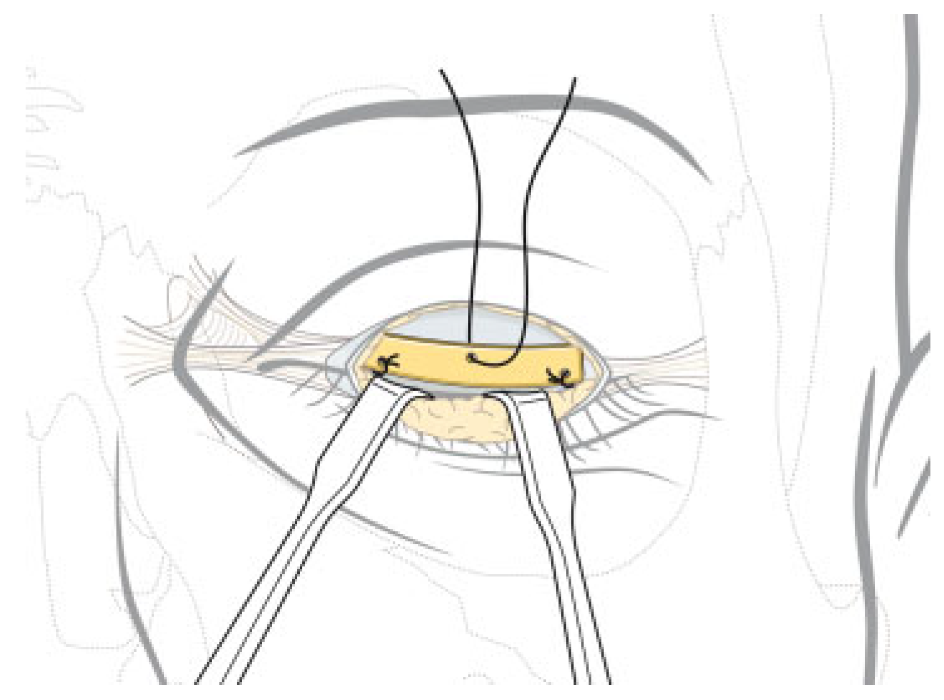
The implantation of a static weight to load the upper lid and achieve eye closure has been successfully performed, since the 1960s (Figure 3).[44] Gold weight implantation in conjunction with lateral tarsorrhaphy has been shown to generate complete eye closure in 83% of patients after one procedure.[45] In this report by Tan, 14% of patients required a revision procedure to optimize their lid weight, with two cases being malpositioned or infected. Platinum may confer a greater benefit than gold due to the higher density and superior biocompatibility of platinum.[4] One review of 100 patients receiving thin profile upper lid implants demonstrated a reduction in capsule formation and extrusion compared with gold.[46]
The development of ectropion in patients with facial paralysis contributes to exposure keratitis by leading to corneal desiccation through worsened lagophthalmos and lacrimal dysfunction.[47,48] The position of the lower lid should be assessed, noting any widening of the lid aperture. Lateral ectropion can be surgically addressed by a lateral tarsal strip procedure or a lateral transorbital canthopexy, while laxity in the medial portion of the lower lid can be corrected by performing a transcutaneous, transcaruncular, or precaruncular medial canthopexy. Decreased morbidity has been noted with the medial canthopexy.[49,50]
Dynamic reanimation of the eye may be accomplished by nerve transfers, such as cross-facial nerve grafting (CFNG), hypoglossal nerve transfer, and direct orbicularis oculi neurotization, or by muscle transfer procedures, borrowing from the frontalis, temporalis, or free muscle flaps.[51,52,53] Evidence suggests that better results are obtained with nerve transfer procedures, rather than by using muscle transfers.[53]

Brow

Brow ptosis leads to cosmetic deformity as well as functional derangements by exacerbating dermatochalasis, thereby encroaching upon the upper visual field. This can be successfully mitigated with surgical brow elevation.[54] Techniques include various open brow approaches, as well as the endoscopic method. While both can achieve similar degrees of brow elevation,[55] the endoscopic lift has the advantage of less sensory disturbance, alopecia, and a faster recovery.[56] Better results can be attained using adjunctive procedures to address hyperactivity seen on the contralateral side of the brow.[57]

Midface

Treatment of the midface aims to alleviate the obstruction of the nasal airway and to counteract the forces of gravity on the malar soft tissues.

Cheek

Paralysis of the midface is of concern, mainly as it relates to nasal valve stenosis and the exacerbation of ectropion by dependent stretching of the canthal ligaments. Surgical procedures to counteract this are aimed at lifting the soft tissues of the middle and lower face. The extended minimal access cranial suspension lift, described by Verpaele et al, is a simple technique that was designed to improve the aging neck and lower two-thirds of the face.[58] While it bolsters the support of the lower lid by lifting the midface, it can lead to a redundancy of lower lid skin. In the study by Elner et al, mobilization of the suborbicularis oculi fat and periosteum was performed to provide midfacial lifting and to minimize ectropion.[59]
Chan and Byrne describe the difference between patients with a neutral vector, where a line dropped vertically from the anterior edge of the lower lid contacts the malar skin, and those with a negative vector, where the malar skin lies posterior to the coronal plane of the lower lid.[60] These authors note that patients with a negative vector are likely to require more extensive measures to prevent ectropion, such as a midface lift, than patients with a neutral vector, who may be correctable with a tarsal strip procedure.

Nose

Symptomatic stenosis of the nasal valve can be easily corrected with suturing techniques, though there are concerns that the benefits of these procedures may be temporary.[61,62] Alex and Nguyen described a minimally invasive suture suspension targeted at the mid and lower face that served to improve the nasal airway, verbal articulation, cosmesis, and oral sphincter competence.[63] A percutaneously placed Gore-Tex sling to suspend the nasolabial fold demonstrated comparable improvements.[64]
Other procedures have shown benefit in terms of their ability to improve nasal breathing. While clearly improving facial symmetry, rhytidectomy has the potential to augment the nasal airway as well.[65,66] Finally, functional septorhinoplasty may be of benefit in select patients with facial paralysis who complain of nasal obstruction, though this may be due to preexisting nasal–septal deformity, or nasal valve collapse.

Lower Face

Disability in the lower face resulting from facial paralysis consists mainly of oral incompetence, manifesting as ptyalism, and difficulty with eating and drinking, poor articulation, and loss of smile and expressivity. Static slings, suspended from the zygomatic arch or deep temporal fascia to support the oral commissure and upper lip, lead to the elevation of these structures and the recreation of the nasolabial fold (Figure 4).[67] Such slings can be constructed from fascia lata, Gore-Tex, or Alloderm (LifeCell Corporation, Bridgewater, NJ),[68] and are most favorable in patients with long-standing paralysis, extensive medical comorbidities, and those who are otherwise poor-operative candidates.
Facial lifting procedures mentioned earlier, such as the extended minimal access cranial suspension can serve a similar purpose. Lower lip reanimation can also be accomplished by means of a Palmaris longus tendon transfer, which is easily performed in the outpatient setting.[69]

Primary Neurorrhaphy

The decision to perform a dynamic reconstruction over static repositioning hinges mainly on the timeframe of the paralysis. For acute traumatic injuries, such as iatrogenic nerve sectioning or penetrating wounds, primary repair of the nerve renders the best outcome.[41] The best results are accomplished with a tension-free coaptation,[70] as tension on the neurorrhaphy seems to diminish perfusion and neural regeneration.[71] An interpositional nerve graft should be used when primary repair is not feasible. Common donor sites include the great auricular or sural nerves. Coaptation of the nerve at a site more proximal than the stylomastoid foramen should be avoided, when possible, as the arrangement of nerve fibers in this region is less favorable and can lead to greater synkinesis.[72,73]

Cross-Facial Nerve Graft

When primary repair or interpositional nerve grafting are not feasible, nerve transfers represent the next rung on the ladder of facial reanimation in cases less than 2 years out from injury. The CFNG, first introduced by Scaramella,[74] relies on peripheral branches of the contralateral, intact facial nerve to innervate corresponding areas of the paralyzed hemiface to produce spontaneous and purposeful facial movement. Use of the CFNG is limited to cases in which nerve injury has occurred within 2 years, before the facial musculature has atrophied, and while motor end plates remain intact.
Criticism of the CFNG revolves around its unpredictability,[75] due to the long distance axons must travel to their targets, while the muscles continue to atrophy. To combat this, the “babysitter” procedure, described by Terzis and Konofaos, has been recommended when the CFNG is performed on patients who suffered their nerve injury greater than 6 months before surgery. The theory behind this technique states that a partial hypoglossal nerve transfer accomplished in the same setting as the CFNG will provide more rapid neural input, preventing loss of motor end plates while the CFNG matures.[41]
The “babysitter” procedure, studied in 20 patients with facial paralysis, resulted in full contraction and symmetrical smile in 10% of patients, and a near symmetrical smile in 65%.[76] The CFNG does carry the risk of disrupting the nonparalyzed donor facial nerve, though this can be mitigated by mapping the nerve and transferring nondominant branches.[77]

Nerve Transposition

When transfer of the contralateral facial nerve is not an option, ipsilateral transfer of hypoglossal, masseteric, spinal accessory, ansa cervicalis, recurrent laryngeal, and phrenic motor nerve fibers is possible. Hypoglossal nerve transfer is the most commonly described, followed by the masseter motor nerve. While the hypoglossal nerve transfer historically entails its complete transection,[78] modification of this technique with transfer of 50% of the hypoglossal nerve fibers provides similar outcomes, but with less tongue weakness and swallowing dysfunction.[79,80]
The modern-day hypoglossal nerve transfer provides reanimation to a House–Brackmann grade II of VI, yielding spontaneous motion with resting facial symmetry (Figure 5).[5,79] Either the superior or inferior half of the nerve can be transferred with similar results.[81] Success has also been shown in hypoglossal nerve transfers performed for facial nucleus palsy.[82]
The masseter nerve represents a good candidate for nerve transfer due to its good size match, length, easy dissection, and minimal impact on masticatory function when dissected intramuscularly to a distal segment.[83] Transfer of the masseter nerve has been shown to result in a more rapid return of facial function compared with hypoglossal transfer with nerve grafting.[84] Faria et al studied 10 patients who underwent masseter nerve transfer, and observed the return of voluntary symmetrical smiling in 90% of cases.[85] The observation of physiologic activity of the masseter during spontaneous smile lends additional strength to its candidacy as a workhorse donor for nerve transfer procedures.[86]
The nerve to the masseter can be reliably found approximately 3 cm anterior to the tragus, approximately 1 cm inferior to the zygomatic arch, and approximately 1.5 cm deep to the SMAS.[87] Another method of locating the nerve uses the “subzygomatic triangle,” which is formed from the zygomatic arch, the temporomandibular joint, and the course of the frontal branch of the facial nerve.[88] The authors of this cadaveric study who described the triangle were able to identify the nerve in an average time of 10.2 minutes. Dissection is not without risk, however, as any functioning upper branches of the facial nerve can be potentially injured.[89]
Combination reinnervation procedures to provide separate neural input to the upper and lower face have been described as well. In a case of radical parotidectomy, Volk et al used an interpositional nerve graft of the great auricular nerve to restore continuity of the proximal facial nerve to the distal upper facial branches, and a simultaneous hypoglossal facial nerve interpositional jump graft technique (instead of classic cross-nerve suturing technique) to the lower distal facial nerve branches.[90] This strategy demonstrated no synkinesis between the upper and lower facial branches and no significant tongue morbidity.

Regional Muscle Transfer

Dynamic muscle transfer can be performed by transposing regional muscle or by free muscle flap transfer. The mainstay of regional muscle transposition is the temporalis muscle transfer, originally described for lower facial reanimation, involving rotation of the temporalis over the zygomatic arch and anchoring it to the oral commissure. Unfortunately, several untoward effects are common, such as bulkiness in the region of the zygomatic arch, depression of the temple, chronic temporomandibular joint dysfunction, and a lack of orthodromic muscle contraction.[52,91,92]
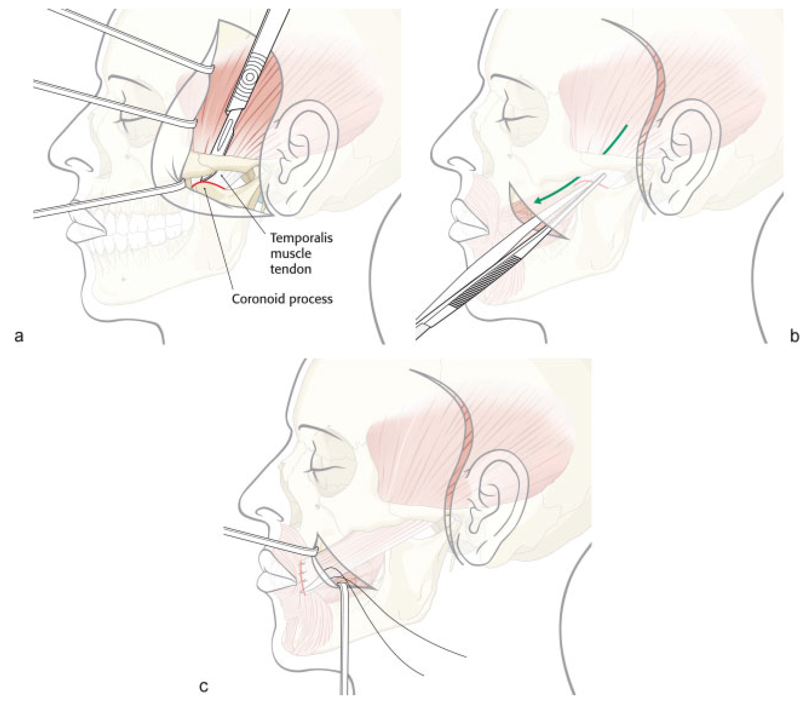
An improvement on the temporalis muscle transfer is the temporalis tendon transfer, initially developed by McLaughlin[93] and further developed by Labbe and Huault.[94] The technique involves removing the temporalis tendon from its insertion on the coronoid process of the mandible and reattaching it to the oral commissure through a transbuccal or nasolabial fold incision (Figure 6).[95] A modification of the procedure involves removal of the coronoid process, through which fascia lata can be passed and then anchored to the facial musculature.[96] Transfer of the tendon avoids excess tissue density in the region of the zygomatic arch, excessive temple depression, and renders a high degree of patient satisfaction.[97]
Masseter muscle transposition is thought to be less advantageous than the use of temporalis tendon due to the limited range of tissue rotation and subsequent masticatory difficulties. A cadaver study suggested the use of a partial masseter transfer, as the dissection of six cadavers revealed a favorable branching pattern of the masseter nerve, with each branch traveling in concert with a vessel pair, creating a neurovascular pedicle on which a partial muscle transposition can be based.[98]
Other options for local muscle transposition include the anterior belly of the digastrics muscle for depressor anguli oris and depressor labii inferioris paralysis. Attachment of the anterior digastric to these target muscles serves to restore depressor function and oppose action of the lip elevators, rendering a more symmetric smile.[3,52]

Free-Tissue Transfer

Free-tissue transfer was first developed for facial reanimation by Harii et al, using a gracilis free flap.[99] A two-stage procedure is described[100] in which a CFNG is performed until a positive Tinel test can be elicited in the paralyzed nasolabial fold, usually 4 to 6 months following surgery. The free muscle flap is placed at this point, with coaptation of the flap’s motor nerve to the distal portion of the CFNG. Fifty percent of patients in Chuang group had a satisfactory result, but revision procedures were frequently needed to enhance the result.[100]
Single-staged free-tissue transfer can be accomplished using a latissimus dorsi free flap with immediate coaptation of the thoracodorsal nerve to the contralateral facial nerve. Using this procedure, 72% of patients in a 33 patient cohort showed at least symmetry at rest.[101] A larger study of 351 single-stage latissimus dorsi transfers demonstrated identifiable muscle contraction in 87% of cases, with an average time to onset of 6.48 months.[102]
Single-stage gracilis free flap reanimation procedures have been described as well, with innervation to the ipsilateral masseter nerve.[103,104] A comparison between 40 patients receiving single-stage latissimus dorsi free flaps innervated by the contralateral facial nerve and 10 patients undergoing single-stage gracilis flaps coapted to the ipsilateral masseter nerve was performed by Biglioli et al.[105] The authors noted voluntary and spontaneous smiling in 92.5% of latissimus flaps, whereas the gracilis group demonstrated spontaneous smile in only 10%. Evidence supporting free gracilis flaps innervated by the masseter nerve has shown that this technique leads to greater dynamic displacement of the oral commissure, as well as increased contraction velocity compared with those innervated by CFNG techniques.[106]

Selection of Neural Input

It is not surprising that use of the contralateral facial nerve seems to provide superior spontaneous emotional expressivity over the masseter nerve and this finding has been corroborated.[107] As mentioned earlier, there is evidence that the masseter has some physiologic activity during spontaneous smile,[86] and spontaneous activity can be found in free muscle flaps supplied by the masseter nerve.[108] Therefore, it is recommended by some authors that the masseter nerve be relegated to salvage cases, after a prior, failed unilateral reanimation based on a CFNG to a gracilis flap, or a singlestage latissimus flap.[109,110]
Double innervations have been performed as well. Biglioli et al implemented gracilis free flaps that were coapted to both the ipsilateral masseter nerve and the contralateral facial nerve simultaneously.[111] This technique provided voluntary flap contraction at 3.8 months postoperatively, and spontaneous contraction at 7.2 months.
In addition to the gracilis and latissimus dorsi, the pectoralis minor muscle has been used for free-tissue transfer, in the setting of facial paralysis, with good results.[112] Another option is the free abductor hallucis muscle, which was studied in 45 patients.[113] All muscle flaps demonstrated contractile activity, as measured on EMG, 6 months after surgery. After 2 years, when graded with the Toronto Facial Grading System and the Facial Nerve Function Index, the affected side demonstrated statistically significant improvements. Complications occurred in 9.8% of patients, consisting of one total flap loss (2.2%), suboptimal flap function due to postoperative infection or hematoma (4.4%) and a hypertrophic scar formation (2.2%). Donor nerves included buccal branches of the contralateral facial nerve or the mylohyoid branch of the ipsilateral trigeminal nerve, with a faster onset of muscle activity in patients with flaps innervated by the mylohyoid nerve. Donor site morbidities include partial loss of abduction function of the big toe and medial donor foot numbness. Normal daily walking was achieved by 3 months after surgery.
Another potential option from the lower extremity is the extensor digitorum brevis muscle. A cadaveric study examined the neurovascular supply to this muscle to assess its potential for transfer as a free muscle flap for facial reanimation.[114] The conclusions drawn were that the muscle could be useful in restoring eyelid function, as well as that of the nasal ala and upper lip with the distal tendon of the extensor digitorum brevis, which splits into four branches. Furthermore, it has a long vascular pedicle based on the dorsalis pedis artery and it is not bulky.
More complex reconstructive efforts are described as well, including the simultaneous use of an anterolateral thigh free flap and a temporalis tendon transfer after radical parotidectomy.[115] The thigh flap was used to provide contour and volume, while the temporalis tendon transfer was performed with the aid of fascia lata harvested from the thigh, to support the lower lip. In addition, cable grafts were taken from the motor nerve to the vastus lateralis to graft the facial nerve branches in an interpositional fashion.

Bilateral Paralysis

Cases of bilateral facial nerve paralysis carry with them the additional challenge of having no functional facial nerve from which to supply innervation. In addition to using the masseter nerve for bilateral simultaneous gracilis transfers,[116,117] end-to-side anastomosis between the hypoglossal nerve and the motor branch to the gracilis has been used on bilateral sides of the face in this situation.[118] The spinal accessory nerve can be used for simultaneous bilateral gracilis-based free-tissue reanimation as well.[119]

Nonoperative Therapy

Options for the rehabilitation of patients suffering from facial paralysis who cannot undergo or do not desire surgery do exist. Physical therapy, in the form of neuromuscular facial retraining can be performed utilizing techniques such as biofeedback, where a patient practices with the assistance of a mirror or an EMG device,[120] with both strategies performing with comparable success.[121] A review of 160 patients with facial paralysis who underwent therapy with alternative techniques, such as biofeedback, massage, and meditation demonstrated improvement in patients’ scores on a facial grading scale that were durable with continued treatment.[122]
In addition, the injection of botulinum toxin A has been used to combat synkinesis in facial paralysis patients, and has been shown to improve patients’ quality of life scores.[120] Low-dose botulinum toxin injection in 11 patients successfully eliminated synkinesis in 7 cases, after no more than 3 applications, though long-term follow-up was lacking.[123] Botulinum toxin can also be injected into the normal side of the face, weakening it, while simultaneously strengthening the paralyzed half, thereby improving symmetry as an adjunct to unsuccessful prior surgical therapy.[124] Some beneficial effects were noted to persist at 6 months even after the pharmacologic effects of the toxin had run their course.

Future Developments

Future directions for management of the paralyzed face include the use of nerve conduits to aid in guiding axonal growth to the desired target muscle. Artificial nerve graft channels can be manufactured from a wide range of materials, both biological and synthetic in origin, such as silicone, polyamide, collagen, and polylactide, with biodegradable polymers having the advantage of absorption without a remnant foreign body. Current trends in nerve conduit engineering are geared toward designing channels that can engage in biochemical signaling and create a scaffold for neurogenesis, directing axonal growth through cell binding domains and the release of neurotrophic factors while supporting the adhesion of glial cells.[125,126]
The development of an implantable, dynamic, artificial muscle may represent a future option for facial reanimation. An electroactive polymer artificial muscle, constructed from silicone, was studied in a gerbil model, where implants were stimulated electrically, for several weeks, successfully creating motion. While significant further research is needed, the artificial muscle implant was well tolerated, without appreciable inflammation or adverse effect.[127]
Other research in prosthetic facial reanimation has been directed toward providing a system, triggered by the nonparalyzed hemiface, and modulated by a processor, that can deliver electrical stimulation to denervated facial musculature via an electrode array, thereby eliciting contraction and preventing muscle atrophy.[128] There is some encouraging evidence to suggest that electrical stimulation may even be able to reverse some of the changes seen in chronic muscle denervation.[129] Others have considered using a low-powered electrode array to maintain muscle density, in a “babysitter” role, in cases where spontaneous recovery is anticipated.[128]

Conclusion

The management of facial paralysis continues to evolve. When the nerve is transected, direct coaptation leads to the best outcome, followed by interpositional nerve grafting. In cases where motor end plates are still intact but a primary repair or graft is not feasible, a nerve transfer should be employed. When complete muscle atrophy has occurred, regional muscle transfer or free flap reconstruction is an option. When dynamic reanimation cannot be undertaken, static procedures offer some benefit. Adjunctive tools such as botulinum toxin injection and biofeedback can be helpful. Several new treatment modalities lie on the horizon which hold potential to alter the current treatment algorithm.

References

  1. May, M. Anatomy for the clinician. In The Facial Nerve; May, M., Schaitkin, B., Eds.; Thieme Medical Publishers: New York, NY, USA, 2000; pp. 19–56. [Google Scholar]
  2. Bernstein, L.; Nelson, R. Surgical anatomy of the extraparotid distribution of the facial nerve. Arch Otolaryngol 1984, 110, 177–183. [Google Scholar] [CrossRef] [PubMed]
  3. Tate, J.; Tollefson, T. Advances in facial reanimation. Curr Opin Otolaryngol Head Neck Surg 2006, 14, 242–248. [Google Scholar] [CrossRef]
  4. Bergeron, C.; Moe, K. The evaluation and treatment of upper eyelid paralysis. Facial Plast Surg 2008, 24, 220–230. [Google Scholar] [CrossRef] [PubMed]
  5. Meltzer, N.E.; Alam, D.S. Facial paralysis rehabilitation: state of the art. Curr Opin Otolaryngol Head Neck Surg 2010, 18, 232–237. [Google Scholar] [CrossRef] [PubMed]
  6. House, J. Facial nerve grading systems. Laryngoscope 1983, 93, 1056–1069. [Google Scholar] [CrossRef] [PubMed]
  7. Brackmann, D.; Barrs, D. Assessing recovery of facial function following acoustic neuroma surgery. Otolaryngol Head Neck Surg 1984, 92, 88–93. [Google Scholar] [CrossRef]
  8. Terzis, J.; Noah, M. Analysis of 100 cases of free-muscle transplantation for facial paralysis. Plast Reconstr Surg 1997, 99, 1905–1921. [Google Scholar] [CrossRef]
  9. Burres, S.; Fisch, U. The comparison of facial grading systems. Arch Otolaryngol Head Neck Surg 1986, 112, 755–758. [Google Scholar] [CrossRef]
  10. Murty, G.; Diver, J.; Kelly, P.; O’Donoghue, G.; Bradley, P. The Nottingham system: objective assessment of facial nerve function in the clinic. Otolaryngol Head Neck Surg 1994, 110, 156–161. [Google Scholar] [CrossRef]
  11. Ross, B.; Frader, G.; Nedzelski, J. Development of a sensitive clinical facial grading system. otolaryngol. Head Neck Surg 1996, 114, 380–386. [Google Scholar] [CrossRef]
  12. Sittel, C.; Stennert, E. Prognostic value of electromyography in acute peripheral facial nerve palsy. Otol Neurotol 2001, 22, 100–104. [Google Scholar] [CrossRef]
  13. Darrouzet, V. Management of facial paralysis resulting from temporal bone fractures: Our experience in 115 cases. Otolaryngol Head Neck Surg 2001, 125, 77–84. [Google Scholar] [CrossRef]
  14. Coker, N. Acute paralysis of the facial nerve. In Head and Neck Surgery—Otolaryngology; Bailey, B, Ed.; J.B. Lippincott Company: Philadelphia, PA, 1993; pp. 1711–1728. [Google Scholar]
  15. Chang, C.; Case, S. Management of facial nerve injury due to temporal bone trauma. Am J Otol 1999, 20, 96–114. [Google Scholar] [CrossRef]
  16. Brodie, H.; Thomson, T. Management of complications from 820 temporal bone fractures. Am J Otol 1997, 18, 188–197. [Google Scholar]
  17. McKennan, K.; Chole, R. Facial paralysis in temporal bone trauma. J Trauma 1992, 13, 167–172. [Google Scholar]
  18. Ghorayeb, B.; Yeakley, J. Temporal bone fractures: longitudinal or oblique? The case for oblique temporal bone fractures. Laryngoscope 1992, 102, 129–134. [Google Scholar] [CrossRef]
  19. Aguilar, E., III; Yeakley, J.; Ghorayeb, B.; et al. High resolution CTscan of temporal bone fractures: association of facial nerve paralysis with temporal bone fractures. Head Neck Surg 1987, 9, 162–166. [Google Scholar] [CrossRef]
  20. Dahiya, R.; Keller, J.; Litofsky, N.; et al. Temporal bone fractures: otic capsule sparing versus otic capsule violating clinical and radiographic considerations. J Trauma 1999, 47, 1079–1083. [Google Scholar] [CrossRef]
  21. Little, S.; Kesser, B. Radiographic classification of temporal bone fractures: clinical predictability using a new system. Arch Otolaryngol Head Neck Surg 2006, 132, 1300–1304. [Google Scholar] [CrossRef] [PubMed]
  22. Quaranta, A.; Campobasso, G.; Piazza, F.; et al. Facial nerve paralysis in temporal bone fractures: outcomes after late decompression surgery. Acta Otolaryngol 2001, 121, 652–655. [Google Scholar] [CrossRef] [PubMed]
  23. Cannon, C.; Jahrsdoerfer, R. Temporal bone fractures. Review of 90 cases. Arch Otolaryngol 1983, 109, 285–288. [Google Scholar] [CrossRef] [PubMed]
  24. Nosan, D.; Benecke, J.J.; Murr, A. Current perspective on temporal bone trauma. Otolaryngol Head Neck Surg 1997, 117, 67–71. [Google Scholar] [CrossRef] [PubMed]
  25. Coker, N.; Kendall, K.; Jenkins, H.A.; et al. Traumatic intratemporal facial nerve injury: management rationale for preservation of function. Otolaryngol Head Neck Surg 1987, 97, 262–269. [Google Scholar] [CrossRef]
  26. Fisch, U.; Mattox, D. Microsurgery of the Skull Base; Georg Thieme Verlag: New York, NY, 1991. [Google Scholar]
  27. Gantz, B.; Gmuer, A.; Holliday, M.; et al. Electroneurographic evaluation of the facial nerve. Method and technical problems. Ann Otol Rhinol Laryngol 1984, 93 Pt 1, 394–398. [Google Scholar] [CrossRef]
  28. House, J.; Brackmann, D. Facial nerve grading system. Otolaryngol Head Neck Surg 1985, 93, 146–147. [Google Scholar] [CrossRef] [PubMed]
  29. Ulug, T.; Arif Ulubil, S. Management of facial paralysis in temporal bone fractures: a prospective study analyzing 11 operated fractures. Am J Otolaryngol 2005, 26, 230–238. [Google Scholar] [CrossRef]
  30. Lambert, P.; Brackmann, D. Facial paralysis in longitudinal temporal bone fractures: a review of 26 cases. Laryngoscope 1984, 94, 1022–1026. [Google Scholar] [CrossRef]
  31. Bozorg Grayeli, A.; Mosnier, I.; Julien, N.; et al. Long-term functional outcome in facial nerve graft by fibrin glue in the temporal bone and cerebellopontine angle. Eur Arch Otorhinolaryngol 2004, 262, 404–407. [Google Scholar] [CrossRef]
  32. Ornelas, L.; Padilla, L.; Di Silvio, M.; et al. Fibrin glue: an alternative technique for nerve coaptation–Part, I. Wave amplitude, conduction velocity, and plantar-length factors. J Reconstr Microsurg 2006, 22, 119–122. [Google Scholar] [CrossRef]
  33. Ornelas, L.; Padilla, L.; Di Silvio, M.; et al. Fibrin Glue: an alternative technique for nerve coaptation–Part, I.I. Nerve regeneration and histomorphometric assessment. J Reconstr Microsurg 2006, 22, 123–128. [Google Scholar] [CrossRef]
  34. Salinas, R.A.; Alvarez, G.; Daly, F.; Ferreira, J. Corticosteroids for Bell’s palsy (idiopathic facial paralysis). Cochrane Database Syst Rev 2010, 17, CD001942. [Google Scholar]
  35. Hadlock, T. Facial paralysis: research and future directions. Facial Plast Surg 2008, 24, 260–267. [Google Scholar] [CrossRef]
  36. Danner, C.J. Facial nerve paralysis. Otolaryngol Clin North Am 2008, 41, 619–632. [Google Scholar] [CrossRef]
  37. Lockhart, P.; Daly, F.; Pitkethly, M.; Comerford, N.; Sullivan, F. Antiviral treatment for Bell’s palsy (idiopathic facial paralysis). Cochrane Database Syst Rev 2009, 7, CD001869. [Google Scholar]
  38. Bodénez, C.; Bernat, I.; Willer, J.C.; Barré, P.; Lamas, G.; Tankéré, F. Facial nerve decompression for idiopathic Bell’s palsy: report of 13 cases and literature review. J Laryngol Otol 2010, 124, 272–278. [Google Scholar] [CrossRef]
  39. Melvin, T.-A.; Limb, C. Overview of facial paralysis: current concepts. Facial Plast Surg 2008, 24, 155–163. [Google Scholar] [CrossRef]
  40. Chu, E.; Byrne, P. Treatment considerations in facial paralysis. Facial Plast Surg 2008, 24, 164–169. [Google Scholar] [CrossRef] [PubMed]
  41. Terzis, J.; Konofaos, P. Nerve transfers in facial palsy. Facial Plast Surg 2008, 24, 177–193. [Google Scholar] [CrossRef]
  42. Reddy, U.; Woodward, J. Abobotulinum toxin A (Dysport) and botulinum toxin type A (Botox) for purposeful induction of eyelid ptosis. Ophthal Plast Reconstr Surg 2010, 26, 489–491. [Google Scholar] [CrossRef] [PubMed]
  43. Mancini, R.; Taban, M.; Lowinger, A.; et al. Use of hyaluronic acid gel in the management of paralytic lagophthalmos: the hyaluronic acid gel “Gold Weight”. Ophthal Plast Reconstr Surg 2009, 25, 23–26. [Google Scholar] [CrossRef] [PubMed]
  44. Smellie, G. Restoration of the blinking reflex in facial palsy by a simple lid-load operation. Br J Plast Surg 1966, 19, 279–283. [Google Scholar] [CrossRef] [PubMed]
  45. Tan, S.T.; Staiano, J.J.; Itinteang, T.; McIntyre, B.C.; MacKinnon, C.A.; Glasson, D.W. Gold weight implantation and lateral tarsorrhaphy for upper eyelid paralysis. J Craniomaxillofac Surg 2013, 41, e49–e53. [Google Scholar] [CrossRef] [PubMed]
  46. Silver, A.L.; Lindsay, R.W.; Cheney, M.L.; Hadlock, T.A. Thin-profile platinum eyelid weighting: a superior option in the paralyzed eye. Plast Reconstr Surg 2009, 123, 1697–1703. [Google Scholar] [CrossRef] [PubMed]
  47. Razfar, A.; Afifi, A.; Manders, E.; et al. Ocular outcomes after gold weight placement and facial nerve resection. Otolaryngol Head Neck Surg 2009, 140, 82–85. [Google Scholar] [CrossRef] [PubMed]
  48. Bergeron, C.; Moe, K. The evaluation and treatment of lower eyelid paralysis. Facial Plast Surg 2008, 24, 231–241. [Google Scholar] [CrossRef]
  49. Liebau, J.; Schulz, A.; Arens, A.; Tilkorn, H.; Schwipper, V. Management of lower lid ectropion. Dermatol Surg 2006, 32, 1050–1057. [Google Scholar] [PubMed]
  50. Moe, K.S.; Kao, C.-H. Preauricular medial canthopexy. Arch Facial Plast Surg 2005, 7, 244–250. [Google Scholar] [CrossRef]
  51. Frey, M.; Giovanoli, P.; Tzou, C.-H.J.; Kropf, N.; Friedl, S. Dynamic reconstruction of eye closure by muscle transposition or functional muscle transplantation in facial palsy. Plast Reconstr Surg 2004, 114, 865–875. [Google Scholar] [CrossRef]
  52. Boahene, K. Dynamic muscle transfer in facial reanimation. Facial Plast Surg 2008, 24, 204–210. [Google Scholar] [CrossRef]
  53. Terzis, J.K.; Karypidis, D. Blink restoration in adult facial paralysis. Plast Reconstr Surg 2010, 126, 126–139. [Google Scholar] [CrossRef]
  54. Meltzer, N.; Byrne, P. Management of the brow in facial paralysis. Facial Plast Surg 2008, 24, 216–219. [Google Scholar] [CrossRef] [PubMed]
  55. Keller, G.; Mashkevich, G. Endoscopic forehead and brow lift. Facial Plast Surg 2009, 25, 222–233. [Google Scholar] [CrossRef]
  56. Moody, F.; Losken, A.; Bostwick, J.I.; Trinei, F.; Eaves, F. Endoscopic frontal branch neurectomy corrugator myectomy and brow lift for forehead asymmetry after facial nerve palsy. Plast Reconstr Surg 2001, 108, 218–224. [Google Scholar] [CrossRef]
  57. Ducic, Y.; Adelson, R. Use of the endoscopic forehead-lift to improve brow position in persistent facial paralysis. Arch Facial Plast Surg 2005, 7, 51–54. [Google Scholar] [CrossRef]
  58. Verpaele, A.; Tonnard, P.; Gaia, S.; Guerao, F.; Pirayesh, A. The third suture in MACS-lifting: making midface-lifting simple and safe. J Plast Reconstr Aesthet Surg 2007, 60, 1287–1295. [Google Scholar] [CrossRef]
  59. Elner, V.; Mauffray, R.; Fante, R.; Harris, M.; Morton, A.; Hassan, A. Comprehensive midfacial elevation for ocular complications of facial nerve palsy. Arch Facial Plast Surg 2003, 5, 427–433. [Google Scholar] [CrossRef] [PubMed]
  60. Chan, J.Y.K.; Byrne, P. Management of facial paralysis in the 21st century. Facial Plast Surg 2011, 27, 346–357. [Google Scholar] [CrossRef]
  61. Friedman, M. A simplified technique for airway correction at the nasal valve area. Otolaryngol Head Neck Surg 2004, 131, 519–524. [Google Scholar] [CrossRef] [PubMed]
  62. Nuara, M.; Mobley, S. Nasal valve suspension revisited. Laryngoscope 2007, 117, 2100–2106. [Google Scholar] [CrossRef]
  63. Alex, J.; Nguyen, D. Multivectored suture suspension: a minimally invasive technique for reanimation of the paralyzed face. Arch Facial Plast Surg 2004, 6, 197–201. [Google Scholar] [CrossRef]
  64. Alam, D.S. Rehabilitation of long-standing facial nerve paralysis with percutaneous suture-based slings. Arch Facial Plast Surg 2007, 9, 205–209. [Google Scholar] [CrossRef] [PubMed]
  65. Capone, R.; Sykes, J. The effect of rhytidectomy on the nasal valve. Arch Facial Plast Surg 2005, 7, 45–50. [Google Scholar] [CrossRef]
  66. Ramakrishnan, Y.; Alam, S.; Kotecha, A.; Gillett, D.; D’Souza, A. Reanimation following facial palsy: present and future directions. J Laryngol Otol 2010, 124, 1146–1152. [Google Scholar] [CrossRef] [PubMed]
  67. Rosson, G.; Redett, R. Facial palsy: anatomy, etiology, grading, and surgical treatment. J Reconstr Microsurg 2008, 24, 379–389. [Google Scholar] [CrossRef] [PubMed]
  68. Fisher, E.; Frodel, J. Facial suspension with acellular human dermal allograft. Arch Facial Plast Surg 1999, 1, 195–199. [Google Scholar] [CrossRef]
  69. Alexander, A.J.; de Almeida, J.R.; Shrime, M.G.; Goldstein, D.P.; Gilbert, R.W. Novel outpatient approach to lower lip reanimation using a palmaris longus tendon sling. J Otolaryngol Head Neck Surg 2011, 40, 481–488. [Google Scholar]
  70. Humphrey, C.; Kriet, J. Nerve repair and cable grafting for facial paralysis. Facial Plast Surg 2008, 24, 170–176. [Google Scholar] [CrossRef]
  71. Agnew, S.P.; Dumanian, G.A. Technical use of synthetic conduits for nerve repair. J Hand Surg Am 2010, 35, 838–841. [Google Scholar] [CrossRef]
  72. Myckatyn, Y.; Mackinnon, S.E. The surgical management of facial nerve injury. Clin Plast Surg 2003, 30, 307–318. [Google Scholar] [CrossRef]
  73. Samii, M.; Matthies, C. Indication, technique and results of facial nerve reconstruction. Acta Neurochir (Wien) 1994, 130, 125–139. [Google Scholar] [CrossRef]
  74. Scaramella, L. Anastomosis between the two facial nerves. Laryngoscope 1975, 85, 1359–1366. [Google Scholar] [CrossRef] [PubMed]
  75. Lee, E.; Hurvitz, K.; Evans, G.; Wirth, G. Cross-facial nerve graft: past and present. J Plast Reconstr Aesthet Surg 2008, 61, 250–256. [Google Scholar] [CrossRef]
  76. Terzis, J.K.; Tzafetta, K. The “Babysitter” procedure: minihypoglossal to facial nerve transfer and cross-facial nerve grafting. Plast Reconstr Surg 2009, 123, 865–876. [Google Scholar] [CrossRef] [PubMed]
  77. Galli, S.K.; Valauri, F.; Komisar, A. Facial reanimation by cross-facial nerve grafting: report of five cases. Ear Nose Throat J 2002, 81, 25–29. [Google Scholar] [CrossRef] [PubMed]
  78. Conley, J.; Baker, D. Hypoglossal-facial nerve anastomosis for reinnervation of the paralyzed face. Plast Reconstr Surg 1979, 61, 63–72. [Google Scholar] [CrossRef]
  79. Rochkind, S.; Shafi, M.; Alon, M.; Salame, K.; Fliss, D. Facial nerve reconstruction using a split hypoglossal nerve with preservation of tongue function. J Reconstr Microsurg 2008, 24, 469–474. [Google Scholar] [CrossRef]
  80. Arai, H.; Kiyoshi, S.; Yanai, A. Hemihypoglossal-facial nerve anastomosis in treating unilateral facial palsy after acoustic neuroma resection. J Neurosurg 1995, 82, 51–54. [Google Scholar] [CrossRef]
  81. Hayashi, A.; Nishida, M.; Seno, H.; et al. Hemihypoglossal nerve transfer for acute facial paralysis. J Neurosurg 2013, 118, 160–166. [Google Scholar] [CrossRef]
  82. Corrales, C.; Gurgel, R.; Jackler, R. Rehabilitation of central facial paralysis with hypoglossal-facial anastomosis. Otol Neurotol 2012, 33, 1439–1444. [Google Scholar] [CrossRef]
  83. Klebuc, M.J.A. Facial reanimation using the masseter-to-facial nerve transfer. Plast Reconstr Surg 2011, 127, 1909–1915. [Google Scholar] [CrossRef]
  84. Hontanilla, B.; Marré, D. Comparison of hemihypoglossal nerve versus masseteric nerve transpositions in the rehabilitation of short-term facial paralysis using the Facial Clima evaluating system. Plast Reconstr Surg 2012, 130, 662e–672e. [Google Scholar] [CrossRef]
  85. Faria, J.C.M.; Scopel, G.P.; Ferreira, M.C. Facial reanimation with masseteric nerve: babysitter or permanent procedure? Preliminary results. Ann Plast Surg 2010, 64, 31–34. [Google Scholar] [CrossRef]
  86. Schaverien, M.; Moran, G.; Stewart, K.; Addison, P. Activation of the masseter muscle during normal smile production and the implications for dynamic reanimation surgery for facial paralysis. J Plast Reconstr Aesthet Surg 2011, 64, 1585–1588. [Google Scholar] [CrossRef]
  87. Borschel, G.; Kawamura, D.; Kasukurthi, R.; Hunter, D.; Zuker, R.; Woo, A. The motor nerve to the masseter muscle: an anatomic and histomorphometric study to facilitate its use in facial reanimation. J Plast Reconstr Aesthet Surg 2012, 65, 363–366. [Google Scholar] [CrossRef]
  88. Collar, R.M.; Byrne, P.J.; Boahene, K.D.O. The subzygomatic triangle: rapid, minimally invasive identification of the masseteric nerve for facial reanimation. Plast Reconstr Surg 2013, 132, 183–188. [Google Scholar] [CrossRef] [PubMed]
  89. Faria, J.C.M.; Scopel, G.P.; Busnardo, F.F.; Ferreira, M.C. Nerve sources for facial reanimation with muscle transplant in patients with unilateral facial palsy. Ann Plast Surg 2007, 59, 87–91. [Google Scholar] [CrossRef] [PubMed]
  90. Volk, G.F.; Pantel, M.; Streppel, M.; Guntinas-Lichius, O. Reconstruction of complex peripheral facial nerve defects by a combined approach using facial nerve interpositional graft and hypoglossal-facial jump nerve suture. Laryngoscope 2011, 121, 2402–2405. [Google Scholar] [CrossRef] [PubMed]
  91. Conley, J. Facial rehabilitation: new potentials. Clin Plast Surg 1979, 6, 421–431. [Google Scholar] [CrossRef]
  92. Conley, J.; Baker, D.; Selfe, R. Paralysis of the mandibular branch of the facial nerve. Plast Reconstr Surg 1982, 70, 569–576. [Google Scholar] [CrossRef]
  93. McLaughlin, C. Surgical support in permanent facial paralysis. Plast Reconstr Surg 1946, 11, 302–314. [Google Scholar] [CrossRef]
  94. Labbe, D.; Huault, M. Lengthening temporalis myoplasty and lip reanimation. Plast Reconstr Surg 2000, 105, 1289–1297. [Google Scholar] [CrossRef] [PubMed]
  95. Boahene, K.; Farrag, T.Y.; Ishii, L.; Byrne, P. Minimally invasive temporalis tendon transposition. Arch Facial Plast Surg 2011, 13, 8–13. [Google Scholar] [CrossRef]
  96. Sidle, D.M.; Fishman, A.J. Modification of the orthodromic temporalis tendon transfer technique for reanimation of the paralyzed face. Otolaryngol Head Neck Surg 2011, 145, 18–23. [Google Scholar] [CrossRef] [PubMed]
  97. Byrne, P.; Kim, M.; Boahene, K.; Millar, J.; Moe, K. Temporalis tendon transfer as part of a comprehensive approach to facial reanimation. Arch Facial Plast Surg 2007, 9, 234–241. [Google Scholar] [CrossRef]
  98. Hontanilla, B.; Qiu, S.S. Transposition of the hemimasseteric muscle for dynamic rehabilitation of facial paralysis. J Craniofac Surg 2012, 23, 203–205. [Google Scholar] [CrossRef] [PubMed]
  99. Harii, K.; Ohmori, K.; Torii, S. Free gracilis muscle transplantation, with microneurovascular anastomoses for the treatment of facial paralysis: a preliminary report. Plast Reconstr Surg 1976, 57, 133–143. [Google Scholar] [CrossRef]
  100. Chuang, D. Free tissue transfer for the treatment of facial paralysis. Facial Plast Surg 2008, 24, 194–203. [Google Scholar] [CrossRef]
  101. Harii, K.; Asato, H.; Yoshimura, K.; Sugawara, Y.; Nakatsuka, T.; Ueda, K. One-stage transfer of the latissimus dorsi muscle for reanimation of a paralyzed face: a new alternative. Plast Reconstr Surg 1998, 102, 941–951. [Google Scholar] [CrossRef]
  102. Takushima, A.; Harii, K.; Asato, H.; Kurita, M.; Shiraishi, T. Fifteen-year survey of one-stage latissimus dorsi muscle transfer for treatment of longstanding facial paralysis. J Plast Reconstr Aesthet Surg 2013, 66, 29–36. [Google Scholar] [CrossRef]
  103. Guelinckx, P.; Sinsel, N. Muscle transplantation for reconstruction of a smile after facial paralysis past, present, and future. Microsurgery 1996, 17, 391–401. [Google Scholar] [CrossRef]
  104. Bianchi, B.; Copelli, C.; Ferrari, S.; Ferri, A.; Bailleul, C.; Sesenna, E. Facial animation with free-muscle transfer innervated by the masseter motor nerve in unilateral facial paralysis. J Oral Maxillofac Surg 2010, 68, 1524–1529. [Google Scholar] [CrossRef]
  105. Biglioli, F.; Colombo, V.; Tarabbia, F.; et al. Recovery of emotional smiling function in free-flap facial reanimation. J Oral Maxillofac Surg 2012, 70, 2413–2418. [Google Scholar] [CrossRef]
  106. Hontanilla, B.; Marre, D.; Cabello, A. Facial reanimation with gracilis muscle transfer neurotized to cross-facial nerve graft versus masseteric nerve: a comparative study using the FACIAL CLIMA evaluating system. Plast Reconstr Surg 2013, 131, 1241–1252. [Google Scholar] [CrossRef] [PubMed]
  107. Gousheh, J.; Arasteh, E. Treatment of facial paralysis. Plast Reconstr Surg 2011, 128, 693e–703e. [Google Scholar] [CrossRef] [PubMed]
  108. Manktelow, R.T.; Tomat, L.R.; Zuker, R. Smile reconstruction in adults with free muscle transfer innervated by the masseter motor nerve: effectiveness and cerebral adaptation. Plast Reconstr Surg 2006, 118, 885–899. [Google Scholar] [CrossRef]
  109. Bianchi, B.; Copelli, C.; Ferrari, S.; Ferri, A.; Sesenna, E. Successful salvage surgery after treatment failures with cross graft and free muscle transplant in facial reanimation. J Craniomaxillofac Surg 2012, 40, 185–189. [Google Scholar] [CrossRef] [PubMed]
  110. Horta, R.; Silva, P.; Bartosh, I.; et al. Facial reanimation with gracilis muscle transplantation and obturator nerve coaptation to the motor nerve of masseter muscle as a salvage procedure in an unreliable cross-face nerve graft. Microsurgery 2011, 31, 164–166. [Google Scholar] [CrossRef]
  111. Biglioli, F.; Bayoudh, W.; Colombo, V.; Pedrazzoli, M.; Rabbiosi, D. Double innervation (facial/masseter) on the gracilis flap, in the middle face reanimation in the management of facial paralysis: A new concept. Ann Chir Plast Esthet 2013, 58, 89–95. [Google Scholar] [CrossRef]
  112. Harrison, D.; Grobbelaar, A. Pectoralis minor muscle transfer for unilateral facial palsy reanimation: an experience of 35 years and 637 cases. J Plast Reconstr Aesthet Surg 2012, 65, 845–850. [Google Scholar] [CrossRef]
  113. Liu, A.-T.; Lin, Q.; Jiang, H.; et al. Facial reanimation by one-stage microneurovascular free abductor hallucis muscle transplantation. Plast Reconstr Surg 2012, 130, 325–335. [Google Scholar] [CrossRef]
  114. Alagöz, M.Ş.; Alagöz, A.N.; Comert, A. Neuroanatomy of extensor digitorum brevis muscle for reanimation of facial paralysis. J Craniofac Surg 2011, 22, 2308–2311. [Google Scholar] [CrossRef]
  115. Revenaugh, P.; Knott, P.; Scharpf, J.; Fritz, M. Simultaneous anterolateral thigh flap and temporalis tendon transfer to optimize facial form and function after radical parotidectomy. Arch Facial Plast Surg 2012, 14, 104–109. [Google Scholar] [CrossRef]
  116. Lindsay, R.W.; Hadlock, T.A.; Cheney, M.L. Bilateral simultaneous free gracilis muscle transfer: A realistic option in management of bilateral facial paralysis. Otolaryngol Head Neck Surg 2009, 141, 139–141. [Google Scholar] [CrossRef] [PubMed]
  117. Bianchi, B.; Copelli, C.; Ferrari, S.; Ferri, A.; Sesenna, E. Facial animation in children with Moebius and Moebius-like syndromes. J Pediatr Surg 2009, 44, 2236–2242. [Google Scholar] [CrossRef] [PubMed]
  118. Hontanilla, B.; Aubá, C. Smile reconstruction through bilateral muscular transplants neurotized by hypoglossal nerves. J Craniofac Surg 2011, 22, 845–847. [Google Scholar] [CrossRef] [PubMed]
  119. Lu, J.C.-Y.; Chuang, D.C.-C. One-Stage reconstruction for bilateral Möbius syndrome. Ann Plast Surg 2013, 70, 180–186. [Google Scholar] [CrossRef]
  120. Husseman, J.; Mehta, R. Management of synkinesis. Facial Plast Surg 2008, 24, 242–249. [Google Scholar] [CrossRef]
  121. Cardoso, J.; Teixeira, E.; Moreira, M.; Favero, F.; Fontes, S.; de Oliveira, A. Effects of exercises on Bell’s palsy: systematic review of randomized controlled trials. Otol Neurotol 2008, 29, 557–560. [Google Scholar] [CrossRef]
  122. Lindsay, R.W.; Robinson, M.; Hadlock, T.A. Comprehensive facial rehabilitation improves function in people with facial paralysis: a 5-year experience at the Massachusetts eye and ear infirmary. Phys Ther 2010, 90, 391–397. [Google Scholar] [CrossRef]
  123. Ito, H.; Nakano, S.; Kusaka, H. Low-dose subcutaneous injection of botulinum toxin type A for facial synkinesis and hyperlacrimation. Acta Neurol Scand 2007, 115, 271–274. [Google Scholar] [CrossRef]
  124. Salles, A.; Toledo, P.; Ferreira, M. Botulinum toxin injection in longstanding facial paralysis patients: improvement of facial symmetry observed up to 6 months. Aesthetic Plast Surg 2009, 33, 582–590. [Google Scholar] [CrossRef] [PubMed]
  125. Chiono, V.; Tonda Turo, C.; Ciardelli, G. Chapter 9: Artificial scaffolds for peripheral nerve reconstruction. Int Rev Neurobiol 2009, 87, 173–198. [Google Scholar] [PubMed]
  126. Cunha, C.; Panseri, S.; Antonini, S. Emerging nanotechnology approaches in tissue engineering for peripheral nerve regeneration. Nanomedicine (Lond Print) 2011, 7, 50–59. [Google Scholar] [CrossRef] [PubMed]
  127. Ledgerwood, L.G.; Tinling, S.; Senders, C.; Wong-Foy, A.; Prahlad, H.; Tollefson, T.T. Artificial muscle for reanimation of the paralyzed face: durability and biocompatibility in a gerbil model. Arch Facial Plast Surg 2012, 14, 413–418. [Google Scholar] [CrossRef]
  128. Griffin, G.R.; Kim, J.C. Potential of an electric prosthesis for dynamic facial reanimation. Otolaryngol Head Neck Surg 2011, 145, 365–368. [Google Scholar] [CrossRef]
  129. Kern, H.; Carraro, U.; Adami, N.; et al. One year of home-based daily FES in complete lower motor neuron paraplegia: recovery of tetanic contractility drives the structural improvements of denervated muscle. Neurol Res 2010, 32, 5–12. [Google Scholar] [CrossRef]
Figure 1. Temporal bone anatomy and intratemporal facial nerve segments.
Cmtr 08 i1f1 g001
Figure 2. Extratemporal facial nerve branches (temporal, zygomatic, buccal, marginal mandibular and cervical branches). Facial nerve branches generally innervate deep to the facial muscles with the exceptions of levator anguli oris, buccinators, and mentalis muscles.
Cmtr 08 i1f1 g002
Figure 3. Demonstration of gold/platinum weight being secured to upper eyelid tarsus.
Cmtr 08 i1f1 g003
Figure 4. An inferior displacement of the affected oral commissure, oral incompetence, upper and lower lip asymmetry are commonly seen with facial paralysis. Upper lip philtral ridge is also commonly displaced to the unaffected side. Static suspension of lower lip can be used to address gross lip asymmetry, drooling and oral incompetence. Fascia lata, Gore-Tex and Alloderm can be used to suspend in a superoposterior vector as seen in the right image.
Cmtr 08 i1f1 g004
Figure 5. Split hypoglossal nerve to facial nerve transfer. Either superior or inferior half of the hypoglossal nerve can be transferred. Less tongue morbidity is noted with this technique compared to classic hypoglossal nerve to facial nerve transfer in which the entire hypoglossal nerve is transferred.
Cmtr 08 i1f1 g005
Figure 6. Dynamic temporalis muscle transfer can be performed by releasing the temporalis muscle tendon from the coronoid process using either a transcutaneous approach (as seen in the diagram) or a transoral approach. Once the temporalis tendon is released from the coronoid process, it can be secured to paralyzed oral commissure. This technique avoids excess tissue bulk noted over the zygomatic arch and temporal hallowing that can result from classic temporalis muscle transfer technique.
Cmtr 08 i1f1 g006
Table 1. A review of medical literature (1900–1990), various causes of facial palsy.
Cmtr 08 i1f1 i001
Cmtr 08 i1f1 i002
Cmtr 08 i1f1 i003