Abstract
Atypical fracture patterns of the facial region have been reported infrequently. An unusual displacement of fractured posterior maxillary segment into the lateral pharyngeal space is described.
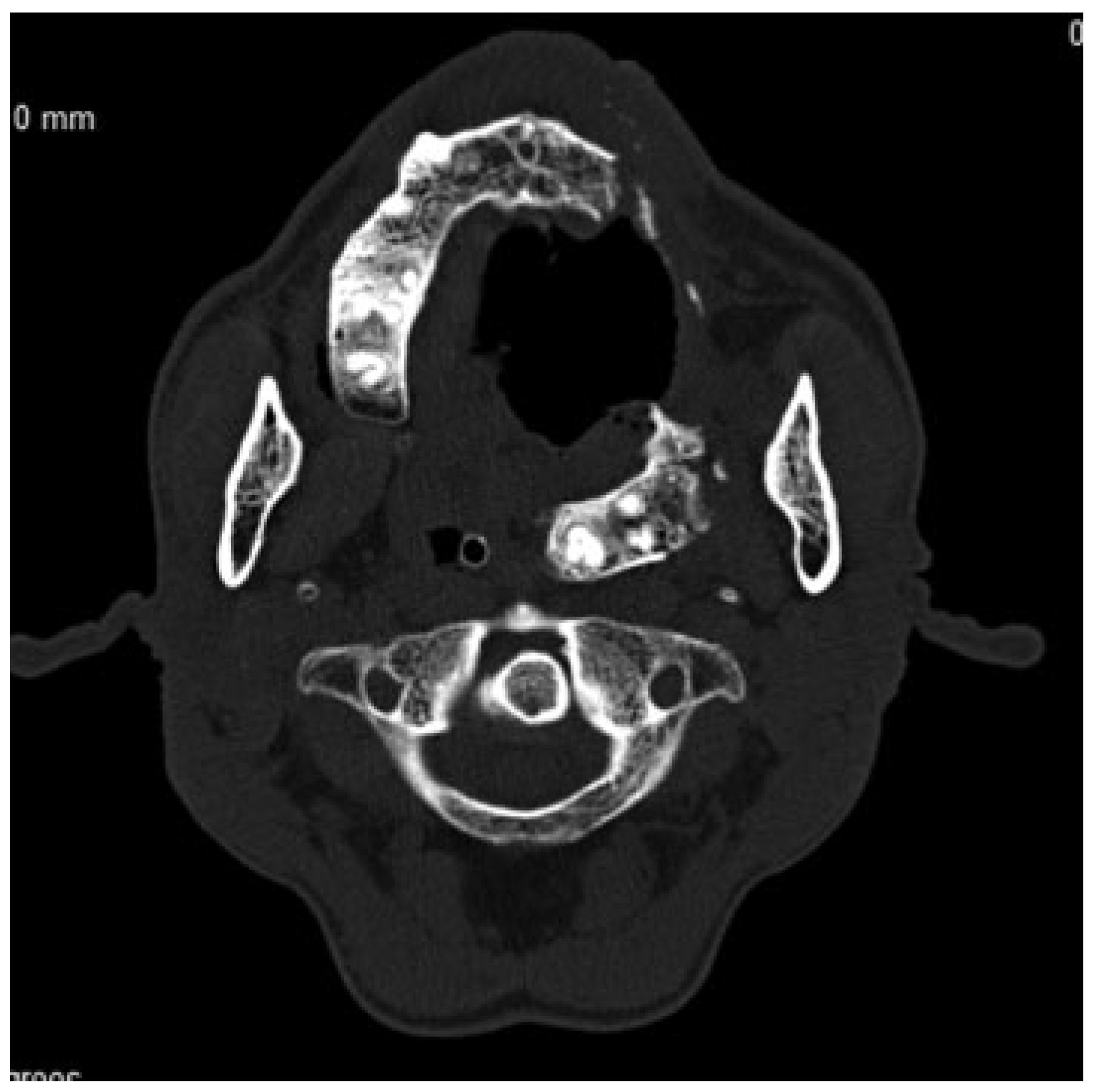
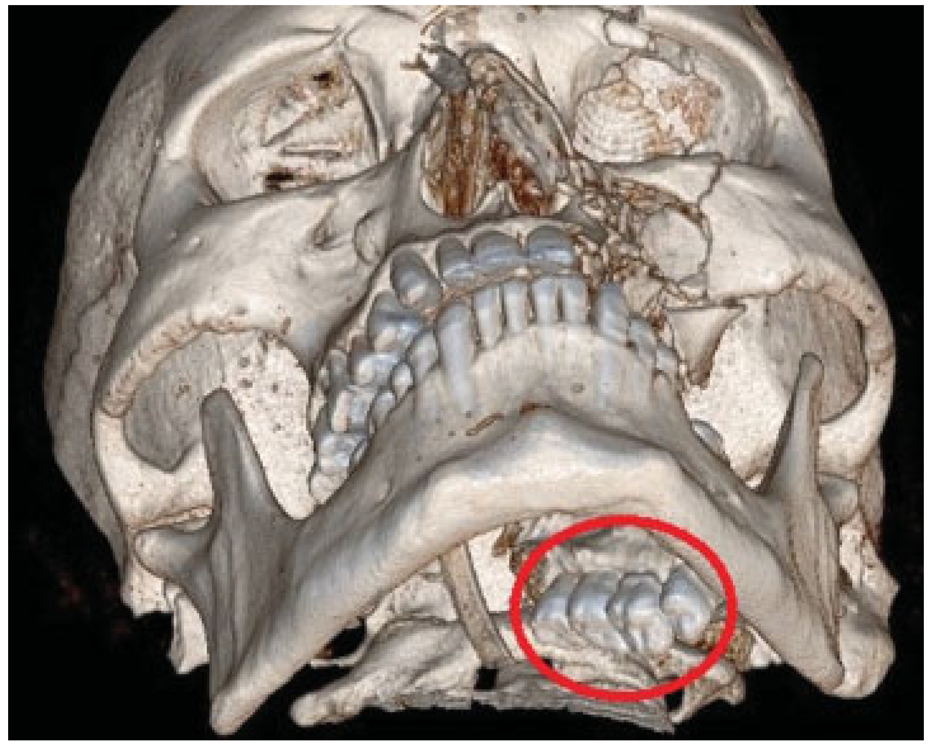
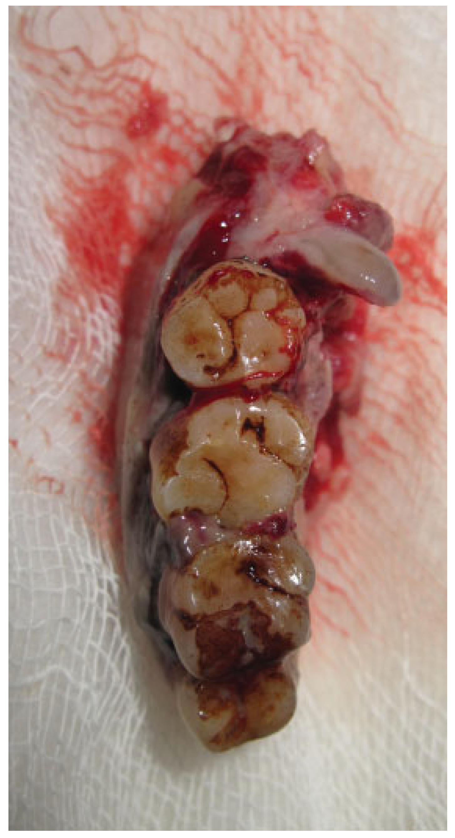
Atypical fracture patterns of the facial region have been reported infrequently [1,2,3]. An unusual displacement of a fractured posterior maxilla is described. A 62-year-old male patient reported to the Department of Dentistry with a complaint of facial pain, restricted mouth opening, and difficulty in swallowing. A history of a road traffic accident 13 days ago was elicited. On examination, the patient appeared distressed and dehydrated. Intraorally, all maxillary teeth on the left side from the lateral incisor along with left posterior maxilla were missing. A large area of the palatal mucosa was missing and the resultant defect was filled with debris, necrotic tissue, and purulent discharge. Severe tenderness was observed on palpation of the left soft palate, pterygomandibular, and lateral pharyngeal region. No neurological deficits were noticed. Neither the patient nor his relative could account for the loss of maxillary teeth. X-rays and computerized tomographic scan showed displacement of the entire left posterior maxillary segment into the lateral pharyngeal space along with a comminuted fracture of the zygomatic complex (►Figure 1 and Figure 2). An incision was made medial to the pterygomandibular raphe and the entire segment was removed by blunt and sharp dissection (►Figure 3). Following a thorough debridement, the resultant defect was allowed to heal by a combination of secondary intention and delayed primary closure. Postoperative period was uneventful and a palatal obturator was subsequently fitted.

Figure 1.
Computerized tomographic scan showing displacement of fractured maxillary segment into lateral pharyngeal space.
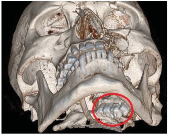
Figure 2.
3D computed tomography scan showing displacement of fractured maxillary segment into lateral pharyngeal space.

Figure 3.
Fractured segment in toto.
References
- GA, G.; Currie, A. Unusual Le Fort I fracture. Br J Oral Maxillofac Surg 2013, 51, 669–670. [Google Scholar]
- Al-Hadad, I.; Burke, G.A.E.; Webster, K. Dentoalveolar fracture of the posterior maxilla. Br J Oral Maxillofac Surg 2009, 47, 165. [Google Scholar] [CrossRef] [PubMed]
- Monaghan, A.M. An unusual Le Fort II fracture. Br J Oral Maxillofac Surg 1991, 29, 256–258. [Google Scholar] [CrossRef] [PubMed]
© 2014 by the author. The Author(s) 2014.