Pseudoaneurysm of the Internal Maxillary Artery: A Rare Complication of Condylar Fracture
Abstract
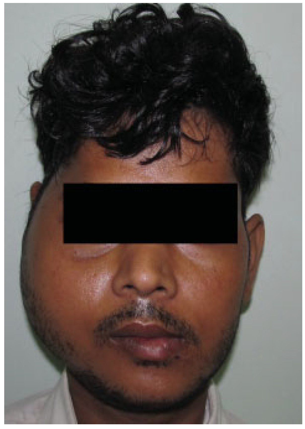
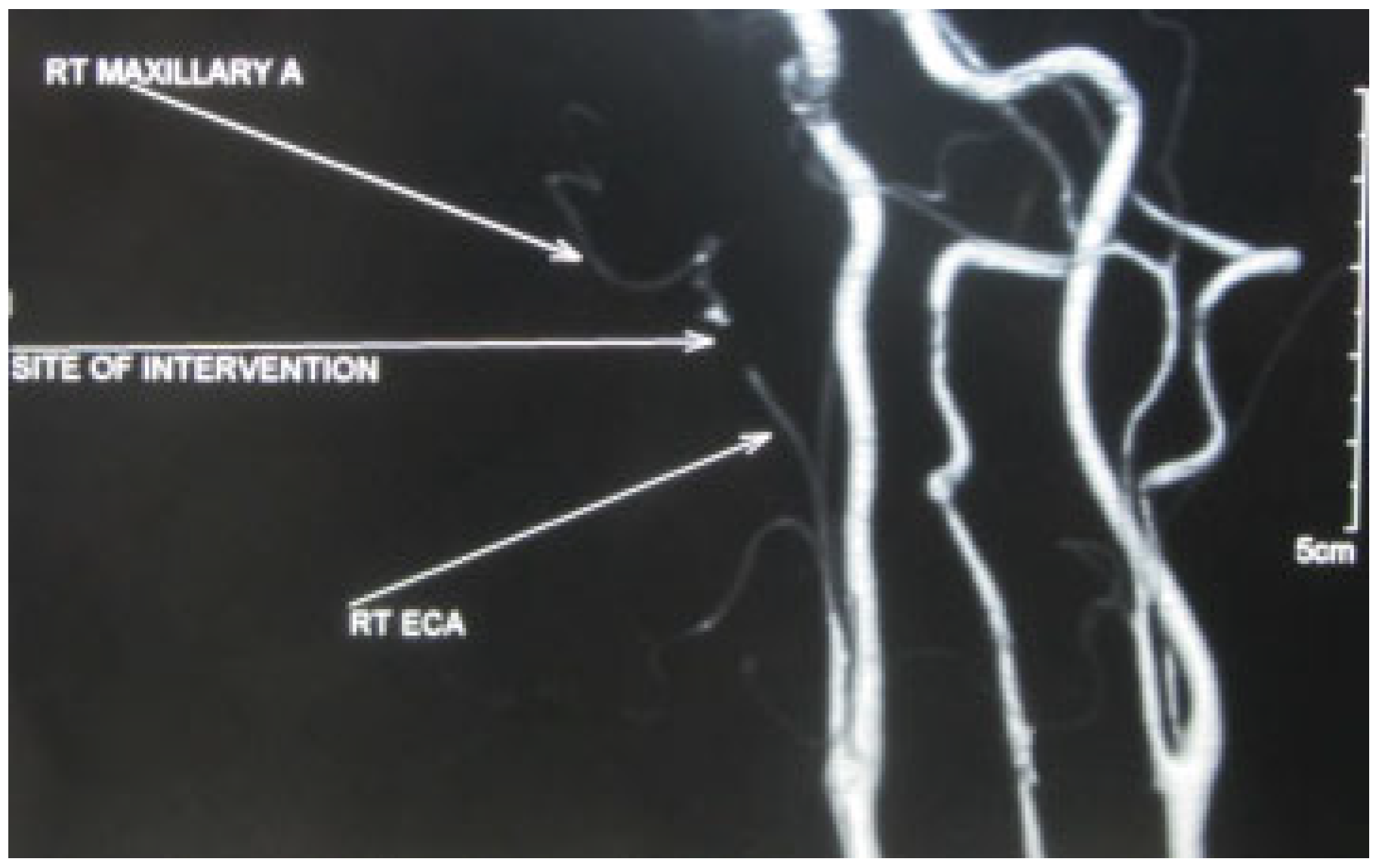
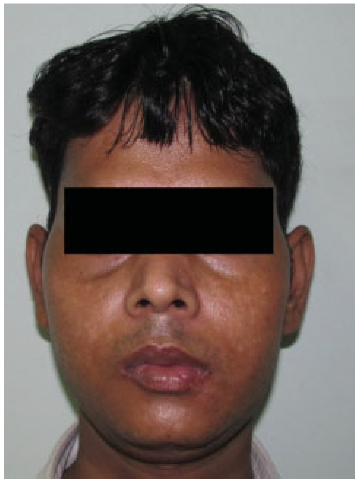
:Case Report
Discussion
Conflicts of Interest
References
- Fernández-Prieto, A.; García-Raya, P.; Burgueño, M.; Muñoz-Caro, J.; Frutos, R. Endovascular treatment of a pseudoaneurysm of the descending palatine artery after orthognathic surgery: Technical note. Int J Oral Maxillofac Surg 2005, 34, 321–323. [Google Scholar] [CrossRef] [PubMed]
- Maheshwari, R.; Paterson, A.W. Pseudoaneurysm of the superficial temporal artery. Br J Oral Maxillofac Surg 2009, 47, 412–413. [Google Scholar] [CrossRef] [PubMed]
- Webber, C.M.; Wind, G.G.; Burton, R.G. Pseudoaneurysm of the superficial temporal artery: Report of a case. J Oral Maxillofac Surg 1997, 55, 166–169. [Google Scholar] [CrossRef] [PubMed]
- El, A.S.; Guo, W.; Loveless, T.; et al. Pseudoaneurysm of the external carotid artery secondary to subcondylar fracture. Int J Oral Maxillofac Surg 2011, 40, 644–646. [Google Scholar] [CrossRef] [PubMed]
- Silva, A.C.; O’Ryan, F.; Beckley, M.L.; Young, H.Y.; Poor, D. Pseudoaneurysm of a branch of the maxillary artery following mandibular sagittal split ramus osteotomy: Case report and review of the literature. J Oral Maxillofac Surg 2007, 65, 1807–1816. [Google Scholar] [CrossRef] [PubMed]
- Hemmig, S.B.; Johnson, R.S.; Ferraro, N. Management of a ruptured pseudoaneurysm of the sphenopalatine artery following a Le Fort I osteotomy. J Oral Maxillofac Surg 1987, 45, 533–536. [Google Scholar] [CrossRef] [PubMed]
- Fan, X.; Mao, Q. Life-threatening oral haemorrhage of a pseudoaneurysm after raising of a fractured zygoma. Br J Oral Maxillofac Surg 2002, 40, 508–509. [Google Scholar] [CrossRef] [PubMed]
- Stiefel, M.F.; Park, M.S.; McDougall, C.G.; Albuquerque, F.C. Endovascular treatment of hemorrhagic alveolar artery pseudoaneurysm after tooth extraction: A case report. J Oral Maxillofac Surg 2010, 68, 2325–2328. [Google Scholar] [CrossRef] [PubMed]

© 2013 by the author. The Author(s) 2013.
Share and Cite
Mohanty, S.; Gulati, U.; Kathuria, S. Pseudoaneurysm of the Internal Maxillary Artery: A Rare Complication of Condylar Fracture. Craniomaxillofac. Trauma Reconstr. 2013, 6, 271-273. https://doi.org/10.1055/s-0033-1349209
Mohanty S, Gulati U, Kathuria S. Pseudoaneurysm of the Internal Maxillary Artery: A Rare Complication of Condylar Fracture. Craniomaxillofacial Trauma & Reconstruction. 2013; 6(4):271-273. https://doi.org/10.1055/s-0033-1349209
Chicago/Turabian StyleMohanty, Sujata, Ujjwal Gulati, and Sanjeev Kathuria. 2013. "Pseudoaneurysm of the Internal Maxillary Artery: A Rare Complication of Condylar Fracture" Craniomaxillofacial Trauma & Reconstruction 6, no. 4: 271-273. https://doi.org/10.1055/s-0033-1349209
APA StyleMohanty, S., Gulati, U., & Kathuria, S. (2013). Pseudoaneurysm of the Internal Maxillary Artery: A Rare Complication of Condylar Fracture. Craniomaxillofacial Trauma & Reconstruction, 6(4), 271-273. https://doi.org/10.1055/s-0033-1349209