Next Article in Journal
Facial Gel Complication After Dental Injection: A Case Report
Previous Article in Journal
Branching Pattern of the Extraosseous Mental Nerve in a Kenyan Population
 
 
Craniomaxillofacial Trauma & Reconstruction is published by MDPI from Volume 18 Issue 1 (2025). Previous articles were published by another publisher in Open Access under a CC-BY (or CC-BY-NC-ND) licence, and they are hosted by MDPI on mdpi.com as a courtesy and upon agreement with Sage.
Article

Complication of Anterior Iliac Bone Graft Harvesting in 372 Adult Patients from May 2006 to May 2011 and a Literature Review

by
Manar Almaiman
1,
Hamed H. Al-Bargi
1,* and
Paul Manson
2
1
Department of Oral and Maxillofacial Surgery, King Fahad Armed Forces Hospital, P.O. Box 12653, Jeddah 21483, Saudi Arabia
2
Department of Plastic and Reconstructive Surgery, John Hopkins University, Baltimore, MA, USA
*
Author to whom correspondence should be addressed.
Craniomaxillofac. Trauma Reconstr. 2013, 6(4), 257-265; https://doi.org/10.1055/s-0033-1357510
Submission received: 1 December 2013 / Revised: 1 December 2013 / Accepted: 1 December 2013 / Published: 7 October 2013

Abstract

:
Autogenous bone graft from the iliac is considered the gold standard graft material in maxillofacial surgery. The common and the rare complications associated with harvesting bone from anterior iliac crest were reviewed; we recommend a safe technique to avoid these complications. A retrospective analysis of 372 adult patients who had undergone anterior iliac bone graft harvesting from May 2006 to May 2011. The patients age range from 21 to 63 years. Out of the 372 patients, 200 were male with age range from 21 to 63 years and 172 were female with age range from 22 to 59 years. Two major complications (fracture and seroma) occurred, a fracture of the anterior superior iliac spine was observed in two patients (0.538%); one male and one female. One female patient (0.269%) developed seroma. One minor complication occurred in three patients (0.806%); one female and two females who suffered from temporary sensory disturbance. All patients (100%) suffered pain maximum for the first 15 days postoperative. In our study; the morbidity after anterior iliac bone graft harvesting was found to be low due to the technique, utilizing the proper instruments, gentle and minimal mobilization of the graft.

Anterior iliac crest is the most commonly used autogenous bone grafting source in oral and maxillofacial surgery. Unfortunately, considerable morbidity is associated with iliac crest harvest with a reported complications rates up to 49% including damage to blood vessels and nerves, joint disruption, fractures, subluxation, herniation of abdominal contents, and delayed iliac abscess.
In maxillofacial surgery, autologous bone grafting from the iliac crest is used for treating a bony defects related to tumors, trauma, inflammation, or aged-related atrophy of the jaw is the gold standard [1].
Regardless the shape or the substance of the graft and the methods of bone harvesting, differences in the incidence and the severity of complication have been described [1].
Major complications have been reported ranging from 0.7 to 25%, including infection, prolonged wound drainage, large hematomas, re-operation, pain lasting more than 6 months, sensory loss, scars, joint subluxation, gait disturbances, sacroiliac joint destabilization, herniation of abdominal muscles and contents, fracture iliac or pelvis, and heterotopic bone formation [2,3,4].
Minor complications are more common, with a reported complications rates ranging from 4 to 49%. These complications included superficial infection, minor wound problems, temporary sensory loss, and mild or transient pain [2].

Materials and Methods

At two different institutions with equal facilities and by the same surgeon, 372 anterior iliac bone graft harvesting procedures were undertaken from the beginning of May 2006 to the end of May 2011.
Total 372 adult patients were fulfilling the following criteria: Bone defects of the jaw secondary to trauma, pathological diseases, and atrophic edentulous or partially edentulous jaw. Patients aged between 21 and 63 years were included in the study, 200 male with age range from 12 to 63 years and 172 female with age range from 22 to 59 years.
Exclusion criteria composed of hematological diathesis, osteoporosis, drug addiction, and aspirin were stopped before surgery.
A minimum time of 1 year for follow-up was maintained, and the evaluation criteria were based on the frequent need for analgesia, the return to same daily activity as before the surgery, the presence of infection, hematoma, temporary, or permanent neurosensory disturbance, abdominal or inguinal hernia development, and postoperative plain films to determine the presence of iliac or pelvic fractures.

Surgical Technique

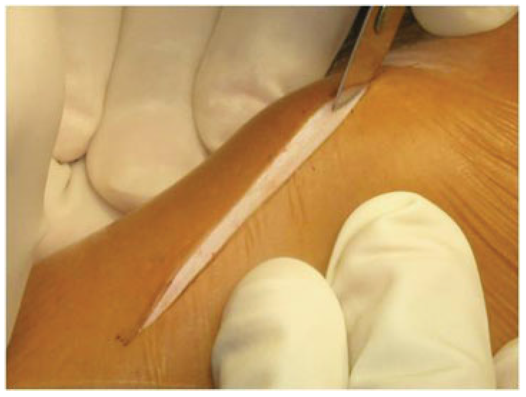
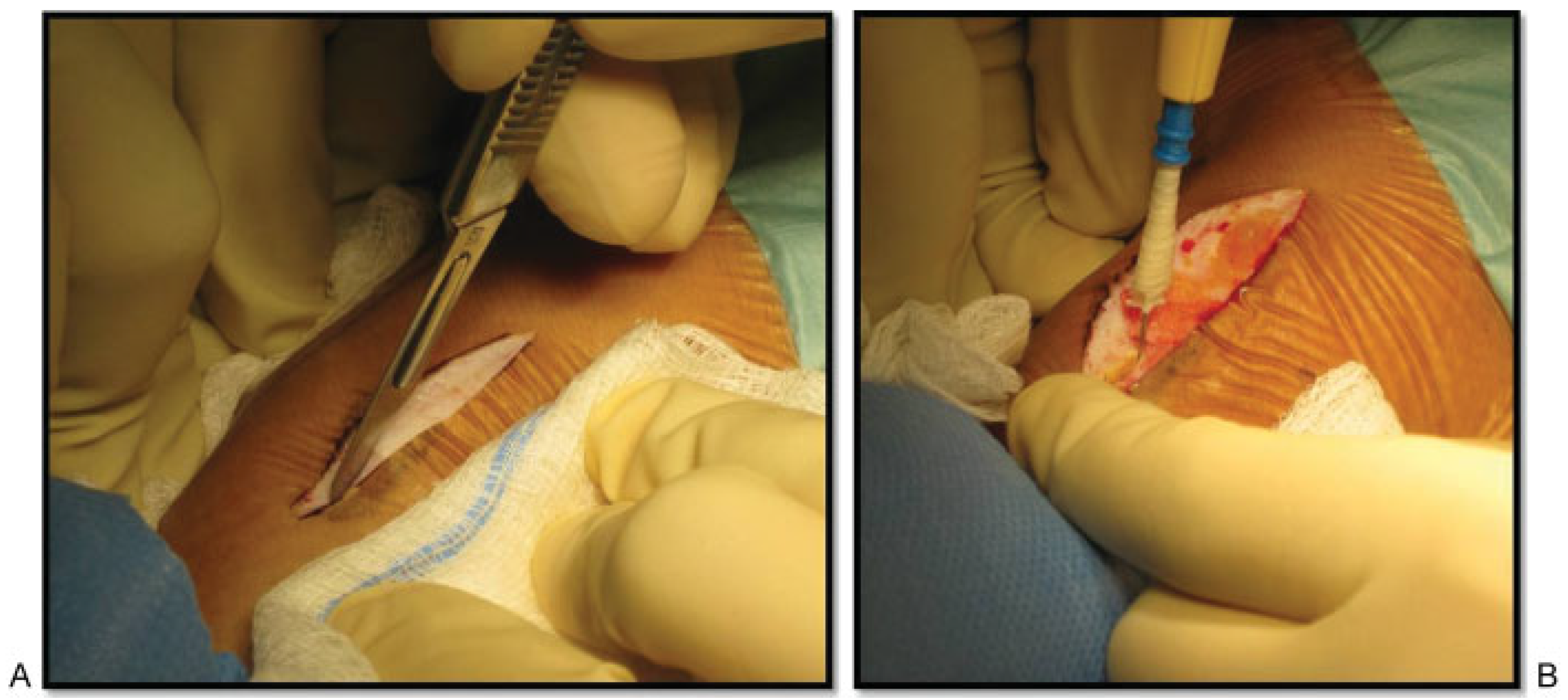
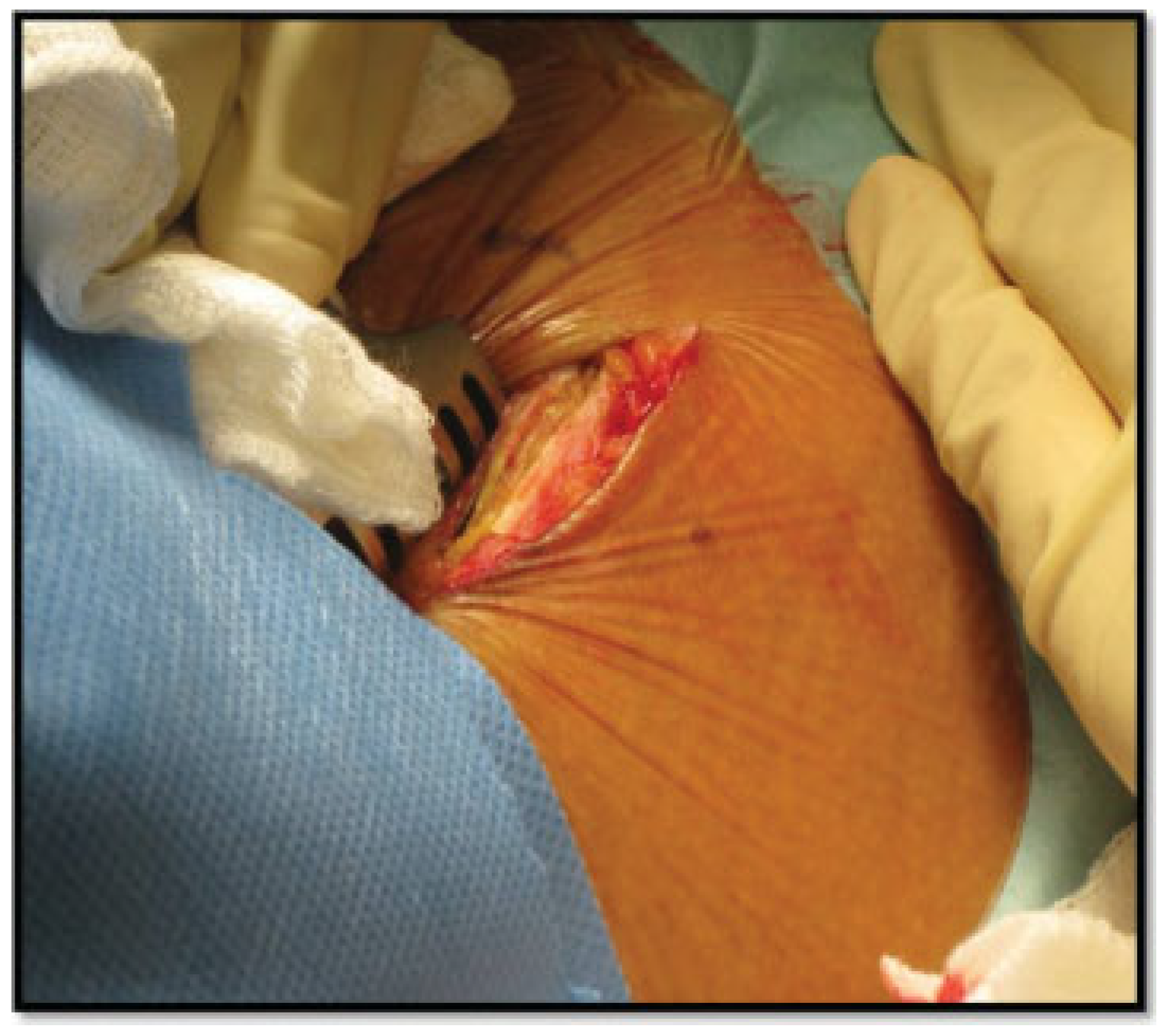
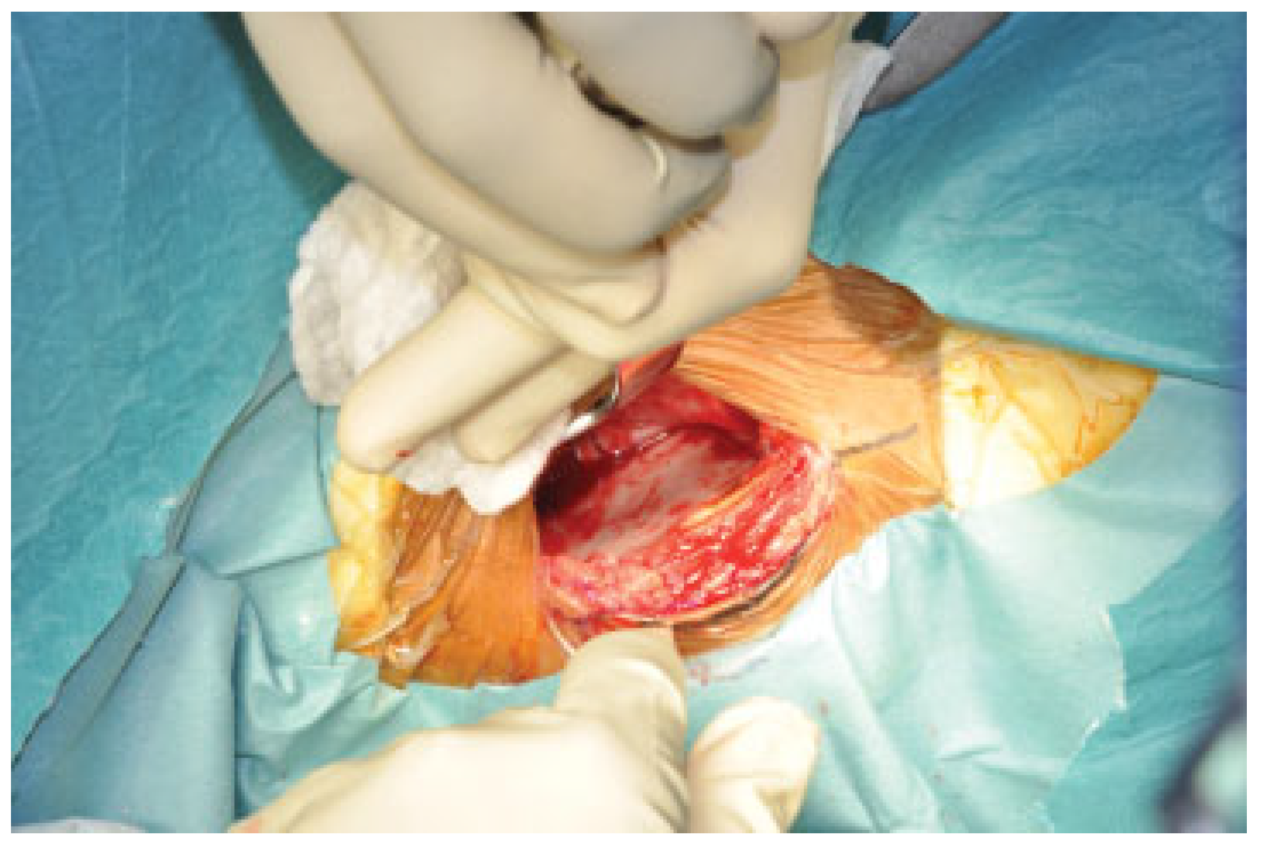
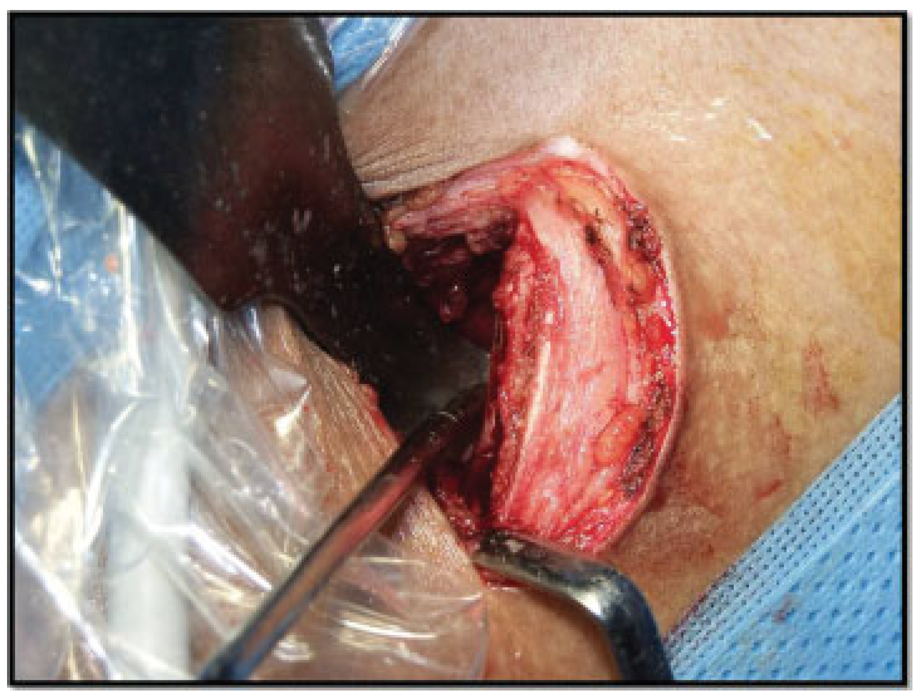
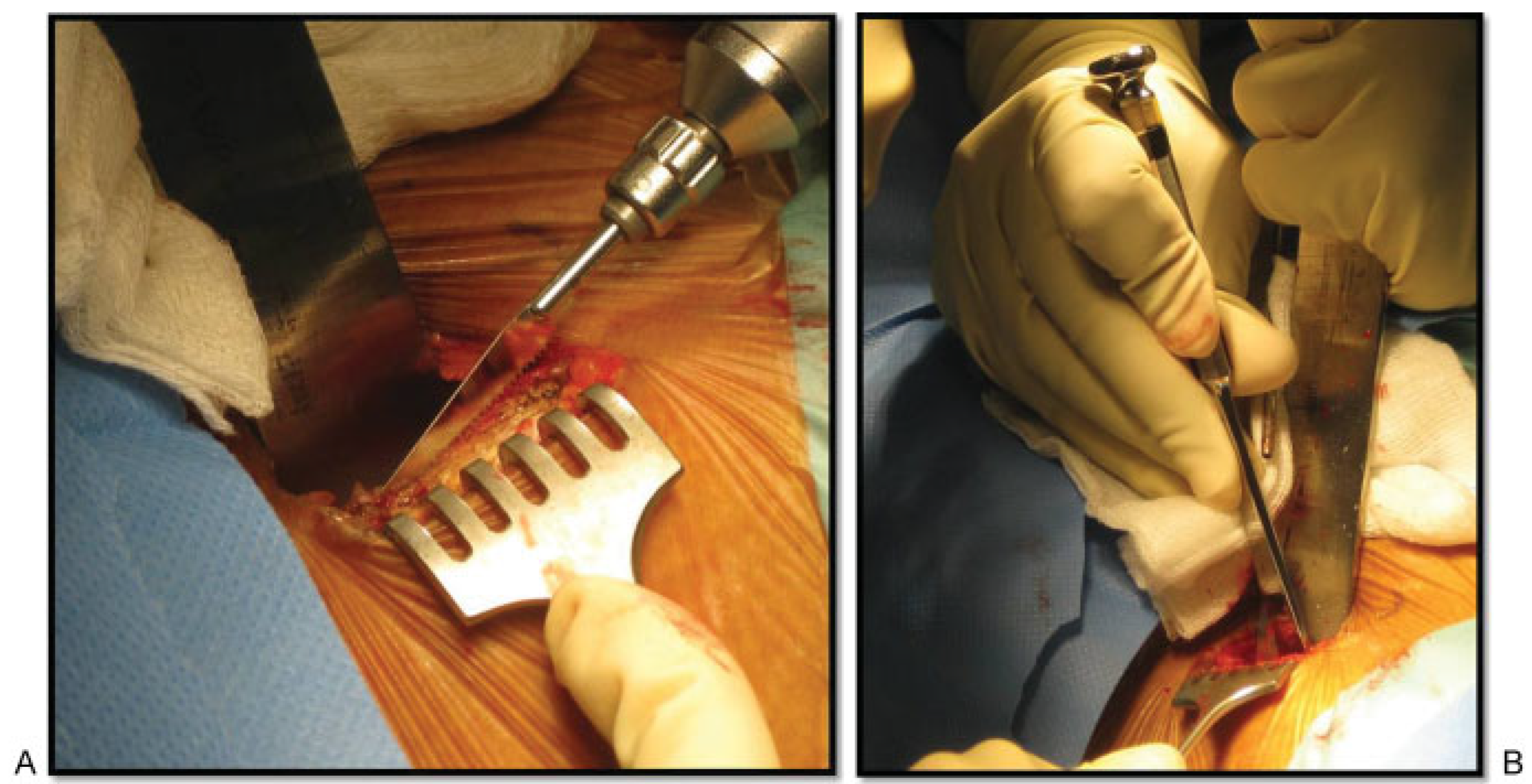
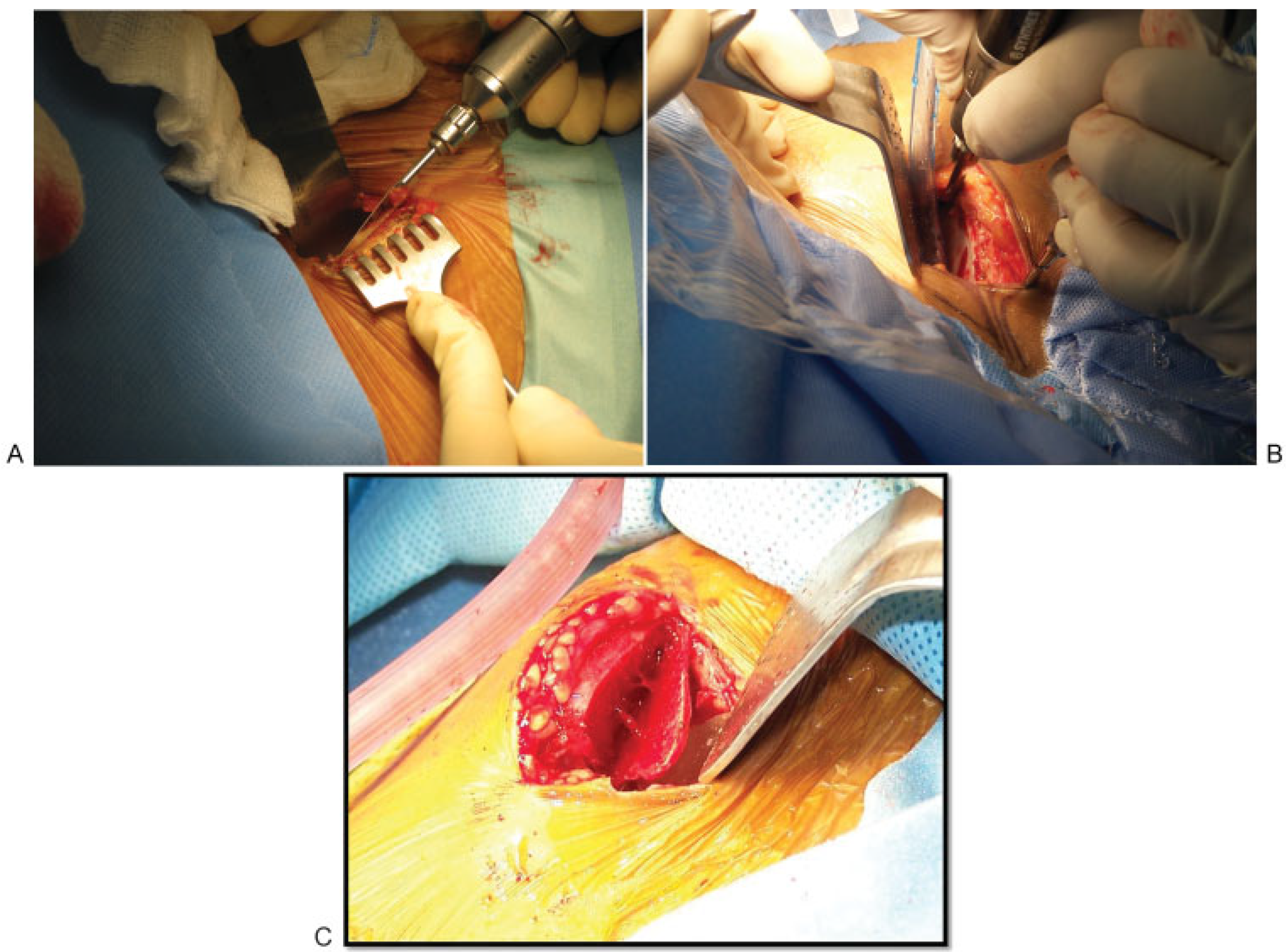
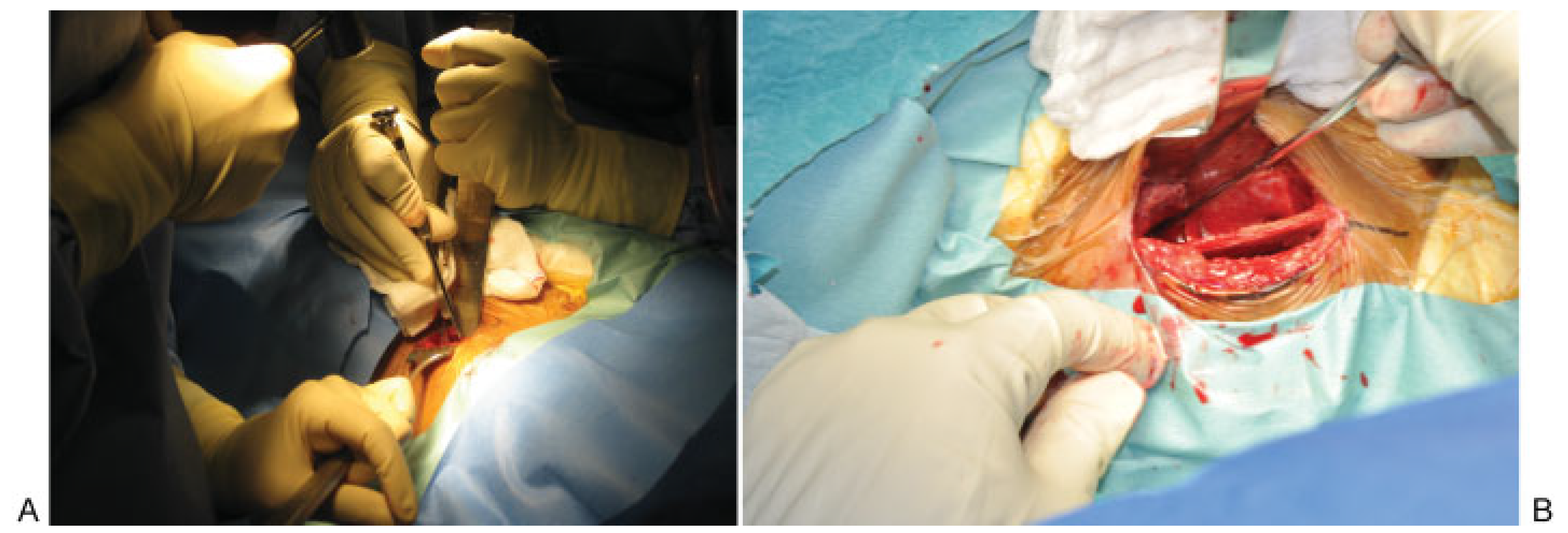
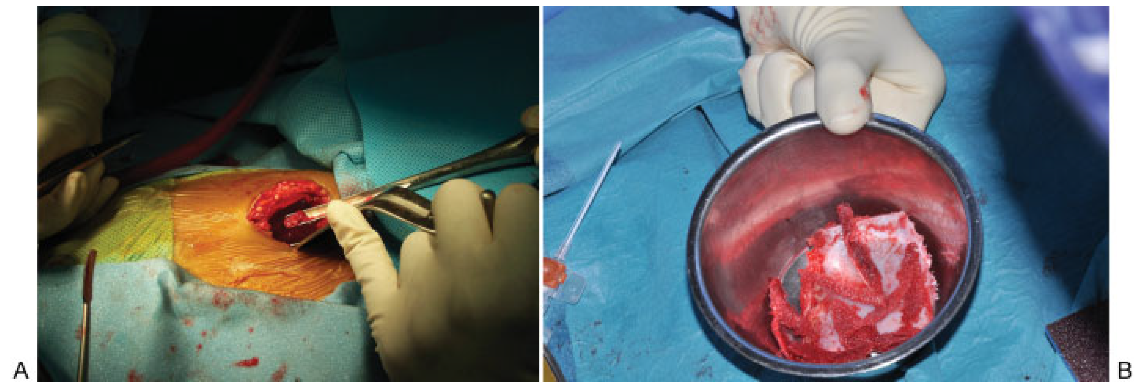
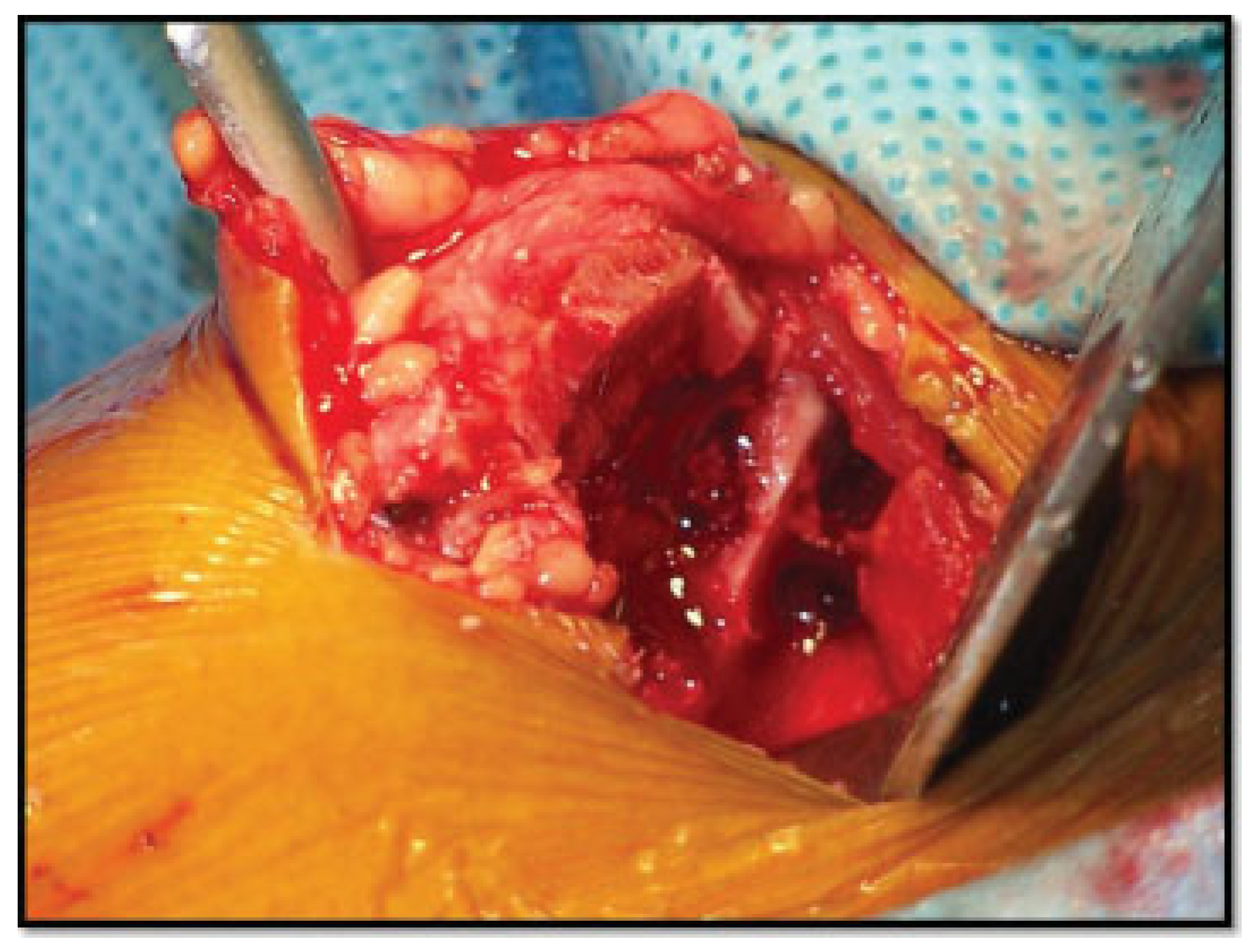
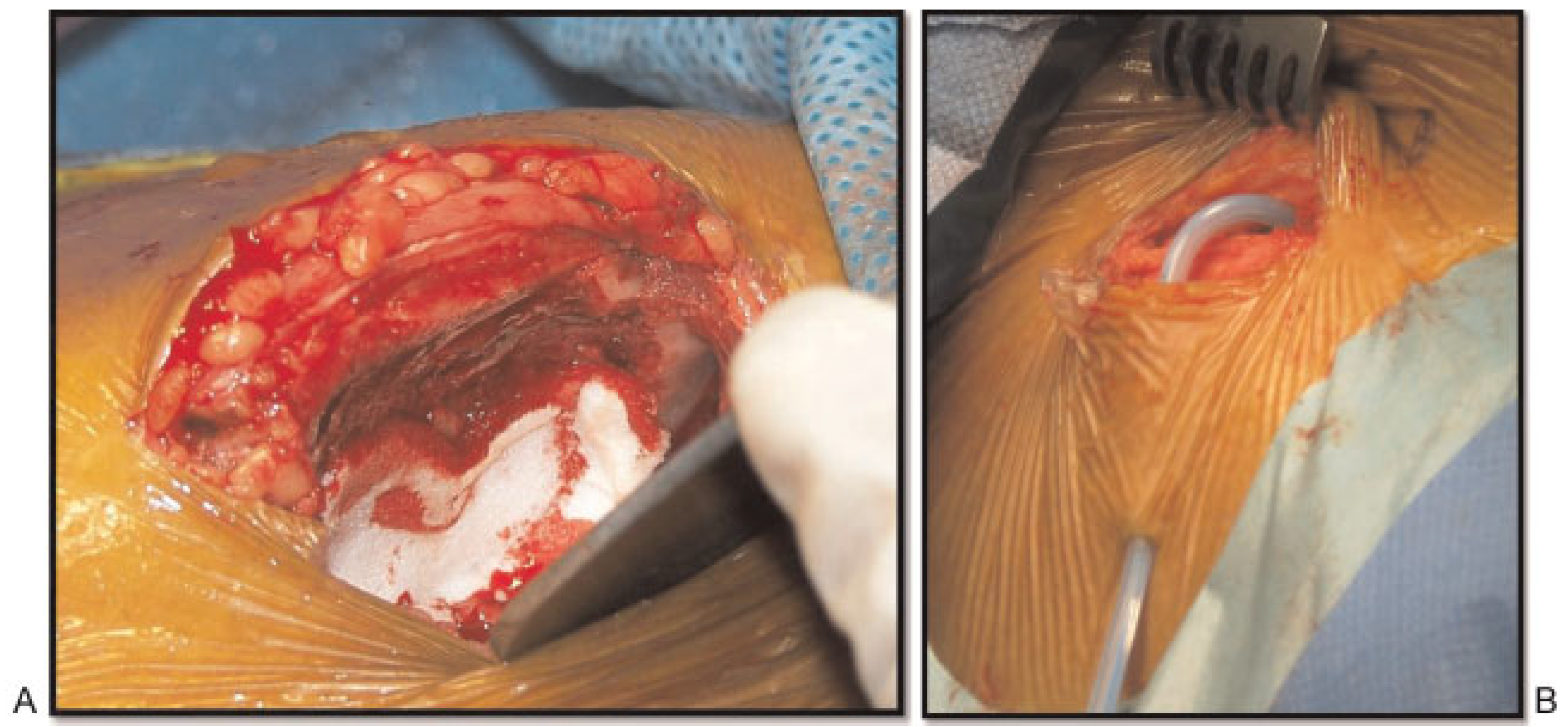
The bony landmark of the anterior superior iliac spine and iliac tubercle is identified (Figure 1). The iliac tubercle is located approximately 6 cm posterior to the anterior superior iliac spine (Figure 2). The incision is designed with the overlying skin retracted medially to prevent the surgical scar from lying directly over the crest (Figure 3). A 4 to 6 cm skin incision is placed 1 to 2 cm posterior to the tubercle of the ilium and 1 cm inferior to the anterior superior iliac spine, along the orientation of the anterior iliac crest (Figure 4). The dissection encounters the skin, subcutaneous tissue (Figure 5A), and Scarpa fascia (Figure 5B). A hypovascular dissection plane without transecting muscle will be found between the tensor fascia lata laterally and the external and transverse abdominal muscles medially (Figure 6). We identify the dense fibrous periosteum of the iliac crests (Figure 7), then sharply transect the periosteum (Figure 8). We reflect the iliacus muscle medially (Figure 9) and expose the medial iliac crest (Figure 10). Bone harvesting can be obtained by different methods using a surgical saw (Figure 11A) and osteotome (Figure 11B). The osteotomy is established first on the iliac tubercle (Figure 12A) followed by two vertical osteotomy lines (according to the desired length) at the medial surfaces of the iliac curvature using reciprocating saw and a horizontal line joining to two vertical lines (Figure 12B). Detaching the bone graft is established by using a straight osteotome starting at the iliac tubercle followed by curved osteotome (Figure 13A), with steady and gentle tapping using the mallet (Figure 13B). The cancellous bone graft can be obtained using bone gauche (Figure 14A, B) A total of 4 × 5 cm of bone can be obtained (Figure 15A,B); this is limited by the proximity to anterior superior iliac spine and tubercle of the ilium. It is important to preserve a 1 to 2 cm of bone posterior to the anterior iliac spine (AIS) distance and a total depth of harvest can be up to 5 cm to minimize the risk of AIS and fracture of tubercle of the ilium (Figure 16). We inspect the surgical wound and obtain hemostasis before closure (Figure 17A,B).
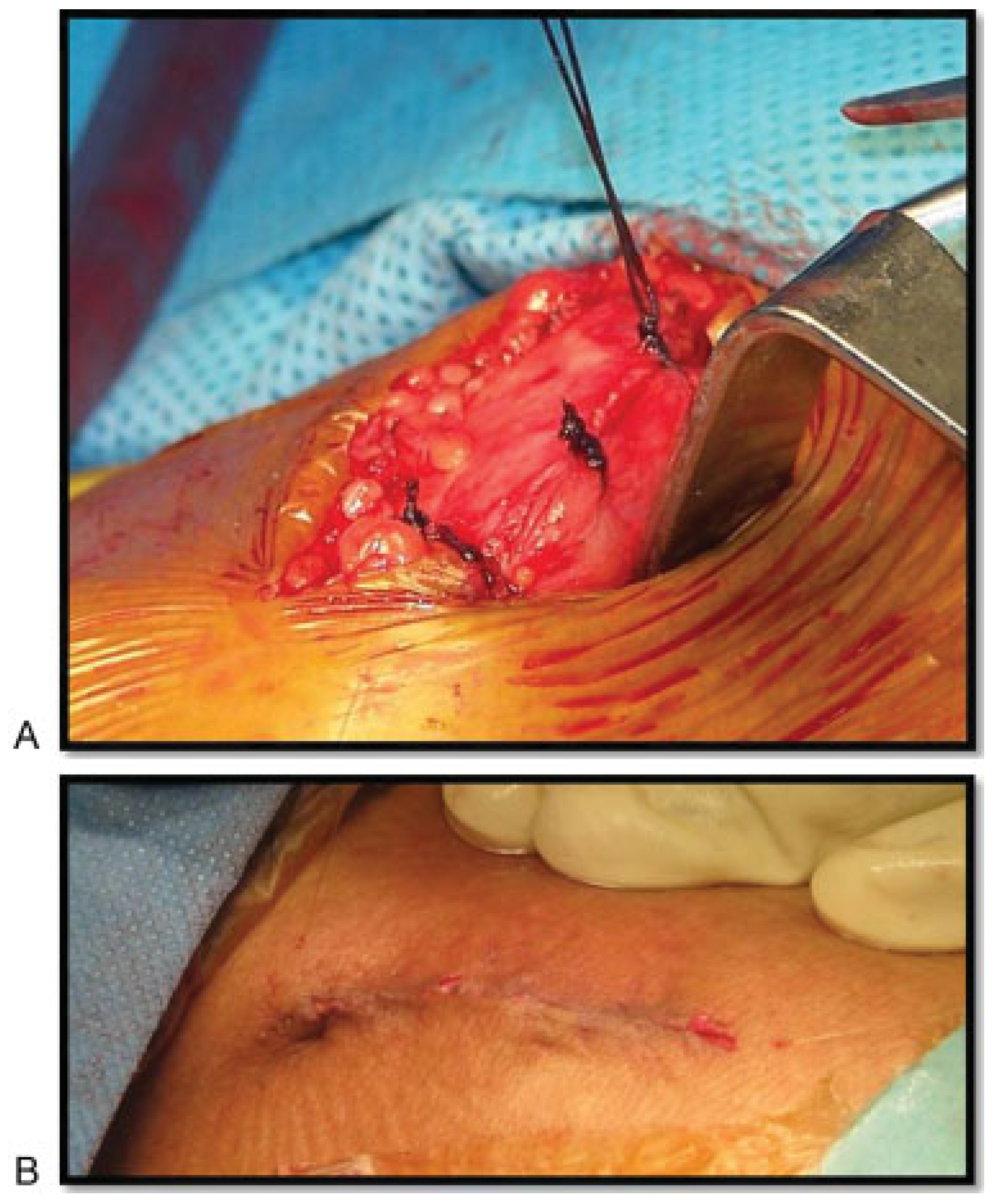
Closure should be achieved to re-approximate the periosteal envelope over the iliac crest followed by subcutaneous and skin closure (Figure 18A,B). The average surgical time is 1 hour.

Postoperative Care

It is important to allow the patient to ambulate on the first postoperative day with assistance, and to provide a meticulous wound care, intravenous antibiotic, and profound pain control.

Result

A total of 372 patients who underwent anterior iliac bone grafting procedures and fulfilled the inclusion criteria participated. All the 372 patients were available for a clinical followup examination. Of the 372 patients, 200 patients (53.763%) were males and 172 patients (46.237%) were female. The main indications for bone graft harvesting were jaw bony defect related to trauma, pathological diseases, and atrophic edentulous or partially edentulous jaw. In the 372 patients, a corticocancellous bone graft harvesting procedures were obtained by the same surgeon, in two different institutions with equal facilities.

Complications

The complications rates are shown in Table 1 (and also see Figure 22).
  • Seroma: Of the 372 patients, 1 patient (0.269%) developed seroma, approximately 25 mL been aspirated (Figure 19A–C).
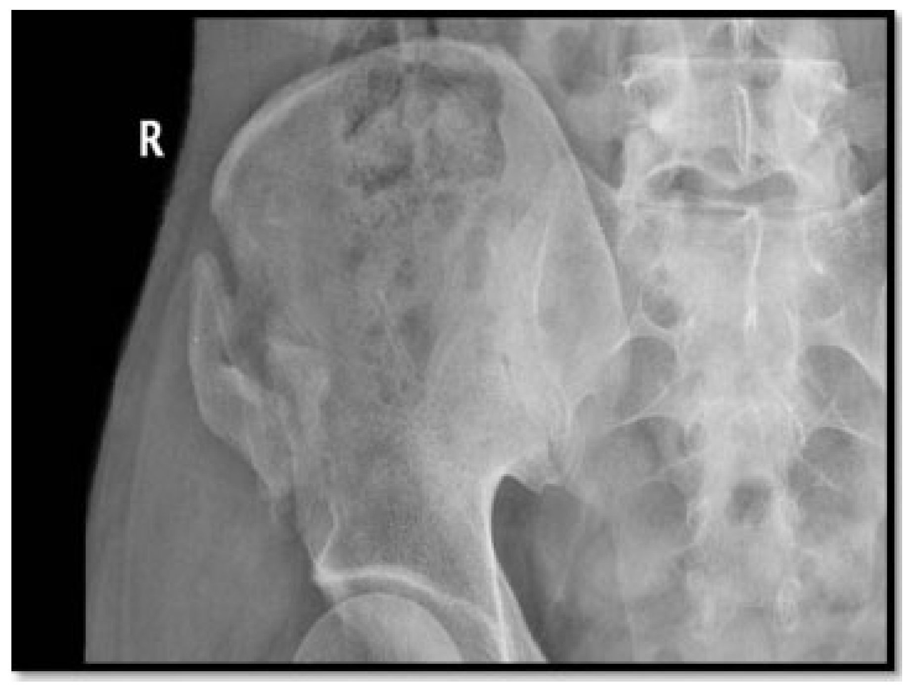
  • Fractures: Of the 372 patients, 2 patients (0.538%) developed fracture iliac, 1 patient developed greenstick fracture of the iliac wing intraoperative and the case was managed with lag screw, and the other patient had the fracture iliac 25 days postoperative secondary to carrying 45 kg of sac of rise and he was managed conservatively (Figure 20).
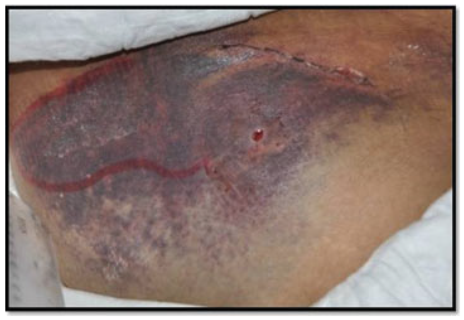
  • Major hematoma: Of the 372 patients, 1 patient (0.269%) developed major hematoma secondary to excessive daily activities and exercises (Figure 21).
  • Temporary neurosensory disturbance: Of the 372 patients, 3 patients (0.806%) developed temporary sensory disturbance, which was managed conservatively.
  • Duration of pain: Of the 372 patients, all the patients (100%) suffered pain within the first 15 days postoperative, and they were managed with a proper analgesic (Figure 22).

Discussion

Many studies have been published on the morbidity of donor site after harvesting anterior iliac bone graft with very different results [1]. Complications associated with anterior iliac crest bone graft harvesting been reported to be range from 1 to 25% including hematoma, seroma, nerve injury, cosmetic deformity, abdominal hernia, pelvic instability ileus, infection, and persistent pain [5].
A variety of possible reasons can related to this wide differences in the results, they might be as a result of different harvesting techniques, different volume of bone graft harvesting, or different methods in evaluation.
Persistent pain percentage range from 0 to 49%, the rate of sensory disturbance of the lateral femoral cutaneous nerve range from 2.9 to 27% and 4.3 to 17% for permanent functional disorders [1].
Eufinger and Leppänen concluded in their study that neither the open technique nor the closed technique was accompanied with a long-term complication [6].
Herford and Dean found in their series of 114 patients that 10% experienced pain for greater than 16 weeks and 23% experience some difficulties in ambulation 6 weeks postoperatively [7].
Ahlmann et al compared the anterior and posterior iliac crest bone harvesting site morbidity in 88 patients, and they concluded that the anterior iliac crest harvesting was associated with more complication rate than the posterior iliac crest approaches, 8 versus 2%, respectively [8].
In a study by Farrow et al, a transient femoral neuropathy after harvesting of bone from the iliac crest have been reported [9]. Another study by Kargel et al reported two cases of femoral nerve palsy as a complication of anterior iliac bone harvesting [10].
A rare complication of anterior iliac bone graft harvesting has been reported. Chou et al report a pseudoaneurysm of the deep circumflex iliac artery as a rare complication at an anterior iliac bone graft donor site treated by coil embolization [11].
A study by De Riu et al published a case of delayed iliac abscess been reported as an unusual complication of an anterior bone graft in orthognathic [12]. Nodarian et al reported a case of liver herniation as a complication of iliac crest bone graft harvesting [13].

Conclusion

Serious damage of nearby structures rarely occurs during iliac crest harvesting, but when such damage occurs, it is often a consequence of proximity of the iliac crest graft site to the major vascular and neurologic structures. However, the frequency of complications with iliac crest harvesting can be reduced by harvesting no more bone than is necessary, adhering to optimal surgical procedures and be aware of the regional anatomy.

References

  1. Schaaf, H.; Lendeckel, S.; Howaldt, H.-P.; Streckbein, P. Donor site morbidity after bone harvesting from the anterior iliac crest. Oral Surg Oral Med Oral Pathol Oral Radiol Endod 2010, 109, 52–58. [Google Scholar] [CrossRef] [PubMed]
  2. Dösoğlu, M.; Orakdöğen, M.; Tevrüz, M.; Göğüsgeren, M.A.; Mutlu, F. Enterocutaneous fistula: A complication of posterior iliac bone graft harvesting not previously described. Acta Neurochir (Wien) 1998, 140, 1089–1092. [Google Scholar] [PubMed]
  3. Fernando, T.L.; Kim, S.S.; Mohler, D.G. Complete pelvic ring failure after posterior iliac bone graft harvesting. Spine 1999, 24, 2101–2104. [Google Scholar] [CrossRef] [PubMed]
  4. Younger, E.M.; Chapman, M.W. Morbidity at bone graft donor sites. J Orthop Trauma 1989, 3, 192–195. [Google Scholar] [CrossRef] [PubMed]
  5. Zouhary, K.J. Bone graft harvesting from distant sites: Concepts and techniques. Oral Maxillofac Surg Clin North Am 2010, 22, 301–316. [Google Scholar] [CrossRef] [PubMed]
  6. Eufinger, H.; Leppänen, H. Iliac crest donor site morbidity following open and closed methods of bone harvest for alveolar cleft osteoplasty. J Craniomaxillofac Surg 2000, 28, 31–38. [Google Scholar] [CrossRef]
  7. Herford, A.S.; Dean, J.S. Complications in bone grafting. Oral Maxillofac Surg Clin North Am 2011, 23, 433–442. [Google Scholar] [CrossRef] [PubMed]
  8. Ahlmann, E.; Patzakis, M.; Roidis, N.; Shepherd, L.; Holtom, P. Comparison of anterior and posterior iliac crest bone grafts in terms of harvest-site morbidity and functional outcomes. J Bone Joint Surg Am 2002, 84-A, 716–720. [Google Scholar] [CrossRef]
  9. Farrow, A.; Morrison, R.; Pickersgill, T.; Currie, R.; Hammersley, N. Transient femoral neuropathy after harvest of bone from the iliac crest. Br J Oral Maxillofac Surg 2004, 42, 572–574. [Google Scholar] [CrossRef]
  10. Kargel, J.; Dimas, V.; Tanaka, W.; et al. Femoral nerve palsy as a complication of anterior iliac crest bone harvest: Report of two cases and review of the literature. Can J Plast Surg 2006, 14, 239–242. [Google Scholar] [CrossRef] [PubMed]
  11. Chou, A.S.-B.; Hung, C.-F.; Tseng, J.H.; Pan, K.T.; Yen, P.S. Pseudoaneurysm of the deep circumflex iliac artery: A rare complication at an anterior iliac bone graft donor site treated by coil embolization. Chang Gung Med J 2002, 25, 480–484. [Google Scholar] [PubMed]
  12. De Riu, G.; Meloni, S.M.; Raho, M.T.; Gobbi, R.; Tullio, A. Delayed iliac abscess as an unusual complication of an iliac bone graft in an orthognathic case. Int J Oral Maxillofac Surg 2008, 37, 1156–1158. [Google Scholar] [CrossRef] [PubMed]
  13. Nodarian, T.; Sariali, E.; Khiami, F.; Pascal-Mousselard, H.; Catonné, Y. Iliac crest bone graft harvesting complications: A case of liver herniation. Orthop Traumatol Surg Res 2010, 96, 593–596. [Google Scholar] [CrossRef] [PubMed]
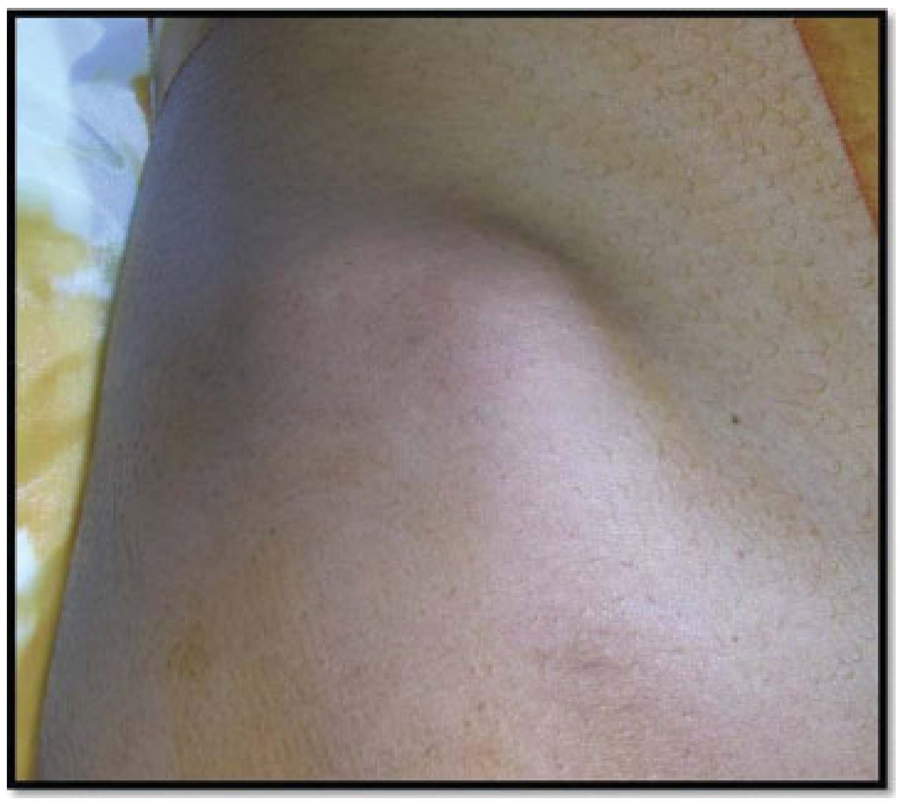
Figure 1. Identify the bony landmark of the anterior superior iliac spine and iliac tubercle.
Cmtr 06 00043 g001
Figure 2. The iliac tubercle location.
Cmtr 06 00043 g002
Figure 3. The overlying skin retracted medially.
Cmtr 06 00043 g003
Figure 4. A 4 to 6-cm skin incision is placed1 to 2 cm posterior to the tubercle of the ilium.
Cmtr 06 00043 g004
Figure 5. (A) The dissection encounters the skin, subcutaneous tissue. (B) The dissection encountered the Scarpa fascia.
Cmtr 06 00043 g005
Figure 6. A hypovascular dissection plane found between the tensor fascia lata laterally and the external and transverse abdominal muscles medially.
Cmtr 06 00043 g006
Figure 7. Identity the dense fibrous periosteum of the iliac crest.
Cmtr 06 00043 g007
Figure 8. Sharp transection of the periosteum.
Cmtr 06 00043 g008
Figure 9. Reflection of the iliacus muscle is medially [2].
Cmtr 06 00043 g009
Figure 10. Exposing the medial iliac crest.
Cmtr 06 00043 g010
Figure 11. (A) Bone harvesting using a surgical saw and (B) bone harvesting osteotomes.
Cmtr 06 00043 g011
Figure 12. A total bone length of 4 to 6 cm can be obtained. (A) Osteotomy is established first on the iliac tubercle. (B) Two vertical osteotomy lines on the medial surfaces of the iliac curvature. (C) The bone harvested from anterior iliac.
Cmtr 06 00043 g012
Figure 13. (A) Detaching the bone graft using a straight osteotome. (B) Bone graft separation.
Cmtr 06 00043 g013
Figure 14. (A) Cancellous bone harvesting using bone gauche. (B) Cancellous bone harvesting.
Cmtr 06 00043 g014
Figure 15. (A) Bone graft length of 4.5 cm. (B) Bone graft width of 5 cm.
Cmtr 06 00043 g015
Figure 16. Total depth of harvest can be up to 5 cm.
Cmtr 06 00043 g016
Figure 17. (A) Surgical wound hemostasis and (B) suction drain.
Cmtr 06 00043 g017
Figure 18. (A) Re-approximate the periosteal envelope over the iliac crest and (B) subcutaneous and skin closure.
Cmtr 06 00043 g018
Figure 19. (A) Aspiration of seroma. (B) Free drainage of seroma from the harvesting site. (C) Aspiration of 25 mL.
Cmtr 06 00043 g019
Figure 20. Fracture iliac wing.
Cmtr 06 00043 g020
Figure 21. Major hematoma.
Cmtr 06 00043 g021
Figure 22. The percentage of anterior iliac crest donor site complications.
Cmtr 06 00043 g022
Table 1. The complications rate of anterior iliac crest donor site.
ComplicationsNumber of patientsPercentage
Bleeding00
Permanent nerve damage00
Seroma10.269
Gait disturbance00
Fractures20.538
Infection00
Major subcutaneous hematoma10.269
Temporary nerve damage30.806
Ileus00
Pain372100