Next Article in Journal
Complication of Anterior Iliac Bone Graft Harvesting in 372 Adult Patients from May 2006 to May 2011 and a Literature Review
Previous Article in Journal
Assessment of Masticatory Function Using Bite Force Measurements in Patients Treated for Mandibular Fractures
 
 
Craniomaxillofacial Trauma & Reconstruction is published by MDPI from Volume 18 Issue 1 (2025). Previous articles were published by another publisher in Open Access under a CC-BY (or CC-BY-NC-ND) licence, and they are hosted by MDPI on mdpi.com as a courtesy and upon agreement with Sage.
Article

Branching Pattern of the Extraosseous Mental Nerve in a Kenyan Population

by
Poonamjeet Kaur Loyal
*,
Fawzia Butt
and
Julius Alexander Ogeng'o
Department of Human Anatomy, University of Nairobi, G.P.O. Chiromo Campus, Riverside Drive off Waiyaki Way, P.O. Box 30197, Nairobi 00100, Kenya
*
Author to whom correspondence should be addressed.
Craniomaxillofac. Trauma Reconstr. 2013, 6(4), 251-256; https://doi.org/10.1055/s-0033-1356756
Submission received: 7 February 2013 / Revised: 8 February 2013 / Accepted: 8 February 2013 / Published: 8 August 2013

Abstract

:
Knowledge of the branching pattern of the mental nerve is an important consideration during placement of tooth implants and reconstructive plates. It is known to display population variations and data for the same is scarce form the sub-Saharan region. With the recent increase in surgical interventions in the mandibular region in Kenya, a detailed description of mental nerve is warranted. A total of 64 mental nerves were dissected and branching pattern was noted. Single mental nerve was present in 60 (93.7%) cadavers while double mental nerves occurred in 3 (4.7%) and accessory in 1 (1.6%). Most common pattern was bifurcation (39%) followed by trifurcation (34%), single (19%), and quadrification (8%). The double and triple branches were seen to further divide into two to three subbranches with diverse patterns. Side symmetry in branching was seen in four (6.25%). These are important anatomical considerations during placement of reconstructive plates for mandibular trauma and administration of mental nerve blocks.

The mental nerve (MN), terminal branch of the inferior alveolar nerve, exits the mental foramen (MF) [1] and divides into three branches to innervate the skin of chin, lower lip, and gingivae [2]. The number of MNs on the ipsilateral side varies from none to four with the most common variation being two [3,4,5,6,7]. Branches of the MN vary from one to four [8,9], with the most common being four branches, namely medial, lateral inferior labial, angular, and mental [10]. These data are, however, mainly in form of case reports, hence the true incidence of such occurrences is difficult to establish. A detailed description regarding the exact branching pattern is important for effective presurgical evaluations in implant surgery, administration of MN block anesthesia, and for osteotomy procedures [11]. Surgical interventions involving the mandibular region have increased in the past decade [12]. The branching pattern displays population differences [9]. There is however no data for the black Kenyan population. The aim of this study was, therefore, to clearly elucidate the branching pattern of the MN in Kenyans to minimize iatro- genic injuries during surgical maneuvers.

Materials and Methods

A total of 64 MNs were dissected at the Department of Human Anatomy, University of Nairobi. All the specimens were from adult black Kenyan. The MN was exposed by three skin incisions and lateral reflection of the skin flap. A midline incision extended from the midpoint of the superior border of the lower lip to the midpoint of the inferior border of the mental symphysis (MS). A lateral incision extended approxi- mately 7 cm from the angle of the lip on either side. An inferior incision was made along the lower border of the mandible from the MS till the lateral extent of the lateral incision. Using blunt dissection, the flap was detached from the bone and reflected laterally until the MN was seen to exit from the MF. The MN was carefully dissected and the branching pattern was carefully displayed. Data collected included the number of MNs, initial branching pattern, terminal branching pattern, and symmetri- cal branching patterns. A branching pattern was classified as high or low depending on whether the MN branched at the level of the MF or at a distance from the MF, respectively. The data collected was analyzed using Microsoft Excel 2007 (Micro- soft Corporation, Redmond, WA). The frequencies and percen- tages of the various branching patterns were determined. The variations noted were photographed using a high resolution digital camera and managed using Photoscape version 3.5 (MOO11 Tech, Korea).

Results

The MN was present in all cadavers that were dissected. The pattern of branching of the MN after its exit from the MF displayed diverse set of variations in the number and branch- ing pattern. A single MN was present in 60 cadavers (93.7%) while double MNs occurred in 3 (4.7%) (Figure 1A,B) and accessory in 1 (1.6%) (Figure 1C). The accessory MN emerged in the region of the second premolar, 4 mm from the MN. When the inferior alveolar nerve branched into two terminal branches which were of similar sizes, the pattern of branching was described as double MNs. Accessory MNs were smaller branches from the inferior alveolar nerve in addition to the MN that had the same area of innervations as the MN.

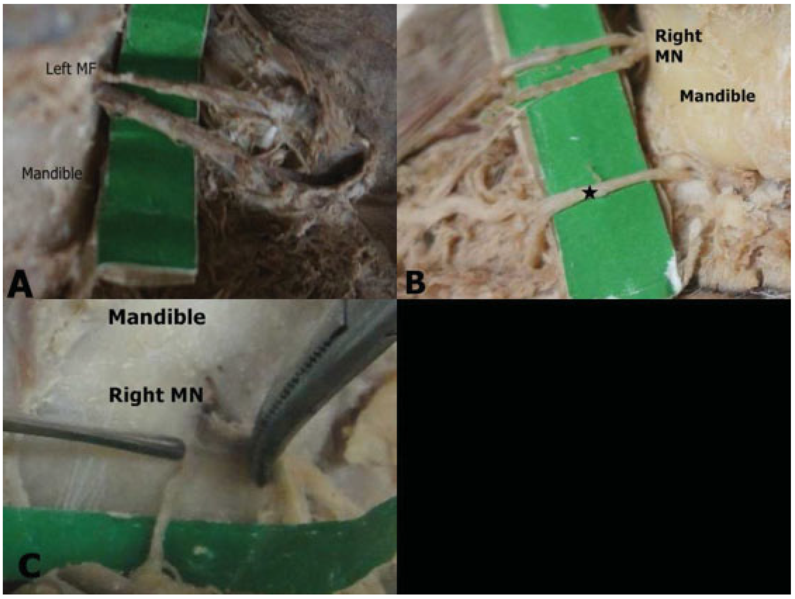
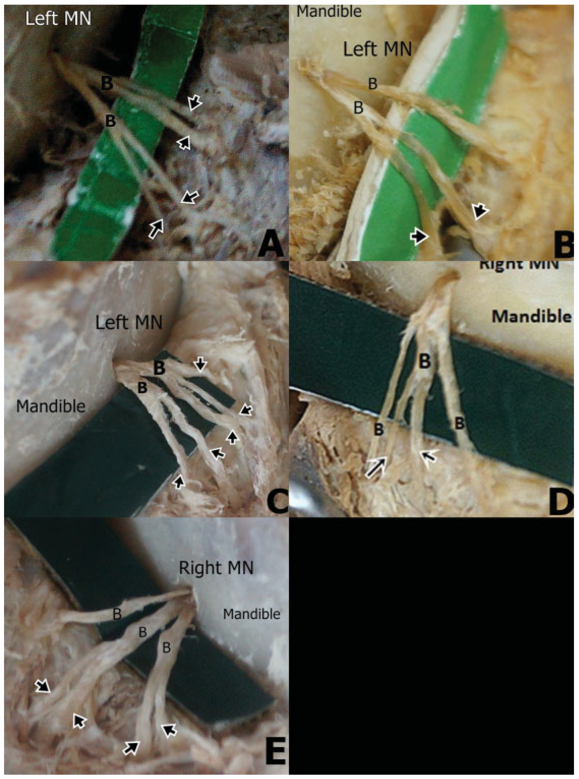
Initial Branching Pattern

The MN displayed a varied branching pattern with the number of branches varying from one to four on the ipsilat- eral side. The most common pattern of branching was divi- sion of the MN into two branches (39%) which displayed a high level division, that is, branching at the level of the MF in 11% (Figure 2A) while in 28%, the branching was seen at a distance from the MF hence a low division branching pattern (Figure 2B). The MN split into three branches in 34% (Figure 2C), continued its course as a single nerve without splitting into any branches in 19% (Figure 2D). In 8% of the cadavers, a quadruple division was observed (Figure 2E).

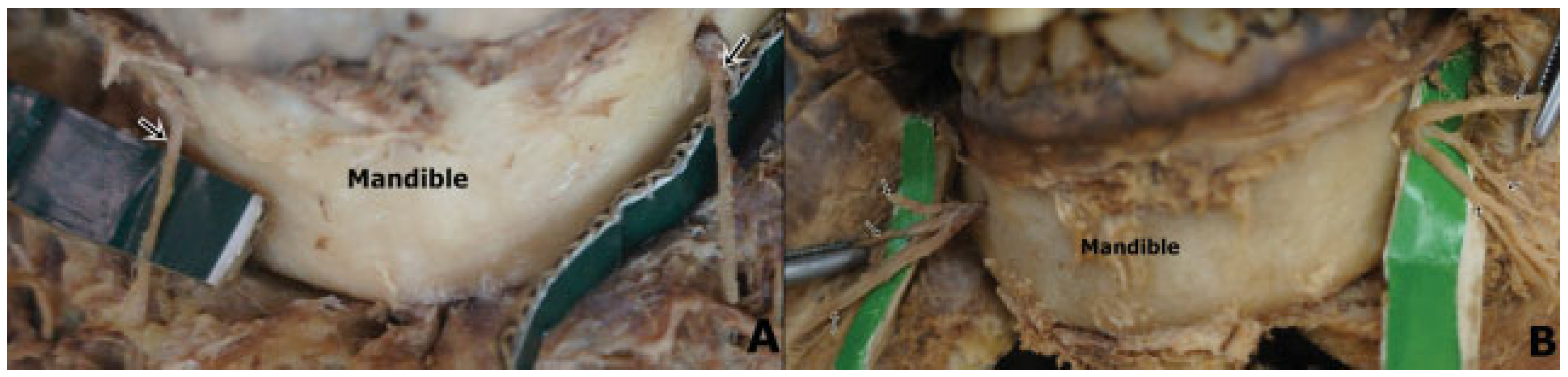
Terminal Branching Pattern

The double and triple branches of the MN further exhibit a diverse pattern of division into subbranches. For the double branches, the most commonly observed redivision pattern was seen in 12.7%, where each of the two main branches of the MN further divided into two smaller branches (Figure 3A), hence a total of four subbranches arising from the two branches of the MN. In 7.8%, one of the two main branches of the MN further redivided into two smaller subbranches (Figure 3B). In 1.56%, five subbranches were seen to arise from the two branches of the MN (Figure 3C). About 9.1% of the triple branching patterns, divided further into two subbranches (Figure 3D) while in 1.56%, four subbranches were observed (Figure 3E).

Symmetry

Significantly high levels of side asymmetry were observed in the same cadaver. Symmetrical branching pattern was ob- served in only 6.25% of the cadavers (Figure 4A,B). In 3.125%, the MN continued as a single branch on both the right and left side on the same cadaver (Figure 4A). Similarly, for another 3.125% of the cases, it split into the triple branch presentation on both the right and the left sides of the same cadaver (Figure 4B).

Discussion

The MN was present in all the 64 hemifaces, which is at variance with previous studies that have reported unilateral and bilateral absences [13]. The prevalence of double MNs in the current study (4.7%) falls in the range of 1 to 10.6% reported in the previous studies [4,14]. It is, nonetheless higher than that reported for the Thai population but lower than in Greeks, Indian, and Japanese population (Table 1). This suggests wide population differences. Separation of the MN earlier than the formation of the MF is a likely explanation for the presence of variations in the number of nerves [4]. The presence of additional MNs has been associated with failed mandibular anesthesia [15]. There are currently no studies done on the failure rates of complete mandibular anesthesia in the sub- Saharan region, but these results suggest a lower rate for Kenyans compared with the Greek, Indian, and Japanese population (Table 1).
Knowledge of the prevalence of accessory MNs is impor- tant during the placement reconstruction plates for mandib- ular fractures and in screwing tooth implants as accessory MNs may be injured during these procedures [11]. The 1.6%, prevalence of accessory MN is comparable to that seen in the Russian and White American population (Table 2). It is however, lower than that reported for Polynesian, Melanesian, Japanese, and Black American population. This suggests wide population differences. The discrepancies observed could also be attributed to the differing sample sizes and to the different study methods whereby dry mandibles [16,17,18] or cone beam computed tomography [4] and cadaveric dissection (current study) were used.
The current results confirmed the previous descriptions of the MN dividing into one to four branches (Table 3). The classic description of division into three branches was only seen in 34% of the cadavers. The most commonly observed branching pattern was division into two branches. These results are at variance with those reported by previous studies where the most commonly observed pattern is divi- sion into three main branches [10,19,20]. The MN divided into four branches in 8% and continued as a single branch 19%. This is lower when compared with the prevalence reported by a French study, at 22% for each branding pattern [19].
It is important to know the branching pattern of the MN since accessory branches can be used for facial nerve grafting. This diverse pattern of branching needs to be considered before biopsy of minor salivary glands, excision of nodules symphyseal bone procedures through the labial route. Dam- age to the branches of the MN can lead to labiomental sensory disturbances which produce severe discomfort [19]. Knowledge of variant branching patterns could be of help in explaining failed MN anesthesia [21] and also referred pain to teeth during cases of injury to any of the cutaneous branches. The branch- ing pattern of the MN becomes an important surgical consid- erate in sliding horizontal osteotomy procedures of the mentum during genioplasties. The MN is also vulnerable to iatrogenic injury during procedures involving placement of alloplastic implants, in horizontal advancement osteotomy also during bone grafting to correct microgenic mandible [22].
The branching pattern further becomes relevant during MN transpositioning and repositioning in mandible endoste- al reconstruction implant surgeries. This is a difficult surgical procedure where the anatomy of the MN often challenges the surgeon during replacement of removable prosthetics, stabi- lizing the anterior residual dentition, and reducing alveolar ride atrophy [23].
In the current study, only 6.25% of the MNs exhibited the same branching pattern bilaterally compared with a French study where 56% homogeneity in branching pattern was observed. This finding has important implications during surgical interventions where side consideration becomes an important factor in avoiding nerve injury. The branching pattern on the right and left side should not be treated in the same way during surgical maneuvers in the area.

Conclusion

Double and accessory MNs were present in 6.3%. The diverse hierarchical pattern of branching of the MN and the high level of asymmetrical branching are important anatomical consid- erations during placement of reconstructive plates for man- dibular trauma and administration of MN blocks.

References

  1. Oguz, O.; Bozkir, M.G. Evaluation of location of mandibular and mental foramina in dry, young, adult human male, dentulous mandibles. West Indian Med J 2002, 51, 14–16. [Google Scholar] [PubMed]
  2. Standring, S.; Berkovitz, K.B. (Eds.) Surface anatomy of the head and neck. In Gray’s Anatomy, 39th ed.; Elsevier: Churchill Livingstone, PA, USA, 2005; pp. 441–442. [Google Scholar]
  3. Mbajiorgu, E.F.; Mawera, G.; Asala, S.A.; Zivanovic, S. Position of the mental foramen in adult black Zimbabwean mandibles: A clinical anatomical study. Cent Afr J Med 1998, 44, 24–30. [Google Scholar] [PubMed]
  4. Naitoh, M.; Hiraiwa, Y.; Aimiya, H.; Gotoh, K.; Ariji, E. Accessory mental foramen assessment using cone-beam computed tomography. Oral Surg Oral Med Oral Pathol Oral Radiol Endod 2009, 107, 289–294. [Google Scholar] [CrossRef] [PubMed]
  5. Balcioglu, H.A.; Kocaelli, H. Accessory mental foramen. N Am J Med Sci 2009, 1, 314–315. [Google Scholar]
  6. Sahin, B.; Ozkan, H.S.; Gorgu, M. An anatomical variation of mental nerve and foramen in a trauma patient. IJAV 2010, 3, 165–166. [Google Scholar]
  7. Ramadhan, A.; Messo, E.; Hirsch, J.M. Anatomical variation of mental foramen. A case report. Stomatologija 2010, 12, 93–96. [Google Scholar]
  8. Kieser, J.; Kuzmanovic, D.; Payne, A.; Dennison, J.; Herbison, P. Patterns of emergence of the human mental nerve. Arch Oral Biol 2002, 47, 743–747. [Google Scholar] [CrossRef]
  9. Fabian, F.M. Position, shape and direction of opening of the mental foramen in dry mandibles of Tanzanian adult black males. Ital J Anat Embryol 2007, 112, 169–177. [Google Scholar]
  10. Hu, K.S.; Yun, H.S.; Hur, M.S.; Kwon, H.J.; Abe, S.; Kim, H.J. Branching patterns and intraosseous course of the mental nerve. J Oral Maxillofac Surg 2007, 65, 2288–2294. [Google Scholar] [CrossRef]
  11. Odero, W.; Garner, P.; Zwi, A. Road traffic injuries in developing countries: A comprehensive review of epidemiological studies. Trop Med Int Health 1997, 2, 445–460. [Google Scholar] [CrossRef]
  12. Greenstein, G.; Tarnow, D. The mental foramen and nerve: Clinical and anatomical factors related to dental implant placement: A literature review. J Periodontol 2006, 77, 1933–1943. [Google Scholar] [CrossRef] [PubMed]
  13. Hasan, T.; Mahmood, F.; Hasan, D. Bilateral absence of mental foramen a rare variation. IJAV 2010, 3, 167–169. [Google Scholar]
  14. Katakami, K.; Mishima, A.; Shiozaki, K.; Shimoda, S.; Hamada, Y.; Kobayashi, K. Characteristics of accessory mental foramina ob- served on limited cone-beam computed tomography images. J Endod 2008, 34, 1441–1445. [Google Scholar] [CrossRef] [PubMed]
  15. Boronat López, A.; Peñarrocha Diago, M. Failure of locoregional anesthesia in dental practice. Review of the literature. Med Oral Patol Oral Cir Bucal 2006, 11, E510–E513. [Google Scholar]
  16. Montagu, M.F.A. The direction and position of the mental foramen in the great apes and man. Am J Phys Anthropol 1954, 12, 503–518. [Google Scholar] [CrossRef] [PubMed]
  17. Riesenfeld, A. Multiple infraorbital, ethmoidal, and mental forami- na in the races of man. Am J Phys Anthropol 1956, 14, 85–100. [Google Scholar] [CrossRef]
  18. Sawyer, D.R.; Kiely, M.L.; Pyle, M.A. The frequency of accessory mental foramina in four ethnic groups. Arch Oral Biol 1998, 43, 417–420. [Google Scholar] [CrossRef]
  19. Alantar, A.; Roche, Y.; Maman, L.; Carpentier, P. The lower labial branches of the mental nerve: Anatomic variations and surgical relevance. J Oral Maxillofac Surg 2000, 58, 415–418. [Google Scholar] [CrossRef]
  20. Alsaad, K.; Lee, T.C.; McCartan, B. An anatomical study of the cutane- ous branches of the mental nerve. Int J Oral Maxillofac Surg 2003, 32, 325–333. [Google Scholar] [CrossRef]
  21. Jha, A.K.; Kumar, N.G. Accessory mental nerve: Case report, review and its role in trigeminal neuralgia. Surg Radiol Anat 2012, 34, 469–473. [Google Scholar] [CrossRef]
  22. Hwang, K.; Lee, W.J.; Song, Y.B.; Chung, I.H. Vulnerability of the inferior alveolar nerve and mental nerve during genioplasty: An anatomic study. J Craniofac Surg 2005, 16, 10–14, discussion 14. [Google Scholar] [CrossRef] [PubMed]
  23. Babbush, C.A. Transpositioning and repositioning the inferior alve- olar and mental nerves in conjunction with endosteal implant reconstruction. Periodontol 2000 1998, 17, 183–190. [Google Scholar] [CrossRef] [PubMed]
  24. Agthong, S.; Huanmanop, T.; Chentanez, V. Anatomical variation of the supraorbital, infraorbital, and mental foramina related to gender and side. J Oral Maxillofac Surg 2005, 63, 800–804. [Google Scholar] [CrossRef] [PubMed]
  25. Zografos, J.; Mutzuri, A. Incidence of double mental foramen in a sample of Greek population. Odontostomatol Proodos 1989, 43, 521–523. [Google Scholar]
  26. Sankar, D.K.; Bhanu, S.P.; Susan, P.J. Morphometrical and morphologi- cal study of mental foramen in dry dentulous mandibles of South Andhra population of India. IJDR 2011, 22, 542–546. [Google Scholar]
Figure 1. Macrographs showing double and accessory mental nerves (MNs). (A) Double MN arising from the left mental foramen (MF). (B) Double MN on the right side with aberrant origin. Whereas, the second MN was seen to arise from an aberrant position (star), the superior MN emerged from the MF before dividing into two branches. (C) Accessory MN on the right side arising from an accessory MF. The two MF were in close proximity being separated by a distance of only 4 mm. Note that, unlike the main MN, the accessory was not seen to further divide into smaller branches.
Cmtr 06 00042 g001
Figure 2. Macrographs showing the initial branching pattern of the mental nerve (MN). (A) Right MN dividing into two separate branches (B) at the mental foramen (MF). Note the high level of division where single MN divides into two branches as it exits the MF. (B) Right MN emerging as a single branch and then dividing into two branches at a distance from the MF. Note that the two branches arise from a common point at the MN. Note also that the two branches do not further divide into smaller branches but continue as single branches to their respective areas of distribution. (C) Left MN dividing into three separate branches after its exit from the MF. The branches continued to their respective areas of distribution without further division. (D) Right MN emerging as a single branch. Note that the single branch continues to its area of distribution without dividing into further branches. (E) Left MN seen to divide into four branches after emergence from the MF.
Cmtr 06 00042 g002
Figure 3. Macrographs showing the terminal branching pattern of the mental nerve (MN). (A) Left MN dividing into two branches (B) which further divides into four smaller branches (arrows). (B) Two branches of the left MN dividing further into two subbranches (arrows). In this case, it is the right branch, which further divides into two subbranches (arrows). (C) MN with two branches and five subbranches (arrows). The left branch divided into three subbranches (arrows) while the right branch divided into two subbranches (arrows) and hence five subbranches in total were seen to arise from this double branch division of the MN. (D) Left MN with three branches and two subbranches (arrows). Note that it is the middle branch which further divides into two subbranches (arrows). (E) Right MN with three separate branches and four subbranches (arrows). The left and middle branches each divide further into two subbranches (arrows). Note that the left branch does not give rise to any subbranches unlike the other two.
Cmtr 06 00042 g003
Figure 4. Macrographs displaying the symmetry in branching pattern of the mental nerve (MN). (A) MN (arrows) arising as a single branch from both the left and right mental foramen. Note that in either case, the MN is not seen to further divide into smaller branches. (B) MN dividing into three branches (arrows) on both the right and left sides after its exit from the mental foramen.
Cmtr 06 00042 g004
Table 1. Frequency of double mental nerves in different populations.
Study (n)Study populationMethodPrevalence (%)
Current study (64)KenyanCadaveric4.7
Zografos and Mutzuri (800) [25]GreekOsteological6.68
Agthong et al. (110) [24]ThaiOsteological1.8
Naitoh et al. 2009 (157) [4]JapaneseOsteological7
Sankar et al. 2011 [26] (90) IndianOsteological8.9
Table 2. Frequency of the accessory mental nerves in different populations.
Study (n)Study PopulationMethodPrevalence (%)
Current study (64)KenyanCadaveric1.6
Montagu 1954 [16] (800)FrenchOsteological2.6
Montagu 1954 [16] (2400)RussianOsteological1.5
Riesenfeld 1956 [17] (430)PolynesianOsteological12.5
Riesenfeld 1956 [17] (484)MelanesianOsteological9.7
Naitoh et al., 2009 [4] (157)JapaneseCone-beam computed tomography7
Sawyer et al., 1998 [18] (510)White AmericanOsteological1.4
Sawyer et al., 1998 [18] (332)Black AmericanOsteological5.7
Table 3. Prevalence of the different branching patterns of the mental nerve in different populations.
Study (n)PopulationNumber of branches
1234
Current study, 2012 (64)Kenyan1939348
Hu et al., 2007 [10] (62)Korean38.861.3
Alsaad et al. 2003 [20] (26)Ireland44.455.6
Alantar et al. 2000 [19] (32)French22282822

